Contents
- Part I - The Wound and the Open Fracture
- Part II - Initial Management of the Open Fracture
- Part III - Antibiotics, Debridement and Irrigation
- Part IV - Skeletal Stabilisation and Wound Cover
- Part V - Gunshot and Ballistic Injuries
- Part VI - Crush Injuries and Crush Syndrome
- Part VII - Infection of the Contaminated and War Wound
- Part VIII - The Mangled Extremity: Salvage versus Amputation
- Part IX - Amputation
- References
Part I - The Wound and the Open Fracture
What makes a fracture “open”
An open fracture is one “where the fracture and the fracture haematoma communicate with the external environment through a traumatic defect in the surrounding soft tissues and overlying skin.”[1] The breach need not lie over the fracture: it may be at a distance and communicate beneath degloved skin, so any fracture with a wound in the same region must be treated as open until surgical exploration proves otherwise.[2] A small laceration oozing venous blood, especially if it carries fat globules, betrays a discharging fracture haematoma.[3]
The open fracture is not simply “a fracture plus a wound.” The wound is a marker of the energy transferred to the limb (kinetic energy = ½mv²), and that energy, not the size of the skin defect, determines the damage: “the size and nature of the external wound may not reflect the damage to the deeper structures.”[4] As the skin tears, a momentary vacuum sucks dirt and debris into deep intermuscular planes and the medullary canal, so every open fracture is contaminated. The skin-wound contamination rate is up to 65%, and bacteria form an antibiotic-resistant biofilm within about five hours.[5] An open wound does not exclude compartment syndrome; swelling can still raise pressure in intact compartments of the same limb.[6]
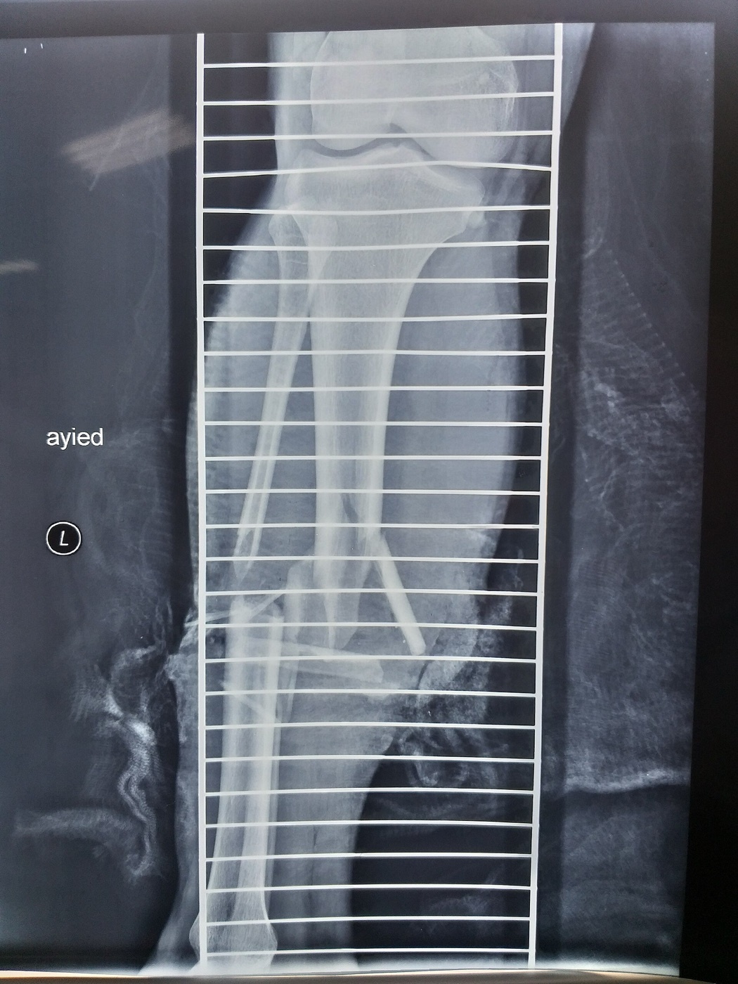
Figure 1. A displaced, comminuted open tibial and fibular shaft fracture. Source: MustafaSalahalden, via Wikimedia Commons, CC BY-SA 4.0.
Tetanus must be addressed in every wound: the patient’s immunisation status is documented and toxoid (with tetanus immunoglobulin for a tetanus-prone wound in an unimmunised patient) is given.[7]
Classifying the open fracture
Gustilo-Anderson (1976, type III subdivided in 1984) is the most-used classification worldwide, based originally on open tibial fractures:[8]
- Type I: wound < 1 cm, clean, minimal soft-tissue damage, simple fracture.
- Type II: wound 1-10 cm, moderate soft-tissue damage and contamination.
- Type IIIA: extensive soft-tissue damage (usually > 10 cm) or any high-energy/heavily contaminated wound, but soft-tissue coverage of bone is still possible.
- Type IIIB: extensive soft-tissue loss with periosteal stripping, requiring a flap for bone cover.
- Type IIIC: any open fracture with an arterial injury requiring repair for limb viability.
Gustilo’s own infection rates were roughly type I 1.9%, type II 8%, type III 41%, though figures vary widely between series (IIIA ~4%, IIIB ~53%, IIIC ~42% in one).[9] The classification has real limitations: interobserver agreement is only about 60%, it depends heavily on wound size, the IIIB category spans a wide spectrum, and it is properly assigned only after debridement.[10] The newer OTA Open Fracture Classification (OTA-OFC) grades five components separately (skin, muscle, arterial, contamination and bone loss), each mild/moderate/severe, which captures within-type variation and predicts treatment, not just infection.[11] The Tscherne classification grades both closed and open soft-tissue injury, and the Ganga Hospital Open Injury Score (GHOIS), devised by the very authors of the Rockwood chapter for type IIIB injuries, scores covering, skeletal and functional tissues separately (1-5 each) plus comorbidities (2 points each): a score ≤ 14 advises salvage, ≥ 17 usually ends in amputation, with 15-16 a “grey zone.”[12]
Part II - Initial Management of the Open Fracture
Every open fracture is a surgical emergency, but about 30% of open-fracture patients have more than one injury, so the bleeding limb must not distract from a full ATLS assessment.[13] The lethal triad of acidosis, hypothermia and coagulopathy is corrected, and damage-control orthopaedics is considered in the unstable patient. Haemorrhage is controlled by compression, a firm dressing and elevation; a bleeding vessel is not blindly clamped in the emergency department (the adjacent nerve is at risk), and uncontrollable bleeding warrants a tourniquet and rapid transfer to theatre.[14] The vascular and neurological status is documented (and re-checked after any realignment). Hard signs of vascular injury (absent pulses, expanding or pulsatile haematoma, bruit or thrill) prompt urgent imaging or exploration, and an ankle-brachial index < 0.9 suggests injury.[15]
The wound is photographed once, then covered with a sterile saline-soaked dressing that is not disturbed again until theatre, because repeatedly redressing the wound in the emergency department raises the infection rate three- to four-fold.[16] The limb is provisionally aligned and splinted to protect the soft tissues and circulation. Routine pre-debridement wound cultures are no longer advised, because they correlate poorly with the organism that later causes infection (most infections are hospital-acquired).[17]
The classic “six-hour rule” for debridement, derived from animal data showing a bacterial infection threshold reached at a mean of about 5 hours, has been substantially challenged.[18] Modern series show no clear difference in infection between debridement before and after six hours when surgery is performed within 24 hours, and an OTA survey found 99.7% of surgeons accept debridement within 12 hours rather than 6.[19] The lesson is that thoroughness matters more than the clock: debride early and urgently, but do not endanger a polytrauma patient to beat an arbitrary deadline.
Part III - Antibiotics, Debridement and Irrigation
Antibiotics: the single strongest modifiable factor
Antibiotics for an open fracture are therapeutic, not prophylactic (the wound is already contaminated), and the agent is matched to the Gustilo type: a first- or second-generation cephalosporin for types I and II, an added aminoglycoside for type III (gram-negative cover), and added penicillin for farmyard, faecal or potentially clostridial wounds.[20] The most important variable is timing: “early timing of antibiotics has been demonstrated as the single most important factor in reducing the infection rate,” ideally within one hour of injury.[21] Prolonged courses breed resistance without benefit, and a 24-hour course is as effective as five days in clean wounds.[22] Local antibiotics (aminoglycoside-PMMA beads in a “bead pouch”, or local vancomycin powder) achieve very high local concentrations and lower infection in severe injuries (one series fell from 12% to 3.7%), but they supplement, never replace, systemic therapy and surgery.[23]
Debridement and irrigation
Debridement is “an active surgical procedure, not just wound washing”: the wound is extended along extensile lines, all foreign material and devitalised tissue are excised, and only living, vascularised tissue is left.[24] Muscle viability is judged by the four Cs (colour, consistency, contractility and capacity to bleed), and “when in doubt, excise.”[25] Bone fragments stripped of all soft-tissue attachment are removed (except articular and cancellous fragments needed for reconstruction). A planned second-look debridement at 48-72 hours is indicated for high-energy, heavily contaminated or late-presenting wounds.[26]
For irrigation, “the solution to pollution is dilution,” but the evidence has overturned old habits. Antiseptics (povidone-iodine, chlorhexidine) are toxic to osteoblasts, chondrocytes and healing tissue and should not be used; adding antibiotics or soap confers no clear benefit; and high-pressure (jet) lavage damages bone, drives bacteria deeper and produces a bacterial rebound, so low-pressure normal saline is preferred.[27] The large FLOW trial compared irrigation pressures and additives and supported low-pressure saline.[28]
Part IV - Skeletal Stabilisation and Wound Cover
Stable skeletal fixation restores length, reduces swelling and pain, and lowers the infection rate; unstable fixation roughly doubles infection in experimental models.[29] The external fixator is the workhorse for the contaminated high-grade open fracture (fast, minimal dissection), with conversion to a definitive intramedullary nail best done within about two weeks (conversion within 28 days carries 3.7% infection versus 22% later).[30] For lower-limb diaphyseal fractures, the reamed intramedullary nail is the definitive treatment of choice; the SPRINT trial and subsequent meta-analyses found no significant outcome difference between reamed and unreamed nailing, with fracture type, not nail type, determining the result.[31]
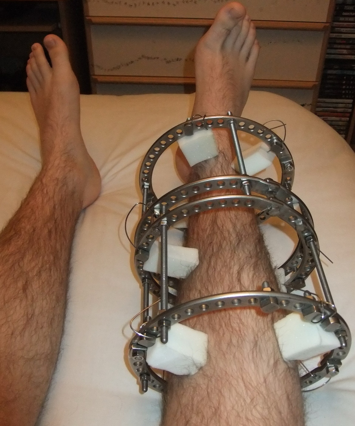
Figure 2. An Ilizarov circular external fixator on the leg, the damage-control workhorse for a contaminated high-grade open fracture. Source: Pagemaker787, via Wikimedia Commons, CC BY-SA 4.0.
The governing modern principle is the orthoplastic approach: orthopaedic and plastic teams working together from the first debridement.[32] Godina’s seminal work showed that microvascular reconstruction within 72 hours sharply reduces flap failure (0.75% vs 12%) and infection (1.5% vs 2%); in “fix and flap” series, muscle-flap cover within 72 hours lowers deep infection (6% vs 29%), giving rise to that doctrine.[33] Soft-tissue cover is chosen along the reconstructive ladder (healing by secondary intention → primary closure → split-skin graft → local/rotational flap → free flap), now often a “reconstructive elevator” in which the surgeon jumps directly to the rung that heals best.[34] For the tibia the classic “thirds” guide applies (proximal-third defects covered by a gastrocnemius flap, middle-third by soleus, distal-third by a free flap), though it is increasingly replaced by perforator-based, vascular-anatomy-led choices.[35] Negative-pressure wound therapy (NPWT) is a useful temporary bridge to cover (one trial: 5.4% vs 28% infection) but is not a substitute for early definitive flap cover.[36]
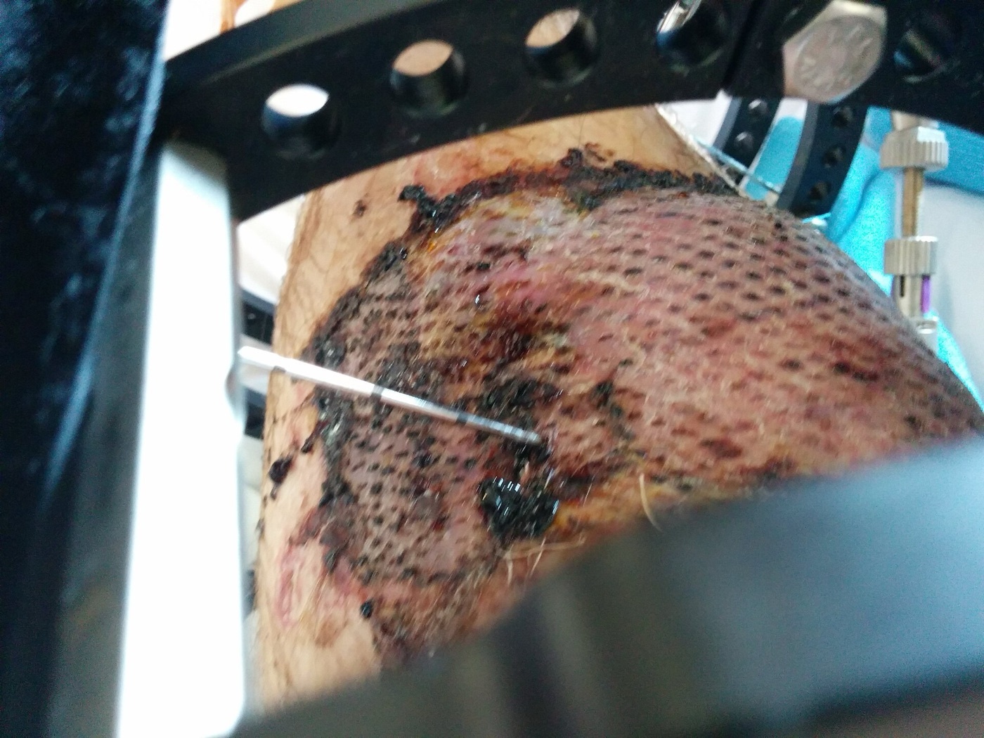
Figure 3. A lower-limb soft-tissue defect resurfaced with a meshed split-thickness skin graft over granulation tissue, a rung of the reconstructive ladder. Source: SOwhoisntme, via Wikimedia Commons, CC0 (public domain).
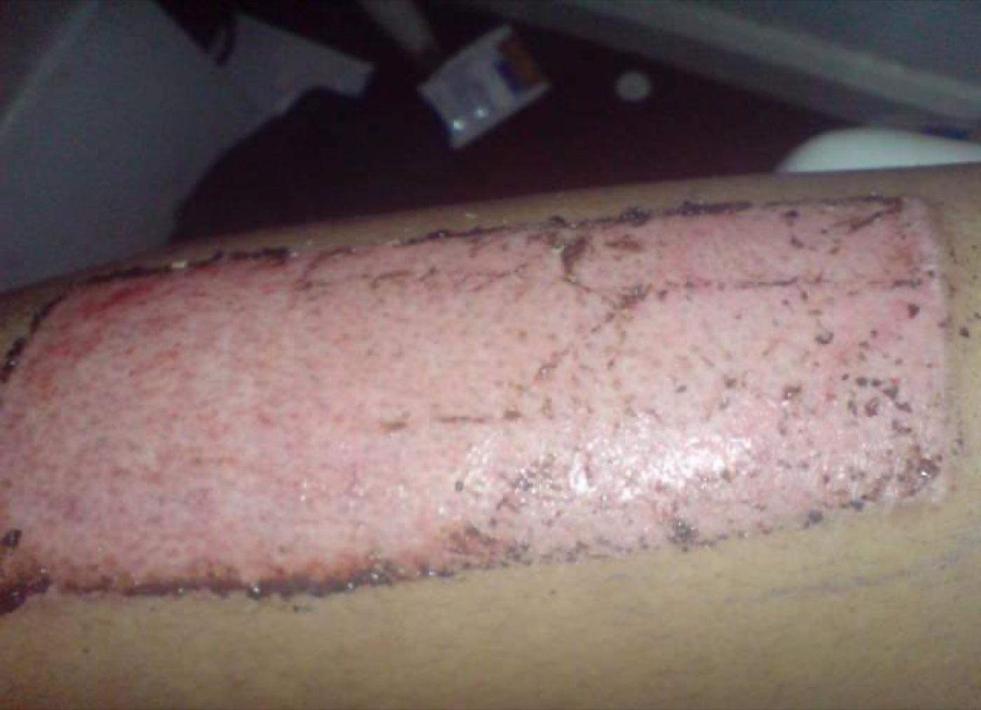
Figure 4. A split-thickness skin-graft donor site (thigh) healing by re-epithelialisation. Source: Kevin308, via Wikimedia Commons, public domain.
Part V - Gunshot and Ballistic Injuries
Wound ballistics
The damage a missile does is governed by the kinetic energy it transfers to the tissue, not merely its speed: “total kinetic energy is the potential for causing damage; transferred kinetic energy is the capacity to cause damage.”[37] Because KE = ½mv², doubling velocity quadruples energy while doubling mass only doubles it, but a bullet that passes straight through deposits only part of its energy, so the useful distinction is low- versus high-energy transfer, not simply low- versus high-velocity.[38] Ballistics divides into internal (within the barrel), external (the trajectory through air) and terminal, and when the target is tissue, terminal ballistics becomes wound ballistics.[39]
A missile produces two cavities. The permanent cavity is the crushed track, roughly the size of the projectile; the temporary cavity is the radial stretch of tissue that lags behind the bullet, reaching a maximum about a millisecond after passage and reaching 10-40 times the permanent-cavity diameter before pulsating shut.[40] A military full-metal-jacket (FMJ) rifle bullet travels nose-first until tissue (800 times denser than air) overcomes its spin stabilisation; once yaw exceeds about 15° it tumbles, presenting its full length, maximising drag and energy transfer, and producing the large temporary cavity.[41] FMJ bullets are mandated for military use by the Hague Convention of 1899, which banned expanding (“dum-dum”) bullets: soft- or hollow-point rounds that mushroom on impact, dramatically increasing energy transfer and tissue destruction (the term commemorates the Dum Dum arsenal near Calcutta).[42] A crucial clinical point: a ricochet or a bullet destabilised by body armour can yaw and fragment so that its wound resembles a dum-dum wound. A fragmenting bullet leaves a radiographic “shower of lead.”[43]
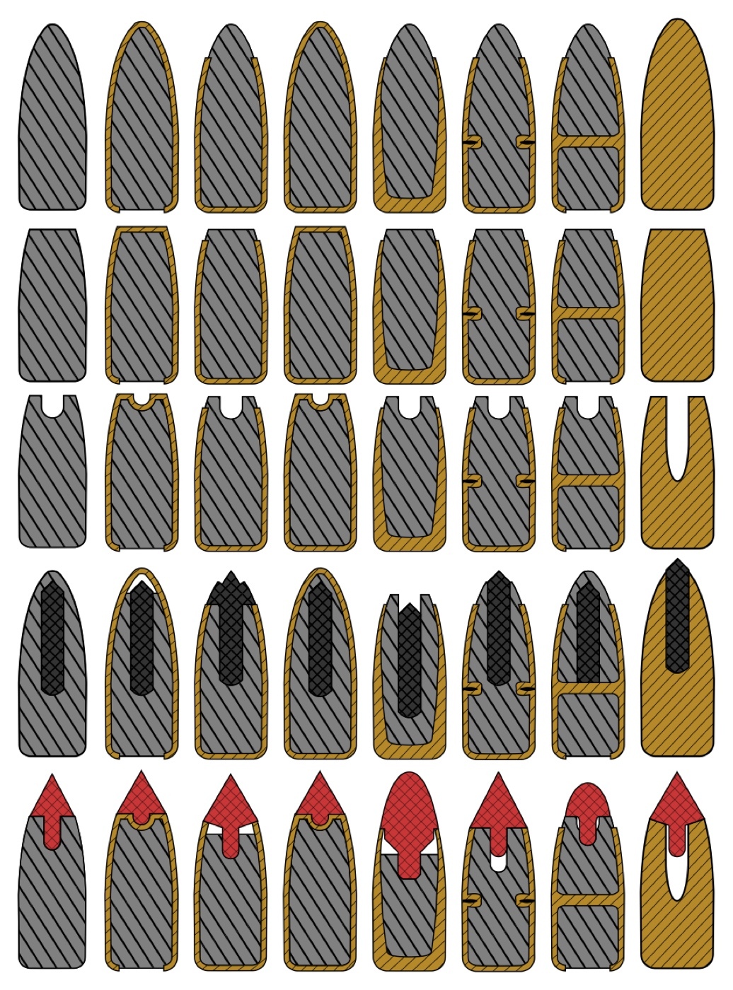
Figure 5. Bullet construction: round-nose, spitzer, full-metal-jacket, soft-point and hollow-point designs (jacket versus core shown by colour). Source: Grasyl, via Wikimedia Commons, CC BY-SA 4.0.
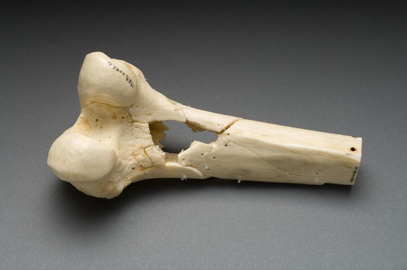
Figure 6. A dry human distal femur perforated by a low-velocity Minié ball: a relatively clean drill-hole defect from limited energy transfer. Source: National Institutes of Health (Ragsdale Gunshot Wound Study), via Wikimedia Commons, public domain (PD-USGov).
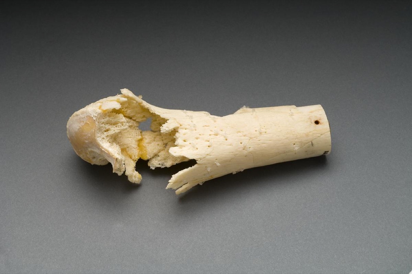
Figure 7. The same bone shattered by a high-velocity 5.56 mm M16 round: extensive comminution from high energy transfer and cavitation. Source: National Institutes of Health (Ragsdale Gunshot Wound Study), via Wikimedia Commons, public domain (PD-USGov).
Blast injury has four mechanisms: primary (the overpressure wave, where tympanic rupture is commonest and “blast lung” most lethal), secondary (fragments, the commonest cause of injury), tertiary (bodily displacement by the blast wind, such as traumatic amputation or being thrown), and quaternary (burns, inhalation).[44] Anti-personnel landmines cause three patterns: traumatic amputation of the foot from a buried blast mine, fragment wounds from a tripwire mine, and severe hand/face injury when a mine is handled.[45]
The Red Cross Wound Score and the management of gunshot fractures
The ICRC Red Cross Wound Score (RCWS) classifies a wound by its features rather than the weapon: E (entry, cm), X (exit, cm), C (cavity, admits two fingers? 0/1), F (fracture: 0 none / 1 simple / 2 comminuted), V (vital structure: neuro, thorax, abdomen, or major-vessel haemorrhage), and M (metallic fragments: 0/1/multiple).[46] From E, X, C and F the wound is Graded 1 (low energy), 2 (high energy) or 3 (massive energy), and typed by injured tissue, a system the text likens to TNM cancer staging.[47]
The central management teaching is that most low-velocity civilian gunshot fractures behave like closed fractures: simple local wound care plus a short antibiotic course suffices, and if surgery is needed the fracture is treated as if closed, the bullet track left unexplored.[48] In contrast, high-velocity, military, shotgun and fragment wounds require formal debridement and are treated as type III open fractures.[49] Retained fragments are generally left alone unless they are painful, periarticular, or intra-articular. An intra-articular bullet must be removed, because lead leaches into the synovium causing lead arthropathy and systemic toxicity.[50] Shotgun wadding is organic and must be sought out and removed as a focus of infection.[51]
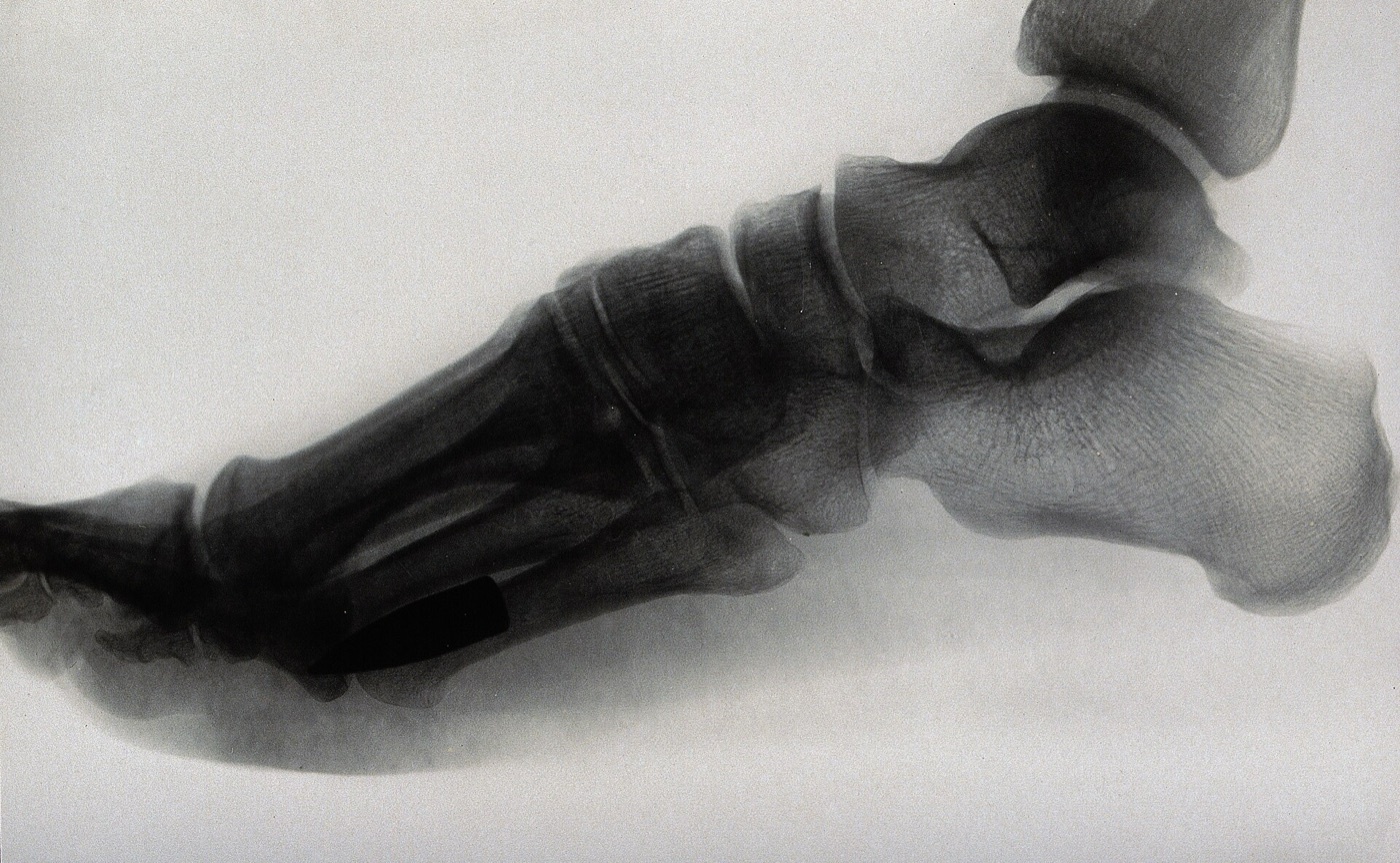
Figure 8. A retained bullet lodged plantar to the metatarsals on a lateral foot radiograph; an intra-articular retained bullet, by contrast, must be removed because of lead arthropathy. Source: Wellcome Collection, via Wikimedia Commons, CC BY 4.0.
Part VI - Crush Injuries and Crush Syndrome
A crush injury is the local tissue damage of prolonged compression; crush syndrome is its systemic, potentially lethal sequel, “the second most common cause of death” after earthquakes and in conflict, behind direct trauma.[52] The mechanism is rhabdomyolysis: prolonged compression and the reperfusion that follows release damage muscle, which leaks potassium, myoglobin, phosphate, urate and creatine kinase into the circulation while sequestering fluid. Reperfusion can cause up to 10 litres of third-space loss per limb and hypovolaemic shock.[53] The consequences are hyperkalaemia (the usual cause of death, by cardiac arrhythmia), hypocalcaemia, metabolic acidosis, and myoglobinuric acute kidney injury: the urine turns “port-wine” coloured and is dipstick-positive for blood without red cells on microscopy, and the CK is typically above 100,000 IU/mL.[54]
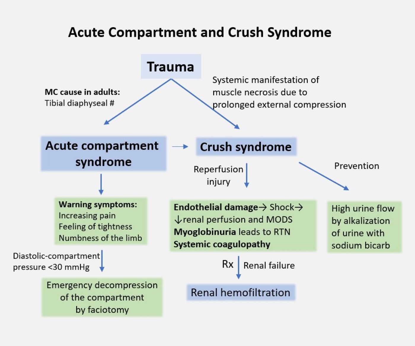
Figure 9. The pathophysiology and management of acute compartment and crush syndrome, summarised. Source: Dr Vijaya Chandar, via Wikimedia Commons, CC BY-SA 4.0.
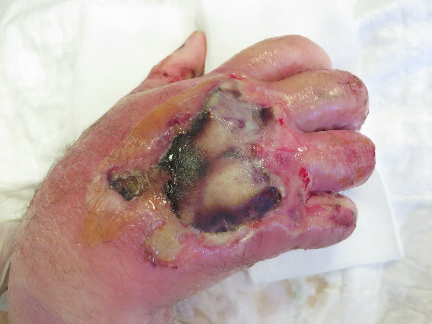
Figure 10. A crush injury of the hand, with necrosis and bullae. Source: AfroBrazilian, via Wikimedia Commons, CC BY-SA 4.0.
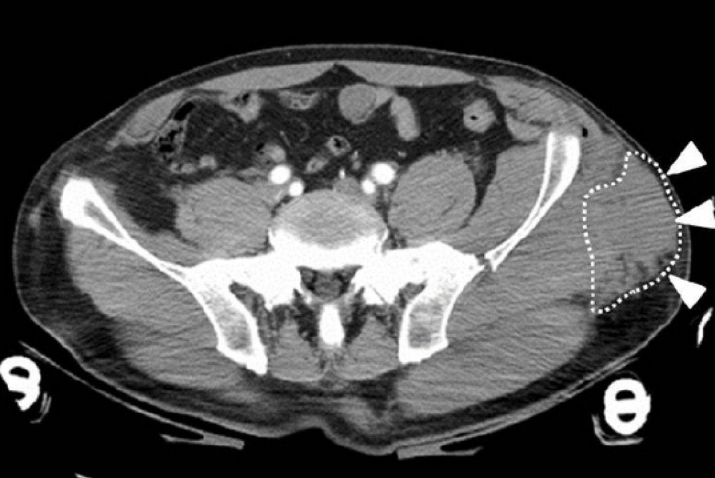
Figure 11. A Morel-Lavallée lesion (closed internal degloving) over the lateral hip on axial CT (arrowed). From Takahara et al. (2014), Case Reports in Orthopedics, doi:10.1155/2014/920317, via Wikimedia Commons, CC BY 3.0.
The primary goal is to prevent acute renal failure. Treatment is aggressive fluid resuscitation begun before extrication if possible, ideally with intravenous access placed before the compressive force is released so the metabolite bolus is diluted; potassium- and lactate-containing fluids are avoided.[55] At least a litre is given before extrication and up to a litre an hour thereafter, with a urine-output target above 100 mL/h, urinary alkalinisation with sodium bicarbonate to a urine pH above 6.5, and mannitol (20%, 1-2 g/kg).[56] Established renal failure requiring dialysis occurs in 50-100% of severe rhabdomyolysis.[57] The surgical decision is governed by the duration of compression: surgical release (fasciotomy) is indicated if compression was relieved within about 6 hours, is controversial at 6-12 hours, and is contraindicated beyond 12 hours, when irreversible muscle death means that opening the limb only exposes necrotic tissue to infection. In a resource-limited setting where follow-up wound care is impossible, fasciotomy should not be performed at all.[58]
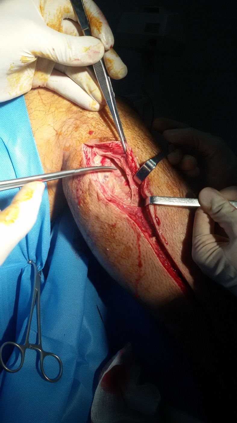
Figure 12. Fasciotomy of the leg for compartment syndrome, the fascial compartments laid open through a longitudinal incision. Source: Armin, via Wikimedia Commons, CC BY-SA 3.0.
Part VII - Infection of the Contaminated and War Wound
All war wounds are grossly contaminated, and the bacterial invasion threshold (10⁶ organisms per gram) falls in the presence of dead muscle, dirt and foreign material; “six hours appears to be the critical period after contamination.”[59] The three great killers of the wounded who survive their initial injury are gas gangrene, tetanus and invasive streptococcal infection, all preventable by the same principle: “the best antibiotic is good surgery,” meaning early wide excision, good drainage, and leaving the wound open for delayed primary closure at about 4-5 days.[60]
Gas gangrene (clostridial myonecrosis) is “a rapidly-spreading oedematous myonecrosis… in muscles contaminated with pathogenic obligatory anaerobes, particularly Clostridium perfringens.”[61] Dead muscle in a deep, closed, anaerobic wound is its ideal medium, and the danger is greatly increased by a wound sutured without debridement, by retained soil, and by a prolonged tourniquet or tight plaster. The incubation is short (usually < 3 days, often < 24 hours), and it announces itself with sudden severe pain, tense oedema, a thin serous discharge, a rapid pulse with little fever, and later bronzed, blistered skin and crepitus from gas in the tissues, though “the extent of gas spread is not the extent of muscle necrosis.”[62] Treatment is radical excision of all dead muscle, often by urgent amputation (“the limit of excision is necrotic muscle, not the extent of tissues containing gas”), with high-dose penicillin and supportive care.[63]
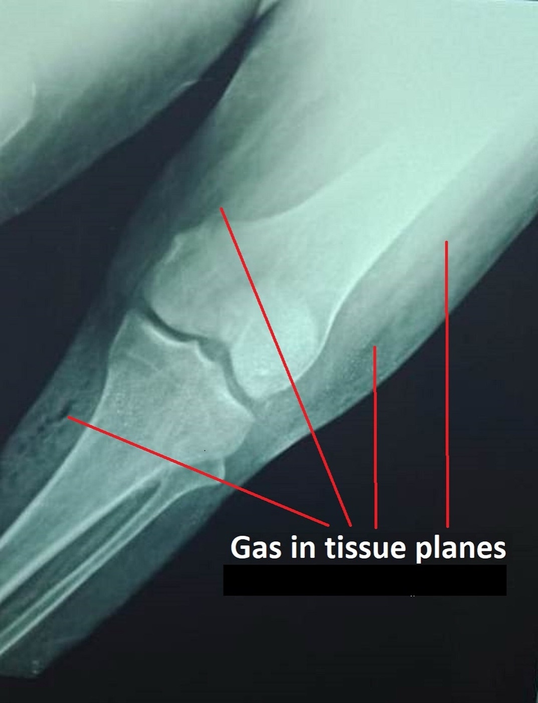
Figure 13. Gas gangrene of the leg: plain radiograph showing gas tracking through the tissue planes. Source: Shahab, via Wikimedia Commons, CC BY-SA 4.0.
Tetanus follows any penetrating wound, especially small deep punctures; Clostridium tetani produces tetanospasmin, which travels up peripheral nerves and causes rigidity, trismus, risus sardonicus and opisthotonus, the patient remaining fully conscious, the greatest danger being asphyxia from laryngeal spasm.[64] Prevention is active immunisation: every wounded patient receives tetanus toxoid and penicillin plus thorough wound excision, and the non-immunised additionally receive human tetanus immunoglobulin (500 IU in adults) with a repeated toxoid course; established tetanus is treated with debridement, penicillin/metronidazole, large-dose immunoglobulin, and spasm control in a dark, quiet room.[65] Necrotising soft-tissue infections (necrotising fasciitis, synergistic gangrene) spare muscle but destroy skin, fat and fascia, and demand aggressive serial excision plus triple antibiotics.[66] In war wounds, antibiotic prophylaxis can be assured only for clostridia and β-haemolytic streptococcus, and penicillin is the drug of choice, begun pre-hospital if possible and continued for about five days until delayed primary closure.[67]
Part VIII - The Mangled Extremity: Salvage versus Amputation
A mangled extremity is “an injury to an extremity so severe that salvage is often questionable and amputation is a possible outcome,” classically a complex fracture with injury to at least two (or, in the Gregory definition, three) of the four systems: bone, soft tissue/skin, vessel and nerve.[68] It is always the result of high-energy trauma, and the great question is whether to reconstruct or amputate.
The OTA criteria for immediate amputation include a life-threatening limb injury, haemodynamic instability, prolonged ischaemia (> 6 hours in the lower limb, > 8 hours in the upper), soft-tissue loss with no flap option, an unreconstructable bone injury, muscle loss across more than two compartments, and tibial bone loss exceeding a third of its length.[69] “Life over limb” supersedes all feasibility when the patient is dying.[70]
The most instructive controversy concerns plantar sensation. Lange held that complete tibial-nerve disruption (an insensate sole) was an absolute indication for amputation. The LEAP study (Bosse et al.) overturned this: more than half of feet insensate at presentation regained normal sensation by two years, and insensate-salvaged feet did no worse than sensate ones, so plantar sensation should not be used to decide amputation.[71] The various limb-salvage scores (MESS [skeletal/soft-tissue energy, ischaemia ×2 if > 6 h, shock, age; an amputation threshold of ≥ 7], PSI, LSI, NISSSA and the Hannover Fracture Scale) share the same flaw on prospective LEAP testing: high specificity but low sensitivity, so a low score reassuringly predicts salvageability but a high score does not reliably mandate amputation.[72] No score should solely dictate amputation.
The outcome data justify caution. LEAP, following 569 patients for two years, found no significant difference in Sickness Impact Profile scores between amputation and reconstruction, and at seven years both groups did poorly, with outcomes driven less by the limb decision than by psychosocial factors: education, income, smoking, social support, self-efficacy and involvement with the legal system.[73] Reconstruction needs roughly twice as many operations; amputation carries a stump-revision rate; and only about half of either group returns to work. (The military METALS study is the exception, finding better function in amputees, attributed to intensive military rehabilitation and advanced prosthetics.)[74] A reasonable strategy is therefore to reconstruct by default, let the limb “declare itself” through serial debridement and external fixation, and accept that delayed amputation does no worse than primary amputation.[75]
Part IX - Amputation
Indications and principles
Only 5-15% of amputations follow trauma (most are for vascular disease).[76] The indications, as Emergency War Surgery lists them, are a traumatic amputation or irreparable vascular injury (the “dead” limb), life-threatening sepsis including clostridial myonecrosis (the “dangerous” limb), and a limb so destroyed that functional recovery is impossible (the “dead loss”).[77] A second surgeon’s opinion before amputating is “always desirable,” and a sensory deficit alone is not an indication, since it is usually a recoverable neurapraxia.[78]
The surgical principles aim at a comfortable, robust residual limb that can drive a prosthesis:[79]
- Preserve length and joints. The energy cost of walking rises as the residual limb shortens and as the level becomes more proximal (transtibial < knee disarticulation < transfemoral); a transfemoral amputee expends as much energy walking at a comfortable pace as an able-bodied person walking flat out.
- Skin and flaps: a long posterior (Burgess) flap for the transtibial level, fishmouth flaps for the transfemoral; atypical flaps are acceptable to preserve length.
- Muscle: myodesis (muscle sutured to bone through drill holes) or myoplasty (opposing muscles sutured together) prevents the wasting, retraction and venous stasis of an unanchored muscle.
- Nerve: traction neurectomy, in which the nerve is drawn down, divided sharply and allowed to retract into muscle, so the inevitable neuroma is buried and less likely to become symptomatic.
- Bone: transected with smooth, bevelled edges (the anterior tibia is bevelled; the fibula is cut about 1.5 cm proximal to the tibia), with every bony prominence covered by muscle.
- Vessels: major arteries double-ligated (a transfixion plus a free tie); a drain is used if the tourniquet is left up to closure.
Levels, the war amputation, and complications
From distal to proximal the levels are toe/ray, transmetatarsal, Lisfranc, Chopart, Syme (ankle disarticulation, end-bearing through the heel pad), transtibial (the workhorse, ideal length ~15 cm from the joint line), knee disarticulation (end-bearing through the femoral condyles), transfemoral (~23-30 cm from the greater trochanter), hip disarticulation and hemipelvectomy.[80] The knee is preserved whenever possible because retaining it markedly improves function.
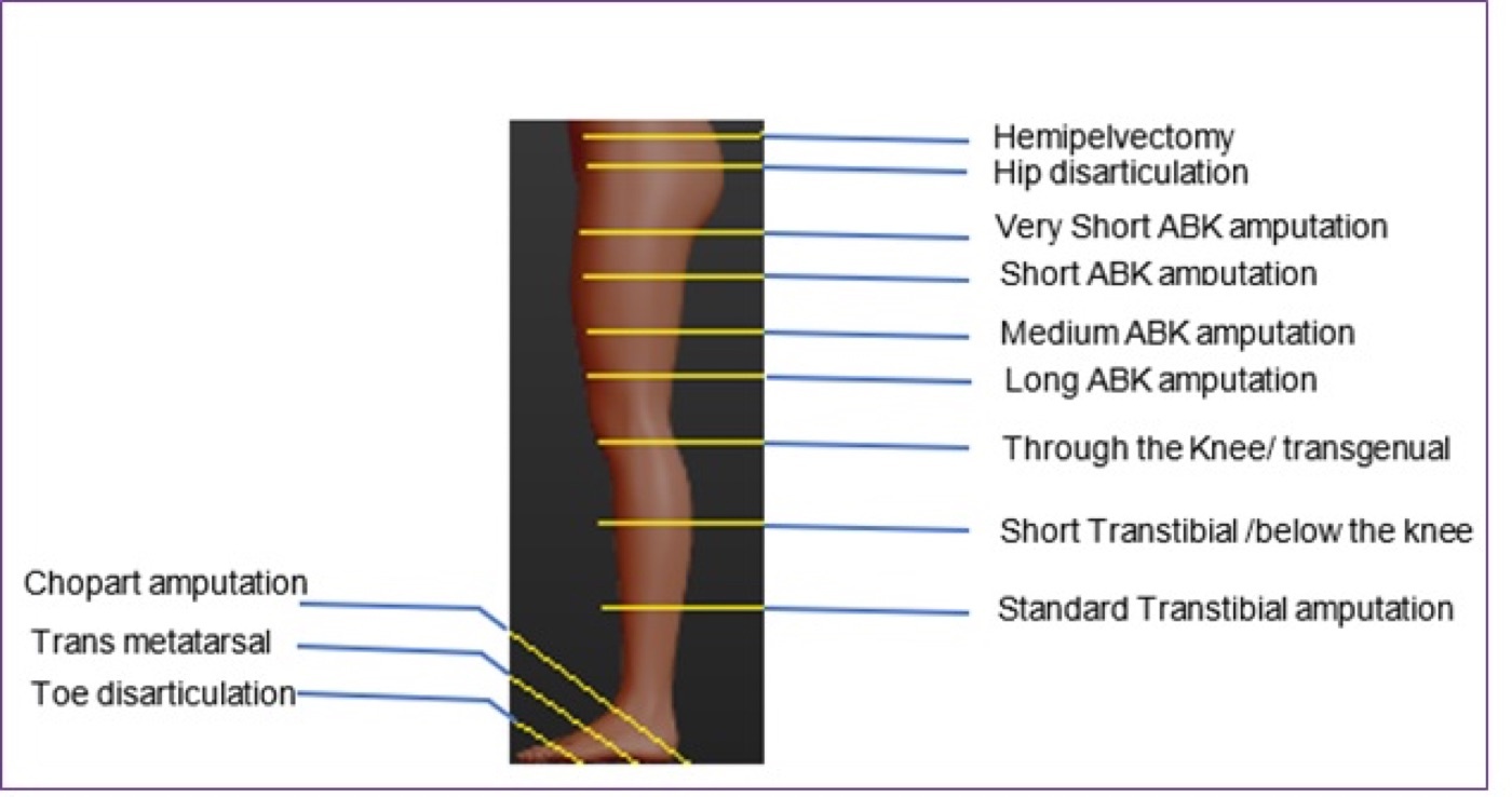
Figure 14. Levels of lower-limb amputation, from hemipelvectomy and hip disarticulation through the transfemoral, through-knee and transtibial levels to the partial-foot (Chopart, transmetatarsal, toe) amputations. Source: Edwin Khundi, via Wikimedia Commons, CC BY-SA 4.0.
The war amputation differs fundamentally: it is performed at the lowest viable level of soft tissue (not at a classic anatomical level), as the first stage of an open, length-preserving amputation, since “the stump is never closed primarily.”[81] Viable “flaps of opportunity” are preserved but not sutured (only held by the dressing), flaps are not fashioned at the first operation, and skin traction or a transportation cast prevents the skin retraction that would otherwise cost length during evacuation.[82] The definitive level and closure are chosen later in a stable environment.
The complications are wound dehiscence and infection, a painful neuroma, heterotopic ossification, and the pain syndromes. The pain syndromes distinguish phantom sensation (awareness of the missing part), phantom pain, and residual-limb (stump) pain from a neuroma or poor prosthetic fit.[83] A rigid (or removable rigid) dressing speeds rehabilitation, controls oedema and reduces revision compared with soft dressings, and graduated weight-bearing begins at about six weeks.[84] Osseointegration, anchoring the prosthesis directly to bone, is an emerging option for those who cannot tolerate a socket, with the skin-implant interface and infection as its main unsolved problem.[85]
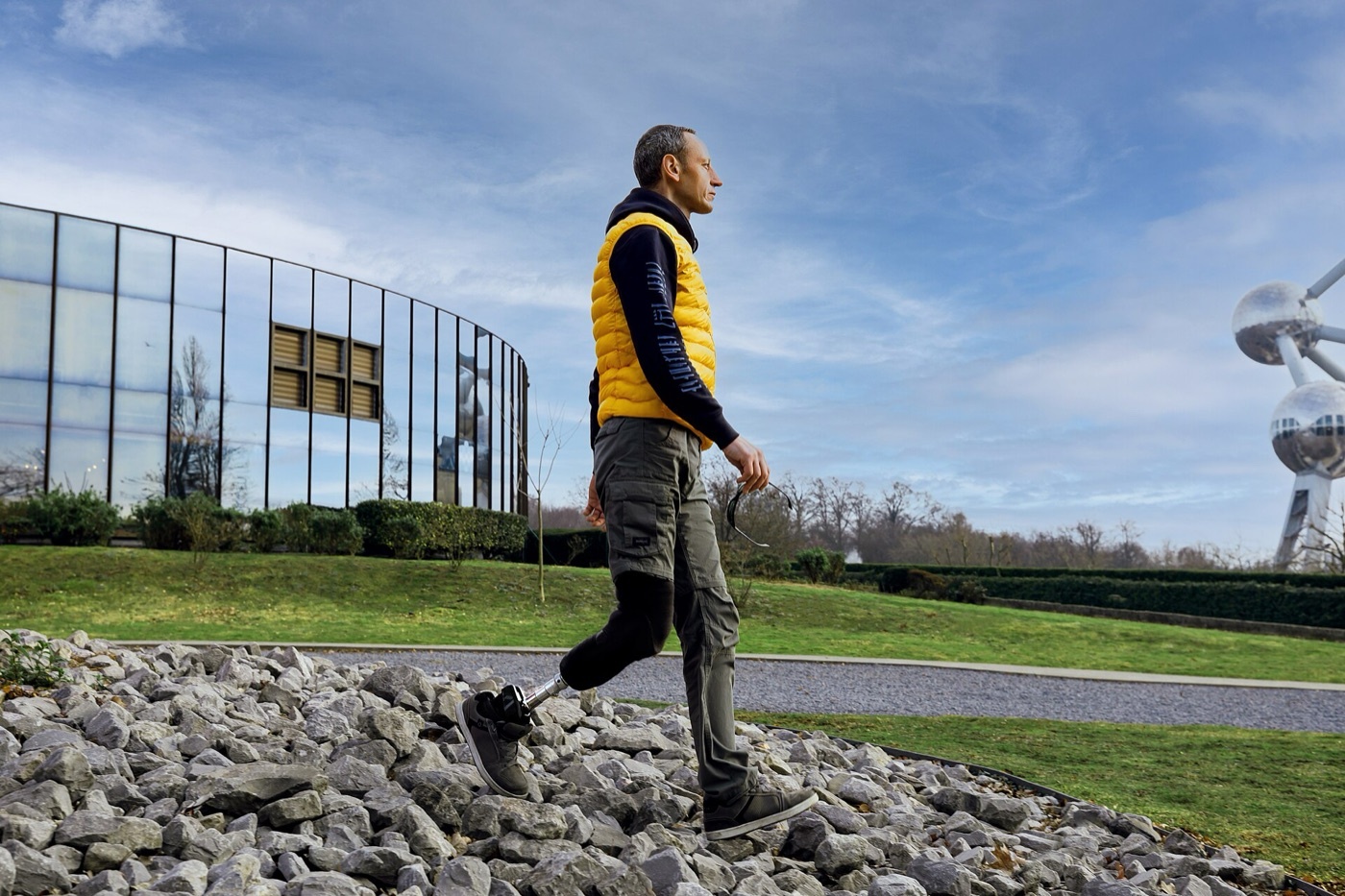
Figure 15. A transtibial (below-knee) amputee walking over rough ground on a prosthesis with an energy-storing foot, the level whose retained knee gives the lowest energy cost of gait. Source: Axiles Bionics, via Wikimedia Commons, CC BY-SA 2.0.
Replantation
Replantation reattaches a completely amputated part by microvascular repair. Its accepted indications are amputation of the thumb, multiple digits, the hand or more proximal levels, any amputation in a child, and a sharp clean amputation; the contraindications are a single finger amputated proximal to the flexor digitorum superficialis insertion in an adult, a severe crush or avulsion mechanism, multiple levels, a medically unfit patient, and prolonged warm ischaemia. A digit (little muscle) tolerates roughly 12 hours of warm or 24 hours of cold ischaemia, whereas a major limb with muscle tolerates only about 4-6 hours of warm ischaemia before myonecrosis forces amputation.[86]
References
-
Rockwood & Green 9e, ch.15 (Rajasekaran et al.), p.835.
-
Rockwood 9e p.835.
-
Rockwood 9e p.842; Skeletal Trauma 5e p.536.
-
Rockwood 9e p.838.
-
Skeletal Trauma 5e pp.534, 542.
-
Rockwood 9e p.838.
-
The mined Rockwood/Skeletal Trauma open-fracture chapters mention only that tetanus status must be documented and prophylaxis started; the immunisation-status criteria and immunoglobulin dosing are standard teaching, detailed in Part VII from the ICRC war-wound protocol.
-
Rockwood 9e p.851; Skeletal Trauma 5e p.534.
-
Rockwood 9e p.851; Skeletal Trauma 5e p.534.
-
Rockwood 9e pp.852-854.
-
Skeletal Trauma 5e pp.534-535, 540.
-
Rockwood 9e pp.855-859.
-
Rockwood 9e pp.839-840.
-
Rockwood 9e p.843.
-
Rockwood 9e p.844; Skeletal Trauma 5e p.536.
-
Rockwood 9e p.843; Skeletal Trauma 5e p.536.
-
Rockwood 9e pp.845-846.
-
Rockwood 9e p.861; Skeletal Trauma 5e p.544.
-
Rockwood 9e p.861.
-
Rockwood 9e p.846; Skeletal Trauma 5e p.543.
-
Skeletal Trauma 5e pp.543-544.
-
Rockwood 9e p.846; Skeletal Trauma 5e p.544.
-
Rockwood 9e p.847; Skeletal Trauma 5e pp.551-552.
-
Rockwood 9e p.861.
-
Rockwood 9e p.865; Skeletal Trauma 5e p.544.
-
Rockwood 9e p.881.
-
Rockwood 9e pp.862-863; Skeletal Trauma 5e p.546.
-
Skeletal Trauma 5e pp.546-547.
-
Rockwood 9e pp.867-868; Skeletal Trauma 5e p.549.
-
Rockwood 9e pp.868-870.
-
Rockwood 9e pp.871-873.
-
Rockwood 9e p.836.
-
Rockwood 9e p.883.
-
Rockwood 9e pp.883-884.
-
Rockwood 9e pp.995-996, 1001-1002.
-
Rockwood 9e pp.886-887.
-
ICRC War Surgery (Giannou & Baldan) p.66.
-
Rockwood 9e p.819; Skeletal Trauma 5e p.579.
-
ICRC p.62-63.
-
Rockwood 9e pp.820-821.
-
Rockwood 9e p.820.
-
Rockwood 9e p.818; ICRC p.63, 69.
-
ICRC pp.65, 68.
-
ICRC p.61; Rockwood 9e p.827.
-
ICRC p.60.
-
ICRC p.87.
-
ICRC pp.91-92.
-
Rockwood 9e pp.822-823; Skeletal Trauma 5e p.582.
-
Rockwood 9e p.823; Skeletal Trauma 5e pp.580-581.
-
Rockwood 9e pp.823-825; Skeletal Trauma 5e p.582.
-
Skeletal Trauma 5e p.579.
-
Skeletal Trauma 5e p.507.
-
Emergency War Surgery (Bowen) p.303; Skeletal Trauma 5e p.507.
-
Emergency War Surgery p.304.
-
Emergency War Surgery pp.304-305; Skeletal Trauma 5e p.507.
-
Emergency War Surgery p.305.
-
Emergency War Surgery p.305.
-
Skeletal Trauma 5e pp.507-508.
-
ICRC p.258.
-
ICRC pp.259, 266.
-
ICRC p.261.
-
ICRC pp.261-262.
-
ICRC p.262.
-
ICRC pp.263-264.
-
ICRC pp.264-265.
-
ICRC p.266.
-
ICRC pp.267-268.
-
Rockwood 9e pp.951, 960.
-
Rockwood 9e pp.955-956.
-
Rockwood 9e p.957.
-
Rockwood 9e pp.958-959.
-
Rockwood 9e pp.961-964.
-
Rockwood 9e pp.964, 974-976.
-
Rockwood 9e pp.975, 979.
-
Rockwood 9e p.1115.
-
Rockwood 9e p.1111.
-
Emergency War Surgery p.346. The “dead/dangerous/dead loss” mnemonic is standard teaching; the source lists the equivalent indications without the phrase.
-
Emergency War Surgery p.346; Rockwood 9e p.1114.
-
Rockwood 9e pp.1111-1119; Atlas of Amputations 4e pp.44-47.
-
Rockwood 9e pp.1114-1125; Atlas of Amputations 4e p.26.
-
Emergency War Surgery pp.347-348.
-
Emergency War Surgery pp.348-350.
-
Rockwood 9e pp.1116, 1127-1128; Atlas of Amputations 4e p.47.
-
Atlas of Amputations 4e pp.50-56; Rockwood 9e pp.1116-1117.
-
Rockwood 9e pp.1128-1130.
-
Replantation is not covered in the mined extracts (which address only war-time digit preservation and the 4-6 h warm-ischaemia limit for major-limb muscle, Emergency War Surgery p.361); the indications, contraindications and digit ischaemia times above are standard hand-surgery teaching.
-
Rockwood 9e pp.835, 851.
-
Rockwood 9e pp.839-846.
-
Rockwood 9e p.861; Skeletal Trauma 5e pp.543-544.
-
Rockwood 9e pp.862-865.
-
Rockwood 9e pp.818-821; ICRC pp.63, 68.
-
Rockwood 9e pp.822-825; Skeletal Trauma 5e pp.580-582.
-
ICRC p.87.
-
Emergency War Surgery pp.303-305; Skeletal Trauma 5e pp.507-508.
-
ICRC pp.261-262, 267.
-
Rockwood 9e pp.955-964, 974-976.
-
Rockwood 9e pp.1111-1119.
-
Emergency War Surgery pp.347-350.
-
Standard hand-surgery teaching; the mined extracts give only the 4-6 h major-limb warm-ischaemia limit (Emergency War Surgery p.361).