Contents
- Orientation: The Extracapsular Mirror-Image of the Femoral Neck
- Part I - Epidemiology
- Part II - Applied Anatomy and the Concept of Stability
- Part III - Classification
- Part IV - Assessment and Imaging
- Part V - Treatment
- Part VI - Complications
- Part VII - A Synthesis: How to Reason About the Trochanteric Fracture
- References
Orientation: The Extracapsular Mirror-Image of the Femoral Neck
The trochanteric fracture is the extracapsular hip fracture, and that one word makes it the mirror-image of the femoral neck fracture of the previous topic. Because it lies outside the capsule, in well-vascularised cancellous metaphyseal bone, it heals readily, and avascular necrosis and nonunion are rare (the opposite of the intracapsular neck).[1] The dominant question here is therefore not “will the head survive?” but “is the fracture stable, and which implant will hold it while it unites?” The answer turns on the stability of the pattern, which is really a question about the posteromedial cortex (the calcar) and the lateral wall. A fracture with an intact posteromedial buttress is stable and suits a sliding hip screw, while a comminuted, reverse-obliquity, or lateral-wall-deficient fracture is unstable and is increasingly fixed with a cephalomedullary nail. Almost all of these fractures are operated on early, so that the frail elderly patient can be mobilised.[2]
Part I - Epidemiology
Trochanteric fractures make up about half of all hip fractures (the extracapsular half, defined as the region between the capsular attachments and a level about 5 cm below the lesser trochanter).[3] Worldwide hip-fracture numbers were estimated at 1.3 million in 1990 and are projected to reach several million by 2050.[4] The patient is typically older and frailer than the femoral-neck-fracture patient (mean age about 80), an osteoporotic woman who has fallen sideways onto the hip. The injury carries the same fragility-fracture significance and a similar mortality.[5]
Part II - Applied Anatomy and the Concept of Stability
The trochanteric region is built of variably cortical and cancellous bone arranged as a trabecular arcade running from the femoral head, around Ward’s triangle, to the lesser trochanter.[6] The key structure is the calcar femorale (first described as Adam’s arch), the dense vertical plate of bone in the posteromedial corner that bears compressive load. It is the buttress that comminutes in unstable fractures.[7] On the other side, the lateral wall (the lateral femoral/greater-trochanteric cortex) is the buttress that a sliding hip screw slides against, and its loss lets the shaft displace medially.[8] The deforming forces give the classic picture: the abductors and short external rotators abduct and externally rotate the greater-trochanter fragment while the adductors and hamstrings pull the shaft medially and proximally, producing the shortened, externally rotated limb in coxa vara.[9] Unlike the femoral neck, the neurovascular structures are rarely at risk and the blood supply to the head is preserved.[10]
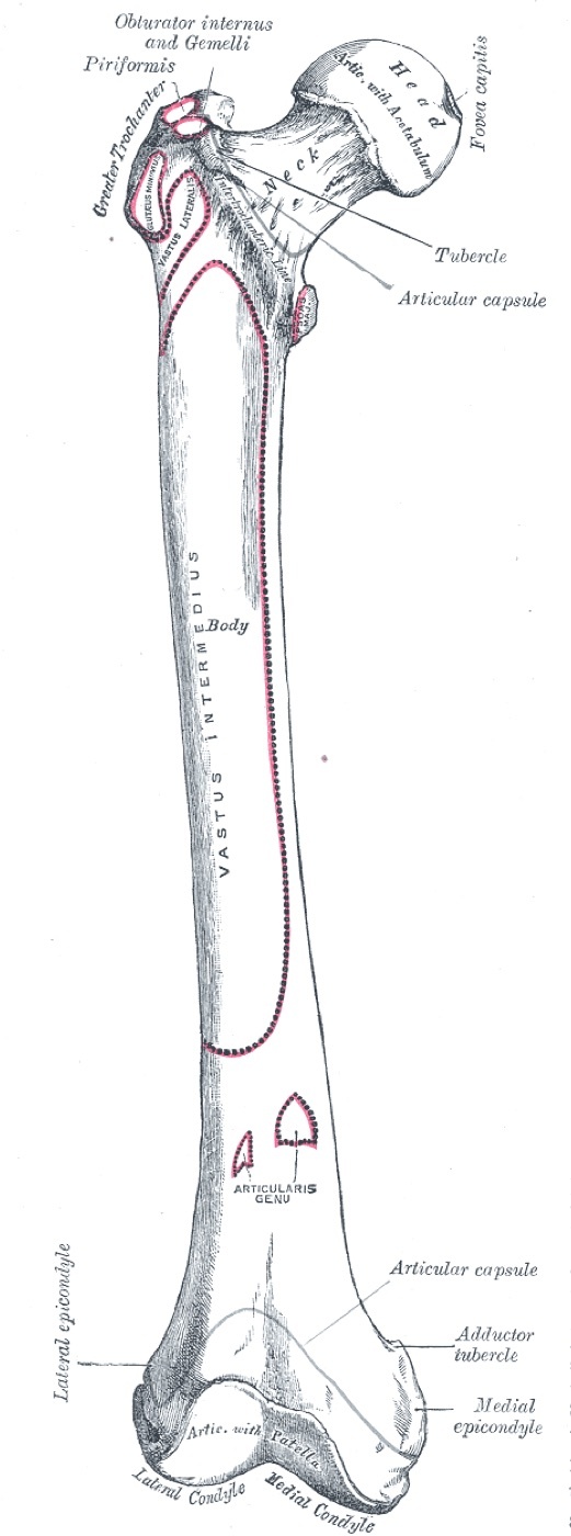
Figure 1. The proximal femur: head, neck, greater and lesser trochanters (the calcar lies along the posteromedial neck). Henry Vandyke Carter, Gray’s Anatomy (1918), public domain, via Wikimedia Commons.
Stability is the organising idea. A stable fracture has an intact (or reconstructable) posteromedial cortex that can resist compressive load once the fracture impacts. An unstable fracture is one with posteromedial (calcar/lesser-trochanter) comminution, a reverse-obliquity pattern, subtrochanteric extension, or an incompetent lateral wall.[11] This matters because stability dictates the implant: a stable fracture can be held by a device that allows controlled collapse onto the medial buttress, whereas an unstable fracture needs a device that resists collapse and medialisation.[12]
Part III - Classification
The working classification is the AO/OTA 31A scheme, in three groups that map directly onto stability:[13]
- 31A1, a simple two-part (pertrochanteric) fracture through the trochanters: the stable pattern.
- 31A2, a comminuted (multifragmentary) pertrochanteric fracture with the lesser and greater trochanters as separate fragments (posteromedial comminution): the unstable pattern.
- 31A3, the reverse-obliquity (or transverse) intertrochanteric fracture at the level of the lesser trochanter, with the fracture line running from proximal-medial to distal-lateral: a distinct unstable pattern that behaves like a subtrochanteric fracture.
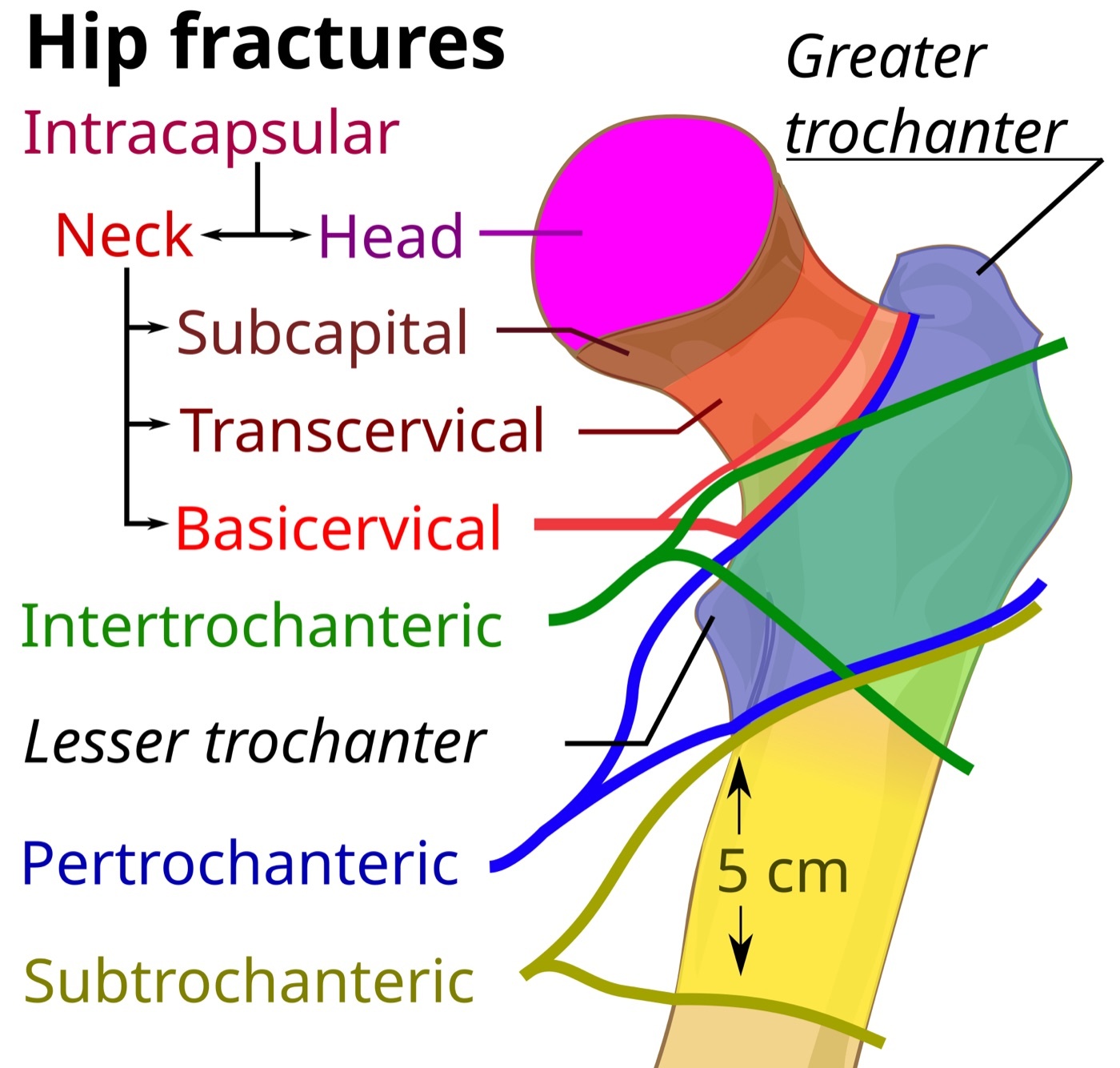
Figure 2. Anatomical classification of proximal femoral fractures: the intertrochanteric/pertrochanteric (extracapsular) zone relative to the trochanters and neck. Mikael Häggström, CC0, via Wikimedia Commons.
The three-group level is reliable and guides treatment, whereas the further sub-subgroups are not reproducible and add little.[14] The older eponymous Evans (and Evans-Jensen) classifications grade the same fractures by stability and by whether the posteromedial buttress can be reconstructed; they remain common teaching but are less reproducible.[15] A basicervical fracture, which runs along the intertrochanteric line and straddles the capsular attachment, is treated as a two-part trochanteric fracture.[16]
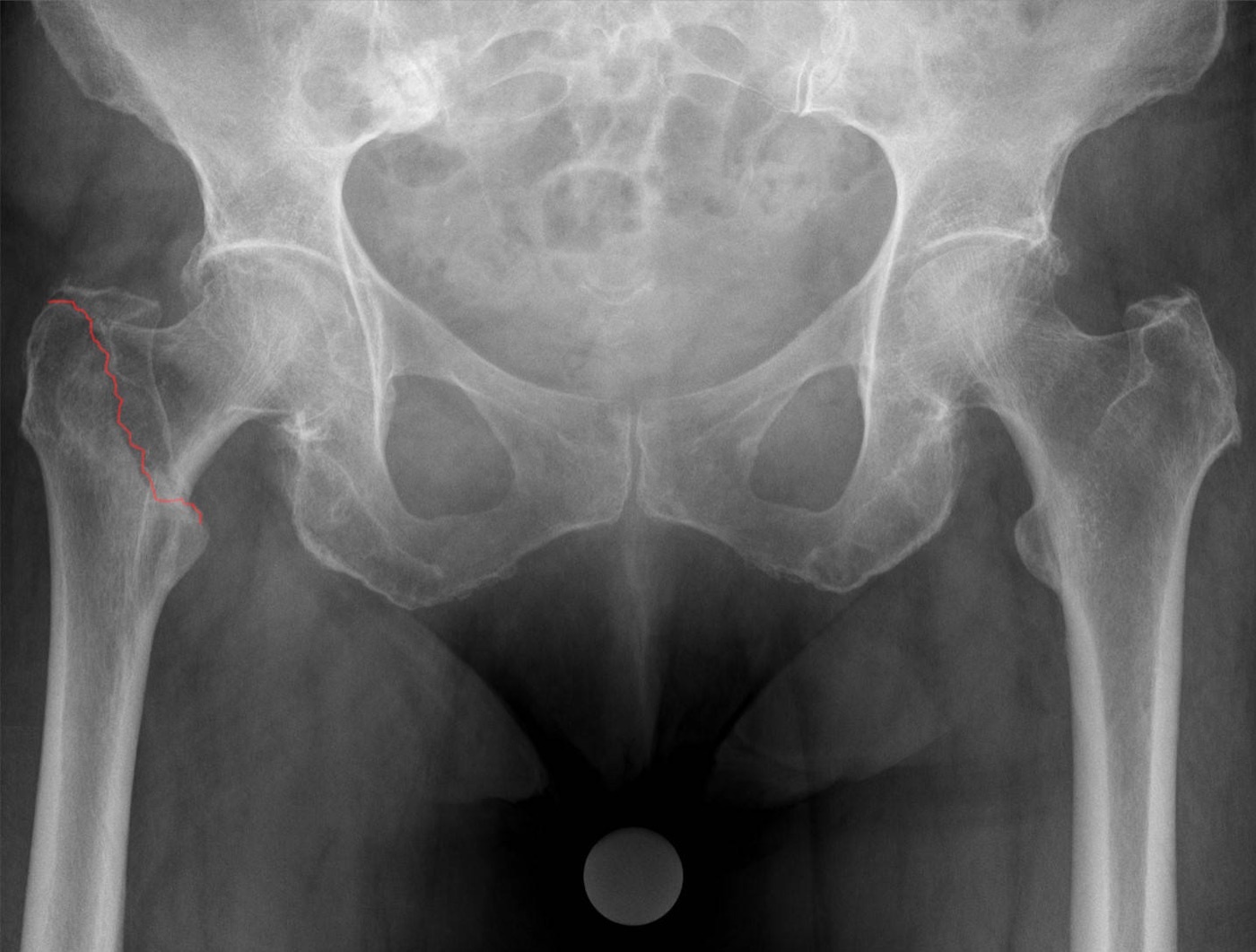
Figure 3. An undisplaced pertrochanteric (extracapsular) fracture on AP radiograph (fracture line in red). Hellerhoff, CC BY-SA 4.0, via Wikimedia Commons.
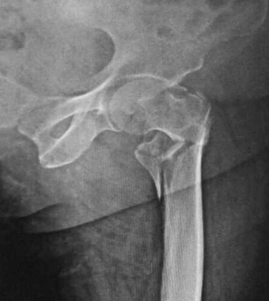
Figure 4. A comminuted (unstable) trochanteric fracture with a separate posteromedial fragment. Memon, Patel & Juva, CC BY 4.0, via Wikimedia Commons.
Part IV - Assessment and Imaging
The mechanism is the low-energy sideways fall of an elderly, often comorbid patient with a history of falls.[17] The presentation is acute hip pain with inability to bear weight and a shortened, externally rotated limb in coxa vara, clinically indistinguishable from a femoral neck fracture.[18] AP pelvis and lateral hip radiographs confirm the diagnosis (the lateral is worth taking, to judge displacement and warn of operative difficulty); a 10°-internal-rotation or traction view helps define the pattern, and MRI (or CT) is used for the occult fracture.[19] The fracture is usually isolated, although about 4% have an associated ipsilateral wrist or shoulder fracture. Significant blood loss (often 500-1000 mL) is common, so the patient is resuscitated and medically optimised for early surgery.[20]
Part V - Treatment
5.1 Operative treatment is the rule
Operative fixation is the treatment of choice for almost all trochanteric fractures, because it controls pain and allows early mobilisation, and these fractures unite reliably.[21] Nonoperative treatment is reserved for the rare exception (a simple undisplaced crack in an active patient, an incomplete fracture on MRI, or a bedbound non-ambulator), since traction for 6 to 8 weeks brings the complications of recumbency and a malunited, shortened limb.[22] Surgery is done early, with prophylactic antibiotics and thromboprophylaxis, on a fracture table that allows traction and image intensification. The two most important steps are the reduction and the placement of the guidewire/lag screw in the femoral head.[23]
5.2 The sliding hip screw (for stable fractures)
The sliding (dynamic) hip screw (SHS/DHS) is the long-standing standard for stable (A1 and many A2) fractures with an intact lateral wall.[24] Its principle is controlled collapse: the lag screw slides within the barrel of a 130-135° side-plate so that weight-bearing impacts the fracture onto the medial buttress, restoring bone contact and uniting with minimal shortening (a fixed, non-sliding implant fails because it cannot collapse).[25] The single most important technical rule is the tip-apex distance (TAD), which should be 25 mm or less (Baumgaertner). TAD is the summed AP and lateral distances from the lag-screw tip to the apex of the femoral head, and a screw placed low-to-central on the AP and central on the lateral, with its tip within about 5 mm of the joint, minimises the risk of cut-out.[26]
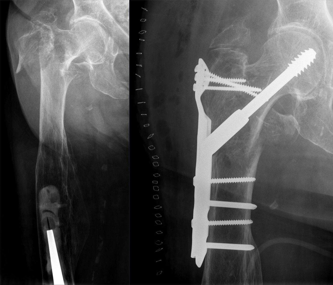
Figure 5. A pertrochanteric fracture (left) fixed with a dynamic hip screw and lateral side plate plus a trochanteric stabilising plate (right). Hellerhoff, CC BY-SA 4.0, via Wikimedia Commons.
5.3 The cephalomedullary nail (for unstable fractures)
For the unstable fracture (a comminuted A2, a reverse-obliquity A3, one with subtrochanteric extension, or one with an incompetent lateral wall), the cephalomedullary nail (the intramedullary hip screw: Gamma, PFN, PFNA, TFN) is increasingly used.[27] Its advantage is that the nail sits against the superior neck and resists the medialisation that an unstable fracture undergoes (medialisation averages about 10% with a sliding hip screw versus 2% with a nail), while its shorter lever arm tolerates the absence of a medial buttress.[28] The reverse-obliquity (A3) fracture is the key indication: the older fixed-angle blade and 95° condylar-screw plates fail in this pattern (one trial reported 7 of 19 failures) and are no longer used, and the AAOS gives strong evidence for an intramedullary nail; by the same logic the standard sliding hip screw is generally avoided in reverse obliquity because it cannot resist the shear (although the Rockwood author argues a sliding hip screw with a stabilising plate remains acceptable).[29]
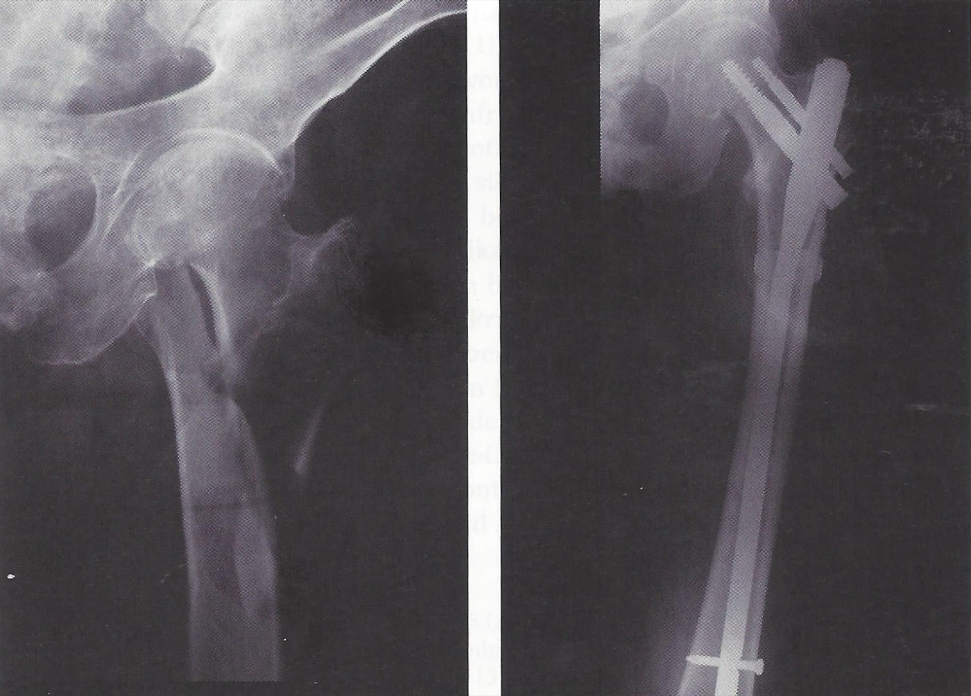
Figure 6. A reverse-obliquity proximal femoral fracture (left) treated with a long cephalomedullary nail and cerclage (right). Mehlauge, CC BY-SA 3.0, via Wikimedia Commons.
5.4 The nail-versus-screw debate
For the stable fracture the choice of nail versus sliding hip screw is genuinely debated.[30] Modern trials show broadly similar outcomes (mortality, union, function), with the sliding hip screw cheaper and the nail carrying a specific risk of peri-implant femoral fracture, and large trials are needed to detect any real difference.[31] The guidelines diverge. The AAOS supports either device for a stable fracture but a cephalomedullary nail for unstable and reverse-obliquity fractures, whereas the UK NICE guidance prefers a sliding hip screw for all trochanteric fractures; the Rockwood author concludes the outcomes are very similar for all patterns and the surgeon should use the implant that works best in their hands.[32] (The notion that loss of the lateral wall mandates a nail is, in the author’s words, “orthopaedic folklore” not borne out by the trials.)[33]
5.5 Reduction principles
Whatever the implant, the fracture is reduced to anatomical or slight valgus, never varus.[34] A valgus reduction carries the lowest risk of cut-out and the least shortening; a varus reduction must never be accepted, because it is mechanically unstable, tilts further into varus, and cuts out.[35] On the lateral view the head, neck, and shaft must be collinear, correcting any sag, and rotation is avoided; the Garden alignment index confirms the reduction. Patients are then mobilised with full weight-bearing as tolerated.[36]
Part VI - Complications
The mortality mirrors hip fractures in general: about 8% at 30 days and 28-38% at one year, mostly from the patient’s medical comorbidities.[37] The characteristic mechanical failure is screw cut-out, the commonest complication of fixation (historically 10-15%, now about 2-3%, and as low as 1% with optimal technique), driven by a high tip-apex distance, a peripheral or varus malreduction, an unstable pattern, and osteoporosis.[38] Malunion (varus and femoral medialisation) and limb shortening are common (10-30%) and impair function (each degree of medialisation adds roughly 1% to the failure risk), which is why a valgus reduction is worth the trouble.[39]
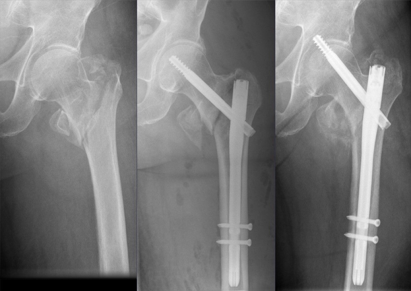
Figure 7. Screw cut-out after cephalomedullary nailing: the cephalic screw migrates through the femoral head toward the acetabulum. Hellerhoff, CC BY-SA 4.0, via Wikimedia Commons.
The other complications are fewer. Peri-implant (femoral shaft) fracture at the nail tip occurs in about 0.5-10% (reduced to about 0.5-1% with modern nail designs), the price of intramedullary fixation; below a side-plate it is rare.[40] Nonunion (1-3%) and avascular necrosis (1-2%) are uncommon because the bone is extracapsular and well-vascularised, the opposite of the femoral neck. A symptomatic cut-out or nonunion is salvaged by revision fixation or, in the older patient, by arthroplasty.[41] Infection (deep 0-1%) and venous thromboembolism complete the list, the latter prevented by early surgery, mobilisation, and chemical-plus-mechanical prophylaxis.[42]
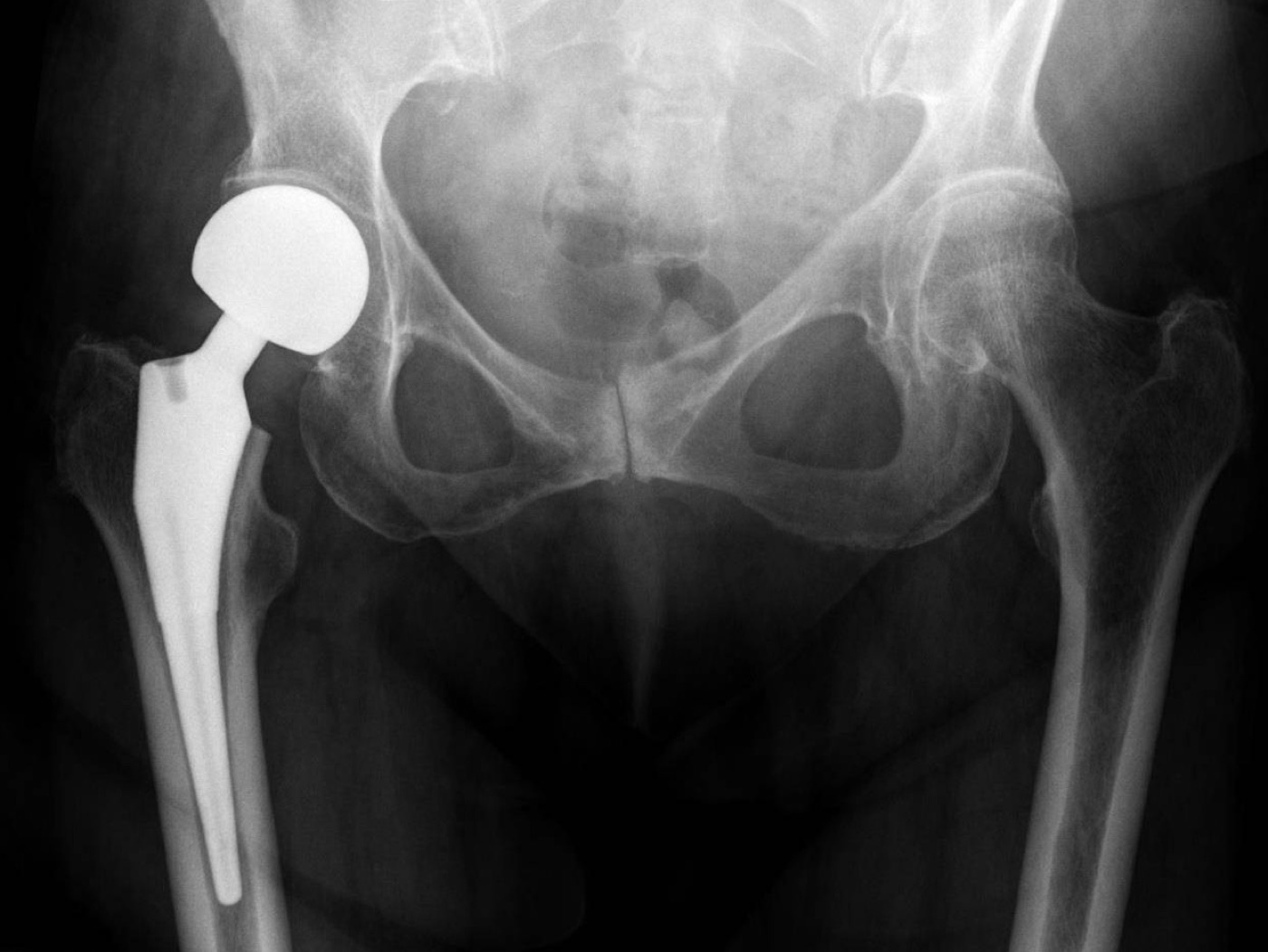
Figure 8. A hip hemiarthroplasty, one arthroplasty option for an unstable proximal-femoral fracture in a frail elderly patient. Jones et al., CC BY 4.0, via Wikimedia Commons.
Part VII - A Synthesis: How to Reason About the Trochanteric Fracture
Start by placing the fracture on the right side of the capsule. It is extracapsular, so the head’s blood supply is safe and the bone unites: the worry is not avascular necrosis but mechanical failure of the fixation. The one question that matters is therefore whether it is stable. A simple two-part fracture (A1) with an intact posteromedial calcar and lateral wall is stable and is held by a sliding hip screw, whose controlled collapse impacts the fracture onto its medial buttress, provided you keep the tip-apex distance under 25 mm with a low-central screw. A comminuted (A2), reverse-obliquity (A3), subtrochanteric-extending, or lateral-wall-deficient fracture is unstable: it resists the sliding screw and is better held by a cephalomedullary nail that buttresses the neck and resists medialisation, and the reverse-obliquity pattern in particular should never be fixed with an old fixed-angle blade plate. Reduce to a slight valgus and never to varus, and mobilise the patient at once. Then watch for the failure this fracture actually produces, screw cut-out, which a good reduction and a low-central screw within a short tip-apex distance largely prevent. The trochanteric fracture is, in the end, a problem of holding well-healing bone in an old skeleton long enough for it to unite, and the craft lies in matching the implant to the stability of the pattern.
References
-
Rockwood & Green’s Fractures in Adults, pp.3689, 3693, 3699, 3736-3741 (extracapsular, well-vascularised cancellous metaphyseal bone, so union is high with nonunion only 1-3% and avascular necrosis only 1-2%, in contrast to the intracapsular neck; operative treatment the rule, with the implant governed by the stability of the pattern); AO Principles of Fracture Management, pp.802-806 (trochanteric fractures unite, with union by about 3 months and AVN rare).
-
Rockwood & Green’s Fractures in Adults, pp.3689, 3693, 3699, 3736-3741 (extracapsular, well-vascularised cancellous metaphyseal bone, so union is high with nonunion only 1-3% and avascular necrosis only 1-2%, in contrast to the intracapsular neck; operative treatment the rule, with the implant governed by the stability of the pattern); AO Principles of Fracture Management, pp.802-806 (trochanteric fractures unite, with union by about 3 months and AVN rare).
-
Rockwood & Green’s Fractures in Adults, pp.3689, 3692 (trochanteric fractures about half of hip fractures, the extracapsular region to ~5 cm below the lesser trochanter; 1.3 million worldwide in 1990 rising toward several million by 2050; patients slightly older and frailer than the intracapsular group, mean age ~80, osteoporotic women falling sideways); Miller’s Review of Orthopaedics, pp.916-917 (geriatric intertrochanteric fragility fracture).
-
Rockwood & Green’s Fractures in Adults, pp.3689, 3692 (trochanteric fractures about half of hip fractures, the extracapsular region to ~5 cm below the lesser trochanter; 1.3 million worldwide in 1990 rising toward several million by 2050; patients slightly older and frailer than the intracapsular group, mean age ~80, osteoporotic women falling sideways); Miller’s Review of Orthopaedics, pp.916-917 (geriatric intertrochanteric fragility fracture).
-
Rockwood & Green’s Fractures in Adults, pp.3689, 3692 (trochanteric fractures about half of hip fractures, the extracapsular region to ~5 cm below the lesser trochanter; 1.3 million worldwide in 1990 rising toward several million by 2050; patients slightly older and frailer than the intracapsular group, mean age ~80, osteoporotic women falling sideways); Miller’s Review of Orthopaedics, pp.916-917 (geriatric intertrochanteric fragility fracture).
-
Rockwood & Green’s Fractures in Adults, pp.3693-3695 (the trabecular arcade around Ward’s triangle; the calcar femorale / Adam’s arch the posteromedial compression buttress, comminuted in unstable fractures; loss of lateral cortical support letting the femur medialise; the abductors/short external rotators displacing the greater trochanter and the adductors/hamstrings the shaft, giving shortening and coxa vara; neurovascular structures rarely at risk).
-
Rockwood & Green’s Fractures in Adults, pp.3693-3695 (the trabecular arcade around Ward’s triangle; the calcar femorale / Adam’s arch the posteromedial compression buttress, comminuted in unstable fractures; loss of lateral cortical support letting the femur medialise; the abductors/short external rotators displacing the greater trochanter and the adductors/hamstrings the shaft, giving shortening and coxa vara; neurovascular structures rarely at risk).
-
Rockwood & Green’s Fractures in Adults, pp.3693-3695 (the trabecular arcade around Ward’s triangle; the calcar femorale / Adam’s arch the posteromedial compression buttress, comminuted in unstable fractures; loss of lateral cortical support letting the femur medialise; the abductors/short external rotators displacing the greater trochanter and the adductors/hamstrings the shaft, giving shortening and coxa vara; neurovascular structures rarely at risk).
-
Rockwood & Green’s Fractures in Adults, pp.3693-3695 (the trabecular arcade around Ward’s triangle; the calcar femorale / Adam’s arch the posteromedial compression buttress, comminuted in unstable fractures; loss of lateral cortical support letting the femur medialise; the abductors/short external rotators displacing the greater trochanter and the adductors/hamstrings the shaft, giving shortening and coxa vara; neurovascular structures rarely at risk).
-
Rockwood & Green’s Fractures in Adults, pp.3693-3695 (the trabecular arcade around Ward’s triangle; the calcar femorale / Adam’s arch the posteromedial compression buttress, comminuted in unstable fractures; loss of lateral cortical support letting the femur medialise; the abductors/short external rotators displacing the greater trochanter and the adductors/hamstrings the shaft, giving shortening and coxa vara; neurovascular structures rarely at risk).
-
Rockwood & Green’s Fractures in Adults, pp.3690-3691, 3693, 3709-3711 (stability defined by the posteromedial cortex/calcar: the two-part A1 pattern is stable, the comminuted A2 [posteromedial/lesser-and-greater-trochanter comminution] is unstable, with reverse obliquity, subtrochanteric extension, and loss of lateral cortical support the unstable features that drive implant choice).
-
Rockwood & Green’s Fractures in Adults, pp.3690-3691, 3693, 3709-3711 (stability defined by the posteromedial cortex/calcar: the two-part A1 pattern is stable, the comminuted A2 [posteromedial/lesser-and-greater-trochanter comminution] is unstable, with reverse obliquity, subtrochanteric extension, and loss of lateral cortical support the unstable features that drive implant choice).
-
Rockwood & Green’s Fractures in Adults, pp.3690-3691 (AO/OTA 31A1 simple two-part “stable”, A2 comminuted “unstable” with the lesser and greater trochanters as third and fourth parts, A3 reversed/transverse at the lesser-trochanter level; the three-group level is reliable while the sub-subgroups are not); AO Principles of Fracture Management, pp.794-796 (31A1 stable simple pertrochanteric, 31A2 unstable multifragmentary with an incompetent lateral wall, 31A3 reverse-oblique intertrochanteric). The proximal-medial-to-distal-lateral geometry of reverse obliquity is the standard descriptor; the mined Rockwood extract terms it the “reversed/transverse (A3)” pattern without that verbatim direction.
-
Rockwood & Green’s Fractures in Adults, pp.3690-3691 (the A1/A2/A3 level reliable and treatment-relevant, the sub-subgroups not; basicervical fractures along the intertrochanteric line, coded AO 31B2.1, treated as a two-part trochanteric fracture). The Evans and Evans-Jensen stability classifications are standard teaching but are not named in the mined Rockwood or Miller extracts (which use AO/OTA 31A); they are noted here as established classifications rather than cited to these sources.
-
Rockwood & Green’s Fractures in Adults, pp.3690-3691 (the A1/A2/A3 level reliable and treatment-relevant, the sub-subgroups not; basicervical fractures along the intertrochanteric line, coded AO 31B2.1, treated as a two-part trochanteric fracture). The Evans and Evans-Jensen stability classifications are standard teaching but are not named in the mined Rockwood or Miller extracts (which use AO/OTA 31A); they are noted here as established classifications rather than cited to these sources.
-
Rockwood & Green’s Fractures in Adults, pp.3690-3691 (the A1/A2/A3 level reliable and treatment-relevant, the sub-subgroups not; basicervical fractures along the intertrochanteric line, coded AO 31B2.1, treated as a two-part trochanteric fracture). The Evans and Evans-Jensen stability classifications are standard teaching but are not named in the mined Rockwood or Miller extracts (which use AO/OTA 31A); they are noted here as established classifications rather than cited to these sources.
-
Rockwood & Green’s Fractures in Adults, pp.3692-3693, 3699 (low-energy sideways fall in the comorbid elderly; shortened/externally rotated limb in coxa vara, indistinguishable from a neck fracture; AP and lateral radiographs, a 10°-internal-rotation or traction view to define the pattern, MRI or CT for the occult fracture; ~4% with an associated ipsilateral distal radius/proximal humerus fracture; blood loss often 500-1000 mL needing resuscitation).
-
Rockwood & Green’s Fractures in Adults, pp.3692-3693, 3699 (low-energy sideways fall in the comorbid elderly; shortened/externally rotated limb in coxa vara, indistinguishable from a neck fracture; AP and lateral radiographs, a 10°-internal-rotation or traction view to define the pattern, MRI or CT for the occult fracture; ~4% with an associated ipsilateral distal radius/proximal humerus fracture; blood loss often 500-1000 mL needing resuscitation).
-
Rockwood & Green’s Fractures in Adults, pp.3692-3693, 3699 (low-energy sideways fall in the comorbid elderly; shortened/externally rotated limb in coxa vara, indistinguishable from a neck fracture; AP and lateral radiographs, a 10°-internal-rotation or traction view to define the pattern, MRI or CT for the occult fracture; ~4% with an associated ipsilateral distal radius/proximal humerus fracture; blood loss often 500-1000 mL needing resuscitation).
-
Rockwood & Green’s Fractures in Adults, pp.3692-3693, 3699 (low-energy sideways fall in the comorbid elderly; shortened/externally rotated limb in coxa vara, indistinguishable from a neck fracture; AP and lateral radiographs, a 10°-internal-rotation or traction view to define the pattern, MRI or CT for the occult fracture; ~4% with an associated ipsilateral distal radius/proximal humerus fracture; blood loss often 500-1000 mL needing resuscitation).
-
Rockwood & Green’s Fractures in Adults, pp.3697-3699, 3713-3714 (operative treatment the rule, controlling pain and allowing early mobilisation; nonoperative only for the rare exception, traction for 6-8 weeks bringing recumbency complications and malunion; surgery early on a fracture table, the reduction and femoral-head guidewire placement the two key steps; prophylactic antibiotics and thromboprophylaxis); AO Principles of Fracture Management, p.806 (early surgery and immediate mobilisation).
-
Rockwood & Green’s Fractures in Adults, pp.3697-3699, 3713-3714 (operative treatment the rule, controlling pain and allowing early mobilisation; nonoperative only for the rare exception, traction for 6-8 weeks bringing recumbency complications and malunion; surgery early on a fracture table, the reduction and femoral-head guidewire placement the two key steps; prophylactic antibiotics and thromboprophylaxis); AO Principles of Fracture Management, p.806 (early surgery and immediate mobilisation).
-
Rockwood & Green’s Fractures in Adults, pp.3697-3699, 3713-3714 (operative treatment the rule, controlling pain and allowing early mobilisation; nonoperative only for the rare exception, traction for 6-8 weeks bringing recumbency complications and malunion; surgery early on a fracture table, the reduction and femoral-head guidewire placement the two key steps; prophylactic antibiotics and thromboprophylaxis); AO Principles of Fracture Management, p.806 (early surgery and immediate mobilisation).
-
Rockwood & Green’s Fractures in Adults, pp.3703, 3720-3728 (the sliding hip screw the gold standard for stable fractures with an intact lateral wall; controlled collapse via the lag screw sliding in a 130-135° plate, impacting onto the medial buttress; tip-apex distance 25 mm or less, low-to-central on AP and central on lateral with the tip ~5 mm from the joint, to prevent cut-out); Miller’s Review of Orthopaedics, p.917 (TAD <25 mm with a centre-centre lag screw, cut-out tied to TAD >25 mm); AO Principles of Fracture Management, pp.802-804 (DHS for stable patterns, central lag screw, TAD <25 mm). The tip-apex distance is the work of Baumgaertner; the mined Rockwood extract gives the ≤25 mm value without writing out the eponym, which is standard teaching.
-
Rockwood & Green’s Fractures in Adults, pp.3703, 3720-3728 (the sliding hip screw the gold standard for stable fractures with an intact lateral wall; controlled collapse via the lag screw sliding in a 130-135° plate, impacting onto the medial buttress; tip-apex distance 25 mm or less, low-to-central on AP and central on lateral with the tip ~5 mm from the joint, to prevent cut-out); Miller’s Review of Orthopaedics, p.917 (TAD <25 mm with a centre-centre lag screw, cut-out tied to TAD >25 mm); AO Principles of Fracture Management, pp.802-804 (DHS for stable patterns, central lag screw, TAD <25 mm). The tip-apex distance is the work of Baumgaertner; the mined Rockwood extract gives the ≤25 mm value without writing out the eponym, which is standard teaching.
-
Rockwood & Green’s Fractures in Adults, pp.3703, 3720-3728 (the sliding hip screw the gold standard for stable fractures with an intact lateral wall; controlled collapse via the lag screw sliding in a 130-135° plate, impacting onto the medial buttress; tip-apex distance 25 mm or less, low-to-central on AP and central on lateral with the tip ~5 mm from the joint, to prevent cut-out); Miller’s Review of Orthopaedics, p.917 (TAD <25 mm with a centre-centre lag screw, cut-out tied to TAD >25 mm); AO Principles of Fracture Management, pp.802-804 (DHS for stable patterns, central lag screw, TAD <25 mm). The tip-apex distance is the work of Baumgaertner; the mined Rockwood extract gives the ≤25 mm value without writing out the eponym, which is standard teaching.
-
Rockwood & Green’s Fractures in Adults, pp.3700-3711, 3733-3734 (the cephalomedullary nail for unstable A2/A3, subtrochanteric extension, or an incompetent lateral wall; the nail buttressing against the neck to resist medialisation, ~10% with the SHS vs ~2% with the nail; the reverse-obliquity A3 fracture failing with fixed-angle blade/95° condylar-screw plates [7 of 19 failures, Sadowski], which are no longer used, with AAOS strong evidence for an intramedullary nail, while the author defends the SHS with a stabilising plate as still acceptable); Miller’s Review of Orthopaedics, p.917 (a cephalomedullary nail for all unstable patterns - reverse oblique, subtrochanteric, comminuted, no intact lateral cortex). The widely taught maxim that “reverse obliquity is a contraindication to the sliding hip screw” reflects this evidence; the Rockwood source frames it as a strong preference for the nail rather than an absolute contraindication.
-
Rockwood & Green’s Fractures in Adults, pp.3700-3711, 3733-3734 (the cephalomedullary nail for unstable A2/A3, subtrochanteric extension, or an incompetent lateral wall; the nail buttressing against the neck to resist medialisation, ~10% with the SHS vs ~2% with the nail; the reverse-obliquity A3 fracture failing with fixed-angle blade/95° condylar-screw plates [7 of 19 failures, Sadowski], which are no longer used, with AAOS strong evidence for an intramedullary nail, while the author defends the SHS with a stabilising plate as still acceptable); Miller’s Review of Orthopaedics, p.917 (a cephalomedullary nail for all unstable patterns - reverse oblique, subtrochanteric, comminuted, no intact lateral cortex). The widely taught maxim that “reverse obliquity is a contraindication to the sliding hip screw” reflects this evidence; the Rockwood source frames it as a strong preference for the nail rather than an absolute contraindication.
-
Rockwood & Green’s Fractures in Adults, pp.3700-3711, 3733-3734 (the cephalomedullary nail for unstable A2/A3, subtrochanteric extension, or an incompetent lateral wall; the nail buttressing against the neck to resist medialisation, ~10% with the SHS vs ~2% with the nail; the reverse-obliquity A3 fracture failing with fixed-angle blade/95° condylar-screw plates [7 of 19 failures, Sadowski], which are no longer used, with AAOS strong evidence for an intramedullary nail, while the author defends the SHS with a stabilising plate as still acceptable); Miller’s Review of Orthopaedics, p.917 (a cephalomedullary nail for all unstable patterns - reverse oblique, subtrochanteric, comminuted, no intact lateral cortex). The widely taught maxim that “reverse obliquity is a contraindication to the sliding hip screw” reflects this evidence; the Rockwood source frames it as a strong preference for the nail rather than an absolute contraindication.
-
Rockwood & Green’s Fractures in Adults, pp.3708-3713 (modern trials show broadly similar mortality/union/function for nail vs sliding hip screw, the SHS cheaper, the nail with a specific peri-implant femoral-fracture risk; AAOS supports either for stable but a cephalomedullary nail for unstable/reverse-obliquity, NICE prefers a sliding hip screw for all, the author concluding outcomes are similar and the implant should be the one the surgeon uses best; the “loss of lateral support mandates a nail” idea called orthopaedic folklore unsupported by the trials).
-
Rockwood & Green’s Fractures in Adults, pp.3708-3713 (modern trials show broadly similar mortality/union/function for nail vs sliding hip screw, the SHS cheaper, the nail with a specific peri-implant femoral-fracture risk; AAOS supports either for stable but a cephalomedullary nail for unstable/reverse-obliquity, NICE prefers a sliding hip screw for all, the author concluding outcomes are similar and the implant should be the one the surgeon uses best; the “loss of lateral support mandates a nail” idea called orthopaedic folklore unsupported by the trials).
-
Rockwood & Green’s Fractures in Adults, pp.3708-3713 (modern trials show broadly similar mortality/union/function for nail vs sliding hip screw, the SHS cheaper, the nail with a specific peri-implant femoral-fracture risk; AAOS supports either for stable but a cephalomedullary nail for unstable/reverse-obliquity, NICE prefers a sliding hip screw for all, the author concluding outcomes are similar and the implant should be the one the surgeon uses best; the “loss of lateral support mandates a nail” idea called orthopaedic folklore unsupported by the trials).
-
Rockwood & Green’s Fractures in Adults, pp.3708-3713 (modern trials show broadly similar mortality/union/function for nail vs sliding hip screw, the SHS cheaper, the nail with a specific peri-implant femoral-fracture risk; AAOS supports either for stable but a cephalomedullary nail for unstable/reverse-obliquity, NICE prefers a sliding hip screw for all, the author concluding outcomes are similar and the implant should be the one the surgeon uses best; the “loss of lateral support mandates a nail” idea called orthopaedic folklore unsupported by the trials).
-
Rockwood & Green’s Fractures in Adults, pp.3715-3719, 3735-3736 (reduce to anatomical or slight valgus, never varus, valgus giving the lowest cut-out risk and least shortening; the lateral view collinear, rotation avoided, the Garden alignment index used; full weight-bearing as tolerated postoperatively).
-
Rockwood & Green’s Fractures in Adults, pp.3715-3719, 3735-3736 (reduce to anatomical or slight valgus, never varus, valgus giving the lowest cut-out risk and least shortening; the lateral view collinear, rotation avoided, the Garden alignment index used; full weight-bearing as tolerated postoperatively).
-
Rockwood & Green’s Fractures in Adults, pp.3715-3719, 3735-3736 (reduce to anatomical or slight valgus, never varus, valgus giving the lowest cut-out risk and least shortening; the lateral view collinear, rotation avoided, the Garden alignment index used; full weight-bearing as tolerated postoperatively).
-
Rockwood & Green’s Fractures in Adults, pp.3736-3742 (mortality ~8% at 30 days, 28-38% at 1 year, mostly from comorbidities; cut-out the commonest fixation complication, historically 10-15%, now ~2-3% and ~1% with optimal technique, driven by high TAD/peripheral or varus malreduction/unstable pattern/osteoporosis; malunion in varus and femoral medialisation, and limb shortening, common at 10-30%, each degree of medialisation adding ~1% failure risk).
-
Rockwood & Green’s Fractures in Adults, pp.3736-3742 (mortality ~8% at 30 days, 28-38% at 1 year, mostly from comorbidities; cut-out the commonest fixation complication, historically 10-15%, now ~2-3% and ~1% with optimal technique, driven by high TAD/peripheral or varus malreduction/unstable pattern/osteoporosis; malunion in varus and femoral medialisation, and limb shortening, common at 10-30%, each degree of medialisation adding ~1% failure risk).
-
Rockwood & Green’s Fractures in Adults, pp.3736-3742 (mortality ~8% at 30 days, 28-38% at 1 year, mostly from comorbidities; cut-out the commonest fixation complication, historically 10-15%, now ~2-3% and ~1% with optimal technique, driven by high TAD/peripheral or varus malreduction/unstable pattern/osteoporosis; malunion in varus and femoral medialisation, and limb shortening, common at 10-30%, each degree of medialisation adding ~1% failure risk).
-
Rockwood & Green’s Fractures in Adults, pp.3737-3742, 3699-3700 (peri-implant femoral fracture at the nail tip ~0.5-10%, reduced to ~0.5-1% with modern designs, rare below a side-plate; nonunion 1-3% and AVN 1-2%, uncommon because the bone is extracapsular and well-vascularised; salvage of cut-out/nonunion by revision fixation or arthroplasty; deep infection 0-1%; VTE prevented by early surgery, mobilisation, and chemical-plus-mechanical prophylaxis).
-
Rockwood & Green’s Fractures in Adults, pp.3737-3742, 3699-3700 (peri-implant femoral fracture at the nail tip ~0.5-10%, reduced to ~0.5-1% with modern designs, rare below a side-plate; nonunion 1-3% and AVN 1-2%, uncommon because the bone is extracapsular and well-vascularised; salvage of cut-out/nonunion by revision fixation or arthroplasty; deep infection 0-1%; VTE prevented by early surgery, mobilisation, and chemical-plus-mechanical prophylaxis).
-
Rockwood & Green’s Fractures in Adults, pp.3737-3742, 3699-3700 (peri-implant femoral fracture at the nail tip ~0.5-10%, reduced to ~0.5-1% with modern designs, rare below a side-plate; nonunion 1-3% and AVN 1-2%, uncommon because the bone is extracapsular and well-vascularised; salvage of cut-out/nonunion by revision fixation or arthroplasty; deep infection 0-1%; VTE prevented by early surgery, mobilisation, and chemical-plus-mechanical prophylaxis).
-
Rockwood & Green’s Fractures in Adults, pp.3689, 3693, 3736-3741.
-
Rockwood & Green’s Fractures in Adults, pp.3690-3691, 3709-3711.
-
Rockwood & Green’s Fractures in Adults, pp.3690-3691; AO Principles of Fracture Management, pp.794-796.
-
Rockwood & Green’s Fractures in Adults, pp.3693, 3709-3711.
-
Rockwood & Green’s Fractures in Adults, pp.3703, 3720.
-
Rockwood & Green’s Fractures in Adults, pp.3720-3726; Miller’s Review of Orthopaedics, p.917.
-
Rockwood & Green’s Fractures in Adults, pp.3707-3711, 3733-3734; Miller’s Review of Orthopaedics, p.917.
-
Rockwood & Green’s Fractures in Adults, pp.3700-3701, 3711.
-
Rockwood & Green’s Fractures in Adults, pp.3708-3713.
-
Rockwood & Green’s Fractures in Adults, pp.3715-3719.
-
Rockwood & Green’s Fractures in Adults, p.3737.
-
Rockwood & Green’s Fractures in Adults, pp.3738-3739.
-
Rockwood & Green’s Fractures in Adults, pp.3740-3741.
-
Rockwood & Green’s Fractures in Adults, p.3736.