Contents
- Orientation: An Articular Injury Where Alignment and Soft Tissue Matter Most
- Part I - Definition, Epidemiology, and Mechanism
- Part II - Applied Anatomy and the Three-Column Concept
- Part III - Associated Injuries
- Part IV - Imaging
- Part V - Classification
- Part VI - Treatment Principles and Nonoperative Care
- Part VII - Operative Treatment
- Part VIII - Complications
- Part IX - Outcomes and Controversies
- Part X - A Synthesis: How to Reason About the Tibial Plateau Fracture
- References
Orientation: An Articular Injury Where Alignment and Soft Tissue Matter Most
The tibial plateau fracture is an intra-articular fracture of the proximal tibia, and three themes run through its management.[1] First, the fracture is two diseases: a lateral plateau fracture (the commonest, a lower-energy valgus injury, the classic “bumper fracture”) and a medial plateau fracture (a high-energy injury that behaves as a variant of a knee dislocation, with the worst prognosis and the highest rate of vascular and nerve injury). Second, the soft-tissue envelope governs the timing and safety of surgery: high-energy bicondylar fractures must often be temporised in a knee-spanning external fixator until the swelling and fracture blisters resolve, because operating through compromised skin (especially through a single extensile incision) was historically catastrophic. Third, and counter to intuition, the long-term outcome depends more on restoring the mechanical axis, the meniscus, and knee stability than on a perfect articular reduction to the last millimetre. CT is essential to plan the operation, and the operative principle for a depressed fracture is to elevate the articular surface, fill the metaphyseal void, and support it with a raft of subchondral screws beneath a buttress plate.[2]
Part I - Definition, Epidemiology, and Mechanism
Tibial plateau fractures make up about 1-2 % of all long-bone fractures (or roughly 18.6 % of all tibial fractures), in a bimodal distribution: high-energy injuries in young men (motor-vehicle crashes, falls from height, pedestrians struck) and low-energy fragility fractures in the osteoporotic elderly.[3] The mechanism is a combination of axial load and a coronal-plane force, and the direction of that force shapes the pattern. A valgus force (favoured by the knee’s normal 5-7° of valgus and the tendency to be struck laterally) drives the lateral femoral condyle into the lateral plateau and produces a lateral split, depression, or split-depression (the “bumper fracture”); young dense bone splits while osteoporotic bone depresses. A varus force produces the less common but higher-energy medial plateau fracture, which sheers off en bloc, is often a fracture-dislocation, and carries a high risk of neurovascular injury. When axial load predominates, both condyles fail and a bicondylar pattern results.[4]
Part II - Applied Anatomy and the Three-Column Concept
The two plateaus are asymmetric, and the difference explains the fracture patterns.[5] The medial plateau is larger, concave, stronger, and bears more load, while the lateral plateau is smaller, convex, weaker, and sits slightly higher, which is why lateral fractures are commoner and medial fractures take more energy. The articular surface slopes posteriorly (commonly taught as about 10°, with wide individual variation), so alignment is judged against the uninjured side. The menisci sit on the plateaus (the lateral covered more than the medial) and must be preserved. The proximal fibula buttresses the lateral plateau, so a fibular head fracture signals a more severe lateral injury. The AO three-column concept divides the plateau into medial, lateral, and posterior columns, each a segment of articular surface with its supporting metaphyseal bone, which guides the approach and the placement of buttress plates. Crucially, the posterior (especially posteromedial) fragment is the one that AP-based classifications miss and that lateral plates fail to support.[6]
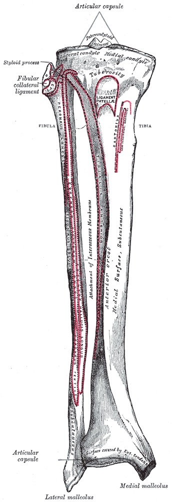
Figure 1. Bony anatomy of the proximal tibia: the lateral and medial condyles, the intercondylar eminence, and the tibial tuberosity. Gray’s Anatomy plate, public domain, via Wikimedia Commons.
The neurovascular and soft-tissue anatomy dictates the surgical hazards.[7] The popliteal artery trifurcates a short distance below the joint (its bifurcation lies about 27-62 mm distal to the lateral plateau), and overdissection toward the fibula in a posterior approach endangers it; the common peroneal nerve wraps the fibular neck and is at risk in posterolateral approaches and from a retractor placed around the neck. The proximal anteromedial tibia is subcutaneous, with a thin soft-tissue cover that is easily compromised in high-energy fractures, which is why surgical timing and approach selection are central to avoiding the wound catastrophes that dominate this injury’s complication profile.[8]
Part III - Associated Injuries
The associated injuries separate the benign lateral fracture from the limb-threatening high-energy one.[9] Compartment syndrome is the dreaded complication, concentrated in high-energy patterns: Wahlquist found a 67 % rate when a medial fracture entered the joint lateral to the spines and exited the medial metaphysis, and although series disagree on whether Schatzker IV (53 % in Stark’s data) or Schatzker VI is the higher risk, the rule is serial examination, a low threshold for compartment pressures in the obtunded patient, and fasciotomy on clinical grounds (severe pain on passive stretch is the sensitive sign; paraesthesia and paralysis are late). Vascular injury accompanies the medial fracture-dislocation, screened with the ankle-brachial index (a value below 0.9 prompts CT angiography). The common peroneal nerve may be injured. Meniscal tears (about 80 %) and ligament injuries (about 40 %) are common on MRI (the lateral meniscus with the lateral split-depression in particular), but knee stability is examined only after the fracture is fixed, because pre-fixation laxity is usually just loss of bony support. Avulsion fractures (Segond, reverse Segond) flag cruciate injury.[10]
Part IV - Imaging
Plain radiographs (AP, lateral, and a 10-15° caudal “plateau” view that profiles the articular surface) make the diagnosis, but CT is essential.[11] CT with coronal, sagittal, and three-dimensional reconstruction characterises the articular fragments, reveals depression and posterior shear that plain films understate, improves the reliability of classification, and frequently changes the operative plan; it is best obtained after a spanning external fixator or splint has restored length. CT angiography is added when the ankle-brachial index is below 0.9. MRI is more sensitive than CT for meniscal and ligamentous injury but is not a routine acute study and can be over-sensitive, so management decisions rest on the intraoperative stability examination after fixation.[12]
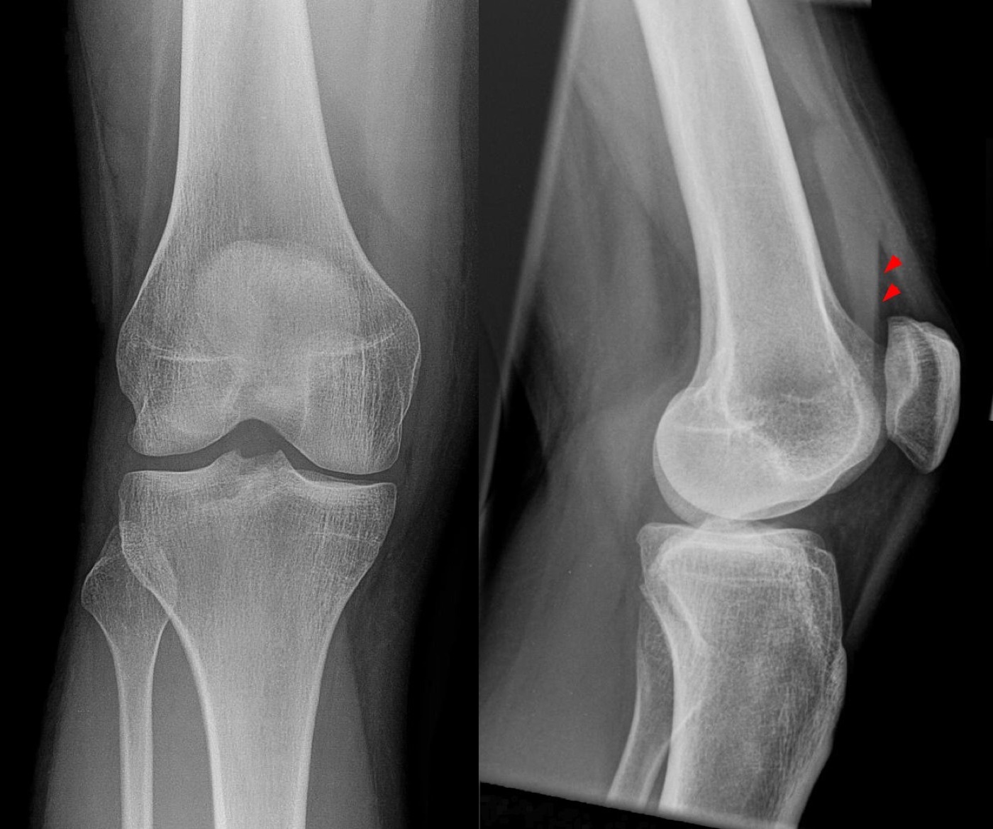
Figure 2. Occult tibial plateau fracture: a lipohaemarthrosis (fat-fluid level, arrowheads) in the suprapatellar recess signals an intra-articular fracture despite a near-normal AP view. Image by Hellerhoff, CC BY-SA 3.0, via Wikimedia Commons.
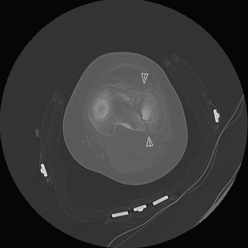
Figure 3. Axial CT of the proximal tibia showing a fracture line through the plateau (arrowheads); CT defines fragment geometry and articular involvement. Image by LottieLattes, CC BY-SA 4.0, via Wikimedia Commons.
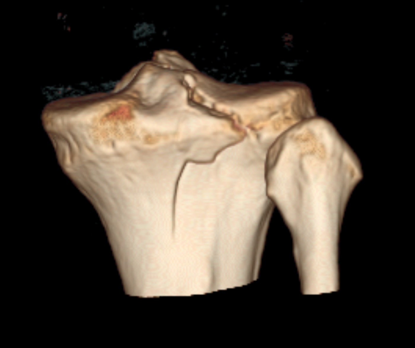
Figure 4. 3D CT reconstruction of a tibial plateau fracture, demonstrating the split through the articular surface. Image by James Heilman, MD, CC BY-SA 4.0, via Wikimedia Commons.
Part V - Classification
The Schatzker classification is the most widely used.[13] Type I is a pure lateral split (a wedge, in young dense bone); type II a lateral split-depression (the commonest operative fracture); type III a pure central lateral depression (in the osteoporotic elderly, and probably rare in its pure form); these three are lateral, lower-energy fractures. Type IV is a medial plateau fracture, a high-energy fracture-dislocation with the worst prognosis and the highest risk of vascular injury; type V is bicondylar with the metaphysis and diaphysis still in continuity; and type VI is a plateau fracture with metaphyseal-diaphyseal dissociation, the highest-energy pattern with the greatest soft-tissue and compartment-syndrome risk. The classification’s weakness is that it is AP-radiograph-based and misses posterior (coronal-shear) fragments.[14]
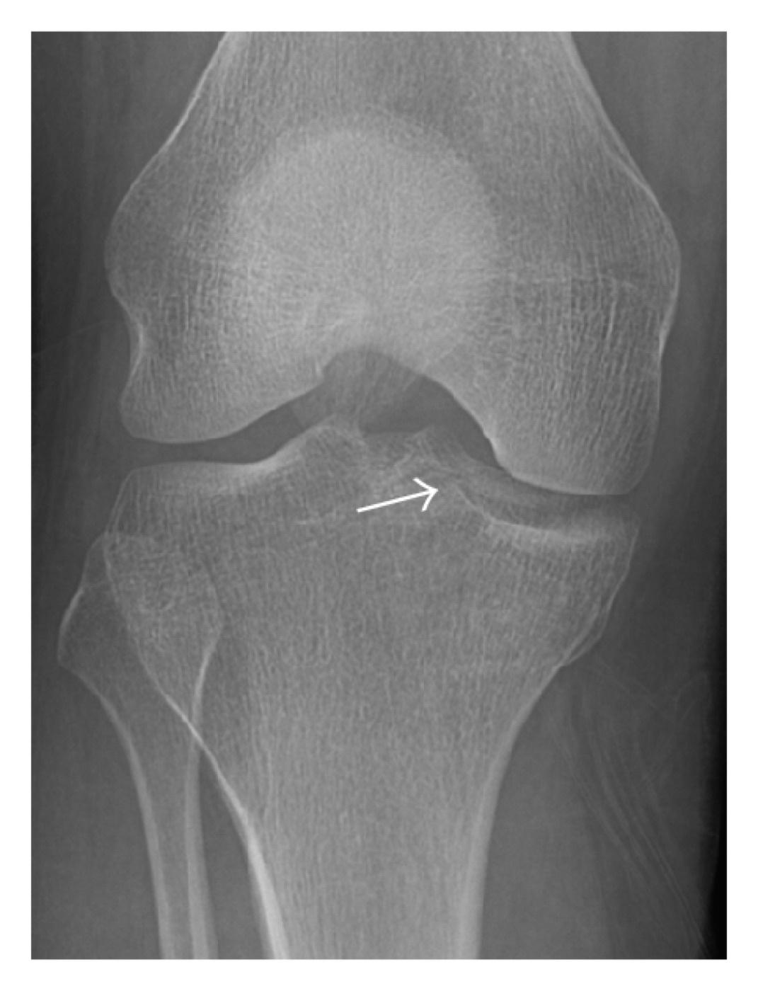
Figure 5. Subtle lateral tibial plateau fracture (arrow) on an AP knee radiograph, the kind easily missed without CT. Image by Jarraya et al., CC BY 3.0, via Wikimedia Commons.
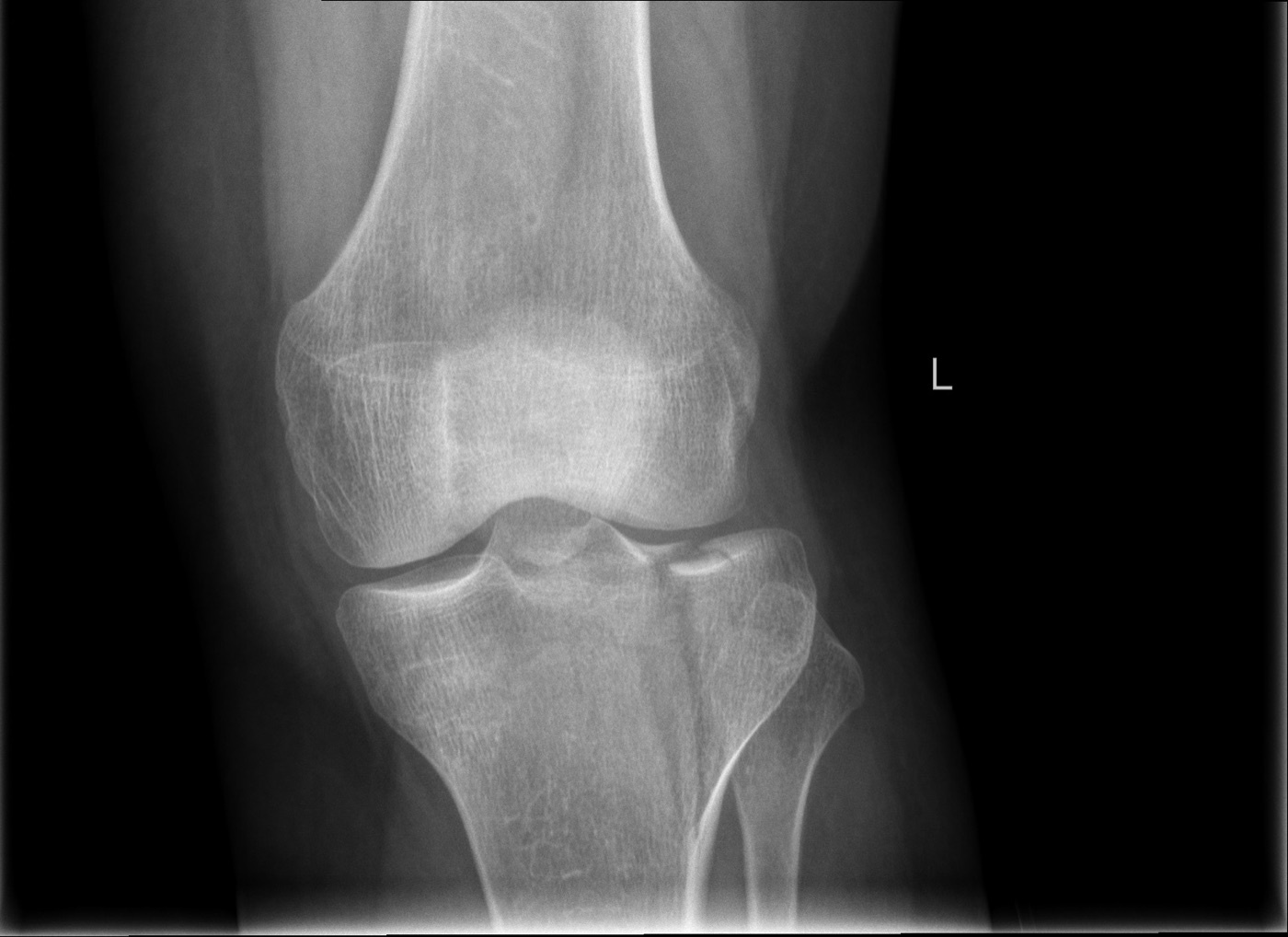
Figure 6. AP knee radiograph showing a lateral tibial plateau fracture with articular depression. Image by LottieLattes, CC BY-SA 4.0, via Wikimedia Commons.
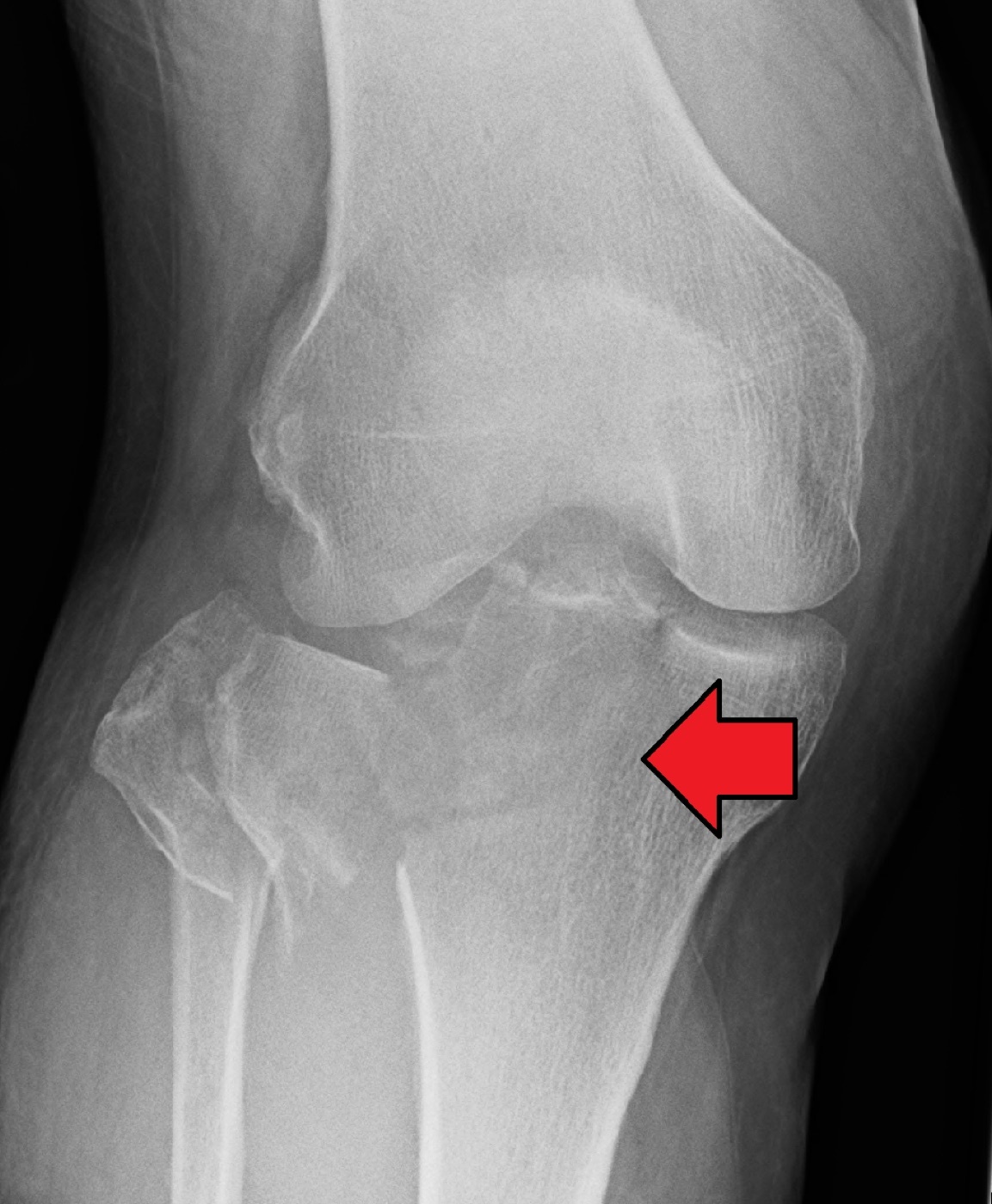
Figure 7. Severe, comminuted, displaced tibial plateau fracture (arrow) on a lateral radiograph, a high-energy injury pattern. Image by James Heilman, MD, CC BY-SA 4.0, via Wikimedia Commons.
Two further systems complete the picture.[15] The AO/OTA system codes the proximal tibia as 41: 41A extra-articular (not a true plateau fracture), 41B partial articular (part of the joint remains continuous with the shaft, broadly Schatzker I-IV), and 41C complete articular (the joint is separated from the shaft, bicondylar, Schatzker V-VI). The three-column (Luo) classification, built on axial CT, divides the plateau into medial, lateral, and posterior columns and was developed precisely because the posterior column carries more outcome weight than the older systems recognised; a column counts as fractured when articular depression is accompanied by a cortical split, and recognising the posterior column has reduced rates of reduction loss.[16]
Part VI - Treatment Principles and Nonoperative Care
The goals of treatment are to restore the articular surface, the mechanical axis, and the condylar width, to fix stably enough for early motion, and to preserve the meniscus.[17] Nonoperative treatment suits the truly nondisplaced or minimally displaced fracture with a stable knee, the low-demand or medically unfit patient, and is delivered in a hinged brace with protected weight bearing for roughly 4-8 weeks. The reported thresholds vary (Honkonen tolerated 5 mm of widening and 3 mm of step-off, Waddell under 10 mm), and the modern teaching is that fixed millimetre cut-offs are simplistic: the decision rests on whether the fracture pattern will deform and whether the knee is stable, with medial fractures watched far more closely because they collapse into varus, which is poorly tolerated. The classic operative articular step-off goal of under 2-3 mm is standard teaching, but the stronger predictors of arthritis are malalignment, meniscectomy, and instability rather than the step itself.[18]
Part VII - Operative Treatment
7.1 The lateral fractures (Schatzker I-III)
A Schatzker I split is often fixed with percutaneous lag screws alone (commonly 6.5 mm, placed parallel to the joint within about 1.5 cm of the subchondral surface), reserving a lateral buttress plate for a large or comminuted split.[19] A Schatzker II split-depression is the commonest operation: through an anterolateral approach, a submeniscal arthrotomy exposes the joint, the depressed segment is elevated from below (the femoral condyle a template), the metaphyseal void is filled with graft or a bone substitute, and the reduction is held with a lateral buttress (or locked) plate and a raft of subchondral screws placed just beneath the articular surface to resist settling. A Schatzker III pure depression is elevated and supported with rafting screws (a raft technique, sometimes arthroscopically assisted), the full height of the compartment restored because these collapse into valgus. Bone-void fillers include autograft, allograft, and calcium phosphate cement, the last being stiffer in compression (and placed after the screws, since it is brittle once set).[20]
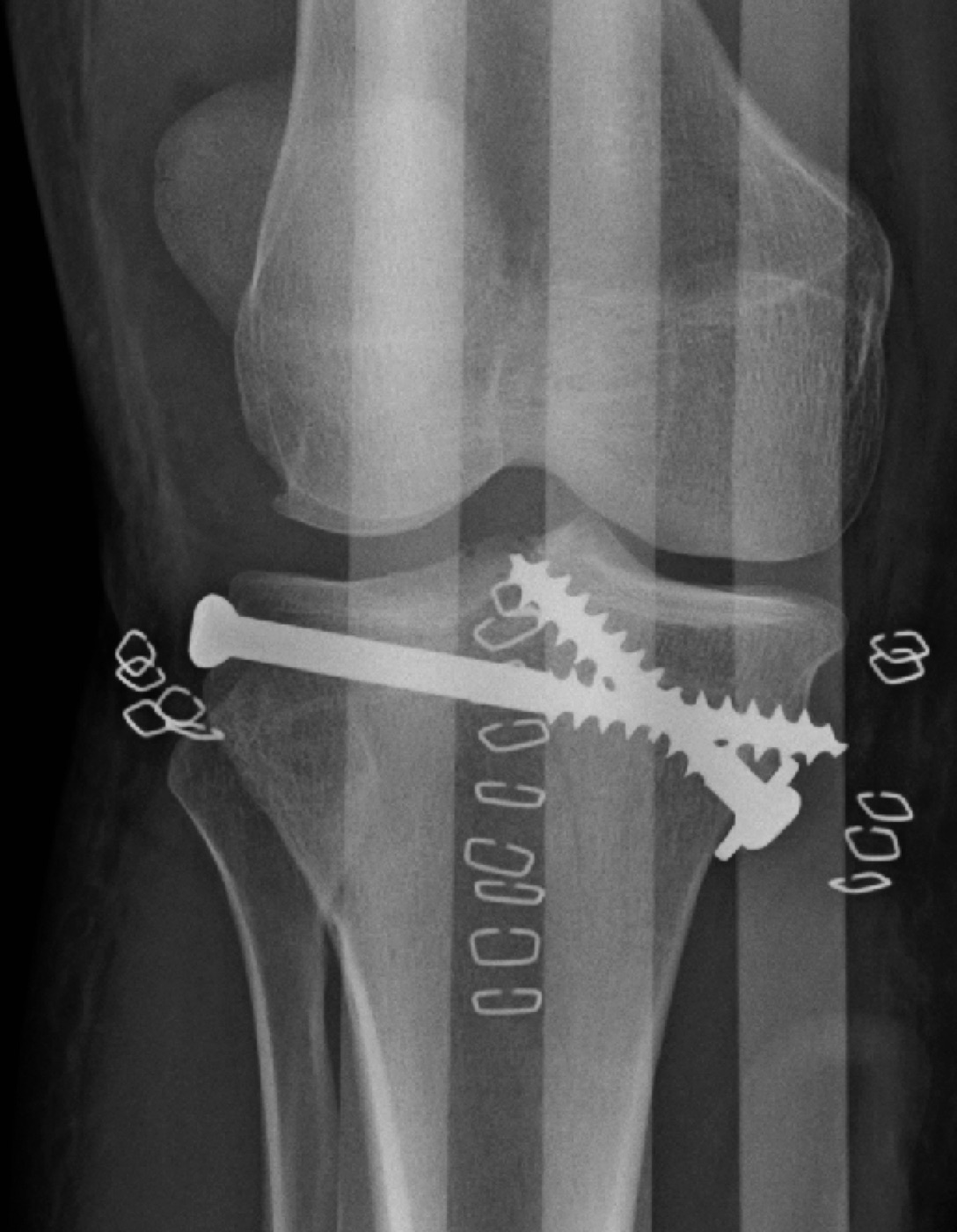
Figure 8. Post-operative radiograph after ORIF of a tibial plateau fracture: a lateral buttress plate with an interfragmentary screw. Image by James Heilman, MD, CC BY-SA 4.0, via Wikimedia Commons.
7.2 The medial fracture (Schatzker IV)
The Schatzker IV medial plateau fracture is treated through a posteromedial approach with an antiglide/buttress plate placed at the apex of the shear fragment to resist the varus collapse the loss of the medial buttress invites.[21] Because it is a high-energy fracture-dislocation, the surgeon is vigilant for vascular, nerve, and ligament injury (including the posterolateral corner, which must be repaired), and examines knee stability after fixation. The deep interval runs between the pes anserinus and the medial head of gastrocnemius, with the gastrocnemius retracted to protect the popliteal structures.[22]
7.3 The bicondylar fractures (Schatzker V-VI) and the staged protocol
The bicondylar and shaft-dissociated fractures are the hardest, and the central decisions are timing and how many plates.[23] High-energy fractures with a compromised soft-tissue envelope are managed in a staged protocol: an initial knee-spanning external fixator restores length and alignment while the swelling and fracture blisters settle (the return of skin wrinkling, at roughly 10-14 days, signals that definitive surgery is safe). Definitive fixation is then performed through two separate limited incisions (an anterolateral and a posteromedial) about 180° apart, never through a single extensile midline incision: the historical “dead bone sandwich” of dual plates through one incision produced infection rates of 73-87 %. A displaced posteromedial coronal-shear fragment must be buttressed directly (a lateral locked plate alone fails to hold it, with high subsidence), so dual plating is used when the medial side is comminuted or displaced; a lateral locked plate alone is reserved for the fracture whose medial column is minimally displaced. A circular or hybrid external fixator is the alternative for the most severe soft-tissue injuries, with pins kept at least 15 mm from the joint to avoid septic arthritis.[24]
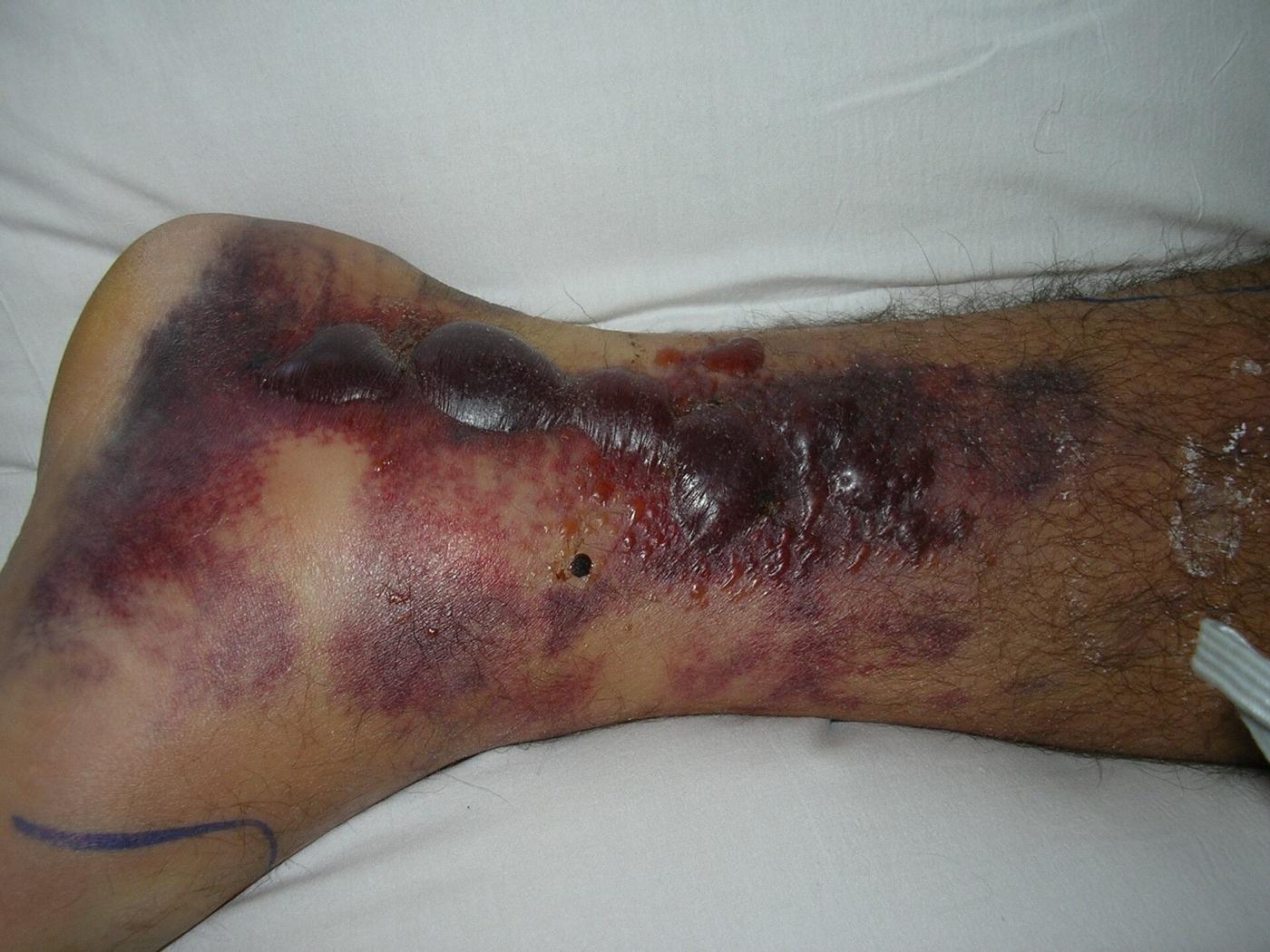
Figure 9. Haemorrhagic fracture blisters over a fractured lower limb, a compromised soft-tissue envelope that mandates staged management. Image by Cindy L. Budge, public domain, via Wikimedia Commons.
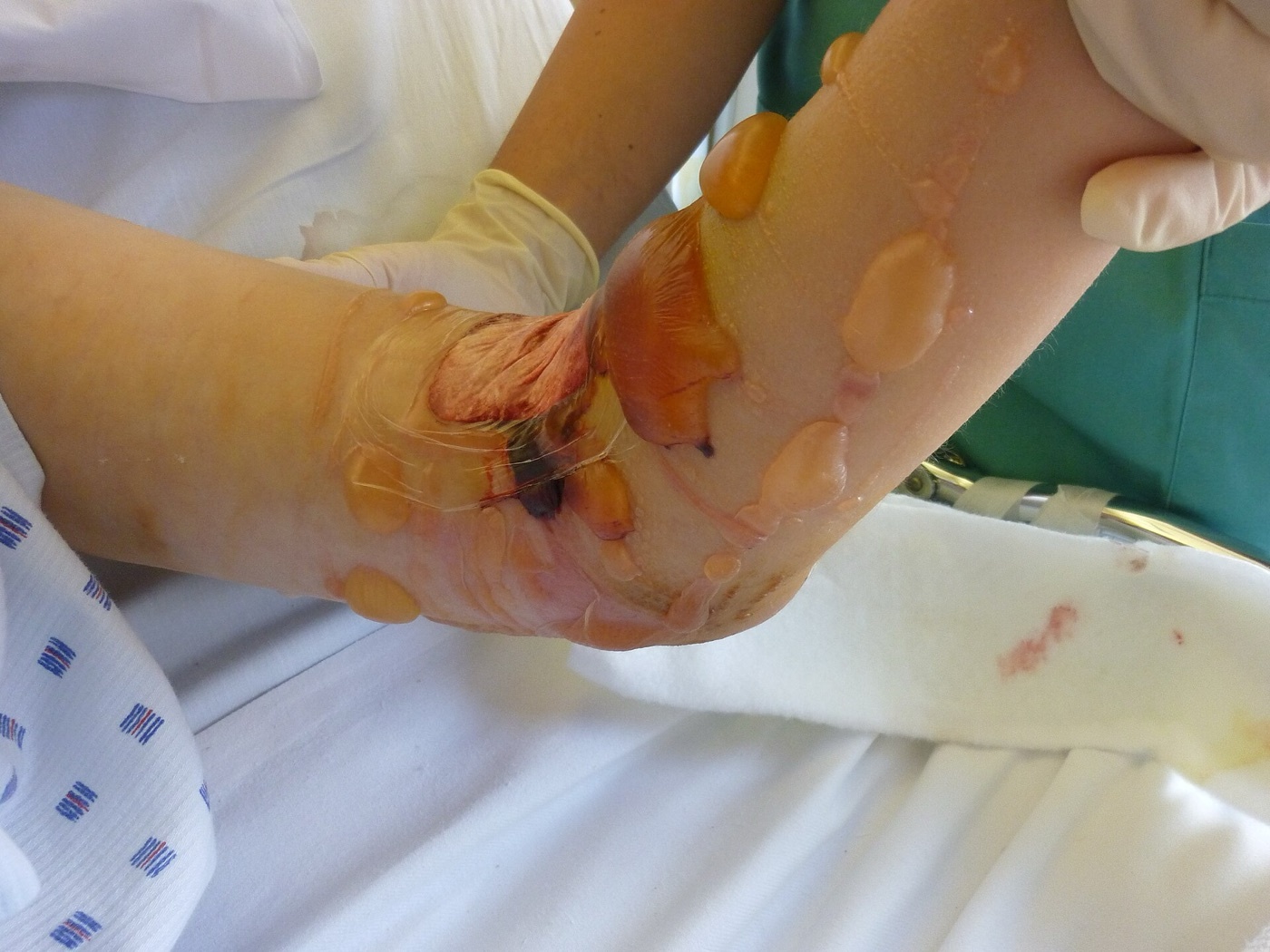
Figure 10. Extensive soft-tissue swelling and blistering of the lower leg, the degree of soft-tissue compromise that mandates delayed definitive fixation. Image by Lklundin, CC BY-SA 4.0, via Wikimedia Commons.
After fixation, the knee is moved early (immobilisation beyond about two weeks produces stiffness), with weight bearing protected until the fracture shows meaningful healing at roughly 6-12 weeks.[25] Throughout, the operative philosophy is the most limited approach that achieves an anatomic articular reduction, a restored axis, and enough stability for early motion, and the failure to achieve stable fixation that allows early motion gives a worse result than nonoperative treatment.[26]
Part VIII - Complications
Wound breakdown and deep infection are the signature complications, concentrated in high-energy bicondylar fractures.[27] The historical 73-87 % infection of single-incision dual plating drove the modern staged, limited-approach, dual-incision strategy, which has cut infection to roughly 3-11 % in contemporary series; the commonest organism is MRSA, and independent risk factors are open fracture, compartment syndrome, and the complex bicondylar pattern. Compartment syndrome complicates around 7-10 % overall (higher in Schatzker VI). Loss of reduction is common, especially in the elderly (about 31 % overall, rising to 79 % over age 60) and when a coronal fragment is fixed with a lateral plate alone. Post-traumatic arthritis correlates with malalignment, meniscectomy, and instability far more than with the articular step (about 36 % after high-energy fractures at 5-11 years, and 68 % over age 60), and conversion to total knee arthroplasty is several times more likely than in the matched population (about 7.3 % at 10 years, a 5.3-fold increase; Wasserstein). Other complications are stiffness (manipulation or arthroscopic lysis if 90° is not reached by 12 weeks), nonunion at the metaphyseal-diaphyseal junction, malunion (valgus from the lateral side, varus from the medial), prominent hardware on the subcutaneous tibia, peroneal palsy, and venous thromboembolism (standard teaching).[28]
Part IX - Outcomes and Controversies
Outcome tracks the fracture, and the gradient is clear: the lateral split has the best results, the medial and bicondylar fractures the worst, and varus is tolerated less well than valgus.[29] The proximal tibia is, encouragingly, more forgiving of articular incongruity than the ankle or hip, so severe arthrosis is less frequent than after a pilon or acetabular fracture. The live controversies are: how much articular step-off is acceptable (with the evidence pointing to alignment, meniscus, and stability mattering more than the step); whether a lateral locked plate alone suffices for a bicondylar fracture (it does not when a posteromedial coronal fragment is present); whether to fix or primarily replace the osteoporotic elderly plateau; and whether early definitive fixation is safe in selected high-energy patterns rather than the staged protocol. The single most repeated lesson is that knee stability and limb alignment, not millimetres of step-off, determine the long-term result.[30]
Part X - A Synthesis: How to Reason About the Tibial Plateau Fracture
Approach the plateau fracture by first deciding which fracture it is. A lateral, lower-energy fracture (Schatzker I-III) is reduced (a split with screws, a depression elevated and grafted with a raft of subchondral screws and a lateral buttress plate) and does well. A medial fracture (Schatzker IV) is a high-energy fracture-dislocation: buttress it from the posteromedial side and hunt for the vascular, nerve, and posterolateral-corner injuries that travel with it. A bicondylar fracture (Schatzker V-VI) is, above all, a soft-tissue problem: stage it in a spanning external fixator, wait for the wrinkle sign, then fix it through two limited incisions, buttressing any posteromedial coronal fragment directly rather than trusting a lateral plate alone. Get the CT, restore the mechanical axis and the meniscus, fix stably enough to move the knee early, and watch relentlessly for compartment syndrome. And hold the central truth that, for this fracture, alignment and stability matter more than a perfect articular step.
References
-
Rockwood & Green’s Fractures in Adults, pp.4256, 4282, 4291, 4335 (lateral valgus “bumper” fractures vs high-energy medial fracture-dislocations; the soft-tissue envelope governing timing; arthrosis correlating with malalignment and meniscectomy rather than step-off, Rademakers 27 % vs 9 %; the elevate-graft-raft-buttress principle); AO Principles of Fracture Management, pp.898, 900, 916 (medial plateau fractures more severe and associated with fracture-dislocations; the three-column concept; knee stability the most important factor for outcome and the causes of post-traumatic arthritis).
-
Rockwood & Green’s Fractures in Adults, pp.4256, 4282, 4291, 4335 (lateral valgus “bumper” fractures vs high-energy medial fracture-dislocations; the soft-tissue envelope governing timing; arthrosis correlating with malalignment and meniscectomy rather than step-off, Rademakers 27 % vs 9 %; the elevate-graft-raft-buttress principle); AO Principles of Fracture Management, pp.898, 900, 916 (medial plateau fractures more severe and associated with fracture-dislocations; the three-column concept; knee stability the most important factor for outcome and the causes of post-traumatic arthritis).
-
Rockwood & Green’s Fractures in Adults, pp.4255, 4256, 4257 (1-2 % of long-bone fractures, bimodal distribution, the valgus mechanism with 5-7° knee valgus producing the lateral “bumper fracture”, the higher-energy varus medial-plateau fracture often a fracture-dislocation, and axial load producing bicondylar patterns; the MCL acting as a hinge so the lateral plateau fractures); AO Principles of Fracture Management, pp.897, 898 (proximal tibia ≈18.6 % of tibial fractures; medial plateau fractures sheared off en bloc and associated with more severe injuries and fracture-dislocations).
-
Rockwood & Green’s Fractures in Adults, pp.4255, 4256, 4257 (1-2 % of long-bone fractures, bimodal distribution, the valgus mechanism with 5-7° knee valgus producing the lateral “bumper fracture”, the higher-energy varus medial-plateau fracture often a fracture-dislocation, and axial load producing bicondylar patterns; the MCL acting as a hinge so the lateral plateau fractures); AO Principles of Fracture Management, pp.897, 898 (proximal tibia ≈18.6 % of tibial fractures; medial plateau fractures sheared off en bloc and associated with more severe injuries and fracture-dislocations).
-
Rockwood & Green’s Fractures in Adults, pp.4282, 4283 (medial plateau larger/concave/stronger/load-bearing, lateral smaller/convex/higher/weaker so lateral fractures commoner; posterior slope with wide variation; greater lateral meniscal coverage; the fibula buttressing the lateral plateau so a fibular fracture indicates a more severe injury); AO Principles of Fracture Management, pp.898, 900 (the asymmetric plateaus, the three-column concept of medial/lateral/posterior columns guiding approach and buttress plating, the posteromedial ridge as the strongest part). The ~10° posterior tibial slope is standard teaching; the source gives slope ranges rather than the single figure.
-
Rockwood & Green’s Fractures in Adults, pp.4282, 4283 (medial plateau larger/concave/stronger/load-bearing, lateral smaller/convex/higher/weaker so lateral fractures commoner; posterior slope with wide variation; greater lateral meniscal coverage; the fibula buttressing the lateral plateau so a fibular fracture indicates a more severe injury); AO Principles of Fracture Management, pp.898, 900 (the asymmetric plateaus, the three-column concept of medial/lateral/posterior columns guiding approach and buttress plating, the posteromedial ridge as the strongest part). The ~10° posterior tibial slope is standard teaching; the source gives slope ranges rather than the single figure.
-
AO Principles of Fracture Management, pp.900, 903, 905 (the popliteal artery trifurcation 27-62 mm distal to the lateral plateau, the common peroneal nerve at the fibular neck at risk in posterolateral approaches and from a fibular-neck retractor); Rockwood & Green’s Fractures in Adults, pp.4281, 4283 (the peroneal nerve on the posterior fibular neck, the subcutaneous anteromedial tibia and thin soft-tissue cover at risk in high-energy fractures).
-
AO Principles of Fracture Management, pp.900, 903, 905 (the popliteal artery trifurcation 27-62 mm distal to the lateral plateau, the common peroneal nerve at the fibular neck at risk in posterolateral approaches and from a fibular-neck retractor); Rockwood & Green’s Fractures in Adults, pp.4281, 4283 (the peroneal nerve on the posterior fibular neck, the subcutaneous anteromedial tibia and thin soft-tissue cover at risk in high-energy fractures).
-
Rockwood & Green’s Fractures in Adults, pp.4258, 4259, 4260, 4266, 4270, 4290 (compartment syndrome highest in high-energy/medial patterns, Wahlquist 67 %, Stark Schatzker IV 53 % vs VI 18 %, serial exams and clinical fasciotomy; ABI normal >0.9 and <0.9 → CT arteriogram; the peroneal nerve; meniscal/ligament injury frequency and examining stability after fixation; Segond/reverse-Segond as cruciate-injury markers); AO Principles of Fracture Management, pp.897, 898 (severe pain on passive stretch the sensitive compartment-syndrome sign with paraesthesia/paralysis late, single-compartment presentation; MRI 80 % meniscal and 40 % ligamentous tears; absent pulses indicating arterial injury).
-
Rockwood & Green’s Fractures in Adults, pp.4258, 4259, 4260, 4266, 4270, 4290 (compartment syndrome highest in high-energy/medial patterns, Wahlquist 67 %, Stark Schatzker IV 53 % vs VI 18 %, serial exams and clinical fasciotomy; ABI normal >0.9 and <0.9 → CT arteriogram; the peroneal nerve; meniscal/ligament injury frequency and examining stability after fixation; Segond/reverse-Segond as cruciate-injury markers); AO Principles of Fracture Management, pp.897, 898 (severe pain on passive stretch the sensitive compartment-syndrome sign with paraesthesia/paralysis late, single-compartment presentation; MRI 80 % meniscal and 40 % ligamentous tears; absent pulses indicating arterial injury).
-
Rockwood & Green’s Fractures in Adults, pp.4261, 4263, 4265, 4266 (AP/lateral and the 10-15° caudal plateau view; CT routinely obtained, demonstrating more displacement than plain films, improving classification reliability and changing operative plans; CT angiography for ABI <0.9; MRI more sensitive for meniscal/ligament injury but of unestablished treatment value); AO Principles of Fracture Management, p.898 (plain films insufficient; preoperative CT with coronal/sagittal/3-D reconstruction the standard, ideally after reduction/splinting; MRI not a routine acute study and can be oversensitive, decisions resting on intraoperative stability).
-
Rockwood & Green’s Fractures in Adults, pp.4261, 4263, 4265, 4266 (AP/lateral and the 10-15° caudal plateau view; CT routinely obtained, demonstrating more displacement than plain films, improving classification reliability and changing operative plans; CT angiography for ABI <0.9; MRI more sensitive for meniscal/ligament injury but of unestablished treatment value); AO Principles of Fracture Management, p.898 (plain films insufficient; preoperative CT with coronal/sagittal/3-D reconstruction the standard, ideally after reduction/splinting; MRI not a routine acute study and can be oversensitive, decisions resting on intraoperative stability).
-
Rockwood & Green’s Fractures in Adults, pp.4266, 4267, 4268, 4269, 4270, 4271, 4272 (Schatzker I lateral split, II lateral split-depression the commonest operative pattern, III pure lateral depression in the elderly and rare in pure form, IV medial plateau as a high-energy fracture-dislocation, V bicondylar with intact metaphysis, VI metaphyseal-diaphyseal dissociation; the AP-based limitation missing posterior shear fragments); Miller’s Review of Orthopaedics, p.926 (type IV highest vascular-injury risk; type V bicondylar with intact metaphysis, type VI with metaphyseal-diaphyseal dissociation).
-
Rockwood & Green’s Fractures in Adults, pp.4266, 4267, 4268, 4269, 4270, 4271, 4272 (Schatzker I lateral split, II lateral split-depression the commonest operative pattern, III pure lateral depression in the elderly and rare in pure form, IV medial plateau as a high-energy fracture-dislocation, V bicondylar with intact metaphysis, VI metaphyseal-diaphyseal dissociation; the AP-based limitation missing posterior shear fragments); Miller’s Review of Orthopaedics, p.926 (type IV highest vascular-injury risk; type V bicondylar with intact metaphysis, type VI with metaphyseal-diaphyseal dissociation).
-
Rockwood & Green’s Fractures in Adults, pp.4272, 4273, 4276, 4277 (AO/OTA 41A extra-articular, 41B partial articular, 41C complete articular; the three-column Luo classification on axial CT dividing the plateau into medial/lateral/posterior columns, a column fractured when depression plus a cortical split, and better outcomes when the posterior column is recognised and addressed); AO Principles of Fracture Management, pp.900, 901 (41A/B/C; the three-column concept).
-
Rockwood & Green’s Fractures in Adults, pp.4272, 4273, 4276, 4277 (AO/OTA 41A extra-articular, 41B partial articular, 41C complete articular; the three-column Luo classification on axial CT dividing the plateau into medial/lateral/posterior columns, a column fractured when depression plus a cortical split, and better outcomes when the posterior column is recognised and addressed); AO Principles of Fracture Management, pp.900, 901 (41A/B/C; the three-column concept).
-
Rockwood & Green’s Fractures in Adults, pp.4284, 4286, 4287, 4335 (operative goals of articular reduction, alignment, condylar width, stable fixation for early motion, meniscal preservation; nonoperative indications and hinged-brace/protected weight-bearing; Honkonen 5 mm widening and 3 mm step-off and Waddell <10 mm; millimetre cut-offs simplistic and pattern/stability-driven, medial fractures collapsing into varus; arthrosis correlating with malalignment and meniscectomy, Rademakers 27 % vs 9 %); AO Principles of Fracture Management, pp.901, 916 (operative indications; knee stability the most important outcome factor and the causes of post-traumatic arthritis). The “<2-3 mm” articular step-off operative target is standard teaching, the sources stating malreduction as a step or gap >2 mm and Honkonen’s tolerated 3 mm rather than a single universal goal.
-
Rockwood & Green’s Fractures in Adults, pp.4284, 4286, 4287, 4335 (operative goals of articular reduction, alignment, condylar width, stable fixation for early motion, meniscal preservation; nonoperative indications and hinged-brace/protected weight-bearing; Honkonen 5 mm widening and 3 mm step-off and Waddell <10 mm; millimetre cut-offs simplistic and pattern/stability-driven, medial fractures collapsing into varus; arthrosis correlating with malalignment and meniscectomy, Rademakers 27 % vs 9 %); AO Principles of Fracture Management, pp.901, 916 (operative indications; knee stability the most important outcome factor and the causes of post-traumatic arthritis). The “<2-3 mm” articular step-off operative target is standard teaching, the sources stating malreduction as a step or gap >2 mm and Honkonen’s tolerated 3 mm rather than a single universal goal.
-
Rockwood & Green’s Fractures in Adults, pp.4292, 4299, 4301, 4302, 4305, 4306, 4307, 4308 (Schatzker I percutaneous 6.5-mm screws parallel to and within 1.5 cm of the joint, buttress for large splits; Schatzker II anterolateral submeniscal arthrotomy, elevation from below with the femoral condyle as template, void filling, lateral buttress/locked plate and rafting subchondral screws, multiple small screws better than a few large; Schatzker III elevation with rafting screws restoring compartment height; calcium phosphate cement stiffer in compression, placed after screws); AO Principles of Fracture Management, pp.902, 905, 910, 913 (lag/raft screws and buttress plating, rafting subchondral 2.7/3.5-mm screws, pure depression treatable by screws alone, the lateral column buttressed to prevent valgus, and depressed fractures collapsing without restored compartment height).
-
Rockwood & Green’s Fractures in Adults, pp.4292, 4299, 4301, 4302, 4305, 4306, 4307, 4308 (Schatzker I percutaneous 6.5-mm screws parallel to and within 1.5 cm of the joint, buttress for large splits; Schatzker II anterolateral submeniscal arthrotomy, elevation from below with the femoral condyle as template, void filling, lateral buttress/locked plate and rafting subchondral screws, multiple small screws better than a few large; Schatzker III elevation with rafting screws restoring compartment height; calcium phosphate cement stiffer in compression, placed after screws); AO Principles of Fracture Management, pp.902, 905, 910, 913 (lag/raft screws and buttress plating, rafting subchondral 2.7/3.5-mm screws, pure depression treatable by screws alone, the lateral column buttressed to prevent valgus, and depressed fractures collapsing without restored compartment height).
-
Rockwood & Green’s Fractures in Adults, pp.4308, 4309, 4310, 4311 (posteromedial approach and antiglide plate at the apex for the medial fracture, the fracture-dislocation nature with vascular/nerve/ligament vigilance, the interval between pes anserinus and medial gastrocnemius protecting the popliteal structures); AO Principles of Fracture Management, p.911 (medial column from a varus-extension force, often a fracture-dislocation, a medial buttress plate to prevent varus, and an associated posterolateral corner injury repaired early).
-
Rockwood & Green’s Fractures in Adults, pp.4308, 4309, 4310, 4311 (posteromedial approach and antiglide plate at the apex for the medial fracture, the fracture-dislocation nature with vascular/nerve/ligament vigilance, the interval between pes anserinus and medial gastrocnemius protecting the popliteal structures); AO Principles of Fracture Management, p.911 (medial column from a varus-extension force, often a fracture-dislocation, a medial buttress plate to prevent varus, and an associated posterolateral corner injury repaired early).
-
Rockwood & Green’s Fractures in Adults, pp.4297, 4311, 4312, 4321, 4322, 4328, 4331 (the staged protocol with a spanning external fixator and the wrinkle sign reducing wound complications; dual limited anterolateral and posteromedial incisions ~180° apart vs the single-incision “dead bone sandwich” with 73-87 % infection; the posteromedial coronal fragment requiring direct buttressing with high subsidence if only a lateral locked plate is used; external fixation with pins ≥15 mm from the joint); AO Principles of Fracture Management, pp.901, 902, 909, 913 (staged management for open/vascular/severe soft-tissue/polytrauma, definitive surgery once swelling recovers at 10-14 days, hybrid/ring external fixation, and converting a type C to a type B by reducing the medial fragment first).
-
Rockwood & Green’s Fractures in Adults, pp.4297, 4311, 4312, 4321, 4322, 4328, 4331 (the staged protocol with a spanning external fixator and the wrinkle sign reducing wound complications; dual limited anterolateral and posteromedial incisions ~180° apart vs the single-incision “dead bone sandwich” with 73-87 % infection; the posteromedial coronal fragment requiring direct buttressing with high subsidence if only a lateral locked plate is used; external fixation with pins ≥15 mm from the joint); AO Principles of Fracture Management, pp.901, 902, 909, 913 (staged management for open/vascular/severe soft-tissue/polytrauma, definitive surgery once swelling recovers at 10-14 days, hybrid/ring external fixation, and converting a type C to a type B by reducing the medial fragment first).
-
Rockwood & Green’s Fractures in Adults, pp.4299, 4322 (early motion since immobilisation beyond two weeks causes stiffness, weight bearing protected to 6-12 weeks, and the philosophy of the most limited approach achieving articular reduction, alignment, and stability for early motion); AO Principles of Fracture Management, pp.914, 916 (toe-touch weight bearing for 6-8 weeks or 10-12 in the highest-energy fractures, and that failure to achieve stable fixation allowing early mobilisation gives poorer outcomes than nonoperative treatment).
-
Rockwood & Green’s Fractures in Adults, pp.4299, 4322 (early motion since immobilisation beyond two weeks causes stiffness, weight bearing protected to 6-12 weeks, and the philosophy of the most limited approach achieving articular reduction, alignment, and stability for early motion); AO Principles of Fracture Management, pp.914, 916 (toe-touch weight bearing for 6-8 weeks or 10-12 in the highest-energy fractures, and that failure to achieve stable fixation allowing early mobilisation gives poorer outcomes than nonoperative treatment).
-
Rockwood & Green’s Fractures in Adults, pp.4328, 4330, 4331, 4332, 4333, 4334, 4335 (single-incision dual plating infection 73-87 % falling to ~3-11 % with the staged limited-approach strategy, MRSA commonest, risk factors open/compartment-syndrome/bicondylar; loss of reduction 31 % overall and 79 % over 60 and with coronal fragments under a lateral plate; arthrosis correlating with malalignment/meniscectomy not step-off, ~36 % at 5-11 years and 68 % over 60; stiffness with lysis if 90° not reached by 12 weeks; nonunion at the metaphyseal-diaphyseal junction; valgus/varus malunion; prominent subcutaneous hardware; the ~7.3 % conversion to total knee arthroplasty at 10 years, a 5.3-fold increase, from Wasserstein et al., RG pp.4279-4280); AO Principles of Fracture Management, p.916 (wound complications the major early problem, nonunion and deep infection commoner after dual-plate ORIF of high-energy bicondylar fractures, arthrofibrosis with lysis if 90° not reached by 12 weeks). Venous-thromboembolism prophylaxis is standard teaching not discussed in these extracts.
-
Rockwood & Green’s Fractures in Adults, pp.4328, 4330, 4331, 4332, 4333, 4334, 4335 (single-incision dual plating infection 73-87 % falling to ~3-11 % with the staged limited-approach strategy, MRSA commonest, risk factors open/compartment-syndrome/bicondylar; loss of reduction 31 % overall and 79 % over 60 and with coronal fragments under a lateral plate; arthrosis correlating with malalignment/meniscectomy not step-off, ~36 % at 5-11 years and 68 % over 60; stiffness with lysis if 90° not reached by 12 weeks; nonunion at the metaphyseal-diaphyseal junction; valgus/varus malunion; prominent subcutaneous hardware; the ~7.3 % conversion to total knee arthroplasty at 10 years, a 5.3-fold increase, from Wasserstein et al., RG pp.4279-4280); AO Principles of Fracture Management, p.916 (wound complications the major early problem, nonunion and deep infection commoner after dual-plate ORIF of high-energy bicondylar fractures, arthrofibrosis with lysis if 90° not reached by 12 weeks). Venous-thromboembolism prophylaxis is standard teaching not discussed in these extracts.
-
Rockwood & Green’s Fractures in Adults, pp.4327, 4328, 4335, 4337, 4338 (lateral split the best and medial/bicondylar the worst outcomes, varus less tolerated than valgus; the proximal tibia more forgiving than the ankle/hip; the controversies over acceptable step-off, lateral locked plate vs dual plating with the coronal fragment, fixation vs arthroplasty in the elderly, and staged vs early definitive care); AO Principles of Fracture Management, p.916 (knee stability the most important factor for long-term outcome and arthroplasty being explored in the elderly with unclear indications).
-
Rockwood & Green’s Fractures in Adults, pp.4327, 4328, 4335, 4337, 4338 (lateral split the best and medial/bicondylar the worst outcomes, varus less tolerated than valgus; the proximal tibia more forgiving than the ankle/hip; the controversies over acceptable step-off, lateral locked plate vs dual plating with the coronal fragment, fixation vs arthroplasty in the elderly, and staged vs early definitive care); AO Principles of Fracture Management, p.916 (knee stability the most important factor for long-term outcome and arthroplasty being explored in the elderly with unclear indications).
-
Rockwood & Green’s Fractures in Adults, pp.4256, 4257; AO Principles of Fracture Management, p.898.
-
Rockwood & Green’s Fractures in Adults, pp.4267-4271; Miller’s Review of Orthopaedics, p.926.
-
Rockwood & Green’s Fractures in Adults, pp.4261, 4263; AO Principles of Fracture Management, p.898.
-
Rockwood & Green’s Fractures in Adults, pp.4276, 4277; AO Principles of Fracture Management, pp.900, 901.
-
Rockwood & Green’s Fractures in Adults, pp.4259, 4260, 4270; AO Principles of Fracture Management, p.897.
-
Rockwood & Green’s Fractures in Adults, pp.4302, 4305, 4306; AO Principles of Fracture Management, pp.910, 913.
-
Rockwood & Green’s Fractures in Adults, pp.4308, 4309; AO Principles of Fracture Management, p.911.
-
Rockwood & Green’s Fractures in Adults, pp.4297, 4328; AO Principles of Fracture Management, pp.901, 902.
-
Rockwood & Green’s Fractures in Adults, pp.4311, 4328, 4331.
-
Rockwood & Green’s Fractures in Adults, pp.4328, 4332.
-
Rockwood & Green’s Fractures in Adults, p.4335; AO Principles of Fracture Management, p.916.
-
Rockwood & Green’s Fractures in Adults, pp.4328, 4330, 4332, 4335; AO Principles of Fracture Management, p.916.