Contents
- Orientation: The Commonest Long-Bone Fracture, Governed by Its Soft Tissues
- Part I - Definition, Epidemiology, and Mechanism
- Part II - Applied Anatomy
- Part III - Compartment Syndrome and Neurovascular Injury
- Part IV - Open Fractures
- Part V - Imaging and Classification
- Part VI - Nonoperative Treatment
- Part VII - Reamed Locked Intramedullary Nailing
- Part VIII - Plating and External Fixation
- Part IX - Complications
- Part X - Isolated Fibula Shaft Fractures
- Part XI - Paediatric Tibial Shaft Fractures
- Part XII - A Synthesis: How to Reason About the Tibial Shaft Fracture
- References
Orientation: The Commonest Long-Bone Fracture, Governed by Its Soft Tissues
The tibial shaft fracture is the commonest long-bone fracture, and everything about it follows from the subcutaneous anteromedial border of the tibia: with no muscle over the front of the bone, even closed fractures injure the skin, and a quarter of all tibial shaft fractures are open, more often needing flap coverage than any other site.[1] Two emergencies dominate the early course: compartment syndrome (the tibial shaft is the single commonest cause) and the open fracture (where early antibiotics, debridement, and a planned orthoplastic soft-tissue reconstruction decide the outcome). The definitive treatment of most displaced shaft fractures is the reamed, locked intramedullary nail, with the technical battles fought over the entry point, the proximal and distal malalignment that a metaphyseal fracture invites, and the anterior knee pain that is the commonest nailing complication. The tibia heals slowly and has the highest nonunion rate of any bone, so the soft-tissue injury, not the fracture line, is what the surgeon must read.[2]
Part I - Definition, Epidemiology, and Mechanism
The tibial shaft is the diaphysis between the proximal and distal metaphyses, and its fracture is the commonest long-bone fracture (about 20 per 100,000 per year, highest in teenage boys at 39 per 100,000), with roughly 80 % accompanied by a fibula fracture.[3] The distribution is bimodal: low-energy torsional injuries produce spiral fractures (commoner over 50), while high-energy direct trauma produces transverse, comminuted, or segmental fractures (commoner under 30, typically vehicular, with pedestrians struck the largest group). Because the tibia is subcutaneous, open fractures are common (24-25 %, up to 63 % after motorcycle crashes), and the tibia is the bone most likely to require a IIIB flap. The tibia also has the highest nonunion rate of any bone (about 5 %).[4]
Part II - Applied Anatomy
Three anatomical facts shape the management.[5] First, the anteromedial tibial border is subcutaneous along its whole length: a thin soft-tissue envelope explains the high open-fracture rate and means that a medial plate, which must be perfectly contoured to the bone, risks skin necrosis and exposure, so medial plating is avoided in thin skin. Second, the leg has four compartments (anterior, lateral, superficial posterior, deep posterior), the anterior compartment (containing the deep peroneal nerve and anterior tibial artery) being the one most often affected by compartment syndrome, which is so common here because the leg fascia is thick and unyielding. Third, the blood supply is tenuous: the nutrient artery (a branch of the posterior tibial artery) enters the proximal third and supplies the inner cortex, the outer quarter to third is supplied by the periosteum (disrupted by soft-tissue stripping), and the distal third is a relative watershed that heals poorly. Reaming temporarily disrupts the endosteal supply, but it is re-established by 8-12 weeks, during which the periosteum sustains the cortex.[6]
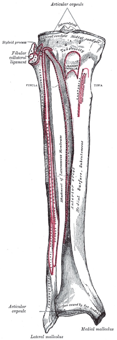
Figure 1. Anterior view of the right tibia and fibula with labelled landmarks; the subcutaneous anteromedial border explains the high open-fracture rate. Gray’s Anatomy (1918), public domain, via Wikimedia Commons.
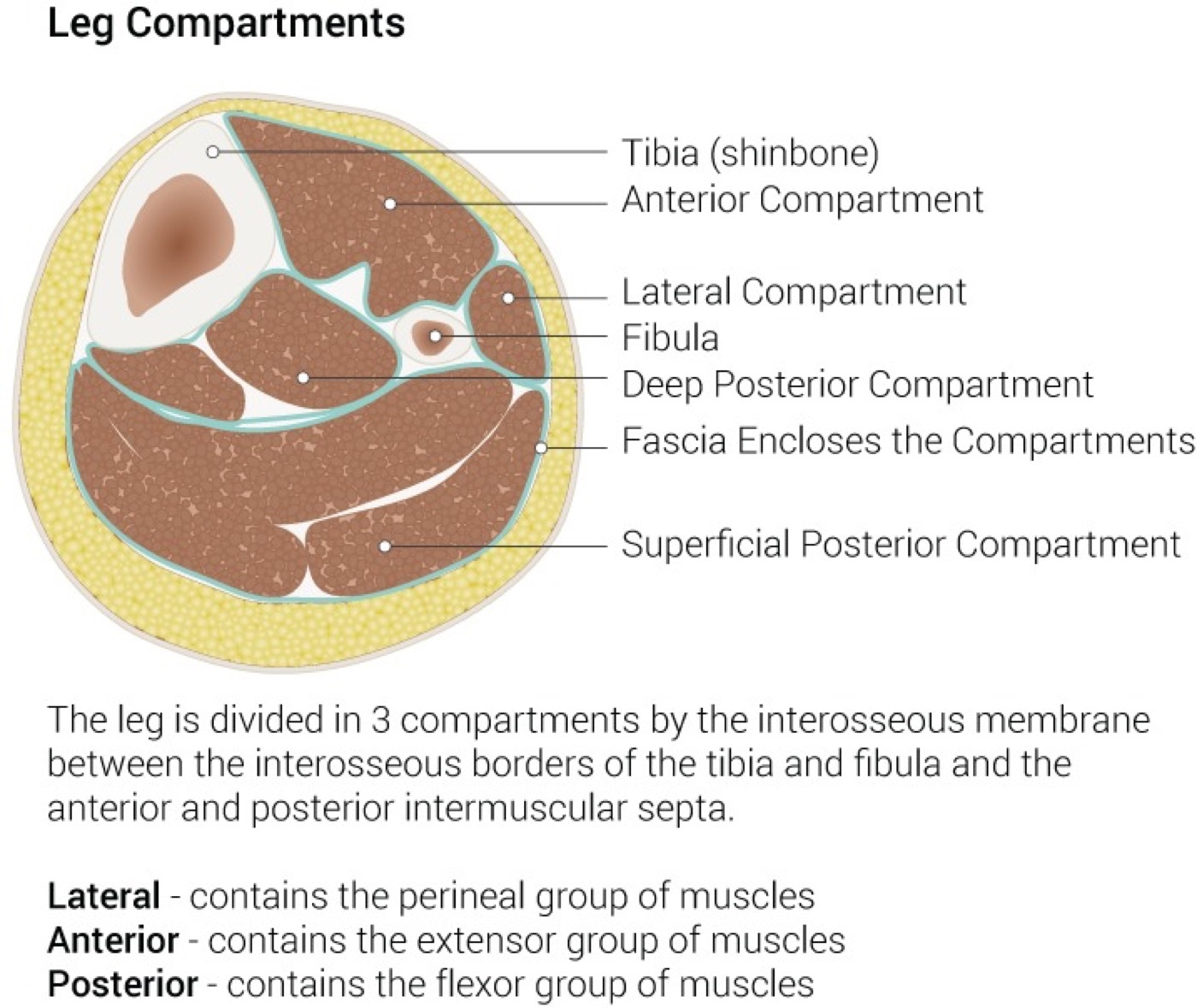
Figure 2. Transverse section of the leg showing the four fascial compartments around the tibia and fibula; the anterior compartment is the one most often affected by compartment syndrome. Illustration by Beckie Palmer (StatPearls), CC BY 4.0, via Wikimedia Commons.
Part III - Compartment Syndrome and Neurovascular Injury
Compartment syndrome is the central early emergency: the tibial shaft causes more than a third of all lower-limb compartment syndromes (and tibial fracture causes it more than any other fracture), with an incidence of roughly 1.5-11 %, classically in a young man with a closed shaft fracture.[7] The diagnosis is clinical: pain out of proportion to the injury and pain on passive stretch of the compartment are the earliest signs, while the textbook “5 P’s” (pulselessness, paralysis) are late and pulses are usually present. When the examination is unreliable (the sedated or obtunded patient), compartment pressures are measured, the operative threshold being a perfusion pressure (diastolic minus compartment pressure) below 30 mmHg, and the treatment is an emergent four-compartment, two-incision fasciotomy. An open fracture does not protect against compartment syndrome. The vascular examination is mandatory: an abnormal pedal pulse after a tibial fracture is a surgical emergency (correct the deformity, re-check, and if it does not return, treat as a vascular injury until proven otherwise), and an ipsilateral femoral and tibial fracture is a floating knee, which carries high rates of vascular injury and open fracture.[8]
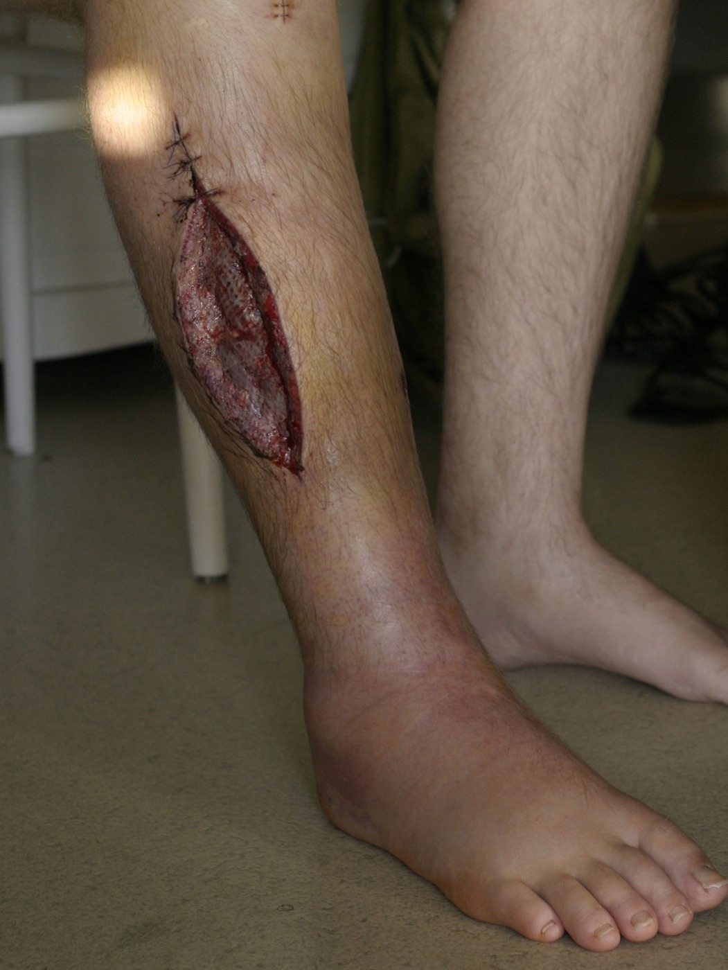
Figure 3. Medial leg fasciotomy wound after decompression for compartment syndrome. CC BY-SA 3.0, via Wikimedia Commons.
Part IV - Open Fractures
Because the tibia is so often open, the Gustilo-Anderson classification is essential: type I is a clean wound under 1 cm; type II a wound of 1-10 cm without extensive soft-tissue damage; type IIIA a high-energy or segmental injury or any wound over 10 cm that can still be closed; type IIIB a wound needing a muscle or skin flap; and type IIIC an injury needing vascular repair to revascularise the limb.[9] The grade correlates with infection (about 1 % for closed and grade I/II, rising to 10-40 % for IIIB) and guides management. The principles are early intravenous antibiotics (a first-generation cephalosporin such as cefazolin, with gram-negative cover added for type III and penicillin for farm-type contamination, given as soon as possible, since modern evidence prioritises antibiotic timing over debridement timing), tetanus prophylaxis, debridement (the historical “6-hour rule” is largely abandoned), low-pressure saline irrigation (the FLOW trial), and early orthoplastic soft-tissue coverage (a “fix-and-flap” approach, with coverage by 5-7 days), much of which the source texts defer to their general open-fracture chapters and which is therefore standard teaching layered onto them.[10]
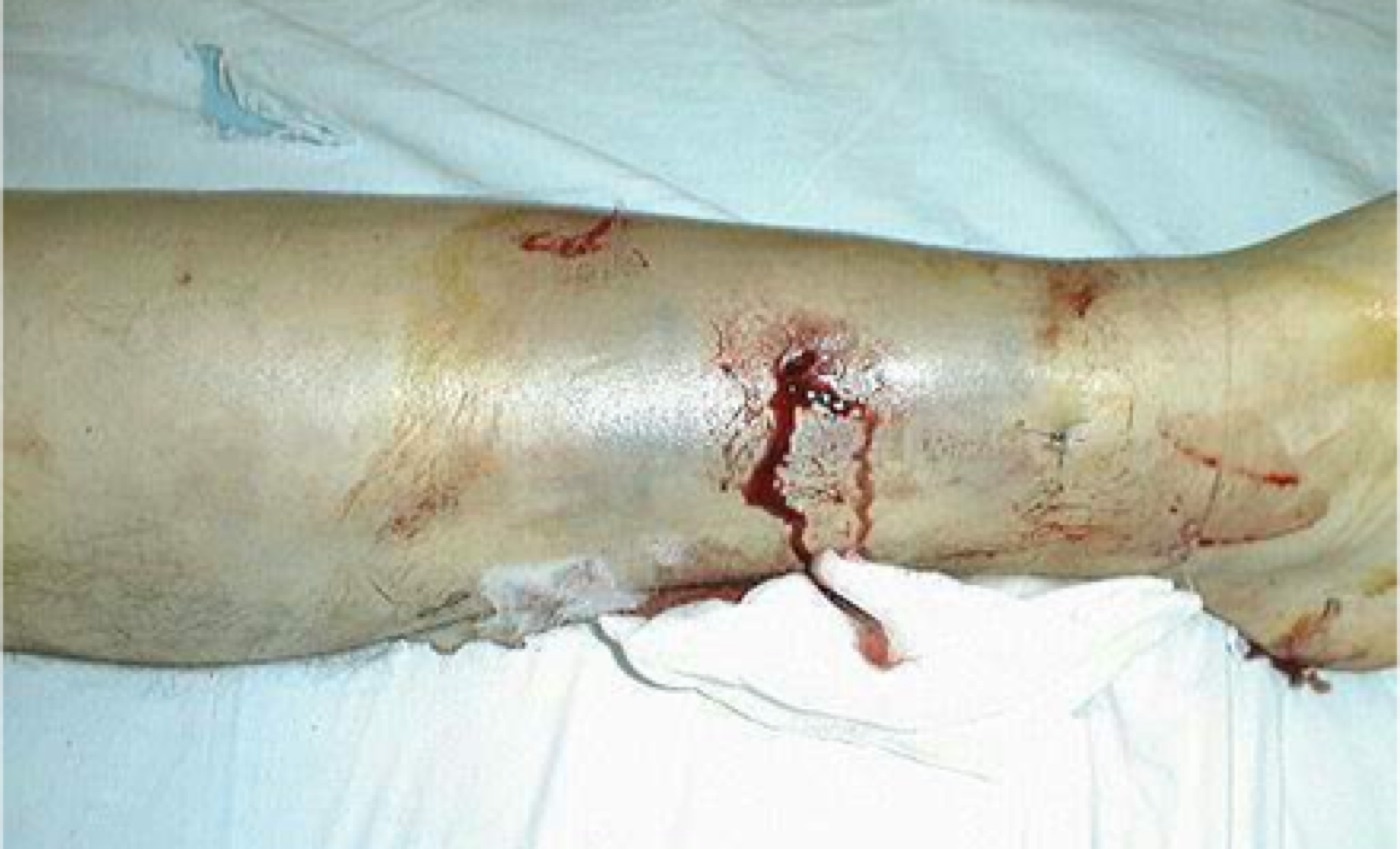
Figure 4. Gustilo-Anderson type I open tibial fracture: a small puncture wound with minimal soft-tissue injury. Image by Doncovska, CC BY-SA 3.0, via Wikimedia Commons.
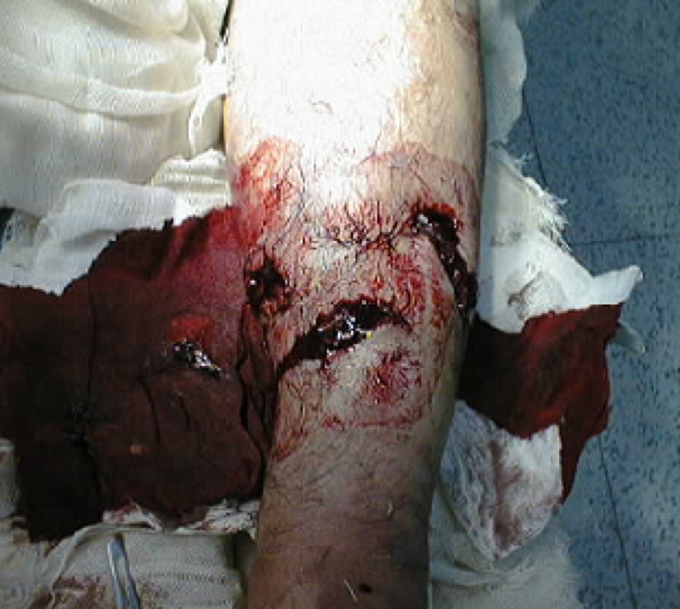
Figure 5. Gustilo-Anderson type II open fracture: a larger laceration with moderate soft-tissue injury. Image by Doncovska, CC BY-SA 3.0, via Wikimedia Commons.
For the most severe open tibias (IIIB and IIIC), the limb-salvage-versus-amputation decision uses scores such as the MESS (a value above 7 suggesting amputation, doubled for ischaemia beyond 6 hours), but these scores have limited validity: they predict salvage potential better than they predict the need for amputation, and the LEAP study found no difference in long-term function between salvage and amputation.[11] The absolute indications for amputation are a warm-ischaemia time beyond 6 hours, a limb attached only by a skin bridge, and a reconstruction that would threaten the patient’s life; a below-knee amputation is preferred, and amputation wounds are not closed primarily.[12]
Part V - Imaging and Classification
Radiographs are AP and lateral views of the whole tibia that must include the knee and the ankle, because a tibial shaft fracture may extend into the plateau proximally or the plafond and malleoli distally.[13] CT is added for the distal third (a spiral distal-third fracture carries an associated posterior malleolar fracture in 25-39 % of cases, and up to 43 % of distal-quarter fractures have intra-articular extension on CT, 14 % of which are missed on plain films) and for proximal extension into the plateau, so that these can be stabilised before nailing displaces them. The diaphyseal tibia is classified by the AO/OTA system as region 42: 42A simple (A1 spiral, A2 oblique with a line over 30°, A3 transverse with a line under 30°), 42B wedge, and 42C complex/multifragmentary; the closed soft-tissue injury is graded by the Tscherne classification (C0-C3), which correlates with healing time.[14]
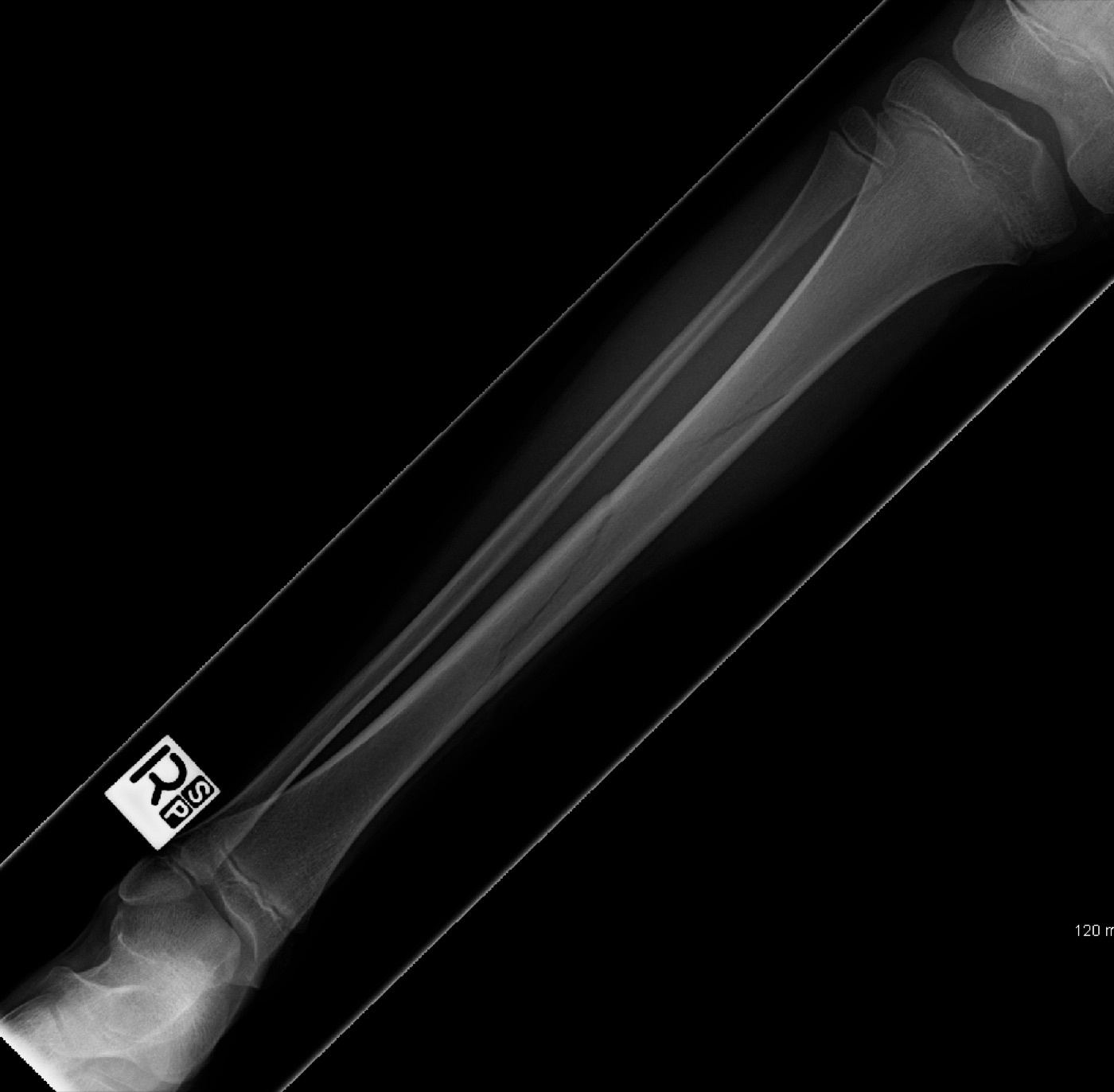
Figure 6. Spiral fracture of the tibial shaft, the low-energy torsional pattern. Image by James Heilman, MD, CC BY-SA 3.0, via Wikimedia Commons.
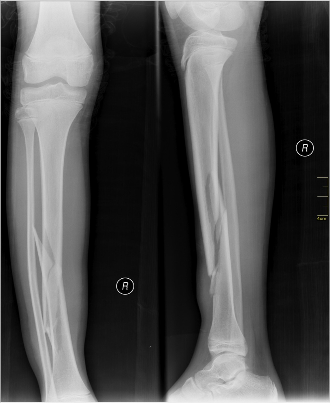
Figure 7. Oblique fracture of the tibial and fibular shafts with a butterfly fragment, AP and lateral radiographs. Image by Nevit Dilmen, CC BY-SA 3.0, via Wikimedia Commons.
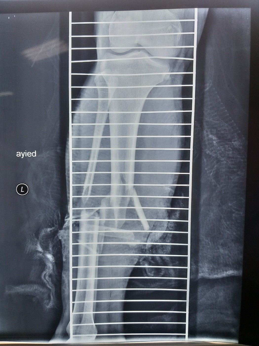
Figure 8. Displaced comminuted fracture of the tibial and fibular shafts, a high-energy pattern. Image by MustafaSalahalden, CC BY-SA 4.0, via Wikimedia Commons.
Part VI - Nonoperative Treatment
Nonoperative treatment is reserved for low-energy, stable, closed fractures whose alignment can be held in a cast.[15] The accepted limits are roughly under 5° of varus or valgus, under 5-10° of anterior or posterior angulation, under 10° of rotation, and under 1 cm (the authors prefer 5-10 mm) of shortening, with more than 50 % cortical apposition the conventional adjunct (varus and valgus being less well tolerated than sagittal-plane angulation). Treatment is a long-leg cast to control rotation, converted at 2-4 weeks to a functional patellar-tendon-bearing (Sarmiento) brace that allows ankle and knee motion and early weight bearing, working by the hydraulic compression of the leg’s soft tissues. Sarmiento’s series of 1,000 braced fractures reported a nonunion rate of only 1.1 %, but the technique is reserved for fractures that will stay aligned, since closed treatment does not correct an inadequate initial reduction and an intact fibula lets the tibia drift into varus; for displaced fractures, intramedullary nailing is superior on union, malunion, and time off work.[16]
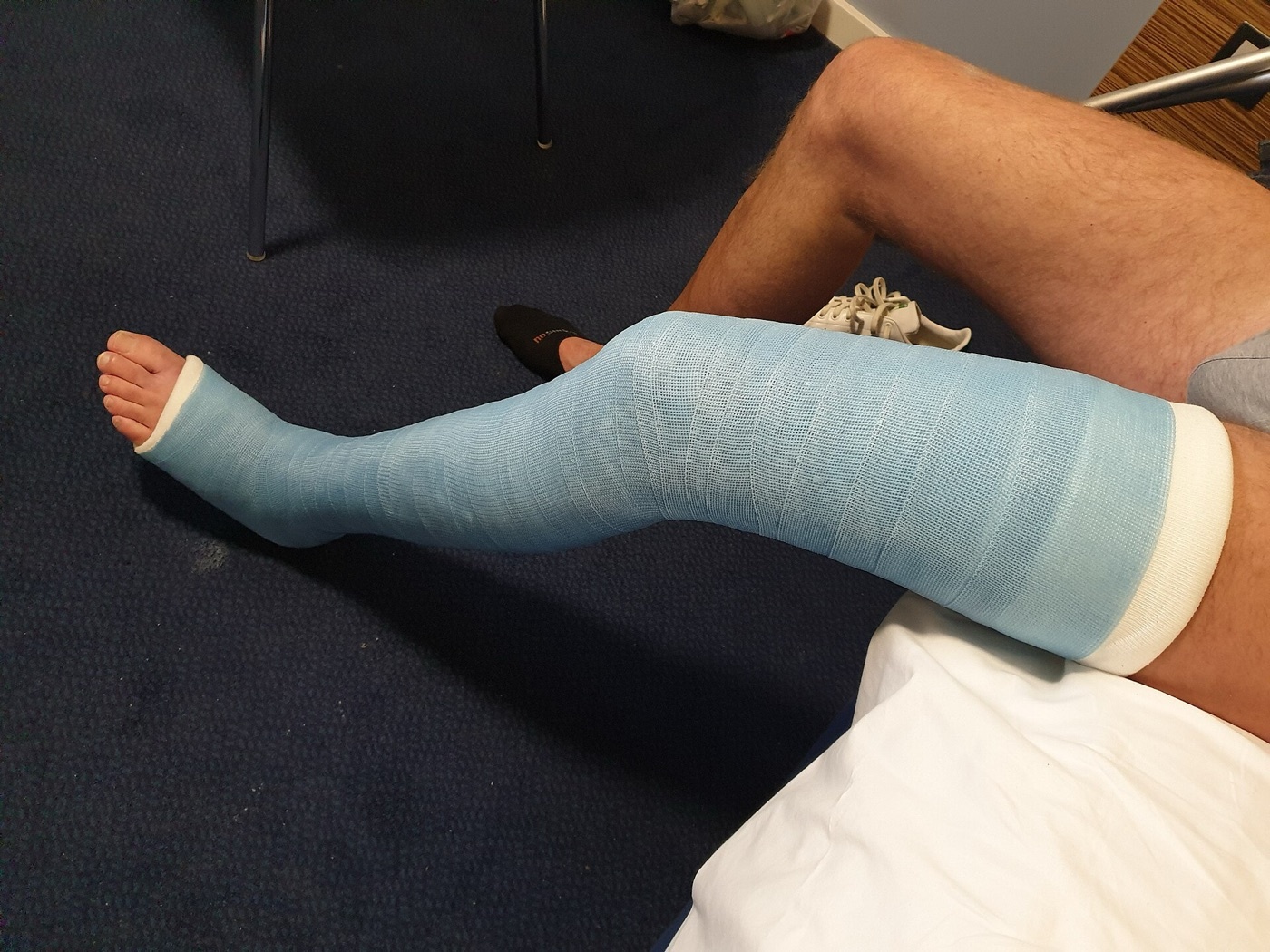
Figure 9. Long-leg cast, the initial immobilisation that controls rotation in nonoperative treatment. Image by Jonuscumgi, CC BY-SA 4.0, via Wikimedia Commons.
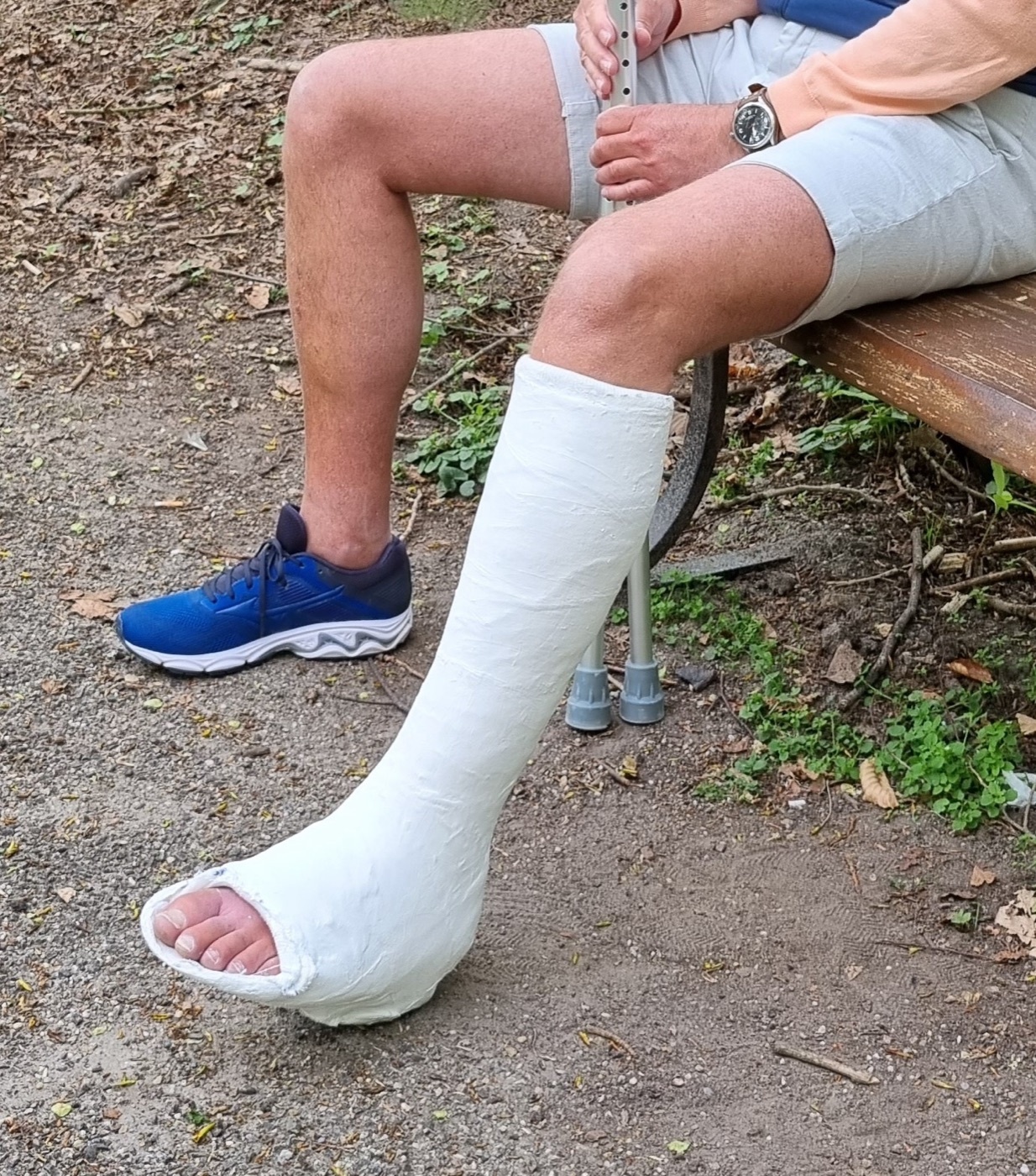
Figure 10. Below-knee (short-leg) walking cast with a toeplate, the form of functional weight-bearing immobilisation. Image by Jonuscumgi, CC BY-SA 4.0, via Wikimedia Commons.
Part VII - Reamed Locked Intramedullary Nailing
The reamed, locked intramedullary nail is the gold standard for most displaced shaft fractures, and the large SPRINT trial established that reaming reduces adverse outcomes in closed fractures, with no clear benefit (and possibly a disadvantage) in open fractures, where reaming is nonetheless safe.[17] The entry point is just medial to the lateral tibial spine on the AP view and just off the articular surface anteriorly, parallel to the anterior cortex, on the lateral (modern nails have a proximal Herzog bend, so the knee is flexed to avoid perforating the posterior cortex). The traditional infrapatellar approaches have characteristic pitfalls (a medial parapatellar start drives the wire too medial and creates valgus; a transtendinous split lets it angle posteriorly and create apex-anterior angulation), which has driven the suprapatellar / semi-extended technique: nailing through the quadriceps tendon with the knee nearly straight neutralises the deforming forces and improves the alignment of proximal (and distal) fractures, with no reproducible difference in knee pain and a theoretical patellofemoral-cartilage risk that has not been borne out. The nail is reamed to about 1-1.5 mm over its diameter (to cortical chatter) and statically locked with two screws proximally and distally, dynamic locking being reserved for a small gap in a simple transverse fracture; rotation is confirmed clinically, not radiographically.[18]
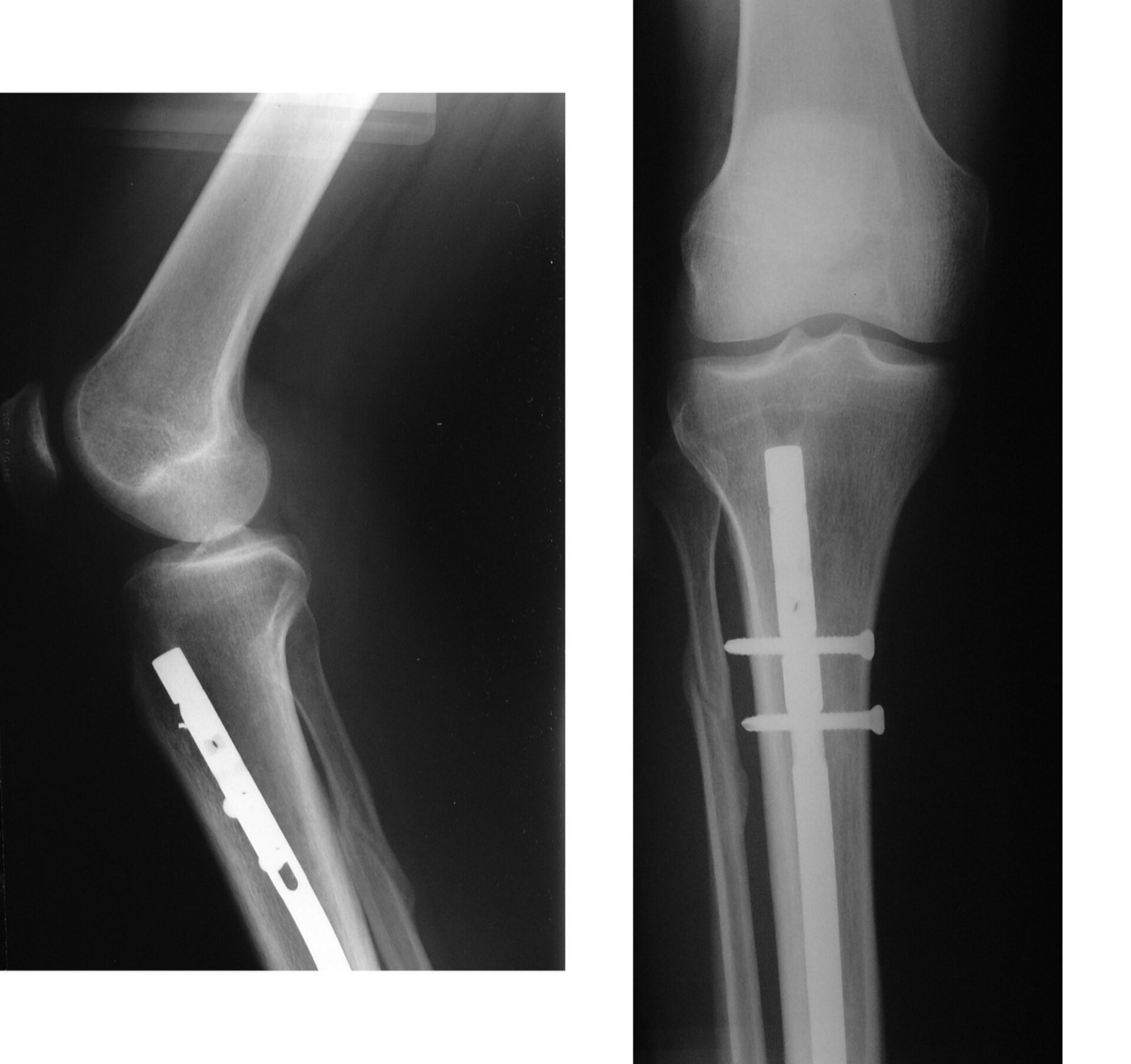
Figure 11. Tibial intramedullary nail with proximal interlocking screws, AP and lateral radiographs. Image by Kolossos, CC BY-SA 3.0, via Wikimedia Commons.
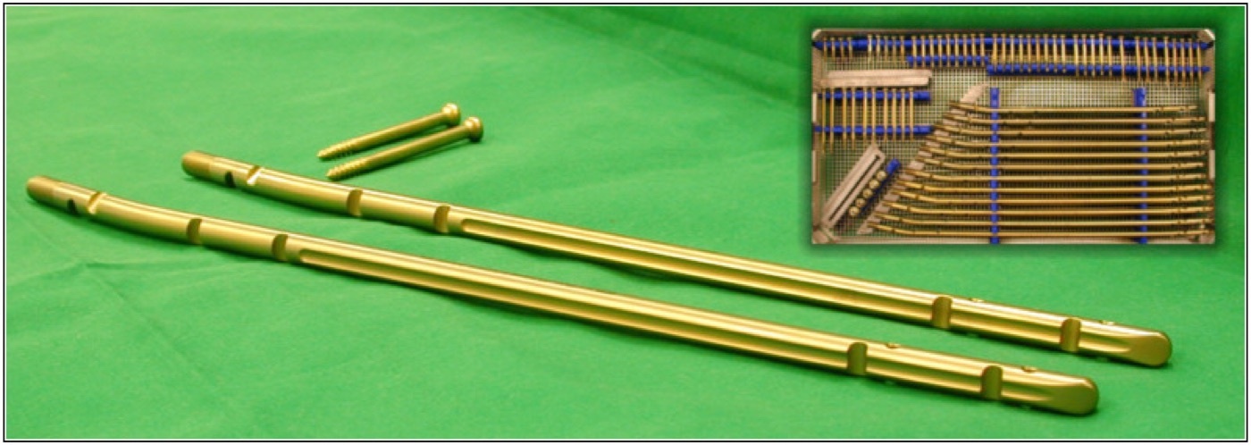
Figure 12. Intramedullary nails with their locking screws (implant hardware). Image by Chriudel, CC BY-SA 3.0, via Wikimedia Commons.
The proximal-third and distal-third extra-articular fractures are the hard ones, because the widened metaphyseal canal does not let the nail reduce the fracture and a proximal fragment tends to drift into valgus and apex-anterior (procurvatum) angulation.[19] These are controlled by a correct (laterally based, anteriorly angled) entry point, the semi-extended / suprapatellar position, blocking (Poller) screws placed on the concave side of the deformity (posterior and lateral for the proximal fragment), provisional unicortical plating, and plating the associated fibula to restore length and alignment in distal fractures. The most common complication of nailing is anterior knee pain (reported in 19-73 %, around 30 %), which often persists even after nail removal and does not differ reliably between the infrapatellar and suprapatellar approaches.[20]
Part VIII - Plating and External Fixation
Plating is reserved for the fractures a nail handles poorly: very proximal or distal metaphyseal-diaphyseal fractures, peri-articular extension, a canal too narrow or deformed to nail, periprosthetic fractures, and cases where rotation is critical.[21] It is done by minimally invasive (MIPO) technique to spare the soft tissues, using a narrow plate (broad plates are too bulky for the tibia), with a bridge plate about three times the length of a comminuted zone and a screw density around 0.5; simple patterns get direct reduction with a lag screw and absolute stability, complex patterns a bridge plate and relative stability. The dominant risk is the subcutaneous soft tissue: definitive plating of open tibias carries about 11 % deep infection, so the timing and approach are chosen to protect the skin. External fixation is a damage-control and severe-open-fracture tool, restoring length and alignment while the soft tissues recover; it is converted to a nail within about two weeks to limit infection, and pin-tract infection is its hallmark. A definitive ring (Ilizarov) fixator is used for severe open fractures with bone loss, where bone transport / distraction osteogenesis or the Masquelet induced-membrane technique reconstructs the defect away from the zone of injury.[22]
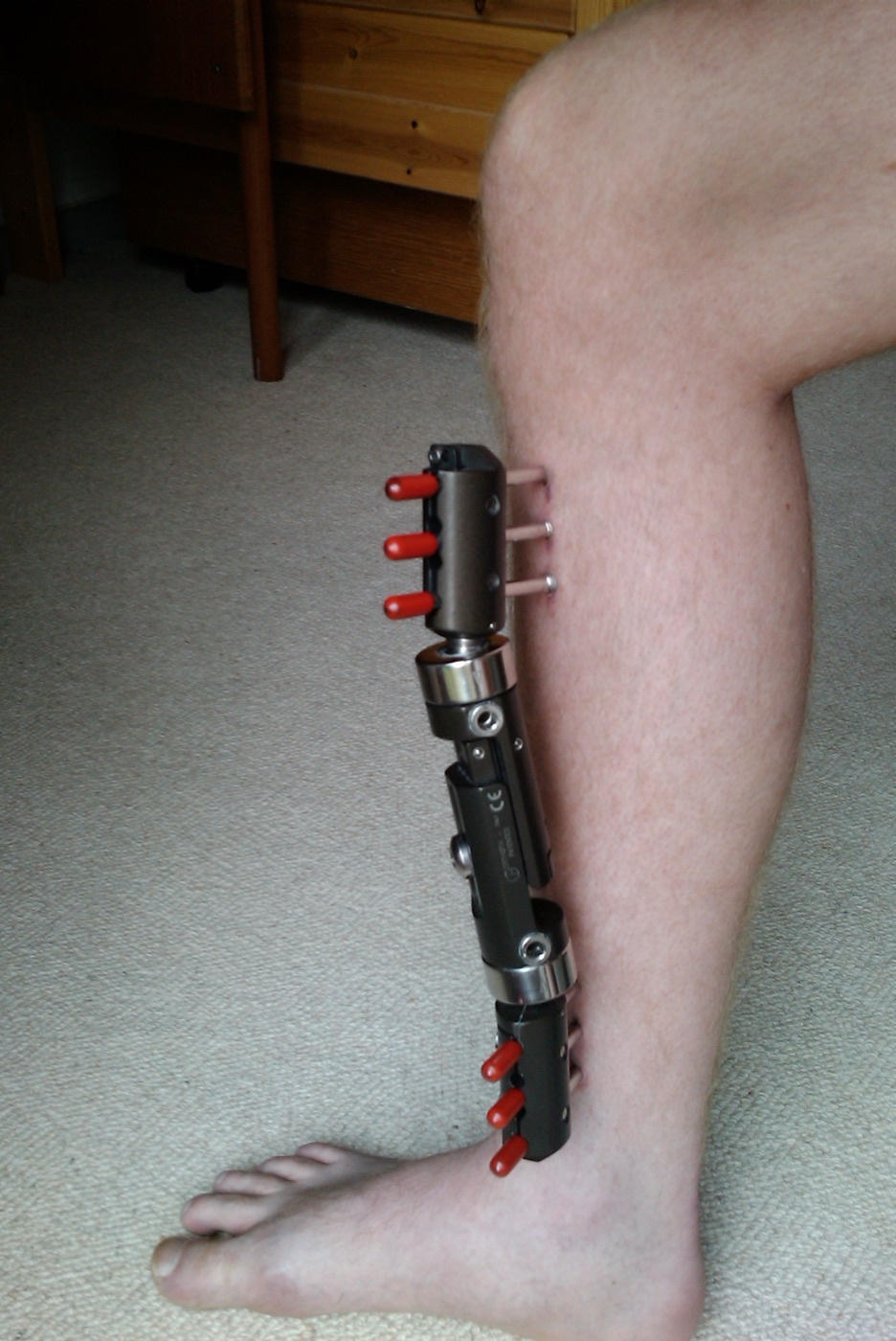
Figure 13. Monolateral external fixator applied to the tibia, used for damage control and severe open fractures. Image by Redhead.dk, CC BY 3.0, via Wikimedia Commons.
Part IX - Complications
The complications of the tibial shaft fracture are the most heavily examined part of the topic.[23] Anterior knee pain is the commonest complication of nailing (19-73 %). Malunion is characteristically the proximal-third valgus-and-procurvatum deformity, though tibial malalignment correlates inconsistently with later arthritis. Nonunion is the signature problem because the tibia heals slowly: delayed union is incomplete healing at 3-6 months and nonunion the failure to heal by 6 months, with rates of about 1-8 % after operative fixation of closed fractures rising to 5.3-24 % in open fractures (the AO quotes up to 14 % overall), the risk factors being an open fracture, high energy, a distal-third location, infection, a fracture gap, and smoking. The treatment ladder runs from dynamization through exchange reamed nailing (the standard for an aseptic nailed nonunion, giving a larger implant and a biological reaming stimulus), bone grafting (including the classic posterolateral graft), rhBMP-2 (approved for the acute open tibia, reducing reoperations) and BMP-7 (for nonunion when autograft is not feasible), to the Masquelet technique and Ilizarov bone transport for segmental defects. Infection complicates about 1 % of closed and grade I/II fractures but 10-40 % of IIIB injuries (S. aureus in about 64 % of deep infections), and an infected nonunion is among the hardest problems in trauma. Other complications include the Volkmann contracture of a missed compartment syndrome (standard teaching) and symptomatic hardware, with about 30 % of patients requesting implant removal.[24]
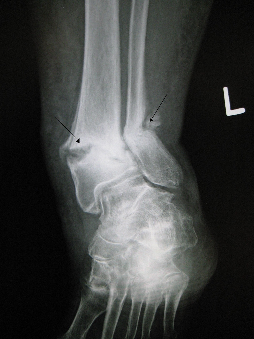
Figure 14. Tibial/fibular nonunion (pseudarthrosis, arrows) with no bridging callus across the fracture. Image by James Heilman, MD, CC BY-SA 3.0, via Wikimedia Commons.
Part X - Isolated Fibula Shaft Fractures
Isolated fibula shaft fractures are rare and, in themselves, of minimal functional consequence, because the fibula carries only 7-16 % of body weight and the intact tibia maintains length and weight bearing, so they are almost always treated nonoperatively with weight bearing as tolerated in a boot.[25] The crucial exception is the Maisonneuve injury: an apparently isolated proximal spiral fibula fracture that is actually the proximal end of a rotational ankle injury, with disruption of the syndesmosis and interosseous membrane and a medial malleolar or deltoid injury, which is unstable and requires syndesmotic and medial fixation. Therefore every apparently isolated fibula fracture must have dedicated ankle imaging (AP, lateral, and mortise views, with external-rotation stress views as needed). A proximal fibular fracture also threatens the common peroneal nerve at the fibular neck (standard teaching) and may accompany lateral knee (LCL) instability.[26]
Part XI - Paediatric Tibial Shaft Fractures
Paediatric tibial shaft fractures make up 4-5 % of all childhood fractures, and three patterns recur.[27] The toddler’s fracture is a nondisplaced spiral fracture of the distal tibial shaft in a child of 1-4 years from a low-energy twist (the classic story is a twist on a sliding board), presenting as a refusal to bear weight; it is often radiographically occult, so it is treated on suspicion and confirmed at 10-14 days by periosteal new bone, healing in a short-leg walking cast in about three weeks. The isolated tibial fracture with an intact fibula is the commonest paediatric tibial shaft fracture and drifts into varus (the intact fibula tethering the lateral side), traditionally held in an above-knee cast with the knee flexed 30° and the ankle in slight plantarflexion. The both-bone (tibia and fibula) fracture instead drifts into valgus and procurvatum because of the bulk of the posterolateral muscles. Acceptable angulation is roughly within 10° for children 6 and over and 15° for younger children, overgrowth is modest (mean about 4.35 mm), open fractures (best prognosis under 12 years) are usually stabilised with external fixation, and unstable high-energy fractures are increasingly fixed with flexible (elastic) nails.[28]
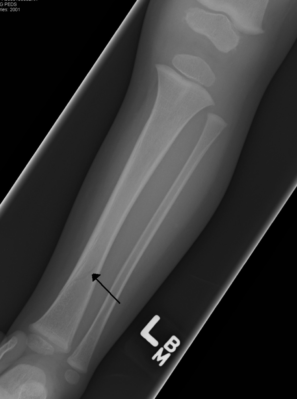
Figure 15. Toddler’s fracture: a subtle, undisplaced spiral fracture of the distal tibial shaft (arrow) in a young child with open physes. Image by James Heilman, MD, CC BY-SA 3.0, via Wikimedia Commons.
A distinct paediatric entity is the Cozen fracture, a proximal tibial metaphyseal fracture in a child under 8 that develops a progressive valgus deformity over the months after injury (from asymmetric overgrowth of the medial physis), even when the fracture was anatomically reduced.[29] The deformity usually appears weeks after the fracture and ceases by about a year; the management of choice is observation (it typically resolves spontaneously, especially under 5 years), with temporary hemiepiphyseal stapling or tension-band plating reserved for a deformity over about 15° that is not remodelling, and corrective osteotomy avoided because of its high recurrence rate, so the family is counselled about the possibility of post-traumatic genu valgum at the time of injury.[30]
Part XII - A Synthesis: How to Reason About the Tibial Shaft Fracture
Reason about the tibial shaft fracture through its soft tissues. The bone is subcutaneous, so first assess and protect the skin, recognise the open fracture (Gustilo grade, early antibiotics, debridement, planned orthoplastic coverage), and exclude the compartment syndrome that this fracture causes more than any other (pain on passive stretch is the early sign; the pulses lie to you). Most displaced shaft fractures are then fixed with a reamed, locked intramedullary nail (reaming helps the closed fracture and is safe in the open one), and the technical work is to choose an entry point and a semi-extended position that prevent the proximal valgus-and-procurvatum malalignment, using Poller screws and a plated fibula when needed, while accepting that anterior knee pain is the price many patients pay. Hold the rule that the tibia heals slowly and nonunites more than any other bone, so watch the distal-third and open fractures, and reach for exchange reamed nailing, BMP, the Masquelet technique, or bone transport when union fails. An isolated fibula fracture is benign unless it is a Maisonneuve, so image the ankle. And in the child, separate the benign toddler’s fracture, the varus-drifting intact-fibula fracture, the valgus-drifting both-bone fracture, and the Cozen fracture that turns valgus over the year and is best simply watched.
References
-
Rockwood & Green’s Fractures in Adults, pp.4349, 4350 (commonest long-bone fracture, ~80 % with a fibula fracture, open in 12-47 %, the bone most likely to need IIIB flap coverage, ~5 % nonunion the highest of all bones, outcomes governed by the soft-tissue injury); AO Principles of Fracture Management, pp.919, 920 (the most common long-bone fracture, the muscle-free subcutaneous anterior tibia so even closed fractures injure the skin, up to 24-25 % open, compartment syndrome more common than with any other fracture, the soft-tissue severity vital to decision-making).
-
Rockwood & Green’s Fractures in Adults, pp.4349, 4350 (commonest long-bone fracture, ~80 % with a fibula fracture, open in 12-47 %, the bone most likely to need IIIB flap coverage, ~5 % nonunion the highest of all bones, outcomes governed by the soft-tissue injury); AO Principles of Fracture Management, pp.919, 920 (the most common long-bone fracture, the muscle-free subcutaneous anterior tibia so even closed fractures injure the skin, up to 24-25 % open, compartment syndrome more common than with any other fracture, the soft-tissue severity vital to decision-making).
-
Rockwood & Green’s Fractures in Adults, pp.4349, 4350 (commonest long-bone fracture, incidence 17/100,000, ~80 % with a fibula fracture; bimodal low-energy spiral over 50 and high-energy transverse/comminuted under 30; vehicular trauma with pedestrians the largest group; open in 12-47 %, up to 63 % after motorcycle crashes, most likely to need a IIIB flap; ~5 % nonunion the highest of all bones); AO Principles of Fracture Management, pp.919, 920 (incidence 20/100,000 overall and 39/100,000 in teenage boys; up to 24-25 % open).
-
Rockwood & Green’s Fractures in Adults, pp.4349, 4350 (commonest long-bone fracture, incidence 17/100,000, ~80 % with a fibula fracture; bimodal low-energy spiral over 50 and high-energy transverse/comminuted under 30; vehicular trauma with pedestrians the largest group; open in 12-47 %, up to 63 % after motorcycle crashes, most likely to need a IIIB flap; ~5 % nonunion the highest of all bones); AO Principles of Fracture Management, pp.919, 920 (incidence 20/100,000 overall and 39/100,000 in teenage boys; up to 24-25 % open).
-
AO Principles of Fracture Management, pp.919, 920, 921, 924 (the muscle-free subcutaneous anterior tibia so most fractures injure the skin, medial plating avoided in thin skin; the four compartments with the thick leg fascia and the anterior compartment most commonly involved; endosteal circulation destroyed by reaming and re-established by 8-12 weeks with the periosteum the main supply meanwhile); Rockwood & Green’s Fractures in Adults, pp.4362, 4363 (triangular cross-section carrying >80 % of load; the four compartments and their contents; nutrient artery from the posterior tibial artery entering the proximal third, outer 25-30 % periosteal, reversal of flow after stripping). The middle-distal-third “watershed” terminology is standard teaching; the source documents the distal-third poor-healing tendency and the proximal nutrient-artery entry.
-
AO Principles of Fracture Management, pp.919, 920, 921, 924 (the muscle-free subcutaneous anterior tibia so most fractures injure the skin, medial plating avoided in thin skin; the four compartments with the thick leg fascia and the anterior compartment most commonly involved; endosteal circulation destroyed by reaming and re-established by 8-12 weeks with the periosteum the main supply meanwhile); Rockwood & Green’s Fractures in Adults, pp.4362, 4363 (triangular cross-section carrying >80 % of load; the four compartments and their contents; nutrient artery from the posterior tibial artery entering the proximal third, outer 25-30 % periosteal, reversal of flow after stripping). The middle-distal-third “watershed” terminology is standard teaching; the source documents the distal-third poor-healing tendency and the proximal nutrient-artery entry.
-
AO Principles of Fracture Management, pp.919, 929 (compartment syndrome more common with the tibia than any other fracture, more than a third of lower-limb cases, up to 9 %, the young man with a closed shaft fracture; pronounced pain not relieved by opiates and pain on passive stretch, pulses usually present; abnormal pedal pulse a surgical emergency and vascular injury until proven otherwise); Rockwood & Green’s Fractures in Adults, pp.4350, 4353, 4421 (incidence 1.5-11 %, pain out of proportion and on passive stretch the earliest signs with pulselessness unusual/late, the perfusion-pressure threshold below 30 mmHg, open fractures still develop it; the floating knee with 21 % vascular injury and 62 % open). The literal “5 P’s” label and the two-incision four-compartment fasciotomy technique are standard teaching, the source detailing the clinical diagnosis and calling for fascial release.
-
AO Principles of Fracture Management, pp.919, 929 (compartment syndrome more common with the tibia than any other fracture, more than a third of lower-limb cases, up to 9 %, the young man with a closed shaft fracture; pronounced pain not relieved by opiates and pain on passive stretch, pulses usually present; abnormal pedal pulse a surgical emergency and vascular injury until proven otherwise); Rockwood & Green’s Fractures in Adults, pp.4350, 4353, 4421 (incidence 1.5-11 %, pain out of proportion and on passive stretch the earliest signs with pulselessness unusual/late, the perfusion-pressure threshold below 30 mmHg, open fractures still develop it; the floating knee with 21 % vascular injury and 62 % open). The literal “5 P’s” label and the two-incision four-compartment fasciotomy technique are standard teaching, the source detailing the clinical diagnosis and calling for fascial release.
-
Rockwood & Green’s Fractures in Adults, pp.4359, 4360 (Gustilo I <1 cm, II 1-10 cm, IIIA high-energy/segmental or >10 cm but closeable, IIIB needing a flap, IIIC needing vascular repair; OTA open-fracture classification; MESS >7 the amputation threshold, doubled for ischaemia >6 hours but not a reliable predictor); AO Principles of Fracture Management, pp.920, 921, 930 (grades guiding management - primary closure I/II, second debridement IIIA, flap IIIB, vascular repair IIIC; infection ~1 % closed/I/II and 10-40 % IIIB; reamed nails safe in open fractures; early combined orthoplastic care and coverage by 5-7 days). Early antibiotics with cefazolin and gram-negative/penicillin cover, tetanus prophylaxis, the abandonment of the 6-hour debridement rule, the FLOW low-pressure-saline conclusion, and the literal “fix-and-flap” phrase are standard teaching, the sources deferring general open-fracture detail to their dedicated chapters.
-
Rockwood & Green’s Fractures in Adults, pp.4359, 4360 (Gustilo I <1 cm, II 1-10 cm, IIIA high-energy/segmental or >10 cm but closeable, IIIB needing a flap, IIIC needing vascular repair; OTA open-fracture classification; MESS >7 the amputation threshold, doubled for ischaemia >6 hours but not a reliable predictor); AO Principles of Fracture Management, pp.920, 921, 930 (grades guiding management - primary closure I/II, second debridement IIIA, flap IIIB, vascular repair IIIC; infection ~1 % closed/I/II and 10-40 % IIIB; reamed nails safe in open fractures; early combined orthoplastic care and coverage by 5-7 days). Early antibiotics with cefazolin and gram-negative/penicillin cover, tetanus prophylaxis, the abandonment of the 6-hour debridement rule, the FLOW low-pressure-saline conclusion, and the literal “fix-and-flap” phrase are standard teaching, the sources deferring general open-fracture detail to their dedicated chapters.
-
Rockwood & Green’s Fractures in Adults, pp.4361, 4362, 4400 (MESS 2-11, >7 the amputation threshold and doubled for ischaemia >6 hours, the scores predicting salvage better than amputation and not in clinical use to decide salvage; the LEAP study showing no functional difference between salvage and amputation; absolute amputation indications of warm ischaemia >6 hours, a distal-only skin bridge, and life-threatening reconstruction, with below-knee preferred and wounds not closed primarily).
-
Rockwood & Green’s Fractures in Adults, pp.4361, 4362, 4400 (MESS 2-11, >7 the amputation threshold and doubled for ischaemia >6 hours, the scores predicting salvage better than amputation and not in clinical use to decide salvage; the LEAP study showing no functional difference between salvage and amputation; absolute amputation indications of warm ischaemia >6 hours, a distal-only skin bridge, and life-threatening reconstruction, with below-knee preferred and wounds not closed primarily).
-
Rockwood & Green’s Fractures in Adults, pp.4354, 4355, 4356, 4360 (AP/lateral full-length tibia plus knee and ankle; CT of the ankle for distal-third spiral fractures with posterior malleolar involvement and of the knee for proximal extension, up to 43 % intra-articular extension in distal-quarter fractures with 14 % missed on plain films; AO/OTA 42A simple [A1 spiral, A2 oblique
30°, A3 transverse <30°], 42B wedge, 42C complex; Tscherne C0-C3 correlating with healing); AO Principles of Fracture Management, p.920 (AP and lateral including the knee and ankle, the 42A/B/C classification).
-
Rockwood & Green’s Fractures in Adults, pp.4354, 4355, 4356, 4360 (AP/lateral full-length tibia plus knee and ankle; CT of the ankle for distal-third spiral fractures with posterior malleolar involvement and of the knee for proximal extension, up to 43 % intra-articular extension in distal-quarter fractures with 14 % missed on plain films; AO/OTA 42A simple [A1 spiral, A2 oblique
30°, A3 transverse <30°], 42B wedge, 42C complex; Tscherne C0-C3 correlating with healing); AO Principles of Fracture Management, p.920 (AP and lateral including the knee and ankle, the 42A/B/C classification).
-
Rockwood & Green’s Fractures in Adults, pp.4365, 4366, 4368, 4369, 4370 (nonoperative for low-energy stable closed fractures; acceptable malalignment of <5° varus/valgus, <5-10° apex anterior/posterior, <10° rotation, <10-12 mm shortening with 5-10 mm preferred, varus/valgus worse than sagittal; long-leg cast to a functional Sarmiento PTB brace by the hydraulic principle; Sarmiento’s 1.1 % nonunion; the intact fibula causing varus; nailing superior for displaced fractures); AO Principles of Fracture Management, p.921 (nonoperative for incomplete/undisplaced low-energy fractures, with more pain and higher malunion/delayed-union than nailing). The “>50 % cortical apposition” rule is standard teaching, the source giving spiral-displacement cut-offs rather than an apposition row.
-
Rockwood & Green’s Fractures in Adults, pp.4365, 4366, 4368, 4369, 4370 (nonoperative for low-energy stable closed fractures; acceptable malalignment of <5° varus/valgus, <5-10° apex anterior/posterior, <10° rotation, <10-12 mm shortening with 5-10 mm preferred, varus/valgus worse than sagittal; long-leg cast to a functional Sarmiento PTB brace by the hydraulic principle; Sarmiento’s 1.1 % nonunion; the intact fibula causing varus; nailing superior for displaced fractures); AO Principles of Fracture Management, p.921 (nonoperative for incomplete/undisplaced low-energy fractures, with more pain and higher malunion/delayed-union than nailing). The “>50 % cortical apposition” rule is standard teaching, the source giving spiral-displacement cut-offs rather than an apposition row.
-
Rockwood & Green’s Fractures in Adults, pp.4373, 4378, 4380, 4385, 4387 (the reamed locked nail the standard, SPRINT showing reaming benefits closed fractures and is safe in open; the entry point medial to the lateral spine and anterior on the lateral with the Herzog curve and knee flexed; the medial-parapatellar valgus and transtendinous apex-anterior pitfalls; the suprapatellar/semi-extended technique neutralising deforming forces with no knee-pain difference and an unproven cartilage risk; reaming to chatter +1-1.5 mm, static two-and-two locking, rotation checked clinically); AO Principles of Fracture Management, pp.921, 922, 924, 927 (reamed nails with better healing and fewer complications than unreamed and safe in open fractures; the entry point and Herzog curve; the semi-extended technique avoiding proximal apex-anterior/valgus; two proximal and two distal locks with the dynamic lock only for a ≤2 mm gap in a 42A3).
-
Rockwood & Green’s Fractures in Adults, pp.4373, 4378, 4380, 4385, 4387 (the reamed locked nail the standard, SPRINT showing reaming benefits closed fractures and is safe in open; the entry point medial to the lateral spine and anterior on the lateral with the Herzog curve and knee flexed; the medial-parapatellar valgus and transtendinous apex-anterior pitfalls; the suprapatellar/semi-extended technique neutralising deforming forces with no knee-pain difference and an unproven cartilage risk; reaming to chatter +1-1.5 mm, static two-and-two locking, rotation checked clinically); AO Principles of Fracture Management, pp.921, 922, 924, 927 (reamed nails with better healing and fewer complications than unreamed and safe in open fractures; the entry point and Herzog curve; the semi-extended technique avoiding proximal apex-anterior/valgus; two proximal and two distal locks with the dynamic lock only for a ≤2 mm gap in a 42A3).
-
Rockwood & Green’s Fractures in Adults, pp.4403, 4404, 4421 (the metaphyseal canal not reducing the fracture, the proximal-third valgus and apex-anterior tendency, prevention by entry point/semi-extended position/Poller screws on the concave side/provisional plating/fibular plating; anterior knee pain 19-73 % the most common complication, persisting after removal, no infrapatellar-vs-suprapatellar difference); AO Principles of Fracture Management, pp.926, 929, 930 (the proximal flexion-valgus deformity from nailing in flexion, Poller screws posterior and lateral, fibular plating enhancing tibial reduction; anterior knee pain up to 30 %).
-
Rockwood & Green’s Fractures in Adults, pp.4403, 4404, 4421 (the metaphyseal canal not reducing the fracture, the proximal-third valgus and apex-anterior tendency, prevention by entry point/semi-extended position/Poller screws on the concave side/provisional plating/fibular plating; anterior knee pain 19-73 % the most common complication, persisting after removal, no infrapatellar-vs-suprapatellar difference); AO Principles of Fracture Management, pp.926, 929, 930 (the proximal flexion-valgus deformity from nailing in flexion, Poller screws posterior and lateral, fibular plating enhancing tibial reduction; anterior knee pain up to 30 %).
-
AO Principles of Fracture Management, pp.920, 921, 924, 925, 926, 928 (plating for proximal/distal extremes, narrow not broad plates, bridge plate ~3× the comminuted zone and screw density 0.5, absolute vs relative stability; external fixation for temporary care with nails better definitively, conversion within 2 weeks, ring fixators with tensioned wires for bone loss and distraction osteogenesis; Masquelet and Ilizarov for bone loss); Rockwood & Green’s Fractures in Adults, pp.4388, 4389, 4395, 4396 (plating for non-nailable canals, periprosthetic and rotation-critical fractures; ~11 % deep infection after acute open-fracture plating; external fixation for damage control and temporisation with conversion within ~2 weeks and pin-tract infection; ring fixation and bone transport for bone loss).
-
AO Principles of Fracture Management, pp.920, 921, 924, 925, 926, 928 (plating for proximal/distal extremes, narrow not broad plates, bridge plate ~3× the comminuted zone and screw density 0.5, absolute vs relative stability; external fixation for temporary care with nails better definitively, conversion within 2 weeks, ring fixators with tensioned wires for bone loss and distraction osteogenesis; Masquelet and Ilizarov for bone loss); Rockwood & Green’s Fractures in Adults, pp.4388, 4389, 4395, 4396 (plating for non-nailable canals, periprosthetic and rotation-critical fractures; ~11 % deep infection after acute open-fracture plating; external fixation for damage control and temporisation with conversion within ~2 weeks and pin-tract infection; ring fixation and bone transport for bone loss).
-
Rockwood & Green’s Fractures in Adults, pp.4403, 4421, 4422, 4423, 4424, 4425 (anterior knee pain the commonest nailing complication; the proximal-third valgus-procurvatum malunion with inconsistent arthritis correlation; delayed union 3-6 months and nonunion at 6 months, closed-operative 1-8 % and open 5.3-24 %, risk factors open/high-energy/distal-third/infection/gap/smoking, the NURD score; exchange reamed nailing for aseptic nonunion, posterolateral bone graft; infection 1.8 % closed and 8-16 % type III with S. aureus 64 %); AO Principles of Fracture Management, p.930 (nonunion up to 14 %, commoner distal-third/open/high-energy, exchange reamed nailing the standard; infection ~1 % closed/I/II and 10-40 % IIIB; ~30 % request hardware removal); Miller’s Review of Orthopaedics, pp.926, 928 (rhBMP-2 for open tibia reducing reoperations, BMP-7 for nonunion when autograft not feasible; fibulectomy and posterolateral bone graft). The Volkmann ischaemic contracture as the sequela of a missed compartment syndrome is standard teaching.
-
Rockwood & Green’s Fractures in Adults, pp.4403, 4421, 4422, 4423, 4424, 4425 (anterior knee pain the commonest nailing complication; the proximal-third valgus-procurvatum malunion with inconsistent arthritis correlation; delayed union 3-6 months and nonunion at 6 months, closed-operative 1-8 % and open 5.3-24 %, risk factors open/high-energy/distal-third/infection/gap/smoking, the NURD score; exchange reamed nailing for aseptic nonunion, posterolateral bone graft; infection 1.8 % closed and 8-16 % type III with S. aureus 64 %); AO Principles of Fracture Management, p.930 (nonunion up to 14 %, commoner distal-third/open/high-energy, exchange reamed nailing the standard; infection ~1 % closed/I/II and 10-40 % IIIB; ~30 % request hardware removal); Miller’s Review of Orthopaedics, pp.926, 928 (rhBMP-2 for open tibia reducing reoperations, BMP-7 for nonunion when autograft not feasible; fibulectomy and posterolateral bone graft). The Volkmann ischaemic contracture as the sequela of a missed compartment syndrome is standard teaching.
-
Rockwood & Green’s Fractures in Adults, pp.4425, 4426 (isolated fibula fractures rare and of minimal functional significance with the fibula carrying 7-16 % of body weight, treated nonoperatively with weight bearing in a boot; the Maisonneuve injury as a proximal spiral fibula fracture with syndesmotic and medial injury requiring fixation, mandating ankle AP/lateral/mortise and external-rotation stress views; proximal fibula fractures with varus knee instability). The common peroneal nerve at the fibular neck in proximal fibula fractures is standard teaching, noted by the source as an injury to assess.
-
Rockwood & Green’s Fractures in Adults, pp.4425, 4426 (isolated fibula fractures rare and of minimal functional significance with the fibula carrying 7-16 % of body weight, treated nonoperatively with weight bearing in a boot; the Maisonneuve injury as a proximal spiral fibula fracture with syndesmotic and medial injury requiring fixation, mandating ankle AP/lateral/mortise and external-rotation stress views; proximal fibula fractures with varus knee instability). The common peroneal nerve at the fibular neck in proximal fibula fractures is standard teaching, noted by the source as an injury to assess.
-
Lovell & Winter’s Pediatric Orthopaedics, pp.5726, 5727, 5728, 5729, 5739 (4-5 % of paediatric fractures; the toddler’s fracture at 1-4 years from a low-energy twist, often occult and confirmed at 10-14 days by periosteal new bone, treated in a short-leg walking cast for ~3 weeks; the intact-fibula fracture the commonest, drifting varus, held in an above-knee cast with 30° knee flexion and 15° ankle plantarflexion; the both-bone fracture drifting valgus and procurvatum from posterolateral muscle bulk; acceptable angulation ≤10° at ≥6 years and ≤15° under 6, overgrowth mean 4.35 mm, external fixation for open fractures and elastic nailing for high-energy fractures).
-
Lovell & Winter’s Pediatric Orthopaedics, pp.5726, 5727, 5728, 5729, 5739 (4-5 % of paediatric fractures; the toddler’s fracture at 1-4 years from a low-energy twist, often occult and confirmed at 10-14 days by periosteal new bone, treated in a short-leg walking cast for ~3 weeks; the intact-fibula fracture the commonest, drifting varus, held in an above-knee cast with 30° knee flexion and 15° ankle plantarflexion; the both-bone fracture drifting valgus and procurvatum from posterolateral muscle bulk; acceptable angulation ≤10° at ≥6 years and ≤15° under 6, overgrowth mean 4.35 mm, external fixation for open fractures and elastic nailing for high-energy fractures).
-
Lovell & Winter’s Pediatric Orthopaedics, p.5722 (the Cozen proximal tibial metaphyseal fracture in a child under 8 developing progressive valgus from medial physeal overgrowth even after anatomic reduction, beginning weeks after injury and ceasing by 12 months; observation the treatment of choice with spontaneous resolution especially under 5 years, temporary hemiepiphyseal stapling/tension-band plating for a deformity >15° not remodelling, osteotomy avoided for its high recurrence, and counselling the family about post-traumatic genu valgum).
-
Lovell & Winter’s Pediatric Orthopaedics, p.5722 (the Cozen proximal tibial metaphyseal fracture in a child under 8 developing progressive valgus from medial physeal overgrowth even after anatomic reduction, beginning weeks after injury and ceasing by 12 months; observation the treatment of choice with spontaneous resolution especially under 5 years, temporary hemiepiphyseal stapling/tension-band plating for a deformity >15° not remodelling, osteotomy avoided for its high recurrence, and counselling the family about post-traumatic genu valgum).
-
Rockwood & Green’s Fractures in Adults, pp.4349, 4350; AO Principles of Fracture Management, pp.919, 920.
-
Rockwood & Green’s Fractures in Adults, pp.4353, 4421; AO Principles of Fracture Management, p.919.
-
Rockwood & Green’s Fractures in Adults, pp.4359, 4360; AO Principles of Fracture Management, pp.920, 930.
-
AO Principles of Fracture Management, pp.920, 921; Rockwood & Green’s Fractures in Adults, pp.4351, 4360.
-
Rockwood & Green’s Fractures in Adults, pp.4354, 4374; AO Principles of Fracture Management, p.920.
-
Rockwood & Green’s Fractures in Adults, pp.4366, 4368, 4370.
-
Rockwood & Green’s Fractures in Adults, pp.4373, 4385; AO Principles of Fracture Management, p.921.
-
Rockwood & Green’s Fractures in Adults, pp.4403, 4404; AO Principles of Fracture Management, p.929.
-
Rockwood & Green’s Fractures in Adults, p.4421; AO Principles of Fracture Management, pp.922, 930.
-
Rockwood & Green’s Fractures in Adults, pp.4423, 4424; AO Principles of Fracture Management, p.930; Miller’s Review of Orthopaedics, pp.926, 928.
-
Rockwood & Green’s Fractures in Adults, pp.4425, 4426.
-
Lovell & Winter’s Pediatric Orthopaedics, pp.5722, 5727, 5728.