Contents
- Orientation: A High-Stress Cortical Fracture with Powerful Deforming Muscles
- Part I - Definition and Epidemiology
- Part II - Biomechanics and the Deforming Forces
- Part III - Classification
- Part IV - Assessment and Imaging
- Part V - Treatment
- Part VI - Complications
- Part VII - A Synthesis: How to Reason About the Subtrochanteric Fracture
- References
Orientation: A High-Stress Cortical Fracture with Powerful Deforming Muscles
The subtrochanteric femur fracture sits in the proximal femoral shaft, and two facts make it harder than the trochanteric fracture above it. First, it lies in a zone of extreme mechanical stress, where the femur carries the highest compressive load (medially) and the highest tensile load (laterally) in the skeleton, in predominantly cortical bone that heals more slowly than the cancellous metaphysis. Nonunion, malunion, and implant failure are real risks.[1] Second, the fracture is pulled apart by powerful, opposing muscle forces. The proximal fragment flexes, abducts, and externally rotates (iliopsoas, the abductors, and the short external rotators), while the distal shaft is adducted and shortened (the adductors). The result is a fracture that is easy to malreduce into varus and apex-anterior angulation, so getting the reduction right is what matters. The answer in almost every case is a long cephalomedullary (intramedullary) nail, a strong, load-sharing implant, placed without leaving the fracture in varus.[2]
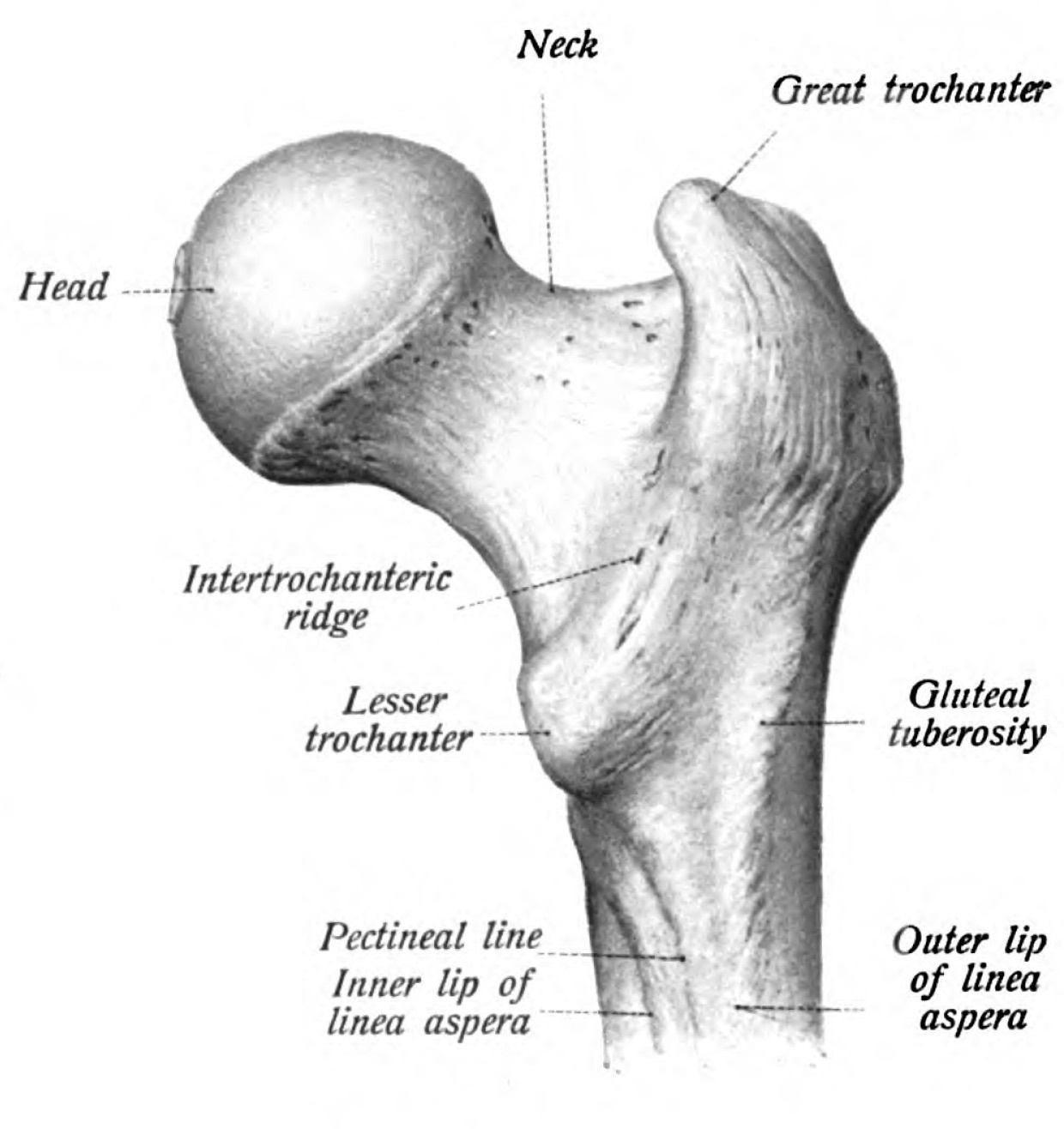
Figure 1. Proximal femur anatomy - head, neck, greater and lesser trochanters, and the subtrochanteric region (the proximal shaft distal to the lesser trochanter). From Sobotta’s Atlas of Human Anatomy (1909), public domain, via Wikimedia Commons.
Part I - Definition and Epidemiology
The subtrochanteric region runs from the lesser trochanter to about 5 cm distal to it (the proximal femoral shaft).[3] The epidemiology is trimodal: high-energy fractures in the young (look for other injuries), low-energy fragility fractures in the osteoporotic elderly, and a distinct subset of atypical fractures associated with long-term bisphosphonate use.[4]
Part II - Biomechanics and the Deforming Forces
The biomechanics explain the difficulty.[5] The subtrochanteric femur is loaded by the highest compressive and tensile forces in the human skeleton (modelled at over 1200 lb/in² medially when the medial cortex is comminuted), so an implant here works in an unforgiving mechanical environment, and the predominantly cortical bone unites more slowly than cancellous metaphyseal bone.[6] The deforming forces are worth knowing in detail. The proximal fragment is flexed by the iliopsoas (on the lesser trochanter), abducted by the gluteus medius (on the greater trochanter), and externally rotated by the short external rotators, while the distal (shaft) fragment is adducted and shortened by the adductors.[7] These opposing pulls drive the fracture into the characteristic varus and apex-anterior (procurvatum) deformity, which the surgeon must actively prevent, because a malreduced subtrochanteric fracture in this high-stress zone fails.[8]
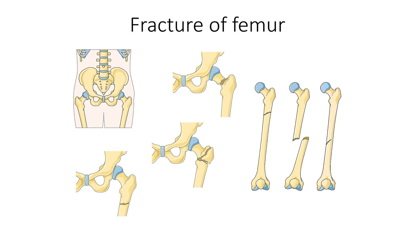
Figure 2. Femoral fracture patterns (neck, intertrochanteric with fragment displacement, and shaft), illustrating how the proximal fragment displaces relative to the shaft under opposing muscle pulls. Illustration by Laboratoires Servier (Servier Medical Art), CC BY-SA 3.0, via Wikimedia Commons.
Part III - Classification
The clinically useful classification is the Russell-Taylor system, which groups fractures by lesser-trochanter involvement and extension into the piriformis fossa, because that historically determined whether a piriformis-entry nail could be used: type I has no extension into the piriformis fossa (IA with the lesser trochanter intact, IB with the lesser trochanter detached), and type II extends into the piriformis fossa.[9] The Seinsheimer classification grades the fracture by the number of fragments and the geometry of the fracture lines.[10] In the AO/OTA scheme the subtrochanteric fracture is coded within the femoral (segment 32) diaphysis with proximal involvement.[11]
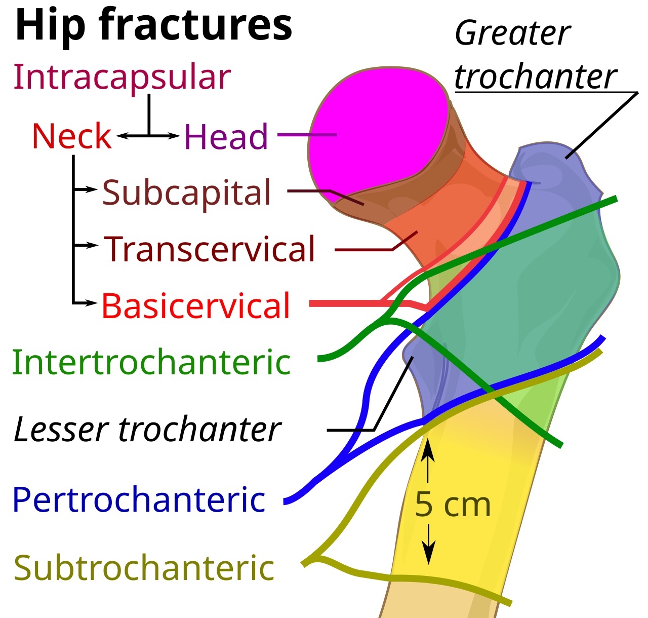
Figure 3. Anatomical zones of proximal femoral fractures, with the subtrochanteric region marked as the ~5 cm zone of proximal shaft distal to the lesser trochanter. Diagram by Mikael Häggström, CC0, via Wikimedia Commons.
A distinct entity is the atypical femoral fracture.[12] Associated with long-term bisphosphonate use, it has a characteristic appearance: a transverse fracture line beginning at the lateral cortex with localised lateral cortical thickening (“beaking”), minimal comminution, and a medial spike, often preceded by prodromal thigh pain and frequently bilateral.[13]
Part IV - Assessment and Imaging
The young patient has a high-energy injury (a fall from height or a road collision, often with other injuries and significant blood loss); the elderly patient has a low-energy fragility fracture; and the atypical fracture may be preceded by weeks of thigh pain in a patient on a bisphosphonate.[14] The limb shows the deformity of the displaced fracture. Imaging is AP and lateral radiographs of the whole femur, including the hip and the knee, with care to exclude an associated femoral neck fracture, and radiographs of the contralateral femur when an atypical fracture is suspected, because these are frequently bilateral.[15]
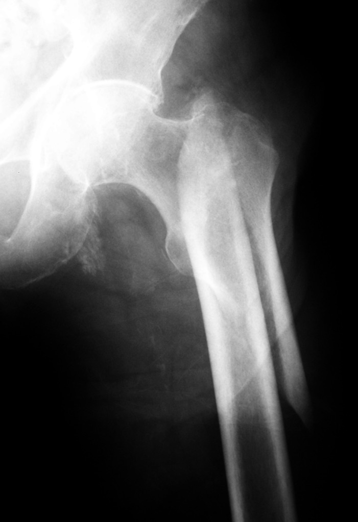
Figure 4. Subtrochanteric femoral fracture on a close-up AP radiograph of the proximal femur, with the fracture line running from just below the trochanters into the proximal shaft. Image by THWZ, CC BY-SA 3.0, via Wikimedia Commons.
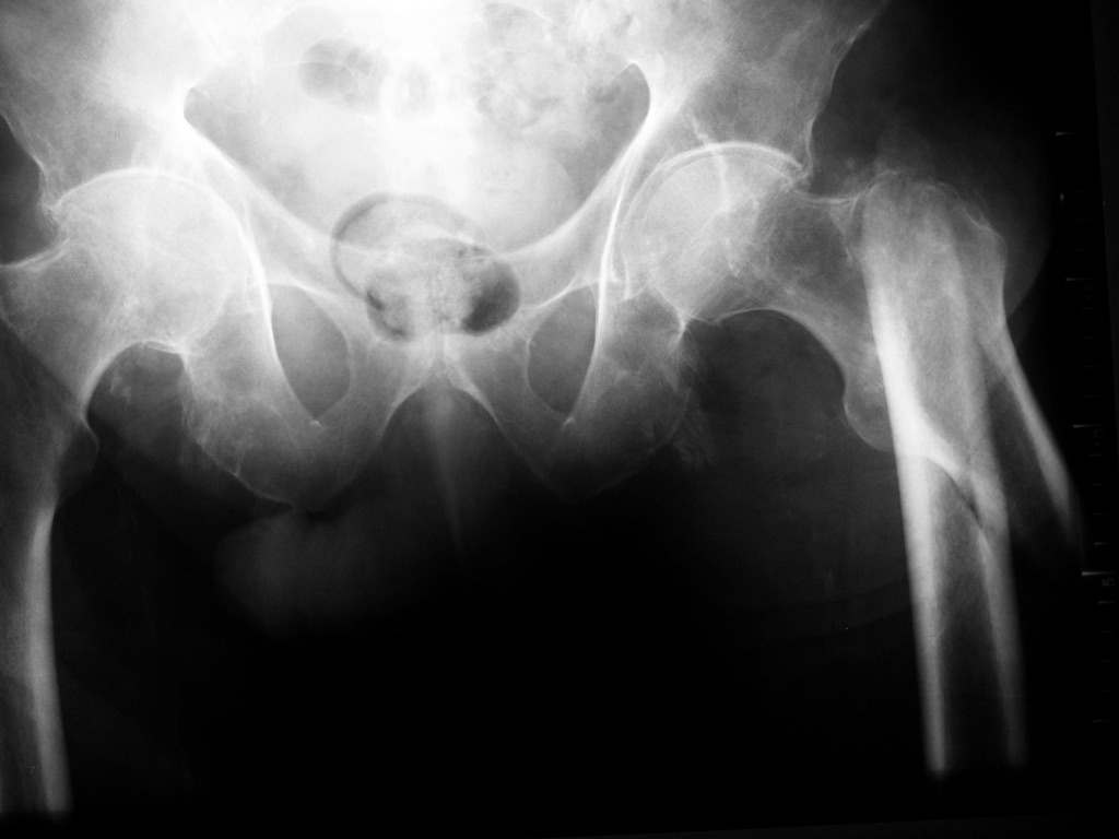
Figure 5. The same subtrochanteric fracture on an AP pelvis radiograph, showing displacement of the distal (shaft) fragment relative to the proximal trochanteric fragment. Image by THWZ, CC BY-SA 3.0, via Wikimedia Commons.
Part V - Treatment
5.1 The cephalomedullary nail is the implant of choice
Subtrochanteric fractures are treated operatively, and the long cephalomedullary (reconstruction) intramedullary nail is the preferred implant for essentially all of them.[16] Its intramedullary, load-sharing position resists the bending load of this high-stress region, and proximal fixation captures the head and neck.[17] The goal is to restore length, alignment, and rotation without leaving the fracture in varus or procurvatum, which is the recurring technical challenge because the proximal fragment is flexed and abducted.[18] The commonest error is a too-lateral entry point (with eccentric reaming), which drives the fracture into varus, so the entry point is taken medial to the tip of the greater trochanter (in line with the canal), and the proximal fragment is controlled with reduction aids: clamps, a Schanz-pin “joystick,” blocking (Poller) screws, a mini-open reduction, or cerclage wires to restore the medial cortex.[19] A 135° device (the sliding hip screw) is contraindicated. Where a nail is unsuitable, a 95° fixed-angle device (the blade plate or dynamic condylar screw) or a proximal femoral locking plate is the alternative, though plates fare less well in this high-stress zone and are largely reserved for nonunion or malunion reconstruction.[20]
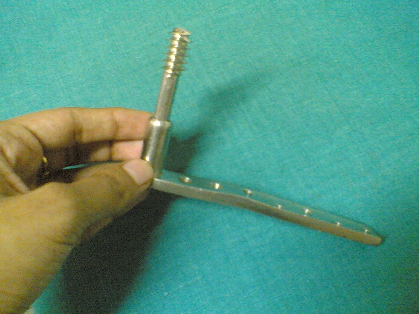
Figure 6. Dynamic condylar screw (DCS): a condylar lag screw fixed to an angled (≈95°) side plate - the fixed-angle plating alternative reserved for cases unsuitable for a nail or for nonunion/malunion reconstruction. Image by Netha Hussain, CC BY-SA 3.0, via Wikimedia Commons.
5.2 The atypical (bisphosphonate-associated) fracture
The atypical femoral fracture is managed by cephalomedullary nailing like other subtrochanteric fractures, but it unites slowly, so healing is followed carefully.[21] The bisphosphonate is stopped, and the contralateral femur is imaged and treated: a symptomatic incomplete atypical fracture (lateral cortical beaking with a “dreaded black line”) is fixed prophylactically with a nail, whereas an asymptomatic incomplete lesion is monitored. Adjuncts such as teriparatide are used to encourage healing.[22]
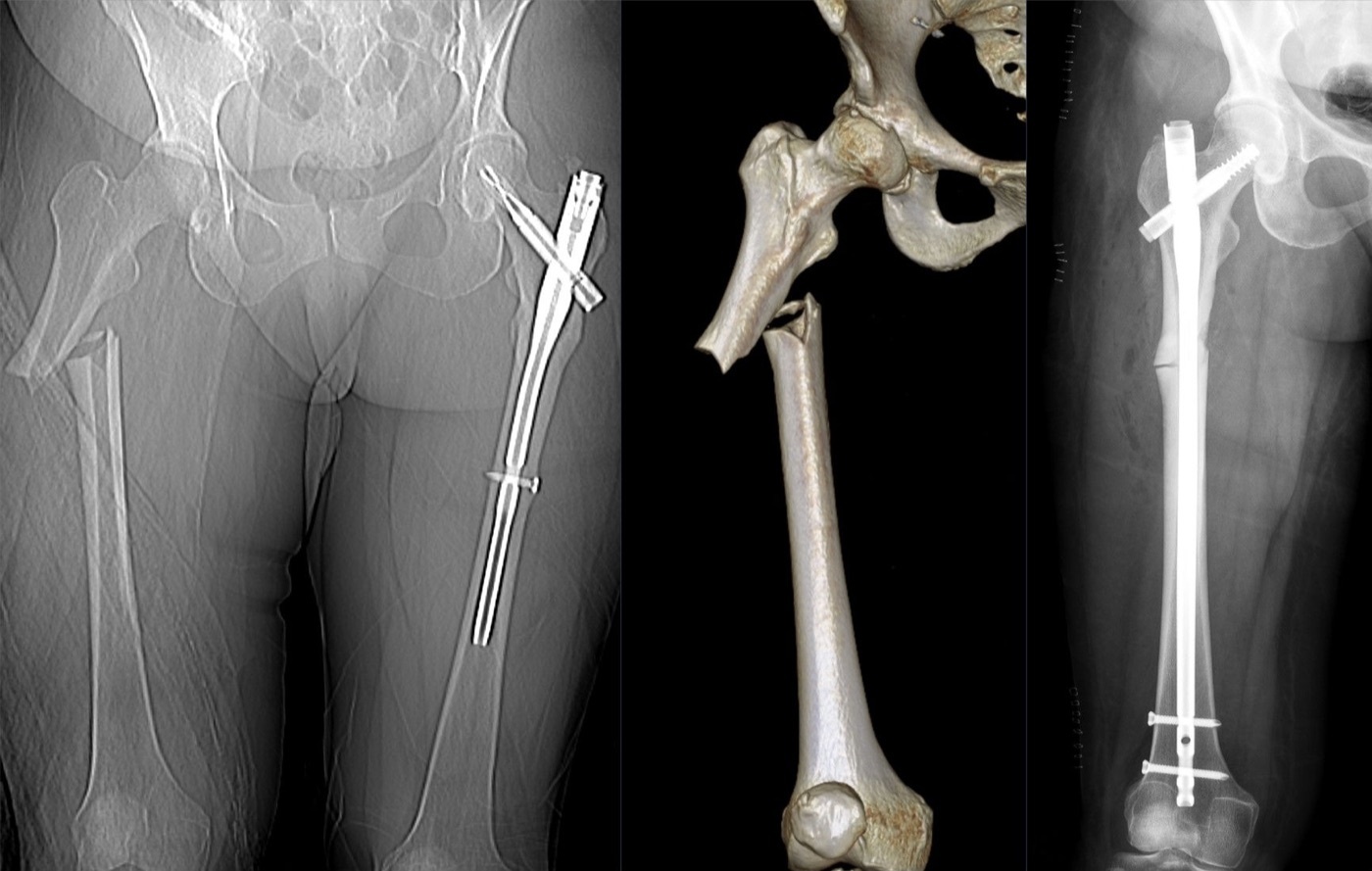
Figure 7. Atypical (bisphosphonate-associated) femoral fracture: a transverse proximal-shaft fracture with lateral cortical thickening (CT topogram and 3D rendering, left and centre) treated with an intramedullary nail (post-operative radiograph, right); note the indwelling nail in the previously fractured contralateral femur, illustrating the bilateral tendency of these fractures. Image by Hellerhoff, CC BY-SA 4.0, via Wikimedia Commons.
Part VI - Complications
The subtrochanteric fracture carries a higher rate of malunion and nonunion than other proximal femoral fractures, owing to its high mechanical stress, its cortical bone, and the ease of malreduction.[23] Malunion is typically into varus and procurvatum (apex-anterior) with shortening and is prevented by a careful, non-varus reduction. Nonunion follows malreduction (especially a varus, gapped medial cortex) and implant overload, and it is salvaged by revision fixation (an exchange nail, or a 95° blade plate with bone grafting) after restoring alignment.[24] Implant failure (nail or screw breakage) accompanies an ununited fracture in this loaded region. With modern cephalomedullary nailing and a good reduction, union rates are high (about 98-100%), and the mortality reflects the elderly fragility population (about a quarter at one year in the older group).[25]
Part VII - A Synthesis: How to Reason About the Subtrochanteric Fracture
This fracture is best approached as a mechanics problem. It sits where the femur is most heavily loaded, in slow-healing cortical bone, and it is torn apart by strong, opposing muscles that flex, abduct, and externally rotate the proximal fragment while adducting and shortening the shaft, so it tends to heal in varus and procurvatum. The job is twofold. Choose a strong, load-sharing implant, which is a long cephalomedullary nail in almost every case, and, more importantly, get the reduction right and hold it out of varus, taking the nail entry medial enough to avoid the lateral-start varus error and using blocking screws, clamps, or a mini-open reduction to corral the flexed-abducted proximal fragment and restore the medial cortex. Avoid the 135° sliding hip screw, and reserve the 95° blade plate for the difficult or failed case. Recognise the atypical, bisphosphonate-associated fracture by its transverse, lateral-cortex, beaked appearance and its prodromal thigh pain, nail it, stop the drug, and check the other femur. The subtrochanteric fracture punishes malreduction with nonunion and implant failure, so the reduction, not the implant alone, is what determines the outcome.
References
-
Rockwood & Green’s Fractures in Adults, pp.3758-3761, 3776, 3780 (the subtrochanteric region carries the highest compressive and tensile forces in the skeleton, in cortical bone, so nonunion/malunion/implant failure are risks; the proximal fragment flexed/abducted/externally rotated and the shaft adducted/shortened; varus malreduction the enemy; the cephalomedullary nail the preferred implant); Miller’s Review of Orthopaedics, pp.918-919 (deforming forces and the tendency to varus and apex-anterior malreduction).
-
Rockwood & Green’s Fractures in Adults, pp.3758-3761, 3776, 3780 (the subtrochanteric region carries the highest compressive and tensile forces in the skeleton, in cortical bone, so nonunion/malunion/implant failure are risks; the proximal fragment flexed/abducted/externally rotated and the shaft adducted/shortened; varus malreduction the enemy; the cephalomedullary nail the preferred implant); Miller’s Review of Orthopaedics, pp.918-919 (deforming forces and the tendency to varus and apex-anterior malreduction).
-
Rockwood & Green’s Fractures in Adults, p.3754 (subtrochanteric region from the lesser trochanter to ~5 cm distal; high-energy young, low-energy elderly, and the bisphosphonate/atypical subset); Miller’s Review of Orthopaedics, pp.918-919 (the subtrochanteric fracture and the atypical bisphosphonate-associated fracture).
-
Rockwood & Green’s Fractures in Adults, p.3754 (subtrochanteric region from the lesser trochanter to ~5 cm distal; high-energy young, low-energy elderly, and the bisphosphonate/atypical subset); Miller’s Review of Orthopaedics, pp.918-919 (the subtrochanteric fracture and the atypical bisphosphonate-associated fracture).
-
Rockwood & Green’s Fractures in Adults, pp.3758-3761 (highest compressive and tensile forces in the skeleton, >1200 lb/in² medially with medial comminution, cortical bone; the proximal fragment flexed by iliopsoas, abducted by the gluteus medius, externally rotated by the short external rotators, and the shaft adducted/shortened by the adductors); Miller’s Review of Orthopaedics, pp.919-920 (the same deforming forces and the resulting varus and apex-anterior malreduction). The explicit “medial compression / lateral tension” assignment, and the predominantly cortical, slower-healing nature of this diaphyseal bone, are standard teaching consistent with the source’s statement that the region bears the highest compressive (medial) and tensile (lateral) forces.
-
Rockwood & Green’s Fractures in Adults, pp.3758-3761 (highest compressive and tensile forces in the skeleton, >1200 lb/in² medially with medial comminution, cortical bone; the proximal fragment flexed by iliopsoas, abducted by the gluteus medius, externally rotated by the short external rotators, and the shaft adducted/shortened by the adductors); Miller’s Review of Orthopaedics, pp.919-920 (the same deforming forces and the resulting varus and apex-anterior malreduction). The explicit “medial compression / lateral tension” assignment, and the predominantly cortical, slower-healing nature of this diaphyseal bone, are standard teaching consistent with the source’s statement that the region bears the highest compressive (medial) and tensile (lateral) forces.
-
Rockwood & Green’s Fractures in Adults, pp.3758-3761 (highest compressive and tensile forces in the skeleton, >1200 lb/in² medially with medial comminution, cortical bone; the proximal fragment flexed by iliopsoas, abducted by the gluteus medius, externally rotated by the short external rotators, and the shaft adducted/shortened by the adductors); Miller’s Review of Orthopaedics, pp.919-920 (the same deforming forces and the resulting varus and apex-anterior malreduction). The explicit “medial compression / lateral tension” assignment, and the predominantly cortical, slower-healing nature of this diaphyseal bone, are standard teaching consistent with the source’s statement that the region bears the highest compressive (medial) and tensile (lateral) forces.
-
Rockwood & Green’s Fractures in Adults, pp.3758-3761 (highest compressive and tensile forces in the skeleton, >1200 lb/in² medially with medial comminution, cortical bone; the proximal fragment flexed by iliopsoas, abducted by the gluteus medius, externally rotated by the short external rotators, and the shaft adducted/shortened by the adductors); Miller’s Review of Orthopaedics, pp.919-920 (the same deforming forces and the resulting varus and apex-anterior malreduction). The explicit “medial compression / lateral tension” assignment, and the predominantly cortical, slower-healing nature of this diaphyseal bone, are standard teaching consistent with the source’s statement that the region bears the highest compressive (medial) and tensile (lateral) forces.
-
Miller’s Review of Orthopaedics, pp.918-919 (Russell-Taylor type I no piriformis-fossa extension [IA lesser trochanter intact, IB detached], type II with piriformis-fossa extension); AO Principles of Fracture Management, p.811 (subtrochanteric fractures coded within the femoral segment 32 diaphysis). The Seinsheimer classification (by fragment number and fracture-line geometry) is standard teaching but is named only as a reference, not described, in the mined Rockwood extract, so it is noted here as an established system rather than cited in detail to that source.
-
Miller’s Review of Orthopaedics, pp.918-919 (Russell-Taylor type I no piriformis-fossa extension [IA lesser trochanter intact, IB detached], type II with piriformis-fossa extension); AO Principles of Fracture Management, p.811 (subtrochanteric fractures coded within the femoral segment 32 diaphysis). The Seinsheimer classification (by fragment number and fracture-line geometry) is standard teaching but is named only as a reference, not described, in the mined Rockwood extract, so it is noted here as an established system rather than cited in detail to that source.
-
Miller’s Review of Orthopaedics, pp.918-919 (Russell-Taylor type I no piriformis-fossa extension [IA lesser trochanter intact, IB detached], type II with piriformis-fossa extension); AO Principles of Fracture Management, p.811 (subtrochanteric fractures coded within the femoral segment 32 diaphysis). The Seinsheimer classification (by fragment number and fracture-line geometry) is standard teaching but is named only as a reference, not described, in the mined Rockwood extract, so it is noted here as an established system rather than cited in detail to that source.
-
Miller’s Review of Orthopaedics, pp.918-920 (the atypical femoral fracture: long-term bisphosphonate association, a transverse fracture, lateral cortical beaking/thickening, prodromal thigh pain, and bilaterality, with contralateral radiographs indicated). The fuller formal feature set (the ASBMR major/minor criteria: lateral-cortex origin, minimal comminution, a medial spike) is standard teaching consistent with the source’s description.
-
Miller’s Review of Orthopaedics, pp.918-920 (the atypical femoral fracture: long-term bisphosphonate association, a transverse fracture, lateral cortical beaking/thickening, prodromal thigh pain, and bilaterality, with contralateral radiographs indicated). The fuller formal feature set (the ASBMR major/minor criteria: lateral-cortex origin, minimal comminution, a medial spike) is standard teaching consistent with the source’s description.
-
Rockwood & Green’s Fractures in Adults, pp.3754-3758 (high-energy young with associated injuries and blood loss, low-energy elderly, the atypical-fracture thigh-pain prodrome; AP and lateral radiographs of the whole femur including hip and knee, excluding an associated femoral neck fracture); Miller’s Review of Orthopaedics, p.918 (contralateral radiographs for the bilateral atypical fracture).
-
Rockwood & Green’s Fractures in Adults, pp.3754-3758 (high-energy young with associated injuries and blood loss, low-energy elderly, the atypical-fracture thigh-pain prodrome; AP and lateral radiographs of the whole femur including hip and knee, excluding an associated femoral neck fracture); Miller’s Review of Orthopaedics, p.918 (contralateral radiographs for the bilateral atypical fracture).
-
Rockwood & Green’s Fractures in Adults, pp.3776-3781, 3788 (the cephalomedullary/intramedullary nail the authors’ preferred treatment for all subtrochanteric fractures; restore length/alignment/rotation; varus driven by a too-lateral start point and eccentric reaming, controlled with reduction aids and mini-open cerclage; plates [95° blade plate, dynamic condylar screw, locking plate] biomechanically inferior with failure rates up to 26-41% and reserved for nonunion/malunion); AO Principles of Fracture Management, pp.814, 819 (entry point medial to the greater-trochanter tip to avoid varus; the 95° blade plate as a plating option); Miller’s Review of Orthopaedics, pp.919-920 (the reconstruction/cephalomedullary nail the device of choice, a 135° device contraindicated, a 95° device the plating alternative, varus and apex-anterior malreduction). The “Poller (blocking) screw” is standard terminology; the mined Rockwood extract describes the blocking concept without that eponym, and teriparatide for atypical fractures is standard adjunctive teaching not detailed in these extracts.
-
Rockwood & Green’s Fractures in Adults, pp.3776-3781, 3788 (the cephalomedullary/intramedullary nail the authors’ preferred treatment for all subtrochanteric fractures; restore length/alignment/rotation; varus driven by a too-lateral start point and eccentric reaming, controlled with reduction aids and mini-open cerclage; plates [95° blade plate, dynamic condylar screw, locking plate] biomechanically inferior with failure rates up to 26-41% and reserved for nonunion/malunion); AO Principles of Fracture Management, pp.814, 819 (entry point medial to the greater-trochanter tip to avoid varus; the 95° blade plate as a plating option); Miller’s Review of Orthopaedics, pp.919-920 (the reconstruction/cephalomedullary nail the device of choice, a 135° device contraindicated, a 95° device the plating alternative, varus and apex-anterior malreduction). The “Poller (blocking) screw” is standard terminology; the mined Rockwood extract describes the blocking concept without that eponym, and teriparatide for atypical fractures is standard adjunctive teaching not detailed in these extracts.
-
Rockwood & Green’s Fractures in Adults, pp.3776-3781, 3788 (the cephalomedullary/intramedullary nail the authors’ preferred treatment for all subtrochanteric fractures; restore length/alignment/rotation; varus driven by a too-lateral start point and eccentric reaming, controlled with reduction aids and mini-open cerclage; plates [95° blade plate, dynamic condylar screw, locking plate] biomechanically inferior with failure rates up to 26-41% and reserved for nonunion/malunion); AO Principles of Fracture Management, pp.814, 819 (entry point medial to the greater-trochanter tip to avoid varus; the 95° blade plate as a plating option); Miller’s Review of Orthopaedics, pp.919-920 (the reconstruction/cephalomedullary nail the device of choice, a 135° device contraindicated, a 95° device the plating alternative, varus and apex-anterior malreduction). The “Poller (blocking) screw” is standard terminology; the mined Rockwood extract describes the blocking concept without that eponym, and teriparatide for atypical fractures is standard adjunctive teaching not detailed in these extracts.
-
Rockwood & Green’s Fractures in Adults, pp.3776-3781, 3788 (the cephalomedullary/intramedullary nail the authors’ preferred treatment for all subtrochanteric fractures; restore length/alignment/rotation; varus driven by a too-lateral start point and eccentric reaming, controlled with reduction aids and mini-open cerclage; plates [95° blade plate, dynamic condylar screw, locking plate] biomechanically inferior with failure rates up to 26-41% and reserved for nonunion/malunion); AO Principles of Fracture Management, pp.814, 819 (entry point medial to the greater-trochanter tip to avoid varus; the 95° blade plate as a plating option); Miller’s Review of Orthopaedics, pp.919-920 (the reconstruction/cephalomedullary nail the device of choice, a 135° device contraindicated, a 95° device the plating alternative, varus and apex-anterior malreduction). The “Poller (blocking) screw” is standard terminology; the mined Rockwood extract describes the blocking concept without that eponym, and teriparatide for atypical fractures is standard adjunctive teaching not detailed in these extracts.
-
Rockwood & Green’s Fractures in Adults, pp.3776-3781, 3788 (the cephalomedullary/intramedullary nail the authors’ preferred treatment for all subtrochanteric fractures; restore length/alignment/rotation; varus driven by a too-lateral start point and eccentric reaming, controlled with reduction aids and mini-open cerclage; plates [95° blade plate, dynamic condylar screw, locking plate] biomechanically inferior with failure rates up to 26-41% and reserved for nonunion/malunion); AO Principles of Fracture Management, pp.814, 819 (entry point medial to the greater-trochanter tip to avoid varus; the 95° blade plate as a plating option); Miller’s Review of Orthopaedics, pp.919-920 (the reconstruction/cephalomedullary nail the device of choice, a 135° device contraindicated, a 95° device the plating alternative, varus and apex-anterior malreduction). The “Poller (blocking) screw” is standard terminology; the mined Rockwood extract describes the blocking concept without that eponym, and teriparatide for atypical fractures is standard adjunctive teaching not detailed in these extracts.
-
Miller’s Review of Orthopaedics, pp.918-920 (atypical fractures nailed, the bisphosphonate stopped, the contralateral side imaged and treated, a symptomatic incomplete fracture with cortical beaking fixed prophylactically while an asymptomatic one is monitored; slow union); Rockwood & Green’s Fractures in Adults, pp.3787-3788 (the bisphosphonate-associated fracture and its slower healing). Teriparatide as a healing adjunct is standard teaching consistent with these sources.
-
Miller’s Review of Orthopaedics, pp.918-920 (atypical fractures nailed, the bisphosphonate stopped, the contralateral side imaged and treated, a symptomatic incomplete fracture with cortical beaking fixed prophylactically while an asymptomatic one is monitored; slow union); Rockwood & Green’s Fractures in Adults, pp.3787-3788 (the bisphosphonate-associated fracture and its slower healing). Teriparatide as a healing adjunct is standard teaching consistent with these sources.
-
Rockwood & Green’s Fractures in Adults, pp.3781-3788 (higher malunion/nonunion than other proximal femoral fractures from the high stress, cortical bone, and malreduction; malunion into varus/procurvatum with shortening, prevented by a non-varus reduction; nonunion from malreduction and overload, salvaged by exchange nailing or a 95° blade plate with grafting; implant failure with nonunion; union ~98-100% with modern nailing and good reduction; ~25% one-year mortality in the elderly, Robinson).
-
Rockwood & Green’s Fractures in Adults, pp.3781-3788 (higher malunion/nonunion than other proximal femoral fractures from the high stress, cortical bone, and malreduction; malunion into varus/procurvatum with shortening, prevented by a non-varus reduction; nonunion from malreduction and overload, salvaged by exchange nailing or a 95° blade plate with grafting; implant failure with nonunion; union ~98-100% with modern nailing and good reduction; ~25% one-year mortality in the elderly, Robinson).
-
Rockwood & Green’s Fractures in Adults, pp.3781-3788 (higher malunion/nonunion than other proximal femoral fractures from the high stress, cortical bone, and malreduction; malunion into varus/procurvatum with shortening, prevented by a non-varus reduction; nonunion from malreduction and overload, salvaged by exchange nailing or a 95° blade plate with grafting; implant failure with nonunion; union ~98-100% with modern nailing and good reduction; ~25% one-year mortality in the elderly, Robinson).
-
Rockwood & Green’s Fractures in Adults, pp.3754, 3758-3761.
-
Rockwood & Green’s Fractures in Adults, pp.3758-3761; Miller’s Review of Orthopaedics, pp.919-920.
-
Miller’s Review of Orthopaedics, pp.918-919.
-
Rockwood & Green’s Fractures in Adults, pp.3776-3780; AO Principles of Fracture Management, p.819; Miller’s Review of Orthopaedics, pp.919-920.
-
Rockwood & Green’s Fractures in Adults, pp.3778-3781; Miller’s Review of Orthopaedics, pp.919-920.
-
Miller’s Review of Orthopaedics, pp.918-920.
-
Miller’s Review of Orthopaedics, pp.918-920; Rockwood & Green’s Fractures in Adults, pp.3787-3788.
-
Rockwood & Green’s Fractures in Adults, pp.3754-3758.
-
Rockwood & Green’s Fractures in Adults, pp.3776, 3781-3788.
-
Rockwood & Green’s Fractures in Adults, pp.3781-3788.
-
Rockwood & Green’s Fractures in Adults, pp.3781-3788.
-
Rockwood & Green’s Fractures in Adults, pp.3758-3761, 3780; Miller’s Review of Orthopaedics, pp.919-920.