Contents
- Orientation
- Part I - Principles: The Injured Patient and Spinal Cord Injury
- Part II - The Incomplete Cord Syndromes
- Part III - The Cervical Spine: Anatomy, Assessment, and Clearance
- Part IV - Upper Cervical Injuries
- Part V - Subaxial (C3-C7) Cervical Injuries
- Part VI - Cervical Treatment Principles
- Part VII - Thoracolumbar Spine: Regions, Assessment, and Classification
- Part VIII - Thoracolumbar Treatment
- Part IX - Paediatric Spine Trauma
- References
Orientation
Spine trauma is the broadest single topic in the traumatology syllabus, and the one where a missed or mishandled injury does the most damage, because the structure at risk, the spinal cord, does not recover what it loses. The subject splits into three problems that must be held apart. First, the patient and the cord: resuscitation, the neurological examination, spinal versus neurogenic shock, and the incomplete cord syndromes. Second, the cervical spine, from the occipitocervical junction down through the subaxial segments. Third, the thoracolumbar spine, with its transition zone, its burst-fracture controversy, and its osteoporotic fractures. A short paediatric section closes the clinical material, since the immature spine fails in ways the adult spine does not.[1]
Two ideas recur throughout. The first is stability: an injury is unstable when it can no longer bear physiological load while protecting the neural elements, and the whole apparatus of classification (the three-column model, the SLIC and TLICS scores, the AOSpine system) exists to answer one question, whether to operate. The second is that the soft tissues and the cord, not just the bone, define the injury. The posterior ligamentous complex decides the fate of many thoracolumbar fractures, the disc decides whether a cervical dislocation can be reduced safely, and the neurological status governs almost everything about urgency.[2]
A Bulgarian glossary and a viva-voce appendix close the document.
Part I - Principles: The Injured Patient and Spinal Cord Injury
Epidemiology and the first hours
Spine fractures are rising in incidence, driven by an ageing, osteoporotic population, and most occur at the junctions of the rigid thoracic cage. Spinal cord injury affects 250,000 to 500,000 people worldwide each year, is traumatic in up to 90 per cent of cases, and typically strikes in the fourth decade.[3] The consequences are lifelong and expensive, with a lifetime cost near 4.7 million dollars for tetraplegia, and they are stratified by level: a cord injury above C5 threatens the diaphragm and spontaneous breathing.[4] Management follows ATLS, with the airway secured under cervical protection. The modern trend has moved away from reflex backboarding and field collars (which cause pressure sores and aspiration and, in the most unstable injuries, do not even reduce motion) toward protocol-driven, examination-based precautions.[5]
The neurological examination: ASIA, complete versus incomplete
Assessment rests on a structured neurological examination scored on the ASIA Impairment Scale (AIS): grade A is complete (no motor or sensory function, including the sacral segments), grades B through D are incomplete (B sensory only, C motor preserved with more than half of the key muscle groups below grade 3, D motor preserved with more than half at grade 3 or above), and grade E is normal.[6] The single most important distinction is complete versus incomplete, and it turns on sacral sparing: any preserved perianal sensation, voluntary anal contraction, or deep anal pressure makes the injury incomplete and sharply improves the prognosis. The AIS becomes reliable only once spinal shock has resolved, so it cannot be assigned in the first hours.[7]
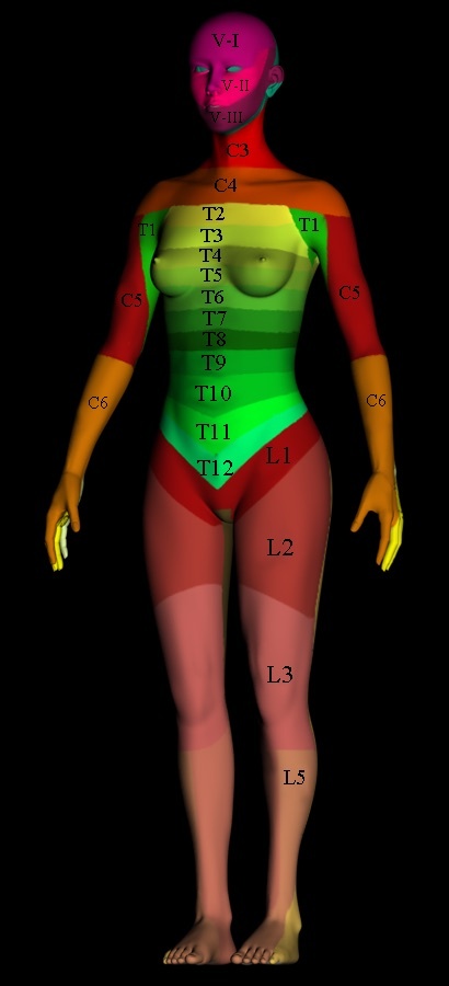
Figure 1. Anterior dermatome map showing the cutaneous segmental levels used to assign the neurological level of injury in the ASIA examination. By Hy9etb, CC BY 3.0, via Wikimedia Commons.
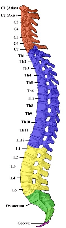
Figure 2. Lateral view of the vertebral column with its cervical, thoracic, lumbar, sacral and coccygeal regions. Coloured plate after Gray’s Anatomy (1918), public domain, via Wikimedia Commons.
Spinal shock versus neurogenic shock
These two entities share a name and a cause (cord injury) but are otherwise distinct, and confusing them is a classic examination trap. Neurogenic shock is a haemodynamic, distributive shock from sympathetic disruption: hypotension with bradycardia (and often hypothermia), most common with cervical or upper-thoracic injury. The bradycardia separates it from the far commoner hypovolaemic shock, in which the patient is tachycardic.[8] Spinal shock is a transient, global loss of motor, sensory, reflex and autonomic function below the lesion. It ends with the return of reflexes, generally between 48 hours and two weeks, the first to return being the polysynaptic bulbocavernosus reflex (anal sphincter contraction on squeezing the glans or tugging the catheter), which tests the S2-S4 conus and marks the end of spinal shock.[9]
Steroids, perfusion, timing, and autonomic dysreflexia
The role of high-dose methylprednisolone is the field’s longest-running controversy. The NASCIS trials showed no benefit in their primary outcomes, only post-hoc subgroup trends, at the cost of unacceptable pneumonia, sepsis and mortality. The modern verdict treats it as experimental at best: several consensus bodies recommend against it, and others reinstate it only as a 24-hour option within 8 hours of injury in selected, healthier patients.[10] It is contraindicated in penetrating injury. Beyond steroids, two levers may protect the cord from secondary injury. The first is perfusion, maintaining a mean arterial pressure around 85 mm Hg for the first week. The second is timely decompression: the STASCIS trial supported decompression within 24 hours, though later analyses softened this to a trend.[11] One delayed complication worth knowing is autonomic dysreflexia, seen after the spinal-shock phase in lesions at or above T6. A noxious stimulus below the lesion, classically a distended bladder or impacted bowel, triggers paroxysmal hypertension, a pounding headache and a baroreflex bradycardia, managed by sitting the patient up, removing the trigger, and giving antihypertensives.[12]
Part II - The Incomplete Cord Syndromes
When a cord injury is incomplete, the pattern of sparing identifies a syndrome with its own prognosis, and these are high-yield. Central cord syndrome is the commonest. It occurs in the older patient with a stenotic, spondylotic cervical canal who hyperextends the neck, injuring the central grey matter; it produces weakness greater in the upper than the lower limbs, with a generally good prognosis for walking but poorer recovery of hand function.[13] Anterior cord syndrome (anterior two-thirds of the cord, the anterior spinal artery territory) loses motor function and pain and temperature while sparing the dorsal columns, and has the worst prognosis. Brown-Séquard syndrome (a hemisection, classically penetrating) loses ipsilateral motor and proprioception with contralateral pain and temperature, and has the best prognosis, with roughly 90 per cent regaining the ability to walk.[14] Posterior cord syndrome (isolated dorsal-column loss) is rare. Below the cord, the conus medullaris syndrome gives early symmetric saddle anaesthesia with mixed upper and lower motor neuron signs, whereas the cauda equina syndrome gives asymmetric, lower-motor-neuron, radicular deficits and is a surgical urgency.[15]
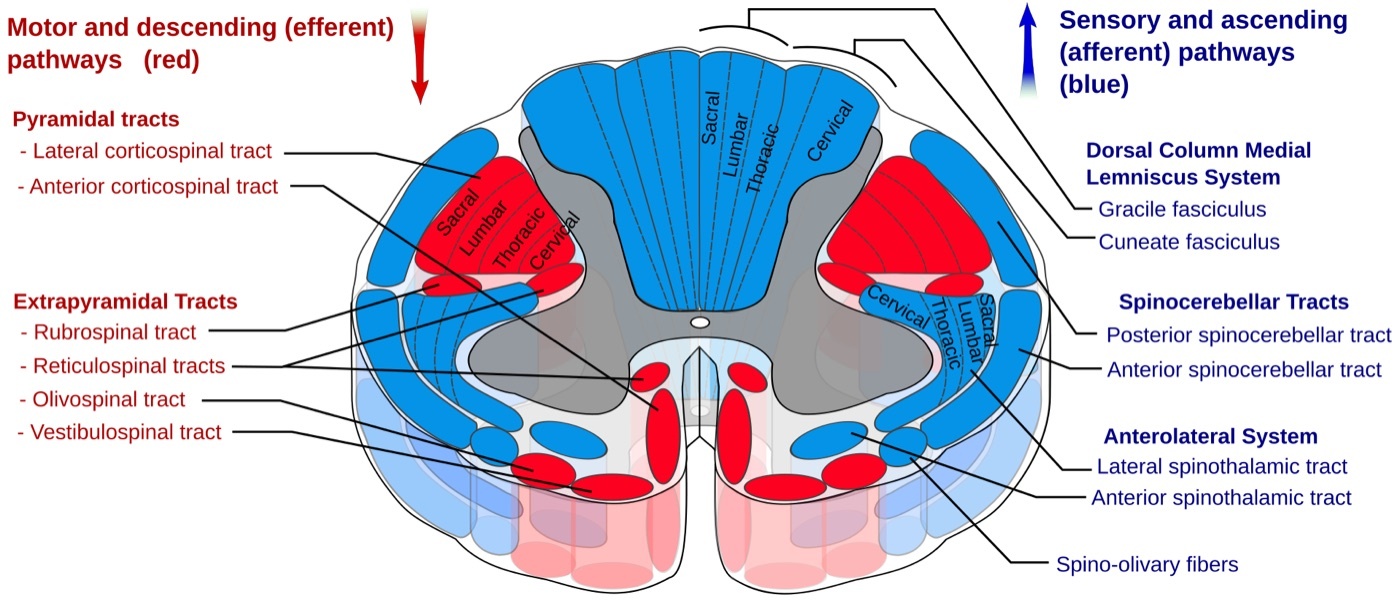
Figure 3. Cross-section of the spinal cord showing the principal ascending (sensory) and descending (motor) tracts; the corticospinal tracts and dorsal columns are ipsilateral while the spinothalamic tracts cross. Polarlys and Mikael Häggström, CC BY-SA 3.0, via Wikimedia Commons.
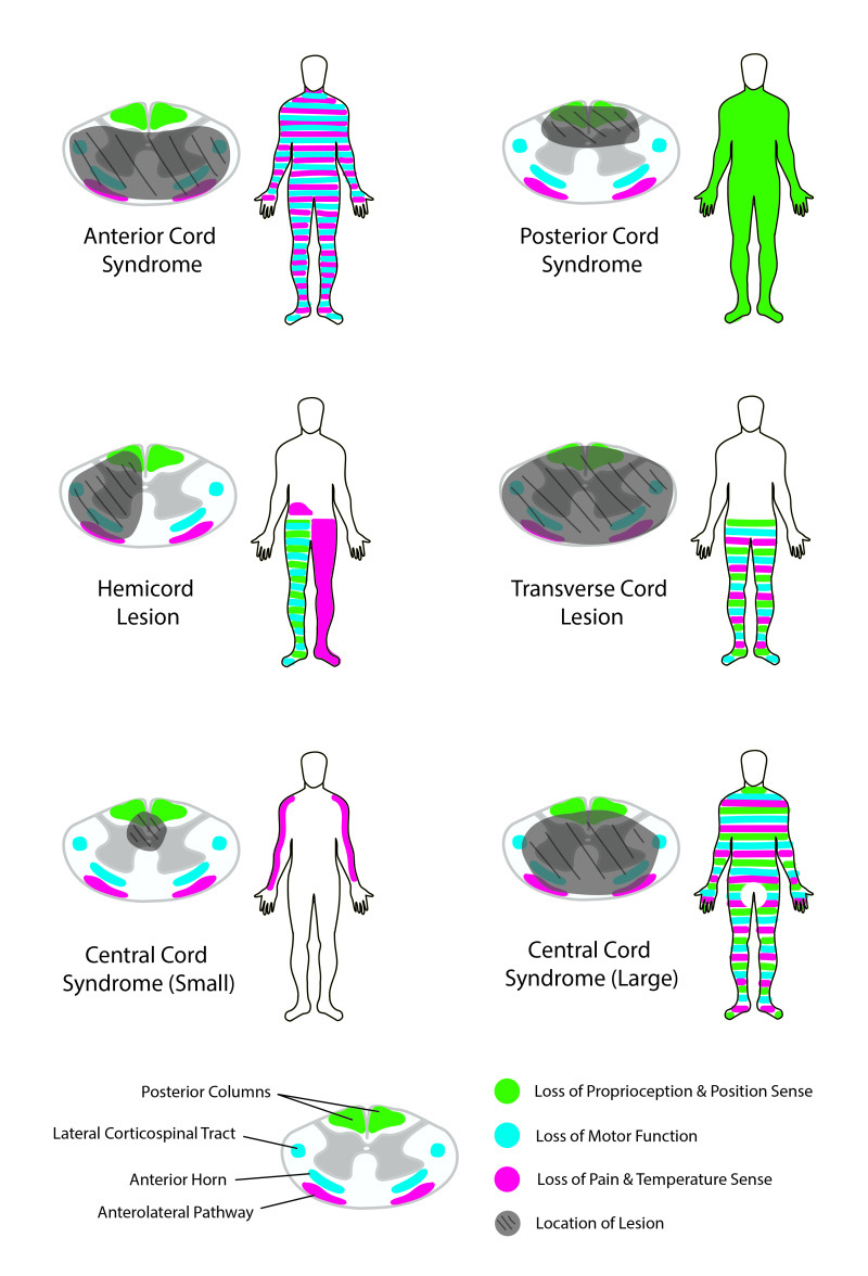
Figure 4. Patterns of incomplete spinal cord injury (anterior, posterior, central, Brown-Séquard and transverse lesions) with the cord cross-section location and the resulting sensorimotor deficits. Rian Kabir, CC BY 4.0, via Wikimedia Commons.
Part III - The Cervical Spine: Anatomy, Assessment, and Clearance
The cervical spine is built in two functionally different parts. The occipitocervical complex (occiput, atlas and axis) is a ligament-dependent, highly mobile region in which the transverse ligament holds the dens against the anterior arch of C1, the alar ligaments restrain rotation, and roughly half of all cervical axial rotation occurs at C1-C2.[16] The subaxial spine (C3-C7) behaves as a series of three-joint complexes (two facet joints and a disc) restrained by the anterior longitudinal ligament and the posterior ligamentous complex, with the vertebral artery running in the foramen transversarium.
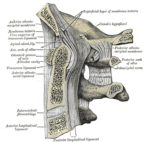
Figure 5. Median sagittal section of the occipito-atlanto-axial region showing the odontoid process (dens) and the transverse ligament that retains it against the anterior arch of the atlas. Gray’s Anatomy (1918), public domain, via Wikimedia Commons.
Mental status governs clearance of the cervical spine, and two validated rules apply to the alert patient. The NEXUS criteria clear the spine without imaging when there is no posterior midline tenderness, no focal neurological deficit, normal alertness, no intoxication, and no distracting injury; the Canadian C-spine rule is more sensitive but more complex.[17] When imaging is needed, CT is the primary screening modality, around 99 per cent sensitive against a plain-film false-negative rate of 30 per cent in the cervical spine. MRI is reserved for an examination that does not match the CT, for suspected cord, ligamentous or disc injury, or for the obtunded patient, where the modern consensus increasingly accepts a high-quality negative CT alone.[18] The radiographic measurements worth knowing are the atlanto-dental interval (normally 3 mm or less in the adult), the prevertebral soft-tissue shadow (about 6 mm at C2, 20 mm at C6), and the occipitocervical relationships used to diagnose dissociation.
Part IV - Upper Cervical Injuries
Occipital condyle fractures and atlanto-occipital dissociation
Occipital condyle fractures are classified by Anderson and Montesano into type I (impaction, stable), type II (basilar skull fracture into the condyle, stable) and type III (avulsion, potentially unstable because it implies alar-ligament and craniocervical injury). CT is the imaging of choice, and the tectorial membrane on MRI determines stability.[19] Atlanto-occipital dissociation amounts to an internal decapitation: a grossly unstable, primarily ligamentous, often fatal distraction injury that is frequently missed on plain films and is diagnosed by the basion-dental and basion-axial intervals (the Harris “rule of 12”) or the Powers ratio (greater than 1). Longitudinal traction is contraindicated, and the treatment is urgent posterior occipitocervical fusion.[20]
Atlas (C1) fractures and the rule of Spence
Because the C1 ring cannot break in a single place, it fractures in at least two, and the classic high-energy axial pattern is the Jefferson burst fracture. The decisive question is the integrity of the transverse ligament. The rule of Spence holds that a combined lateral-mass overhang exceeding about 7 mm (the often-quoted 6.9 mm figure is Miller’s) on the open-mouth view implies transverse-ligament rupture, though this measurement now serves as an adjunct to MRI, which inspects the ligament directly.[21] A C1 fracture with an intact ligament is treated in a collar or halo; one with an incompetent ligament, or persistent C1-C2 instability after healing, calls for posterior C1-C2 fusion.
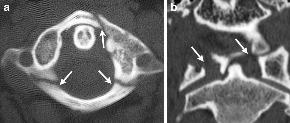
Figure 6. CT of a Jefferson (C1 burst) fracture: the axial image shows fractures of the anterior and posterior arches of the atlas, with lateral-mass spread on the coronal reformat. Utz et al., CC BY 4.0, via Wikimedia Commons.
Odontoid (dens) fractures
The dens fractures in a pattern classified by Anderson and D’Alonzo: type I is an avulsion of the tip (alar ligament), type II is through the waist at the junction of the dens and the body, and type III extends into the cancellous body of C2.[22] The type II fracture is the problem child, with a nonunion rate around 30 per cent and identifiable risk factors: displacement over 5 mm, posterior displacement, angulation over 10 degrees, advanced age, and delayed treatment. Type I is treated in a collar and type III usually heals in an orthosis. The displaced type II is generally operative, by either an anterior odontoid screw (which preserves C1-C2 motion but requires a reducible fracture and a favourable, posterior-oblique fracture line, and is contraindicated in the anteroinferior-to-posterosuperior pattern) or a posterior C1-C2 fusion.[23] The elderly type II fracture is its own controversy, because both operative and nonoperative routes do poorly over 80, and many units accept a stable fibrous nonunion in a hard collar.
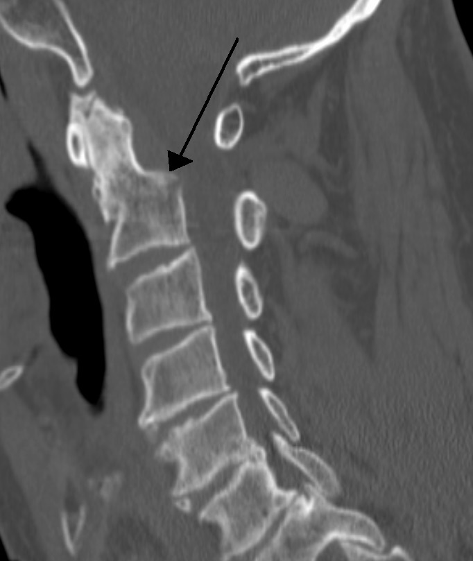
Figure 7. Sagittal CT showing a fracture through the base of the odontoid process (dens) of C2, the Anderson and D’Alonzo type II pattern that carries the highest nonunion risk. James Heilman, MD, CC BY-SA 3.0, via Wikimedia Commons.
Hangman’s fracture and atlantoaxial rotatory injury
The hangman’s fracture (traumatic spondylolisthesis of the axis) is a fracture through the C2 pars, classified by Levine and Edwards: type I is minimally displaced (under 3 mm, no angulation), type II has both angulation and translation from extension then flexion, type IIa is a flexion injury with angulation but little translation that must not be put in traction (which worsens it), and type III adds a C2-C3 facet dislocation.[24] Most are treated nonoperatively, with a collar for type I, a halo for type II, and reduction by extension and compression for IIa; surgery is reserved for type III and for failures. Atlantoaxial rotatory subluxation (Fielding-Hawkins types I-IV) is rare in adults, frequently missed, and diagnosed by a dynamic rotation CT showing a fixed deformity.[25]
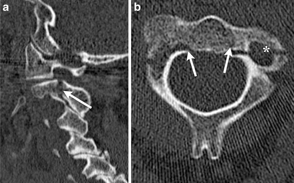
Figure 8. Hangman’s fracture (traumatic spondylolisthesis of the axis): sagittal CT shows the bilateral C2 pars fracture with anterolisthesis of C2 on C3, confirmed on the axial image. Utz et al., CC BY 4.0, via Wikimedia Commons.
Part V - Subaxial (C3-C7) Cervical Injuries
Classification: Allen-Ferguson, SLIC, and AOSpine
Three systems describe subaxial injuries. The Allen and Ferguson mechanistic scheme sorts them by the position of the neck and the mode of failure into six families (compressive flexion, vertical compression, distractive flexion, compressive extension, distractive extension, lateral flexion), each with progressive stages.[26] The SLIC score is the working treatment tool, summing three axes (morphology, discoligamentous complex, and neurological status), with surgery generally indicated at a total of 5 or more. An isolated burst without ligamentous or neurological injury scores only 2 and does not warrant surgery, whereas any neurological deficit, regardless of ligament status, mandates stabilisation.[27] The AOSpine subaxial classification parallels the thoracolumbar one (A compression, B tension-band, C translation, plus a facet “F” modifier).
Facet dislocations and the MRI-before-reduction question
Facet injuries run a continuum from a sprain to a complete bilateral dislocation, and one paradox is worth remembering: a unilateral dislocation can occur without catastrophic ligamentous disruption, whereas a bilateral facet subluxation usually carries it.[28] The central management controversy is whether to obtain an MRI before reducing a dislocation, the concern being a herniated disc that could be driven into the cord. The pragmatic resolution is that closed reduction by serial traction is safe in an awake, cooperative, examinable patient and can proceed without prior MRI, while an MRI comes first when the patient cannot be examined (intoxicated, obtunded) or is neurologically intact. A disc herniation found after reduction is addressed by an anterior approach.[29]
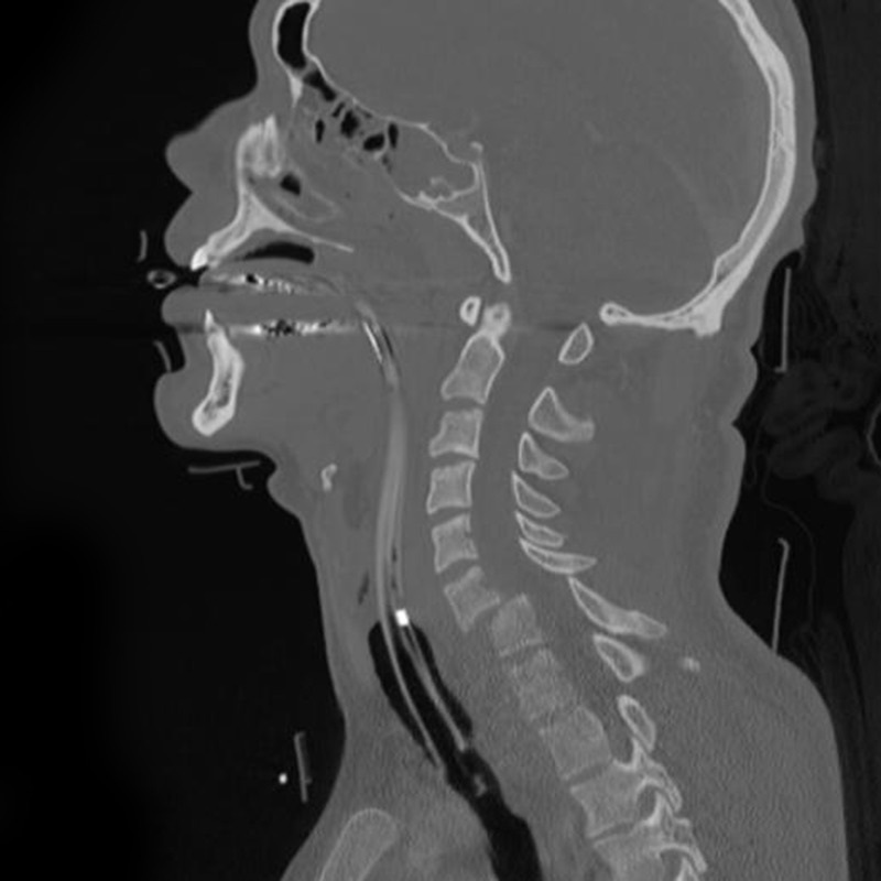
Figure 9. Sagittal CT of a C6-C7 cervical fracture-dislocation with anterolisthesis, the bony correlate of jumped facets. Frank Gaillard (Radiopaedia), CC BY-SA 3.0, via Wikimedia Commons.
Teardrop fractures, central cord, and the ankylosed spine
The flexion teardrop fracture is a severe compressive-flexion injury with a teardrop fragment, frequent posterior translation, and a high rate of complete cord injury. It usually requires anterior corpectomy and fusion, with posterior stabilisation added for the most translated patterns.[30] Central cord syndrome in the stenotic spine, discussed in Part II, is the prototypical cord injury without instability, and its timing of surgery is debated, with many units observing for early recovery and decompressing those who plateau. The most dangerous subaxial setting is the ankylosed spine (ankylosing spondylitis or DISH), which behaves like a fractured long bone: a low-energy injury produces a highly unstable, often three-column fracture, frequently missed, with a high rate of epidural haematoma and a grim mortality (acute mortality 17 to 30 per cent, and for ankylosing spondylitis up to 50 per cent at two years).[31] Inline traction can be catastrophic here because the pre-existing kyphosis must be respected, so treatment is long-segment posterior fixation contoured to the patient’s own deformity. The clay-shoveler’s fracture of the C7 spinous process, by contrast, is a benign avulsion when isolated.
Part VI - Cervical Treatment Principles
Nonoperative options run from collars (soft and rigid) to the halo vest, which controls the upper cervical spine well but the subaxial spine poorly, is tolerated badly by the elderly (with significant mortality), and is applied with four pins at 6 to 8 inch-pounds in the adult.[32] Closed reduction of facet dislocations uses Gardner-Wells tongs with progressive weights in the awake patient. Operative treatment uses anterior approaches (anterior cervical discectomy or corpectomy with plating, ideal for anterior compression and disc herniation) or posterior approaches (lateral mass and pedicle screws, and for C1-C2 the lateral-mass-plus-isthmus or transarticular constructs). The goals are decompression, realignment, stabilisation and fusion, and the evidence (STASCIS) favours decompression within 24 hours where feasible.[33]
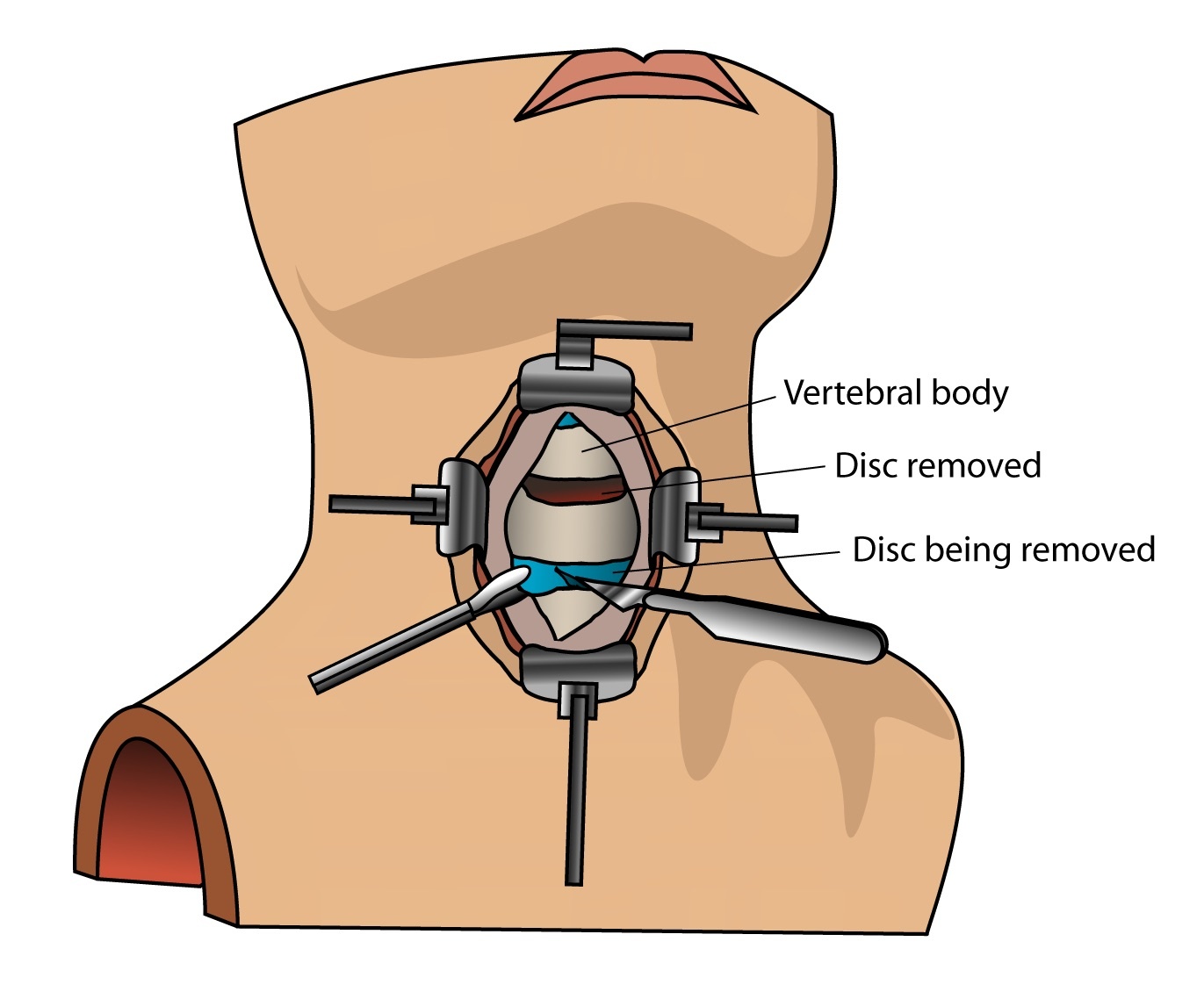
Figure 10. Anterior cervical discectomy and fusion (ACDF): the disc is removed through an anterior approach before interbody grafting and plating, addressing anterior compression. By debivort, CC BY-SA 3.0, via Wikimedia Commons.
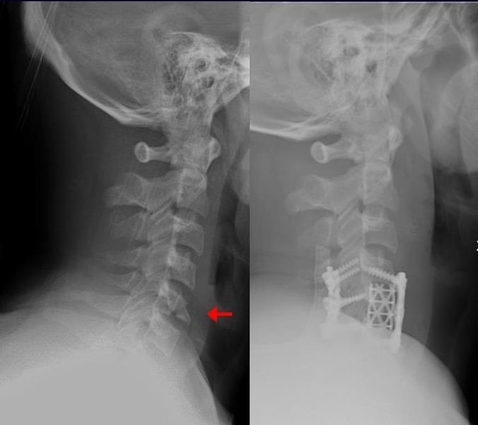
Figure 11. A lower cervical (teardrop) fracture before (left) and after anterior cervical discectomy and fusion with an interbody cage and anterior plate (right). By Moquito 17, CC BY-SA 3.0, via Wikimedia Commons.
Part VII - Thoracolumbar Spine: Regions, Assessment, and Classification
The three regions and the posterior ligamentous complex
The thoracolumbar spine divides into three regions. The rigid, kyphotic thoracic region (T2-T10) is splinted by the rib cage and coronally oriented facets but has a narrow canal and a vascular watershed at T4-T8, so deficits can occur with little canal compromise. The thoracolumbar junction (T11-L2) is the straight transition zone, the commonest fracture site, and houses the conus medullaris. The more mobile, lordotic lumbosacral region (L3-S1) has a wide canal and a resilient cauda equina that tolerate far greater canal compromise.[34] Running through all three is the posterior ligamentous complex (the facet capsules, the interspinous and supraspinous ligaments, and the ligamentum flavum), which acts as the posterior tension band, and whose failure is the hallmark of an unstable injury.[35]
Denis, TLICS, and AOSpine
Three classifications dominate. The Denis three-column model divides the spine into an anterior column (anterior longitudinal ligament and anterior half of the body and disc), a middle column (posterior half of the body and disc, and the posterior longitudinal ligament), and a posterior column (the neural arch and the posterior ligamentous complex). The middle column is the key to stability, an injury is unstable when at least two columns fail, and the four major injury types are compression, burst, flexion-distraction (Chance), and fracture-dislocation.[36] The TLICS score sums morphology (compression 1, burst 1, translational 3, distraction 4), posterior-ligamentous-complex integrity (intact 0, indeterminate 2, injured 3) and neurology (intact 0, root or complete cord 2, incomplete cord or cauda equina 3); a total of 3 or less is treated nonoperatively, 4 is indeterminate, and 5 or more operatively.[37] The revised AOSpine classification grades by type A (compression: A0 minor, A1 wedge, A2 split, A3 incomplete burst, A4 complete burst), type B (tension-band failure: B1 the bony Chance, B2 the posterior ligamentous disruption, B3 the hyperextension injury of the ankylosed spine) and type C (translation, the most unstable), with neurological (N) and modifier (M) qualifiers.[38]
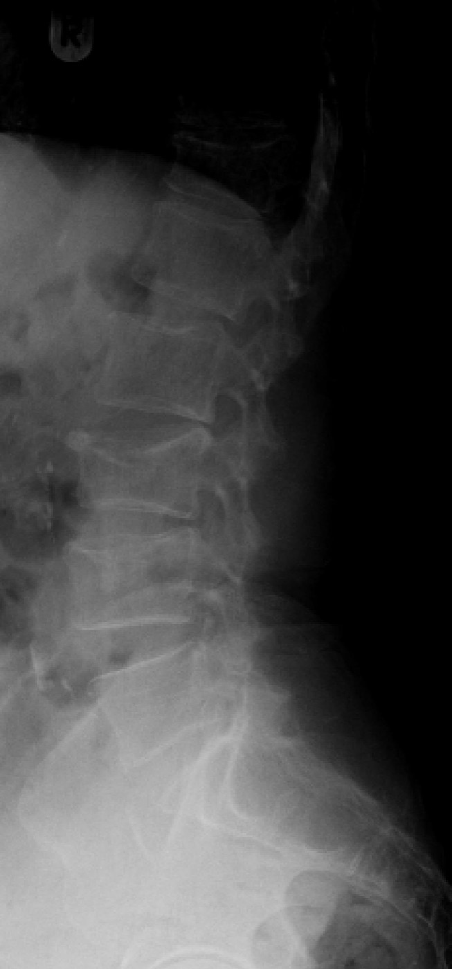
Figure 12. Lateral radiograph of a thoracolumbar compression (anterior wedge) fracture with preserved posterior body height; involvement of the anterior column alone distinguishes it from a burst fracture. Lucien Monfils, CC BY-SA 3.0, via Wikimedia Commons.
Burst and flexion-distraction fractures
The burst fracture is defined by a fracture of the posterior vertebral cortex with a retropulsed fragment in the canal (AOSpine A3 incomplete, A4 complete), recognised by interpedicular widening on the AP film and characterised by CT.[39] The flexion-distraction (Chance) fracture is the seatbelt injury, in which a lap belt focuses the moment along the anterior body and the posterior tension band fails. The bony Chance (B1) passes transversely through a single vertebra, while the ligamentous form (B2) crosses a disc space. Its high-yield association is intra-abdominal injury, found in about 30 per cent of flexion-distraction injuries.[40]
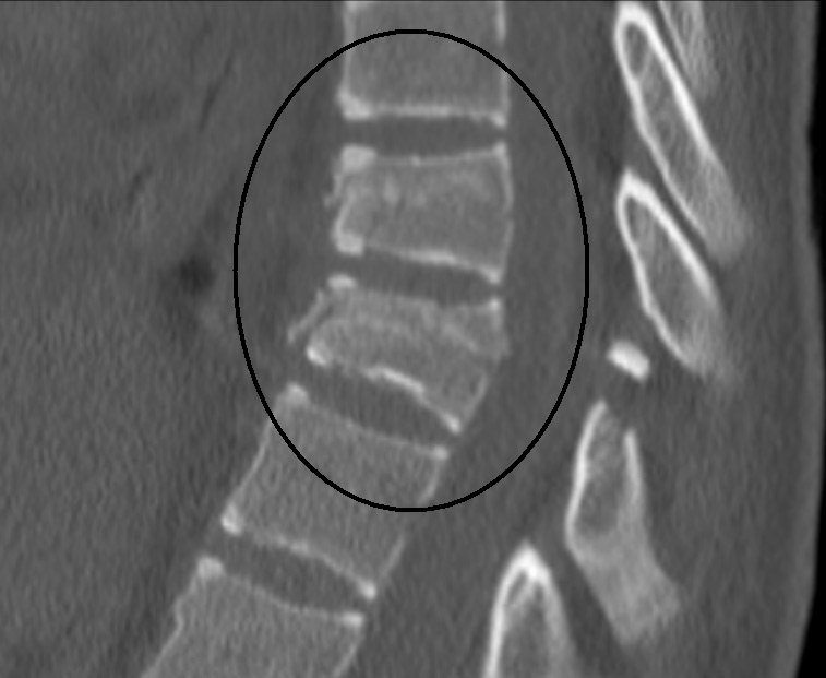
Figure 13. Sagittal CT of a Chance (flexion-distraction) fracture, with a horizontal split running through the vertebral body and posterior elements; the seatbelt mechanism carries a high rate of intra-abdominal injury. James Heilman, MD, CC BY-SA 3.0, via Wikimedia Commons.
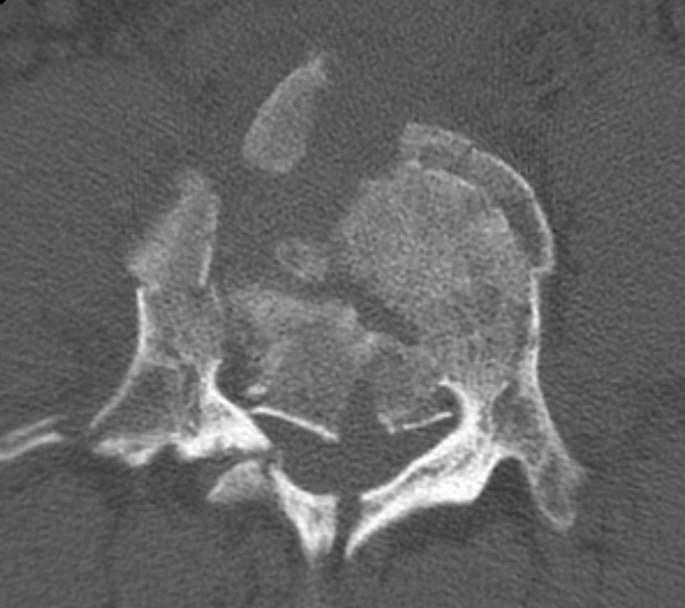
Figure 14. Axial CT of a lumbar burst fracture with comminution of the vertebral body and a retropulsed fragment narrowing the spinal canal. James Heilman, MD, CC BY-SA 4.0, via Wikimedia Commons.
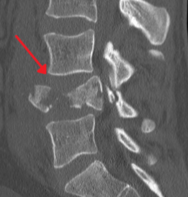
Figure 15. Sagittal CT of the same burst fracture showing loss of vertebral body height and posterior retropulsion into the canal. James Heilman, MD, CC BY-SA 4.0, via Wikimedia Commons.
Part VIII - Thoracolumbar Treatment
Nonoperative care and the burst-fracture controversy
Mechanically stable, neurologically intact type A injuries without significant deformity are treated nonoperatively, traditionally in a brace (a TLSO for T7-L3, a CTLSO higher up, and a hip extension for the low lumbar spine) for 6 weeks to 3 months.[41] The most debated injury is the neurologically intact burst fracture, where the evidence has shifted toward conservatism: a burst fracture without posterior-ligamentous-complex injury can usually be treated nonoperatively, and the Bailey trial found that stable A3 burst fractures did equally well with or without a brace, which suggests these fractures are more inherently stable than once thought.[42] Surgery is generally accepted for a burst fracture with a neurological deficit; in the intact patient the relative indications are kyphosis of 30 degrees or more, over 50 per cent height loss, severe comminution, or an inability to brace. Canal compromise without a deficit has not been shown to need surgery, because the canal remodels.
Operative treatment
Surgery is for unstable injuries (AOSpine B and C) and for neurological deficits, and the dominant approach is posterior pedicle-screw and rod fixation, which gives three-column fixation, deformity correction, indirect canal decompression by ligamentotaxis (up to about 50 per cent), and direct decompression by laminectomy where needed.[43] Short-segment fixation (one level above and below) is reserved for younger patients with good bone, because without anterior column support the cantilever forces on a short construct cause hardware failure and kyphosis. An anterior approach (corpectomy with a structural cage or graft) is added or used alone for severe comminution, marked retropulsion, or kyphosis over 30 degrees, with combined approaches for the most unstable injuries.[44] Type C fracture-dislocations, which carry a 75 per cent or greater rate of neurological injury, are stabilised operatively regardless of neurological status, usually from behind.
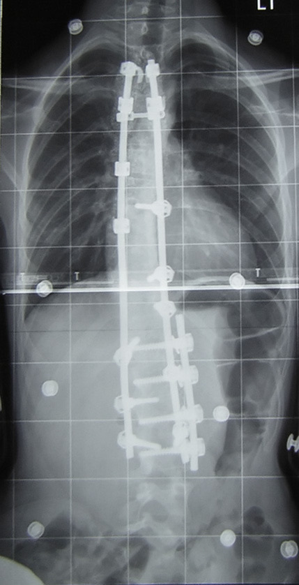
Figure 16. Postoperative radiograph after posterior pedicle-screw-and-rod instrumentation and fusion spanning the injured level. By Silverjonny, public domain, via Wikimedia Commons.
Osteoporotic vertebral compression fractures and penetrating injuries
Osteoporotic vertebral fractures are common (the spine is the commonest osteoporotic fracture site) and are mostly treated nonoperatively, with bracing of uncertain benefit and a place for analgesia, calcitonin and bone-protective medication.[45] Cement augmentation (vertebroplasty and kyphoplasty) is genuinely controversial. The 2009 sham-controlled trials (Kallmes, Buchbinder) and the 2018 Cochrane review and VERTOS IV found no clinically relevant benefit of vertebroplasty over placebo, while other trials (Klazen) found benefit against conservative care; the AAOS recommends against vertebroplasty and offers kyphoplasty only as a limited option.[46] Penetrating (gunshot) injuries are usually mechanically stable and rarely need stabilisation, since the column concept does not apply to them. Surgery is reserved for a progressive deficit from a compressive lesion, retained canal fragments are considered for removal between T12 and L4, and antibiotics are extended when the bowel is traversed.[47] As for sacral fractures, the standard teaching is the Denis zonal classification (zone I lateral to the foramina, zone II through the foramina, zone III central, the last carrying the highest rate of neurological, especially bowel and bladder, injury), with the U-type spinopelvic dissociation managed by lumbopelvic fixation.[48]
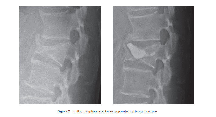
Figure 17. Osteoporotic vertebral compression fracture before (left) and after balloon kyphoplasty with cement augmentation (right). Rei Momomura, CC BY 4.0, via Wikimedia Commons.
Part IX - Paediatric Spine Trauma
The immature spine is different. Greater ligamentous laxity, a relatively large head that raises the fulcrum, incomplete ossification and horizontal facets shift injuries toward the upper cervical spine and the cord in young children, and several normal variants mimic injury.[49] The most important variant is pseudosubluxation of C2 on C3 (and C3 on C4), a physiological hypermobility normal under 8 years, distinguished from a true injury by the Swischuk posterior cervical line, which should pass within about 2 mm of the C2 spinolaminar junction. The atlanto-dental interval may also be wider than in adults (up to 5 mm).[50]
The defining paediatric entity is SCIWORA (spinal cord injury without radiographic abnormality), first described by Pang and Wilberger: an objective cord injury with normal plain films and CT, attributed to the cord’s tenuous blood supply and the column’s greater elasticity than the cord, sometimes with delayed onset, and assessed by MRI (which may still be normal).[51] In the thoracolumbar spine the same patterns occur as in adults, but the flexion-distraction (Chance) injury is again the one to know, with its lap-belt mechanism and high association with intra-abdominal injury, occurring at lower levels than in adults. The apophyseal ring (limbus) fracture, an avulsion of the vertebral ring apophysis with disc herniation in adolescents, is best seen on CT and easily missed on MRI.[52]
References
-
The sources are Rockwood & Green’s Fractures in Adults (9th ed., chapters 46 to 48), supplemented by Miller’s Review of Orthopaedics (9th ed., chapter 8) for high-yield framing and by Lovell & Winter’s Pediatric Orthopaedics for the child. Every claim carries its page citation; genuinely established facts absent from these sources are flagged as standard teaching rather than given a false citation. Two notes on scope: detailed sacral fractures (Denis zones, the U-type) sit at the spine-pelvis border and are treated only briefly here as standard teaching, since neither source’s spine chapters tabulate them; and pelvic ring injuries belong to a separate konspekt topic.
-
RG p.3110; AO concepts throughout. The recurring practical lesson is that the determinants the surgeon controls (timely decompression, restoration of alignment, stable fixation) sit alongside determinants he does not (the energy of the impact and the primary cord injury sustained at that instant).
-
RG p.2929-2930. Spine fractures run about 3:1 male to female, equalising for compression fractures because of female osteoporosis; the rate of neurological deficit after a spine fracture is 5 to 39 per cent.
-
RG p.2930, p.2932. Cervical fracture in a patient over 65 carries a 28 per cent one-year mortality regardless of deficit, rising to 39 per cent over 85.
-
RG p.2931. Cadaveric data show cervical collars do not reduce motion in the most unstable injury models, which is why immobilisation is increasingly individualised.
-
RG p.2933, Table 46-1; Miller p.791; RG p.3095. The neurological level is the most caudal segment with both intact sensation and at least antigravity (grade 3) strength.
-
RG p.2933; Miller p.789, p.791. In a complete injury about 80 per cent of patients recover one nerve-root level and 20 per cent recover two; the more sparing and the faster the recovery, the better the eventual outcome (Miller p.791-792).
-
RG p.2932; Miller p.789-790. Neurogenic shock complicates about 20 per cent of cervical cord injuries; it is treated with volume first, then vasopressors, with atropine for symptomatic bradycardia (the vasopressor detail is standard teaching).
-
RG p.2933-2934; Miller p.792-793. A persistent absence of the bulbocavernosus reflex with an injury below the conus suggests cauda equina syndrome rather than ongoing spinal shock, because an injury below the cord does not produce spinal shock.
-
RG p.2936; Miller p.791. The dosing, when used, is 30 mg/kg over 15 minutes then 5.4 mg/kg/h, run for 24 hours if started within 3 hours or 48 hours if started 3 to 8 hours after injury (Miller p.791).
-
RG p.2932, p.2944; RG p.3117. The Harborview protocol is a MAP of at least 80 mm Hg for 48 hours after admission and 24 hours postoperatively (RG p.3117).
-
Miller p.793-794. The trigger is most often bladder or bowel; an undiagnosed orthopaedic injury can also provoke it. The classic exam answer is bradycardia (a baroreflex response to the surge in blood pressure), though Miller’s Testable Concepts list a reflex tachycardia, so both are described.
-
Miller p.793, Table 8.9; RG p.3073-3074. About half of elderly and almost all young central-cord patients regain independent ambulation (Miller p.810).
-
Miller p.793. The anterior spinal supply derives in part from the artery of Adamkiewicz, which most often arises on the left between T9 and L1.
-
RG p.2934, p.3091-3092. The conus is housed at the thoracolumbar junction; the cauda equina, being nerve roots rather than cord, is more resilient and recovers better.
-
RG p.3047. Flexion and extension are greatest at C4-C5 and C5-C6 (about 20 degrees), and C1-C2 supplies about half of cervical rotation.
-
RG p.2937, p.3076-3077.
-
RG p.2934-2935, p.2941; the prevertebral soft-tissue figures (about 6 mm at C2, 20 mm at C6) are from Miller p.789 (RG gives over 22 mm at C6). Flexion-extension films are not recommended acutely because injured patients guard and the cervicothoracic junction is poorly seen.
-
RG p.3019-3021. Tectorial membrane disruption on MRI is a usual contraindication to nonoperative care.
-
RG p.3013-3015; Miller p.794-795. Atlanto-occipital dissociation accounts for 14 to 18 per cent of blunt-trauma fatalities (RG p.3013); the Powers ratio is named in Miller but not in this RG extract, where the Harris intervals are used.
-
RG p.3022; Miller p.795-796. The transverse ligament alone allows a maximum atlanto-dental interval of about 5 mm; widening beyond that implicates the alar ligaments too. The Dickman classification (intrasubstance tear versus bony avulsion) is standard teaching not named in this RG extract.
-
RG p.3030; Miller p.796. The Grauer subclassification of type II (IIa transverse, IIb anterosuperior-to-posteroinferior favouring a screw, IIc the reverse contraindicating one) refines the operative decision (RG p.3030).
-
RG p.3032-3034; Miller p.796-797. Anterior odontoid-screw union rarely exceeds 85 per cent, and age over 65 is a relative contraindication; posterior C1-C2 fusion (C1 lateral mass plus C2 isthmus screws) is the most versatile construct.
-
RG p.3035-3036; Miller p.797. Coric found nonoperative treatment reliable below about 6 mm of displacement; Vaccaro’s halo failures all had initial angulation over 12 degrees.
-
RG p.3028-3029; Miller p.796.
-
RG p.3046, p.3076. The classic correlates are the unilateral facet dislocation (distractive flexion) and the flexion teardrop (compressive flexion stage III or greater).
-
RG p.3050-3052; Miller p.799, p.803. The full SLIC and AOSpine point tables live in the chapter’s first half and in Miller; kyphosis over 11 degrees at the injured segment strongly suggests posterior-ligamentous-complex disruption (RG p.3049).
-
RG p.3065. Bilateral facet dislocation is itself a marker of less neurological recovery, and bilateral dislocations classically show more than 50 per cent translation (Miller p.811).
-
RG p.3065, p.3068; Miller p.799. Reduction uses Gardner-Wells tongs with sequential weights and a neurological check plus a radiograph after each addition.
-
RG p.3054-3059.
-
RG p.3071-3073. An ankylosing-spondylitis patient with neck pain after any trauma has a cervical fracture until proven otherwise, and the whole spine must be imaged because of noncontiguous injuries.
-
RG p.2986-3013; Miller p.790. Children take 8 to 10 pins at 2 inch-pounds; the safe anterior pin zone is above the eyebrow in the middle-to-lateral third, avoiding the supraorbital nerve.
-
RG p.2986-3013; Miller p.797-799. The C1 lateral mass plus C2 pedicle (Harms) construct is biomechanically the strongest for C1-C2; vertebral-artery anatomy must be checked on CT before C2 screws.
-
RG p.3091-3092. Over half of thoracolumbar injuries follow motor-vehicle accidents and a quarter follow falls over 6 feet; the radiographic indicators of instability are over 50 per cent height loss, over 30 degrees of kyphosis, and 2.5 mm of translation (RG p.3096).
-
RG p.3091. CT has largely supplanted plain films for screening; MRI is complementary, best for the cord and the posterior ligamentous complex, but is criticised for over-calling ligamentous injury and thereby over-calling instability.
-
RG p.3111; Miller p.799, p.803. Denis derived the model from 412 injuries; a neurological deficit itself implies instability.
-
RG p.3112, Table 48-1; Miller p.803. TLICS scores incomplete and cauda equina injuries higher than complete injuries, reflecting their greater potential to benefit from decompression; its weaknesses are that it needs a reliable neurological exam and that the controversial burst-with-questionable-PLC injury “invariably” scores an indeterminate 4.
-
RG p.3113-3116. A combined injury is coded by specifying the associated type A fracture (for example “T12-L1 B2; L1 A3”).
-
RG p.3095, p.3103. Canal compromise is tolerated very differently by region (poorly in the thoracic cord, well below the conus).
-
RG p.3092; Miller p.803-805. The Chapman series found intra-abdominal injury in 30 per cent of 153 flexion-distraction injuries; the seatbelt mechanism mandates a careful abdominal evaluation.
-
RG p.3120. Bracing is followed by upright radiographs to confirm alignment under load and by flexion-extension films at discontinuation.
-
RG p.3120, p.3136; Miller p.803. The Bailey trial enrolled A3 fractures from T11 to L3 in patients under 60 with under 35 degrees of kyphosis.
-
RG p.3121-3124. STASCIS supported decompression within 24 hours, and early stabilisation of thoracic fractures reduces pulmonary morbidity and ICU stay.
-
RG p.3122, p.3124-3125; Miller p.803. A laminectomy alone is contraindicated because it removes the posterior tension band and produces progressive kyphosis.
-
RG p.3149-3150; Miller p.802.
-
RG p.3151-3152. The kyphoplasty balloon can restore some height, but the two procedures are largely equivalent for pain, and kyphoplasty is more expensive.
-
RG p.3155-3160; Miller p.805-806. Methylprednisolone is contraindicated in penetrating cord injury.
-
Standard teaching; the spine chapters of these sources do not tabulate the Denis sacral zones, which sit at the spine-pelvis border (RG p.3168 confirms the absence).
-
Lovell p.5549; Miller p.807. On a backboard the child’s large head flexes the neck, so the torso is elevated 2 to 3 cm to keep the cervical spine aligned.
-
Miller p.807. The C2 dens fracture (often through the basilar synchondrosis) is the commonest paediatric cervical fracture, typically under 6 years, and traction is avoided because it distracts the dens.
-
Miller p.807. The original definition predated MRI and excluded penetrating trauma and congenital abnormality.
-
Lovell p.5561-5572; Miller p.807-808. The paediatric three-column concept applies, and growth and deformity are the long-term concerns.
-
RG p.2932-2934; Miller p.789-790, p.792-793.
-
RG p.2933, p.3093-3094; Miller p.789, p.791.
-
Miller p.793, Table 8.9; RG p.3073-3074.
-
RG p.2936, p.3118-3119; Miller p.791.
-
RG p.3022; Miller p.795-796.
-
RG p.3030, p.3032-3034; Miller p.796-797.
-
RG p.3035-3036; Miller p.797.
-
RG p.3065, p.3068; Miller p.799.
-
RG p.3111; Miller p.799, p.803.
-
RG p.3112, Table 48-1; Miller p.803.
-
RG p.3120, p.3136, p.3151-3152; Miller p.802-803.
-
Miller p.807; Lovell p.5549.