Contents
- Orientation: What “Amyelic” Means and Why It Frames the Whole Topic
- Part I - Epidemiology, Mechanism, and the Anatomy of Vulnerability
- Part II - Initial Assessment and Neurological Evaluation
- Part III - The Concept of Stability
- Part IV - Imaging and Cervical Spine Clearance
- Part V - Upper Cervical Injuries (C0-C2)
- 5.1 Occipitocervical (atlanto-occipital) dissociation
- 5.2 Occipital condyle fractures
- 5.3 Atlas (C1) fractures, the Jefferson burst, and the transverse ligament
- 5.4 Transverse-ligament injury and traumatic atlanto-axial instability
- 5.5 Atlanto-axial rotatory subluxation/fixation
- 5.6 Odontoid (dens) fractures
- 5.7 Hangman’s fracture (traumatic spondylolisthesis of the axis)
- Part VI - Subaxial Cervical Injuries (C3-C7)
- Part VII - Thoracolumbar Injuries
- Part VIII - Principles of Treatment
- Part IX - A Synthesis: How to Reason Through the Amyelic Spine
- References
Orientation: What “Amyelic” Means and Why It Frames the Whole Topic
The konspekt heading for this topic is fractures and dislocations of the spine without cord injury, the classic Bulgarian designation амиелично увреждане (amyelic injury): a structural failure of the vertebral column, bony or ligamentous or both, in a patient whose spinal cord and nerve roots are working normally. The neurological examination is intact. On the modern grading scale this is the patient who is ASIA E (normal motor and sensory function).[1] The drama of the spinal-cord-injured patient, the paralysis, the neurogenic shock, the rehabilitation, belongs to a different chapter. What this topic asks of the candidate is something more subtle and, in daily practice, more common: how to recognise that a column has been mechanically compromised even when the patient can move everything, how to decide whether that column is stable or unstable, and how to protect the cord that is, for the moment, still intact.
This orientation matters because almost every decision in spine trauma turns on it. An unstable injury in a neurologically intact patient is a warning: a column that has lost its restraints and could, with the wrong movement or the wrong missed diagnosis, convert an ASIA E patient into an ASIA A patient. The literature documents exactly this catastrophe. In one occipitocervical-dissociation series an average two-day delay in diagnosis produced secondary neurological deterioration in 29% of the cohort.[2] The amyelic patient is therefore not the easy patient. He is the patient in whom a good outcome is still entirely available and entirely losable.
Two terminological cautions, in the Boychev tradition of precise naming, will run through this summary. First, stability is a mechanical concept distinct from the neurological state. A fracture can be neurologically silent yet mechanically unstable (the dangerous amyelic case), or neurologically devastating yet, once fixed, mechanically sound. Second, the column language (anterior, middle, posterior) and the complex language (the posterior ligamentous complex, гръбначен заден лигаментен комплекс) are the vocabulary in which instability is actually argued. The candidate who masters them masters the topic.
Part I - Epidemiology, Mechanism, and the Anatomy of Vulnerability
1.1 How common, in whom, and where
Spine fractures are rising in incidence. Two forces drive this: an ageing population sustaining osteoporotic fragility fractures, and the persistent toll of high-energy trauma in the young.[3] The injuries are not scattered randomly along the column. They cluster at the junctions of the rigid thoracic cage, the points where a stiff segment meets a mobile one and stress concentrates.[4] The thoracolumbar junction (T11-L2) is the single most commonly injured region of the spine, a relatively straight transition zone caught between the kyphotic, rib-splinted thoracic spine above and the lordotic, mobile lumbar spine below.[5] The cervicothoracic junction is the other classic site, more often injured in high-speed motor-vehicle collisions and sport.[6]
The demographic skew is roughly 3:1 male to female, with one telling exception: in compression fractures the ratio equalises, because postmenopausal osteoporosis brings women into the denominator.[7] Older patients sustain isolated cervical fractures from simple falls, most often compression fractures (an axial-load mechanism) or spinous-process fractures (a hyperextension mechanism).[8] The thoracolumbar literature describes a bimodal age distribution: an early peak of high-energy injuries in men under thirty, and a later peak of low-energy ground-level falls in the elderly with poor bone quality.[9] Motor-vehicle collisions cause more than half of thoracolumbar injuries and falls from over six feet about a quarter.[10]
The stakes remain high even when the cord is spared. The rate of neurological deficit accompanying a spine fracture ranges from 5% to 39%, and that range is precisely the gap the amyelic clinician is trying to stay on the right side of.[11] Mortality after spine fracture is not trivial in the old either. Regardless of neurological status, patients aged sixty-five and over with a cervical fracture carry a 28% one-year mortality, rising to 39% above age eighty-five.[12] The geriatric odontoid fracture, neurologically intact and radiographically unimpressive, is one of the deadlier injuries in orthopaedics precisely because it is so easy to underestimate.
1.2 The anatomy of vulnerability: why each region behaves as it does
The thoracic spine (T2-T10) is the most stable and least often injured region, splinted by the rib cage and locked by the coronal orientation of its facets. Yet it carries a high propensity for cord injury when it does fail, because the canal is narrow with little reserve and the cord between T4 and T8 lies in a vascular watershed between the anterior spinal artery and the artery of Adamkiewicz.[13] Small displacements here can produce large deficits. The thoracic kyphosis places the centre of gravity anterior to the column and so generates a constant kyphotic moment that any injury must be expected to amplify.[14]
The thoracolumbar junction has none of this protection. The lower thoracic ribs are floating, the facet orientation is transitioning from coronal to sagittal, and the straightened sagittal profile concentrates stress. This region houses the conus medullaris, so injuries here can produce mixed upper- and lower-motor-neuron pictures, from isolated bladder dysfunction to complete paralysis.[15] The lumbar region below L3, by contrast, is comparatively forgiving: the lordosis places the weight-bearing axis posterior to the bodies, which minimises kyphotic forces, and the wide canal houses the resilient cauda equina, which tolerates considerable canal compromise without deficit and, when injured, produces lower-motor-neuron root lesions with a better recovery prognosis than cord or conus injuries.[16]
The upper cervical spine is a world of its own. The atlanto-axial joint (C1-C2) provides roughly 47° of rotation, about half of the entire cervical axial rotation, while permitting almost no lateral bending. The offset of the C2 inferior articular processes throws high shear onto the pars interarticularis under axial load, which is exactly why the hangman’s fracture favours that location.[17] The occipitocervical junction has no intervertebral disc and relies almost wholly on ligaments for restraint. Its failure is therefore a ligamentous catastrophe rather than a bony one.[18]
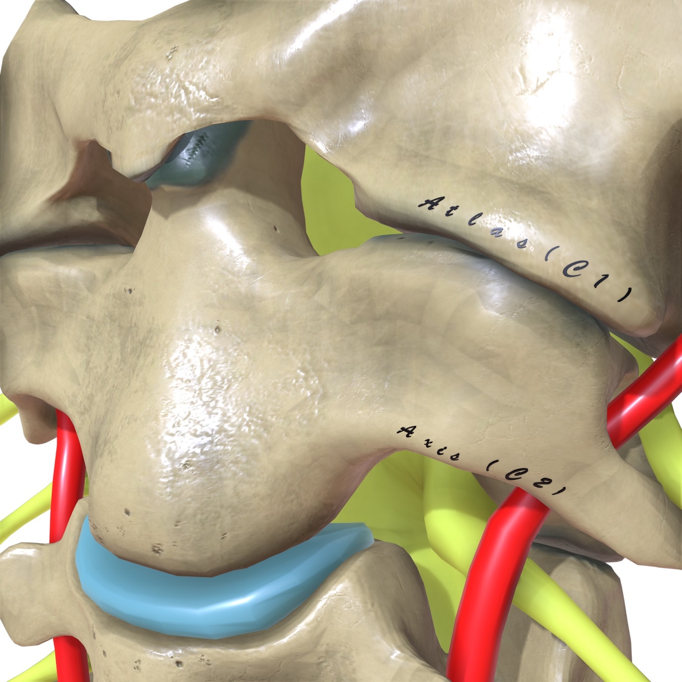
Figure 1. The upper cervical spine: the atlas (C1) and axis (C2) with the odontoid process (dens). The atlanto-axial joint provides roughly half of all cervical rotation. Source: DrJanaOfficial, via Wikimedia Commons, CC BY-SA 4.0.
Part II - Initial Assessment and Neurological Evaluation
2.1 The field and the resuscitation room
Up to 10-20% of spinal-cord-injured patients die before reaching hospital, so the early phase is genuinely decisive.[19] In the unresponsive trauma patient, a spine injury is presumed present until excluded.[20] Traditional rigid immobilisation, “boarded and collared,” is now applied more selectively, because the long backboard itself causes pain, pressure sores, and impaired respiration. The consensus position (NAEMSP and the ACS Committee on Trauma) recommends backboard immobilisation for blunt trauma with altered consciousness, neck or back tenderness, a neurological complaint, or gross deformity. It recommends against it in the fully alert patient (GCS 15) with no tenderness, no neurological complaint, no distracting injury, and no intoxication.[21] One cadaveric finding tempers our faith in hardware: rigid cervical collars do not reduce motion in the most unstable cervical injury model, and they carry their own toll of aspiration and delirium, especially in the elderly.[22]
Assessment follows the ATLS sequence. Two airway and breathing points are spine-specific. An unstable cervical injury can make intubation hazardous, and a cord injury above C5 paralyses the diaphragm. Even lower lesions weaken the intercostal and abdominal musculature, producing the paradoxical breathing pattern of the high cord injury.[23] For these reasons a “neurologically intact” label must be earned by examination and not assumed.
2.2 The two shocks: a distinction the amyelic clinician must own
Nothing exposes a shaky understanding of spine trauma faster than confusing the two shocks, and the distinction is genuinely useful at the bedside.
Neurogenic shock is a distributive shock that follows cord injury through loss of sympathetic outflow, leaving unopposed vagal tone. Its hallmark pairing is hypotension with bradycardia (a systolic pressure below 90 mmHg), often with hypothermia.[24] It occurs in roughly 20% of cervical-level cord injuries and is more frequent with complete than incomplete lesions, its incidence falling as the level descends.[25] The single most useful discriminator is the heart rate: hypovolaemic shock drives a compensatory tachycardia, neurogenic shock does not.[26] In the polytrauma patient the two frequently coexist, and haemorrhage must always be excluded before hypotension is attributed to the cord. Management aims at a mean arterial pressure above 85 mmHg for the first week after injury.[27] For the amyelic patient this section is mostly a rule-out: a neurologically intact patient who is hypotensive is bleeding until proven otherwise, not in neurogenic shock.
Spinal shock is a different and older concept (the term dates to 1841): the transient loss of all motor, sensory, reflex, and autonomic function caudal to a cord injury, caused by loss of afferent input into the reflex arc.[28] Its resolution is signalled by the return of spinal reflexes below the level, generally between 48 hours and two weeks, and marks the moment at which a meaningful ASIA grade can first be assigned.[29] The reflexes return in a characteristic order, cutaneous before deep tendon: deep plantar response, then the bulbocavernosus reflex, then cremasteric, Achilles, Babinski, and patellar.[30] The bulbocavernosus reflex, mediated through the S2-S4 neurons of the conus, is the classic monitor of spinal shock.[31] One corollary matters greatly for the amyelic spine. An injury below the cord (below the conus) cannot cause spinal shock, so in a burst fracture below the conus an absent anal sphincter contraction indicates a cauda equina syndrome, not spinal shock.[32]
2.3 Grading: the motor scale, sacral sparing, and the ASIA Impairment Scale
The motor examination is graded 0 to 5: 5 is full power through a full range against full resistance; 4 is partial resistance; 3 is full range against gravity only; 2 is full excursion only with gravity eliminated; 1 is a palpable contraction without joint movement; 0 is no function.[33] Every patient has anal sphincter tone assessed and a dermatomal sensory examination performed, including perianal sensation. Preserved perianal sensation (sacral sparing) is the single sign that converts a “complete” into an “incomplete” injury, and it is a positive prognostic factor.[34]
The ASIA Impairment Scale (AIS), refined from the older Frankel classification and gradeable only once spinal shock has resolved, runs:[35]
| Grade | Type | Description |
|---|---|---|
| A | Complete | No motor or sensory function preserved, including the sacral segments |
| B | Incomplete | Sensory but not motor function preserved below the level (including sacral segments) |
| C | Incomplete | Motor preserved below the level; more than half of key muscles grade below 3 |
| D | Incomplete | Motor preserved below the level; at least half of key muscles grade 3 or more |
| E | Normal | Normal motor and sensory function |
The ASIA motor score is a 100-point scale built from five key muscle groups per limb, each scored out of five.[36] The single most important sentence for this topic is that the amyelic patient is ASIA E, and the whole task is to keep him there. The grade is explicitly not reliable within 72 hours of injury, a caution that guards against premature prognostication.[37]
The radiographic counterpart of neurological state is the space available for the cord (SAC). In the cervical spine the mean SAC is about 10.5 mm in complete injuries, 13 mm in incomplete injuries, and 17 mm where there is no deficit.[38] This dose-response between canal narrowing and neurological risk is the mechanical justification for treating an unstable amyelic spine as an emergency of prevention.
Part III - The Concept of Stability
3.1 From two columns to three: Holdsworth, McAfee, Denis
Stability is the organising idea of spine trauma, and its modern form was built in stages.[39] Holdsworth first introduced spinal columns, dividing the spine into an anterior column resisting compression and a posterior column resisting tension, and located instability in disruption of the posterior tension band. McAfee was the first to use CT for classification, describing six patterns of increasing instability. Denis, working from 412 thoracolumbar injuries, modified the two-column theory into the three-column model that still dominates teaching.[40]
The three columns are defined as follows:[41]
- Anterior column (преден стълб): the anterior longitudinal ligament and the anterior half of the vertebral body and annulus.
- Middle column (среден стълб): the posterior half of the vertebral body and annulus together with the posterior longitudinal ligament.
- Posterior column (заден стълб): the neural arch, the facet joints, and the entire posterior ligamentous complex.
The Denis rule of instability is compact and examinable: an injury is unstable when at least two of the three columns are disrupted, and any injury extending into the middle column is largely regarded as unstable.[42] The middle column is, in this sense, the linchpin. It is what distinguishes a benign anterior wedge compression (anterior column only) from a burst fracture (anterior and middle columns), and that single distinction reorganised the entire field. Denis also introduced the notion of neurological stability: a neurological deficit implies an injury severe enough to have been mechanically unstable.[43]
3.2 The posterior ligamentous complex: the posterior tension band
The posterior ligamentous complex (PLC) is the structure on which the modern debate about stability actually turns. It comprises the facet joint capsules, the interspinous and supraspinous ligaments, and the ligamentum flavum, together with their bony attachments, so it is more accurately a posterior osseoligamentous complex.[44] Functionally it is a posterior tension band counteracting flexion and kyphotic forces, and failure of this tension band is the hallmark of the unstable injury.[45] In the subaxial cervical spine the equivalent structures, the ligamentum flavum together with the strong interspinous and supraspinous ligaments (the ligamentum nuchae), form the cervical PLC, whose disruption likewise produces mechanical instability.[46]
The PLC has become the practical fulcrum because it is the variable that most often decides operative versus non-operative care in the neurologically intact patient. A burst fracture with an intact PLC is a different animal from the same fracture with a torn PLC, and much of the imaging effort in the amyelic spine, particularly MRI, is directed at answering this one question. It also explains why the upright radiograph is so valuable: an injury that increases its kyphosis when the patient stands has, by that behaviour, declared its posterior tension band incompetent.[47]
3.3 Mechanical versus neurological instability
Two distinct drivers send a patient to surgery, and keeping them separate is essential. Mechanical instability is the loss of the column’s ability to bear physiological loads without progressive deformity or pain. Neurological instability is the presence of (or risk of) cord or root compromise from canal encroachment.[48] The amyelic patient, by definition, presents with the first and not yet the second, and the entire purpose of stabilising him, by brace or by screw, is to prevent the second from ever appearing. The surgical goals follow directly: early mobilisation, restoration of coronal and sagittal balance, the fewest fused segments compatible with stability, and freedom from prolonged bracing.[49]
Part IV - Imaging and Cervical Spine Clearance
4.1 Clearing the cervical spine: NEXUS and the Canadian C-spine rule
A large fraction of trauma patients can have the cervical spine cleared clinically, without any imaging, and doing so spares cost, radiation, and the morbidity of prolonged collar use. Two validated instruments govern the decision.[50]
The NEXUS criteria (National Emergency X-Radiography Utilization Study) permit clearance without imaging when all five low-risk features are present: no posterior midline cervical tenderness, no focal neurological deficit, normal alertness, no intoxication, and no painful distracting injury.[51] The validation study deliberately left the definitions of “intoxication” and “distracting injury” to clinical judgement.[52] The Canadian C-spine rule (CCR) applies to alert (GCS 15), haemodynamically stable patients, layering high-risk factors (which mandate imaging) against low-risk factors (which permit a safe assessment of neck rotation), and clearing the patient who can actively rotate the neck 45° to each side.[53] In head-to-head comparison the CCR is more sensitive but more often misapplied, being the more complex rule.[54] Clinical clearance is reassuringly robust. A meta-analysis of fourteen studies and over 60,000 patients found a 99.8% negative predictive value and 98.1% sensitivity for clearing alert, asymptomatic patients, with the few missed injuries causing no neurological harm.[55]
The full itemised factor list of the Canadian C-spine rule (age 65 or over, a dangerous mechanism, or extremity paraesthesias as high-risk factors; simple rear-end collision, sitting position in the department, ambulation at any time, delayed onset of neck pain, or absence of midline tenderness as low-risk factors permitting a rotation test) is standard teaching but was presented in the mined source as a figure rather than transcribed text.[56]
4.2 A practical four-category algorithm
The authors’ clearance algorithm organises patients by mental status, which is the cornerstone of the whole decision.[57]
- Alert and asymptomatic: Level-1 evidence supports no imaging; remove the collar (apply NEXUS or CCR).
- Short-term cognitive impairment (intoxication, delirium, a distracting injury): keep immobilised until the cause resolves, then reassess; the Confusion Assessment Method is a validated delirium screen.
- Symptomatic (neurological symptoms, midline tenderness, or pain on active motion): proceed to imaging, the modality of choice being multidetector CT.
- Obtunded / long-term cognitive impairment (expected beyond 48-72 hours): the centre of the MRI controversy below.
4.3 The modalities, and what each is for
Plain radiography has been displaced as the acute screening tool by its poor sensitivity. False-negative rates are about 30% in the cervical, 52% in the thoracic, and 14% in the lumbar spine, and it cannot reliably image the occipitocervical and cervicothoracic junctions.[58] The standard cervical series (anteroposterior, lateral, and open-mouth odontoid views) retains a role in follow-up and in dynamic flexion-extension assessment, but not as the primary acute screen.[59]
Multidetector CT is the primary screening modality endorsed by EAST. A 64-slice study with 1 mm collimation and sagittal/coronal reformation achieves about 99% sensitivity for cervical injury, and is the single best test for distinguishing compression from burst, for facet widening, for quantifying canal compromise, and so for determining stability.[60] Dedicated thoracolumbar CT uses 2-3 mm slices reformatted in the sagittal and coronal planes.[61]
MRI is the complement, not the competitor, of CT. Its role is soft tissue: the cord (oedema, compression, continuity), the discs, and above all the integrity of the PLC and the other ligaments, plus epidural haematoma and dural tears.[62] Its great limitation in the amyelic spine is low specificity for clinically relevant tension-band injury. MRI tends to over-call PLC disruption and so risks driving unnecessary surgery, and it is inferior to CT for bony detail.[63] This double-edged quality is exactly why the obtunded-clearance debate exists.
On the use of MRI for ligament assessment the literature openly disagrees. In awake, alert, intact patients with midline tenderness, Resnick et al. (830 patients) found that CT detected all clinically relevant injuries and that complementary MRI added no benefit; Ackland et al. (178 patients) found MRI changed management 22% of the time.[64] For the obtunded patient, James et al. (1,535 patients) identified eleven unstable surgical injuries detectable only by MRI, while Schoenfeld et al. (8,060 propensity-matched patients) found MRI revealed new injuries in only 8%, a minority clinically significant; EAST’s 2016 guidance “conditionally” endorses collar removal after a high-quality negative CT.[65] The authors’ pragmatic position: CT alone suffices to clear the obtunded patient when two independent expert reviewers agree there is no injury; discordance or equivocation triggers MRI.[66]
One practical caution. Acute flexion-extension films are not recommended, because muscle spasm prevents enough motion to disclose instability, and the cervicothoracic junction (up to 15% of cervical injuries) is poorly seen.[67] Their place is at healing, to confirm that a treated injury is stable before the collar comes off.
4.4 The measurements that quantify instability
Plain-film and CT lines convert a vague impression into a decision. On the anteroposterior view, a widened interpedicular distance signals a burst fracture and an increased interspinous distance signals posterior tension-band injury.[68] On the lateral view, the smooth anterior and posterior vertebral lines are inspected for breaches, and retropulsion of a superoposterior fragment into the canal distinguishes a burst from a simple compression.[69] Height loss is measured against the average of the two adjacent vertebrae, and angulation by the Cobb method between the endplates above and below the injured level.[70] The classic instability thresholds, drawn from multiple authors, are vertebral height loss over 50%, kyphotic angulation over 30°, and translation of 2.5 mm.[71]
At the craniocervical junction the most useful measures are the basion-axial interval (BAI) and basion-dental interval (BDI), together known as Harris’s “rule of 12”: each should be no greater than 12 mm.[72] In the subaxial spine, normal alignment is 2-4° of lordosis between adjacent vertebrae, and kyphosis exceeding 11° strongly suggests PLC disruption.[73] The atlanto-dental interval (ADI) and its thresholds are treated under the atlas and transverse-ligament injuries below.
Part V - Upper Cervical Injuries (C0-C2)
5.1 Occipitocervical (atlanto-occipital) dissociation
This is the injury whose missed diagnosis defines the danger of the amyelic spine. It results from high-energy distraction at a junction that has no disc and relies on ligaments, separating the occiput from C1 in what is, anatomically, a closed or internal decapitation (вътрешна декапитация).[74] Traction injury to the medulla and upper cord causes respiratory compromise and frequently immediate death, and the injury is documented in 14-18% of blunt-trauma fatalities.[75] Survivors are at extreme risk. As noted, delayed diagnosis produced secondary neurological decline in 29% of one cohort, and the injury was visible on the initial trauma plain film in only two of seventeen cases.[76] CT or MRI markedly reduces the missed-injury rate, and the diagnosis rests on the BAI and BDI (Harris’s rule of 12).[77]
The Traynelis classification, the most commonly used, is descriptive by direction of occipital displacement: type I anterior, type II axial (longitudinal distraction, with a IIb variant occurring through the C1-C2 articulation), type III posterior.[78] Treatment is operative, and uniquely so. Non-operative management has little role, halo immobilisation serves only as temporary stabilisation, and longitudinal traction is contraindicated (it can distract the already-disrupted junction catastrophically).[79] The standard construct is an occipital plate connected by rods to cervical screws, with the authors favouring C2 pars (isthmus) screws as distal fixation. Bellabarba reported 100% fusion in seventeen patients, though mortality remains high (22% in one survivor series, concentrated in those with associated brain injury).[80]
The classic Powers ratio (basion-posterior-arch distance divided by opisthion-anterior-arch distance, abnormal above 1.0) is standard teaching for anterior dislocation but was not present in the mined source.[81]
5.2 Occipital condyle fractures
Stable patterns arise from axial impaction of the head onto the cervical spine, unstable patterns from distraction with ligamentous avulsion.[82] CT is the imaging modality of choice, as radiographs are unreliable.[83] Two classifications are used in parallel. The Anderson-Montesano system: type I an impaction fracture (stable), type II a basilar skull fracture extending into the condyle (stable), type III a displaced avulsion (unstable, ligamentous), treated by occipitocervical fusion.[84] The Tuli system grades by displacement and ligament status: type I non-displaced (stable), type IIa displaced without ligamentous injury, type IIb displaced with ligamentous disruption (unstable).[85] In practice the authors rely less on the classification than on the integrity of the tectorial membrane on sagittal T2 MRI. Its disruption is a contraindication to non-operative care, signalling occult occipitocervical dissociation.[86] Most condyle fractures heal in a rigid collar over 8-12 weeks, while the unstable type III is fused.[87]
5.3 Atlas (C1) fractures, the Jefferson burst, and the transverse ligament
A mechanical truth governs all C1 fractures: it is not possible to fracture the C1 ring in only one place, so the minimum number of fracture sites is two.[88] Simple posterior arch fractures (hyperextension, the ring pinched between occiput and C2) are the most benign. The Jefferson burst fracture is the classic four-part injury of axial loading, with bilateral fractures of the anterior and posterior arches.[89] Vertebral artery occlusion is rare but reported.[90]
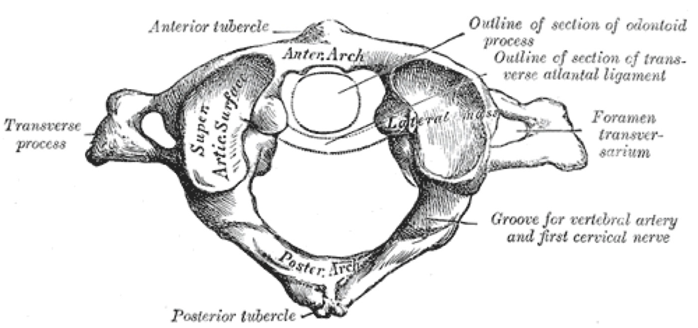
Figure 2. The first cervical vertebra (atlas, C1) from above: anterior and posterior arches, paired lateral masses, and transverse foramina. Because the ring is closed, it cannot fracture in only one place. From Gray’s Anatomy (H. V. Carter, 1918), public domain, via Wikimedia Commons.
The stability of a C1 fracture is decided not by the bone but by the transverse atlantal ligament. The traditional radiographic surrogate is the combined lateral overhang of the C1 lateral masses on C2 on the open-mouth view: the mined source states that an overhang exceeding 7 or 8 mm implies transverse-ligament injury.[91] This is the “rule of Spence” of classic teaching, whose original cadaveric value was 6.9 mm; the source rounds it to 7-8 mm and, importantly, reports contradicting data, Woods et al. found ligament failure at an average overhang of only 3.8 mm, and Radcliff et al. found no correlation between ligament integrity and lateral-mass displacement, so the overhang is now best used as an adjunct to MRI rather than as a standalone rule.[92]
When the transverse ligament fails it permits a maximum of 5 mm of widening at the atlanto-dental interval (ADI), the alar ligaments restraining further displacement.[93] Stable C1 bursts are treated in a rigid collar or CTO for about 12 weeks, with flexion-extension views after healing to exclude residual C1-C2 instability. Unstable patterns are reduced in halo traction and held in a halo vest, and frank transverse-ligament disruption (ADI widening beyond 5 mm) is managed by C1-C2 fusion using C1 lateral-mass and C2 isthmus screws (the fractured arch precludes the classic Brooks and Gallie wiring techniques).[94]
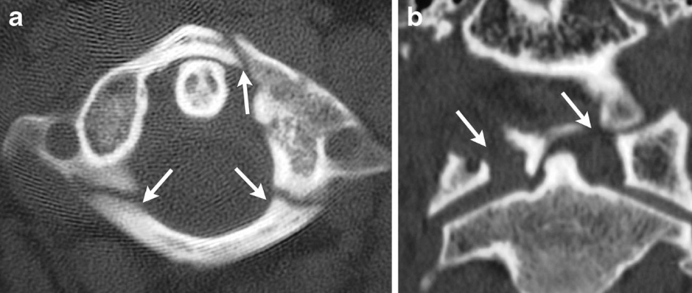
Figure 3. CT of a Jefferson (C1 burst) fracture: axial and coronal images with arrows marking the breaks in the anterior and posterior arches of the atlas. From Utz et al. (2013), Insights into Imaging 5(1):67-75, CC BY 4.0, via Wikimedia Commons.
5.4 Transverse-ligament injury and traumatic atlanto-axial instability
Pure ligamentous C1-C2 instability without fracture follows an abrupt flexion moment shearing the atlanto-axial articulation.[95] The diagnosis is made on the ADI, normally no greater than 2-3 mm in the adult. An ADI of 3-5 mm (somewhat widened or asymmetrical) warrants flexion-extension views to gauge the degree of instability, while an ADI above 5 mm implies disruption of both the transverse and alar ligaments and is a surgical injury.[96] A transverse-ligament injury attached to a small bony avulsion can heal (better in the young) and may be braced if the ADI is held reduced. Isolated transection without bony attachment, and any ADI above 5 mm or any cord injury, is fused (posterior C1 lateral-mass plus C2 isthmus screws).[97]
The Dickman classification of transverse-ligament injury (type I an intrasubstance tear, which will not heal and is fused; type II a bony avulsion off the C1 lateral mass, which may heal in immobilisation) is standard teaching but was not present in the mined source.[98]
5.5 Atlanto-axial rotatory subluxation/fixation
Traumatic rotatory dislocation is rare in adults, arising from combined lateral flexion and forced rotation, and presents with a fixed torticollis (“cock-robin” posture, “cock-robin” наклон на главата) rather than with neurological compromise. It is commonly missed, with diagnostic delays from weeks to years.[99] The diagnosis is confirmed by axial CT with the head maximally rotated to each side.[100] The Fielding-Hawkins classification has four types defined by the anterior displacement and ligament status: type I rotation without anterior widening (transverse ligament intact); type II widening of 3-5 mm (transverse ligament disrupted); type III widening above 5 mm (transverse and alar ligaments disrupted); type IV a posteriorly displaced rotatory dislocation (rare, classically with odontoid erosion).[101] Acute cases often reduce with traction and are immobilised by type (collar for I, CTO for II, halo for III). Failed reduction or true traumatic instability is fused posteriorly even in the neurologically intact patient.[102]
5.6 Odontoid (dens) fractures
The odontoid fracture is the quintessential amyelic injury of the elderly: neurologically intact, radiographically easy to underestimate, and disproportionately lethal. It is increasingly seen as an isolated injury in the elderly after a low-velocity fall with neck hyperextension.[103] The Anderson and D’Alonzo classification, purely descriptive, has three types: type I an avulsion of the tip (alar-ligament avulsion, rare in isolation); type II through the waist at the junction of the dens and the C2 body, a relative vascular watershed and the high-nonunion type; type III extending into the cancellous C2 body, which heals well.[104] Grauer’s subclassification of type II guides screw fixation: IIa transverse and minimally displaced; IIb running anterosuperior to posteroinferior (the ideal pattern for an anterior odontoid screw); IIc running anteroinferior to posterosuperior (a contraindication to odontoid screw, since compression would increase the displacement).[105]
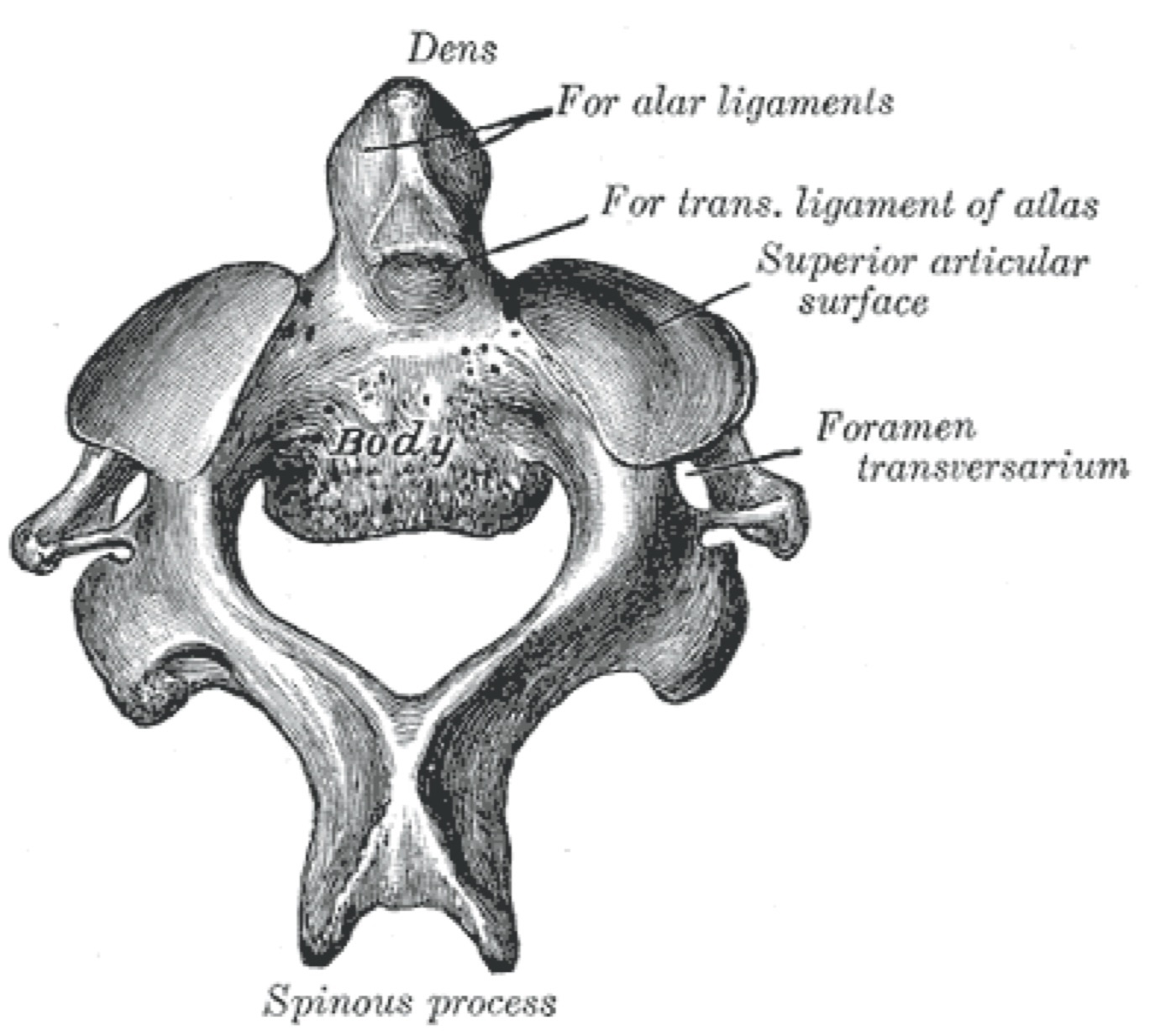
Figure 4. The axis (C2) from above, showing the odontoid process (dens) and the facets for the transverse and alar ligaments. The dens base is the site of the high-nonunion type II fracture. From Gray’s Anatomy (H. V. Carter, 1918), public domain, via Wikimedia Commons.
Nonunion of type II is common, reported around 30% (Greene et al., 340 injuries), with as little as 66% union in a halo and under 50% in some series, and displacement of 50% or more raises the risk of both nonunion and mortality regardless of treatment.[106] The danger of nonunion is not academic. Kepler et al. found a 17% incidence of new neurological deficit after subsequent trauma in the presence of an established odontoid nonunion, the main argument for operating on the majority.[107] Type III fractures fare well externally. Type II nondisplaced fractures may be braced or, given the nonunion risk, treated in a halo, but the halo is hazardous in the elderly, with one cohort reporting roughly 50% mortality and 70% complications, though other series dispute the excess mortality.[108]
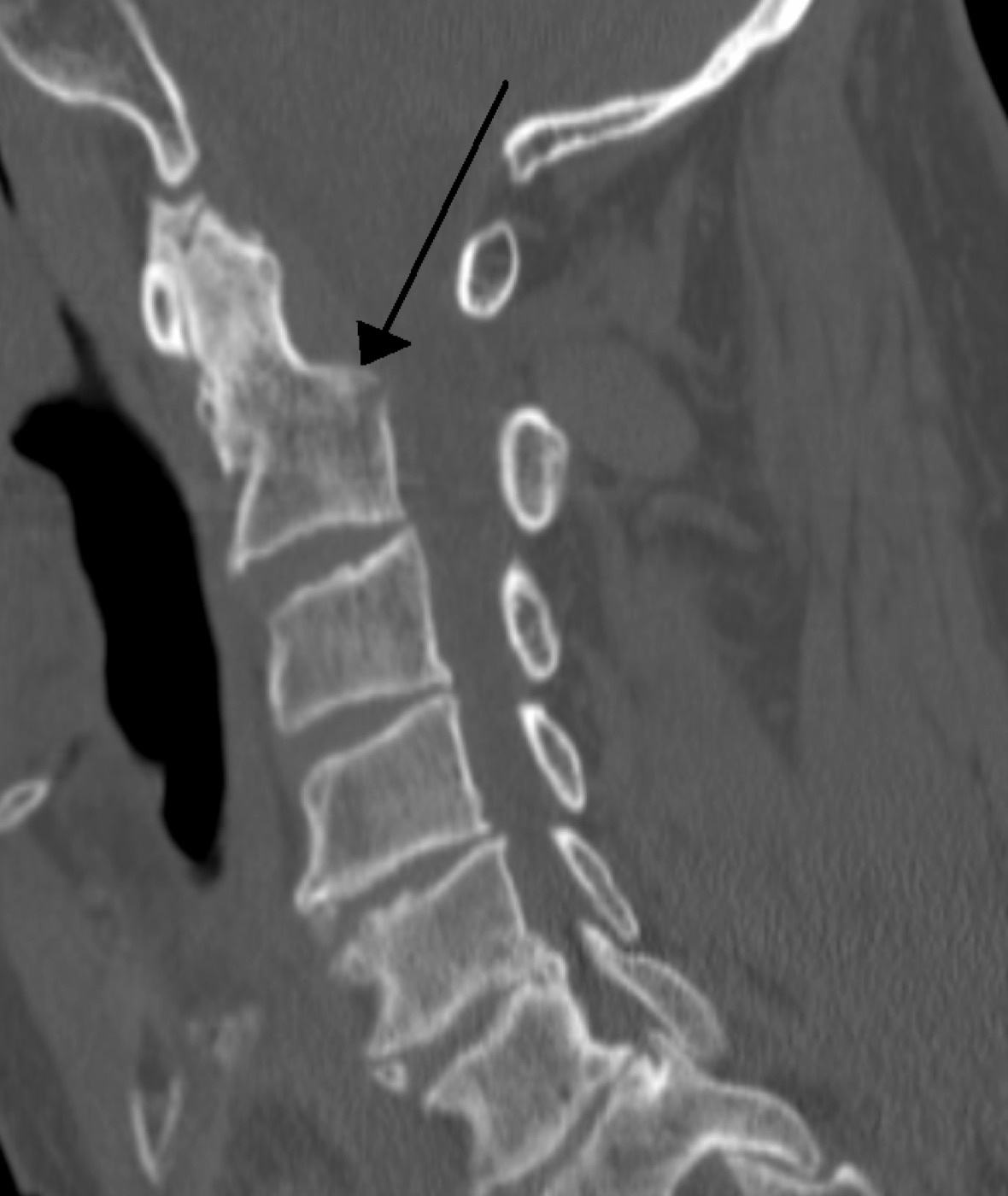
Figure 5. Sagittal cervical CT of a fracture through the base of the dens (arrow): a type II odontoid fracture, the high-nonunion pattern. Source: James Heilman, MD, via Wikimedia Commons, CC BY-SA 3.0.
Operative indications in the younger patient are displacement over 5 mm, angulation over 10°, neurological deficit, substantial comminution, or multisystem trauma.[109] Anterior odontoid screw fixation preserves C1-C2 motion in theory (though atlanto-axial rotation is reduced by about half regardless) and suits type II transverse or IIb oblique patterns. It is contraindicated in Grauer IIc, in barrel-chested or kyphotic patients, and is relatively contraindicated above age 65 (osteopenia and screw cut-out), with union around 82%.[110] Posterior C1-C2 fusion (C1 lateral-mass plus C2 instrumentation, the most versatile because it does not require anatomical reduction) achieves union above 90%.[111] Above age 80, external immobilisation is usual unless there is a neurological deficit.[112]
5.7 Hangman’s fracture (traumatic spondylolisthesis of the axis)
The name is a misnomer, since it is the hanged man, not the hangman, who classically sustains it. It denotes a bilateral fracture through the C2 pars interarticularis with or without spondylolisthesis of C2 on C3.[113] The Levine and Edwards classification is the working scheme:[114]
- Type I: minimally displaced, no translation or angulation, no significant C2-C3 disc injury.
- Type II: both angulation and translation (extension mechanism), with substantial C2-C3 disc injury.
- Type IIa: marked angulation with minimal translation (flexion mechanism). Traction is contraindicated because it accentuates the deformity; reduction is by extension and compression.
- Type III: a pars fracture with dislocation of the C2-C3 facet joints, which mandates surgery.
Starr and Eismont added a type Ia in which a fragment of the posterior C2 body stays in continuity with a pars fragment, carrying a higher incidence of neurological deficit because the displacement narrows rather than expands the canal.[115] Most hangman’s fractures are managed non-operatively: type I in a collar, type Ia in an orthosis, type II by traction then a halo vest, type IIa straight into a halo vest with extension-compression reduction (no traction), and type III operatively because the facet dislocation contraindicates conservative care.[116] When surgery is required the constructs are typically posterior C1-C2-C3 fixation, anterior C2-C3 fusion, or direct pars osteosynthesis with a C2 pedicle screw that preserves motion.[117]
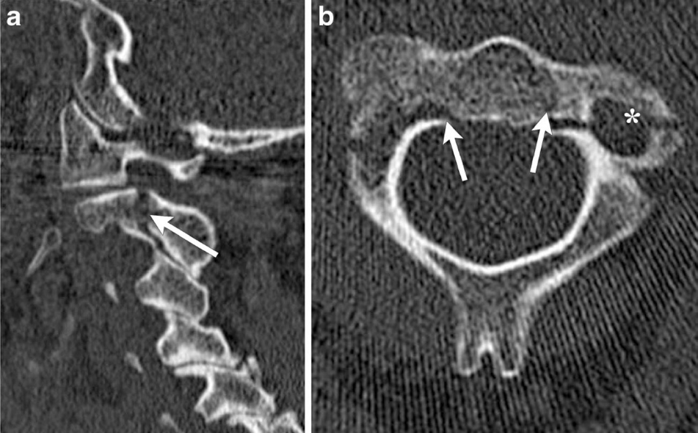
Figure 6. CT of a hangman’s fracture (traumatic spondylolisthesis of the axis): sagittal and axial images with arrows at the bilateral C2 pars fractures. From Utz et al. (2013), Insights into Imaging 5(1):67-75, CC BY 4.0, via Wikimedia Commons.
Part VI - Subaxial Cervical Injuries (C3-C7)
6.1 The mechanistic classification of Allen and Ferguson
The subaxial spine, uniform in configuration and analogous to the thoracolumbar spine, was first systematically classified by Allen and Ferguson (1982, 165 cases) into six force-vector groups, each staged by increasing severity.[118] The system is mechanistic rather than treatment-directing and has never been formally validated for reliability, yet it remains the vocabulary for describing how these injuries happen:[119]
- Compressive flexion (CF), five stages, culminating in the flexion teardrop (an oblique fracture from the anterior body to the inferior endplate, stage 3) and then posterior translation with facet gapping (stages 4-5, anterior and posterior ligamentous failure).
- Vertical compression (VC), three stages, culminating in the comminuted cervical burst fracture with retropulsion (stage 3).
- Distractive flexion (DF), four stages: facet subluxation (1), unilateral facet dislocation (2), bilateral facet dislocation with 50% translation (3), and 100% translation, the “floating vertebra” (4).
- Compressive extension (CE), five stages, from a unilateral posterior-arch (“lateral mass”) fracture to 100% anterior body displacement.
- Distractive extension (DE), two stages: anterior disc-space widening or body avulsion (1), with posterior translation (2).
- Lateral flexion (LF), two stages.
A useful caution from the data: a higher Allen stage does not always mean greater instability, which is part of why scoring systems supplanted purely mechanistic schemes for treatment decisions.[120]
6.2 The SLIC score and the AO Spine subaxial classification
The Subaxial Cervical Injury Classification (SLIC) of Vaccaro, Dvorak, and the Spine Trauma Study Group is one of the few classifications that actually directs treatment, and it serves as the cervical counterpart of the thoracolumbar TLICS.[121] It scores three categories, summing to a maximum of 10:[122]
- Injury morphology: compression/burst, distraction, or rotational/translational (increasing points).
- Discoligamentous complex (DLC): intact, indeterminate, or disrupted.
- Neurological status: intact, root injury, complete cord, or incomplete cord (with a modifier for ongoing cord compression).
The operative thresholds mirror TLICS: a score of 3 or less favours non-operative care, 5 or more favours surgery, and 4 is individualised.[123] The AO Spine subaxial classification parallels its thoracolumbar sibling: type A compression with an intact posterior tension band, type B anterior or posterior tension-band failure without malalignment, type C malalignment/translation, with separate facet (F) injury descriptors and neurological and case modifiers (PLC disruption, disc herniation, arterial injury, osteoporosis, ankylosis).[124] The older Cervical Injury Severity Score (Anderson et al.) and the Bono description system remain in use but are limited by complexity or modest reliability.[125]
6.3 The injury patterns and their management
Compression (wedge) fractures are stable when the facets are not subluxed or widened, there is no body translation, interspinous gapping is minimal, and kyphosis does not exceed 11°. They are braced (rigid collar for C3-C6, cervicothoracic brace for C7-T1) with flexion-extension views at about three months.[126] Burst fractures are high-energy injuries with retropulsion and frequent cord injury. The neurologically intact, minimally comminuted, minimally kyphotic (under 5°) burst with an intact PLC scores as low as SLIC 2 and may be braced, but any neurological deficit mandates surgical stabilisation (anterior corpectomy, cage, and plate, with added posterior fixation if the PLC is disrupted).[127]
Flexion teardrop fractures (Allen CF stage 3 and above) occur in younger patients under high energy and carry a high incidence of complete cord injury. PLC disruption is suggested by kyphosis over 11° or posterior body translation, and most are treated operatively by anterior corpectomy and plating, with posterior fixation added when translation exceeds about 3-3.5 mm with facet widening.[128] It is essential to distinguish this dangerous flexion teardrop from the benign extension-type teardrop (a small anteroinferior avulsion in the elderly), which is usually stable.[129]
Facet fractures without dislocation are usually minimally displaced and mechanically stable, treated in a rigid collar for 6-12 weeks with frequent films. They may harbour occult ligamentous injury, so MRI is increasingly used to separate stable from unstable, and follow-up flexion-extension views confirm stability.[130] When surgery is needed, the only randomised comparison (Kwon et al.) and other series favour the anterior approach (single-level ACDF) over posterior lateral-mass fixation for less pain, higher fusion, better alignment, and lower infection.[131]
Facet dislocations deserve special attention because they embody the central amyelic controversy. They may be unilateral or bilateral, perched or locked. Unilateral dislocations can occur without complete PLC disruption and may be mechanically stable, whereas bilateral injuries predict poorer neurological recovery.[132] The cornerstone of treatment is closed reduction with cranial-tong traction, which is safe and may be performed promptly in the awake, cooperative, serially examinable patient regardless of neurological status, because there is no compelling evidence that closed reduction in such a patient causes neurological deterioration.[133] The danger lies in the disc herniation that reduction can drive into the canal. If the patient cannot be examined (obtunded, head-injured, intoxicated), a pre-reduction MRI is strongly advised; if the patient is awake and intact, a pre-reduction MRI is reasonable; and in the awake patient with cord compromise or a progressive deficit, emergent reduction outweighs any delay for imaging.[134] Definitive fixation is most often anterior (ACDF removes the offending disc), with posterior or combined approaches for highly unstable bilateral or fixed deformities.[135]
The clay-shoveler’s fracture, an avulsion of a lower cervical (classically C7) spinous process from powerful contraction of the attached back muscles, is benign in isolation, though a spinous-process fracture combined with bilateral laminar fractures can signal a “floating” posterior arch at risk of displacing into the canal.[136] Injuries of the ankylosed or spondylotic spine (ankylosing spondylitis, DISH) behave like a fractured long bone. They are almost universally unstable, are frequently missed, and must be immobilised in the pre-injury position because in-line traction of a fixed kyphosis can be catastrophic.[137]
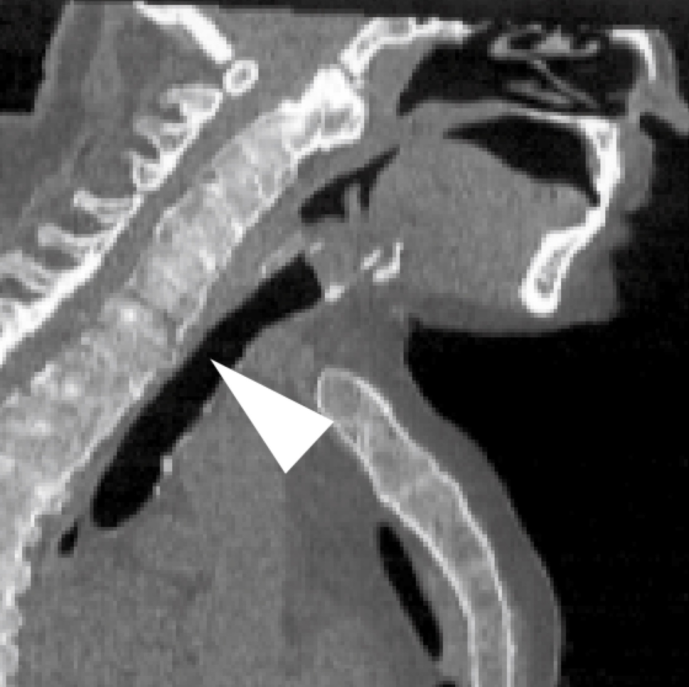
Figure 7. Sagittal CT of an ankylosed (bamboo) spine with a transverse fracture at the cervicothoracic junction: the highly unstable, long-bone-like fracture pattern of the stiff spine. (Used to illustrate an unstable transvertebral cervical pattern.) From Ushijima et al., CC BY 4.0, via Wikimedia Commons.
Part VII - Thoracolumbar Injuries
7.1 The Denis four injury types
Built on the three-column concept, the Denis classification recognises four major injury types, the distinction between the first two being the entire reason the three-column model was needed:[138]
- Compression fracture: failure of the anterior column only; typically stable.
- Burst fracture: failure of the anterior and middle columns under axial load; potentially unstable.
- Flexion-distraction (Chance / seat-belt) injury: tension failure of the posterior and middle (and sometimes anterior) elements.
- Fracture-dislocation: failure of all three columns with translation; the most unstable.
Minor injuries (transverse and spinous-process fractures) matter clinically chiefly as markers. Isolated lower lumbar transverse-process fractures are associated with pelvic and abdominal injury (Lombardo et al., 881 patients), and a transversely oriented spinous-process fracture can be the only sign of a tension-band (distraction) injury.[139]
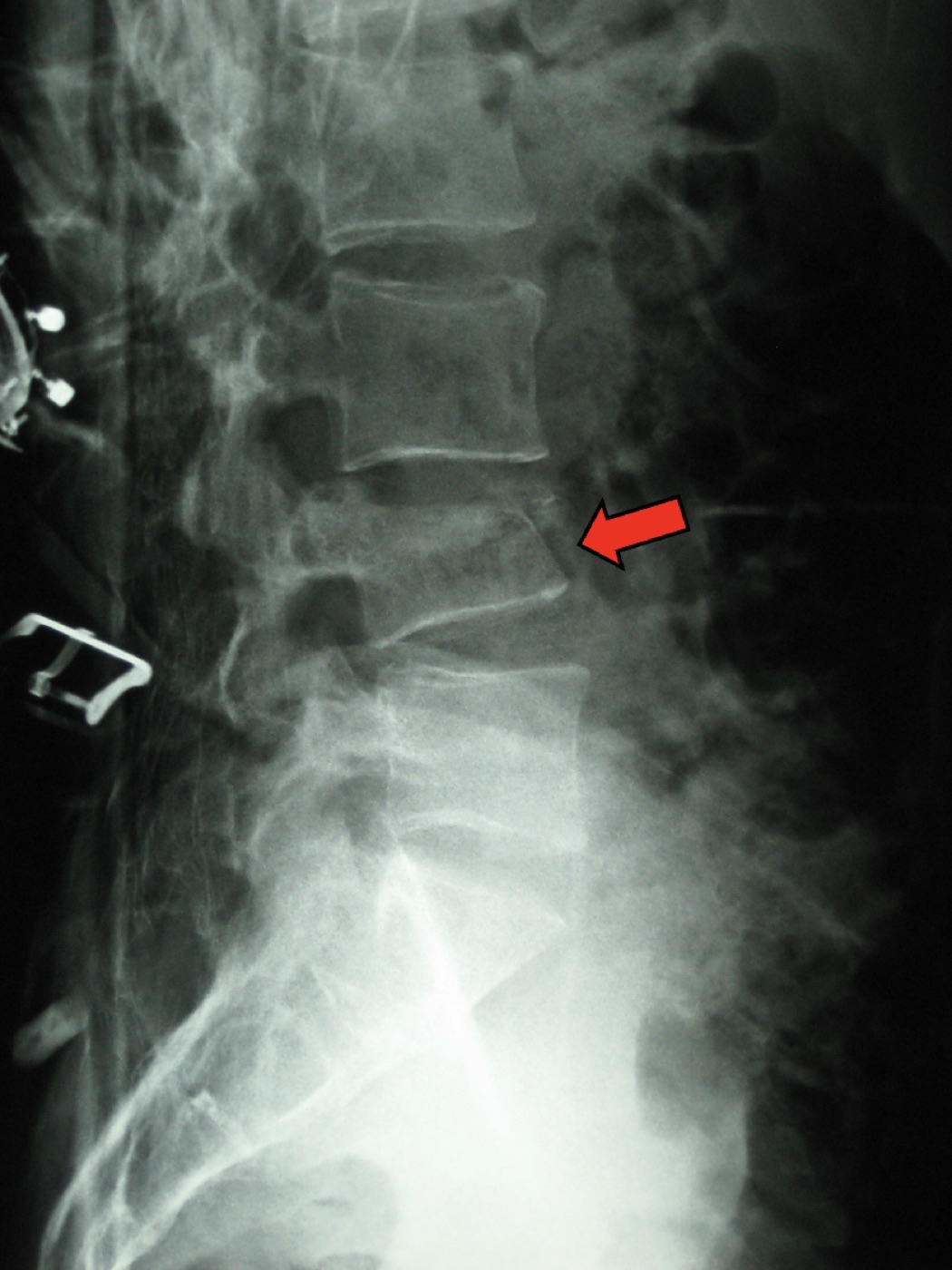
Figure 8. Lateral lumbar radiograph of a compression (wedge) fracture at L4 (arrow): anterior-column failure with an intact posterior cortex, the typically stable Denis compression injury. Source: James Heilman, MD, via Wikimedia Commons, CC BY-SA 3.0.
7.2 The TLICS score
The Thoracolumbar Injury Classification and Severity Score (TLICS) is the thoracolumbar decision tool, summing three components:[140]
| Component | Qualifier | Points |
|---|---|---|
| Morphology | Compression | 1 |
| Burst | 2 | |
| Translational/rotational | 3 | |
| Distraction | 4 | |
| PLC integrity | Intact | 0 |
| Indeterminate | 2 | |
| Injured | 3 | |
| Neurological status | Intact | 0 |
| Nerve root | 2 | |
| Cord/conus, complete | 2 | |
| Cord/conus, incomplete | 3 | |
| Cauda equina | 3 |
The threshold follows the same logic as SLIC: 3 or less is treated non-operatively, 5 or more operatively, and 4 is left to surgeon discretion.[141] The system’s own acknowledged weakness is revealing. By design it leaves the controversial neurologically intact burst with a questionable PLC at the indeterminate score of 4, and it is built on a North American philosophy in which some centres operate on burst fractures even without PLC injury or deficit, which limits its universality.[142]
7.3 The revised AO Spine thoracolumbar classification
The AO Spine system grades in ascending instability through three types with subtypes:[143]
- Type A - compression / axial loading: A0 clinically insignificant (transverse or spinous-process fracture); A1 wedge compression of a single endplate with intact posterior cortex; A2 a split of both endplates with intact posterior cortex; A3 incomplete burst (single endplate plus the posterior cortex); A4 complete burst (both endplates plus the posterior cortex).
- Type B - tension-band failure: B1 the bony (mono-osseous) Chance fracture; B2 posterior tension-band disruption (bony or ligamentous, crossing a motion segment, often with an associated type A); B3 hyperextension failure of the anterior tension band (typically the ankylosed spine).
- Type C - translation/displacement: complete three-column failure, the most unstable.
Neurology is graded N0-N4 (intact; transient; radicular; cauda equina or incomplete cord; complete cord) with NX for the unassessable patient and + for continued compression. Two case modifiers capture the clinical nuance: M1 (indeterminate posterior tension band) and M2 (a patient-specific comorbidity such as ankylosis or osteoporosis that may dictate treatment).[144] The Load-Sharing (McCormack) classification is referenced for deciding when vertebral-body comminution is severe enough to require anterior reconstruction, scoring comminution, fragment apposition, and focal kyphosis. The source named these three components but did not give the numerical 1-3 per-component scoring or the operative threshold.[145]
7.4 The burst-fracture controversy: the heart of the amyelic thoracolumbar question
The treatment of the neurologically intact burst fracture is, in the source’s own words, “probably the most widely debated” question in thoracolumbar trauma, and it is precisely the amyelic question.[146] Stability hinges on the PLC. An unstable burst shows a considerable increase in kyphosis on upright films, and a three-column burst with facet subluxation or equivalent PLC injury is a reason to stabilise even an intact patient.[147] For the intact patient with an intact PLC, by contrast, the indications for surgery are poorly defined, and the case for conservative care is strong. Systematic reviews show equivalent functional outcomes with non-operative treatment, at lower complication rates and lower cost, and canal compromise without deficit has not been linked to worse outcomes, with substantial spontaneous remodelling of the canal (over 50% canal expansion documented at nine months).[148]
The classic instability thresholds, kyphosis of 30° or more, height loss over 50%, and severe comminution or retropulsion, are explicitly relative, not absolute, indications, and there is no numeric canal-compromise percentage that mandates surgery in the intact patient.[149] The Bailey et al. randomised trial sharpened the point. Ninety-six patients with AO A3 bursts (T11-L3, under age 60, kyphosis under 35°, no deficit) had equivalent disability and pain with or without a TLSO brace at three months and two years, suggesting these injuries are inherently more stable than once believed.[150] The geographic and philosophical character of the disagreement is real and worth stating in an examination. The Rockwood authors favour non-operative treatment of the burst without PLC injury, whereas other centres operate routinely.[151]
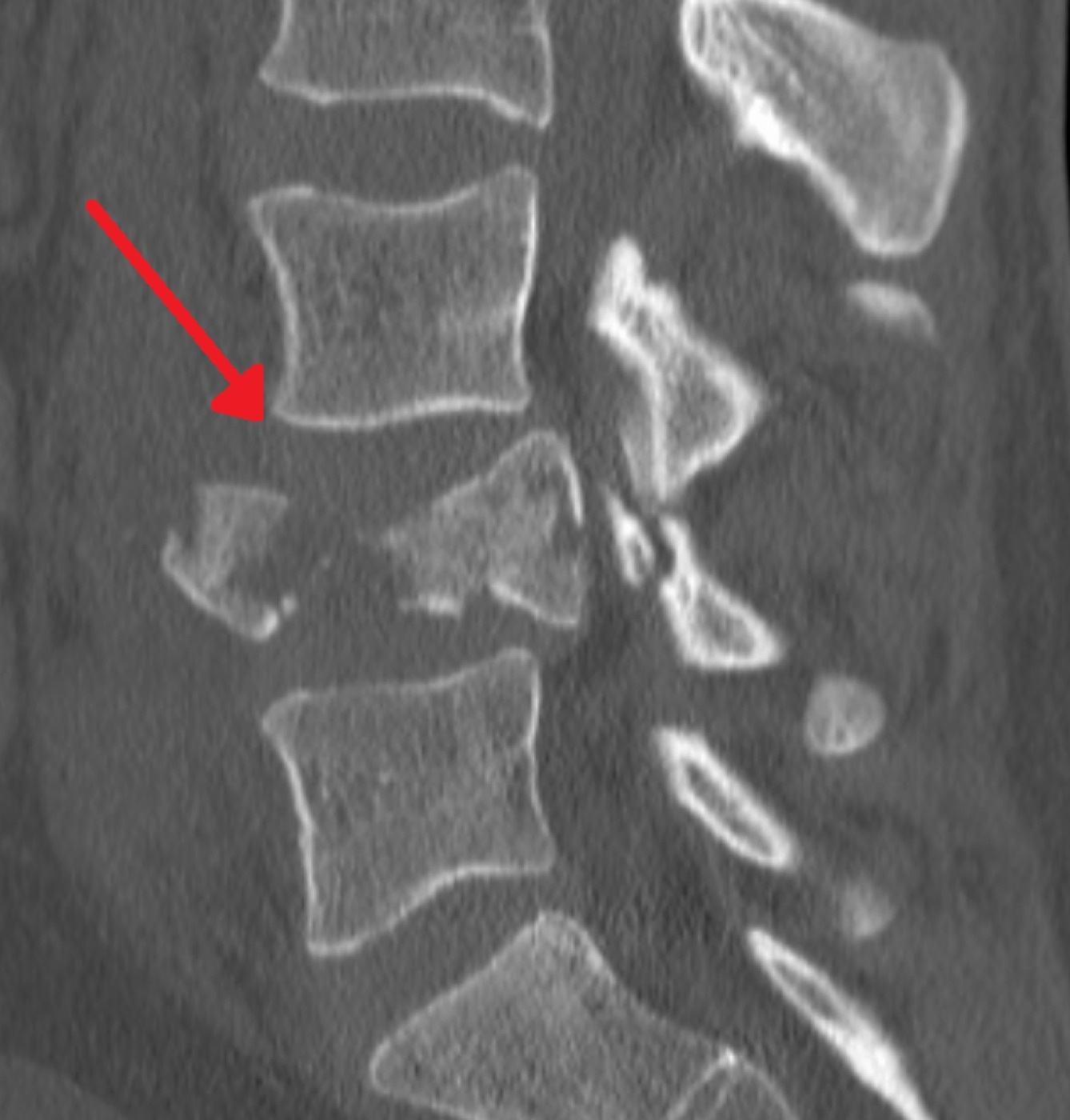
Figure 9. Sagittal CT of an L4 burst fracture: a retropulsed fragment encroaches on the spinal canal (arrow), the hallmark of middle-column failure. Source: James Heilman, MD, via Wikimedia Commons, CC BY-SA 4.0.
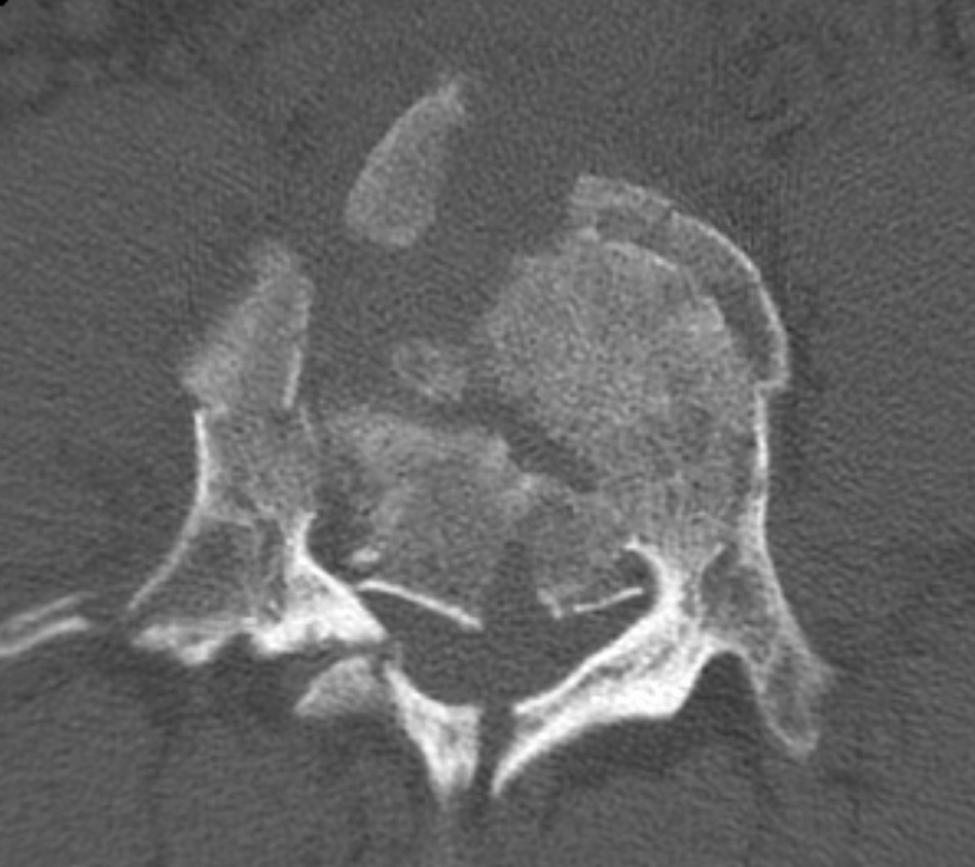
Figure 10. Axial CT of the same L4 burst fracture: comminution of the vertebral body with a retropulsed fragment narrowing the canal. Source: James Heilman, MD, via Wikimedia Commons, CC BY-SA 4.0.
7.5 Flexion-distraction (Chance) and fracture-dislocation
The Chance fracture (B1) is a transverse osseous failure through a single vertebra from the posterior tension band forward, the classic lap-belt injury. Because the rotational axis lies anterior to the body it carries a high association with intra-abdominal injury (in one series 30% of flexion-distraction patients had abdominal injuries), so the seat-belt sign mandates a search for visceral trauma.[152] The purely osseous Chance, being a three-column bony injury with high healing potential, may be treated non-operatively in a hyperextension brace, the rare exception to the rule that AO B and C injuries are operative. The ligamentous or mixed flexion-distraction injury (B2) heals less reliably and is fixed by posterior instrumented fusion.[153] The fracture-dislocation (type C) is the most unstable injury, with neurological injury in at least three-quarters of patients, and is operative regardless of neurological status, addressed first by a posterior approach.[154]
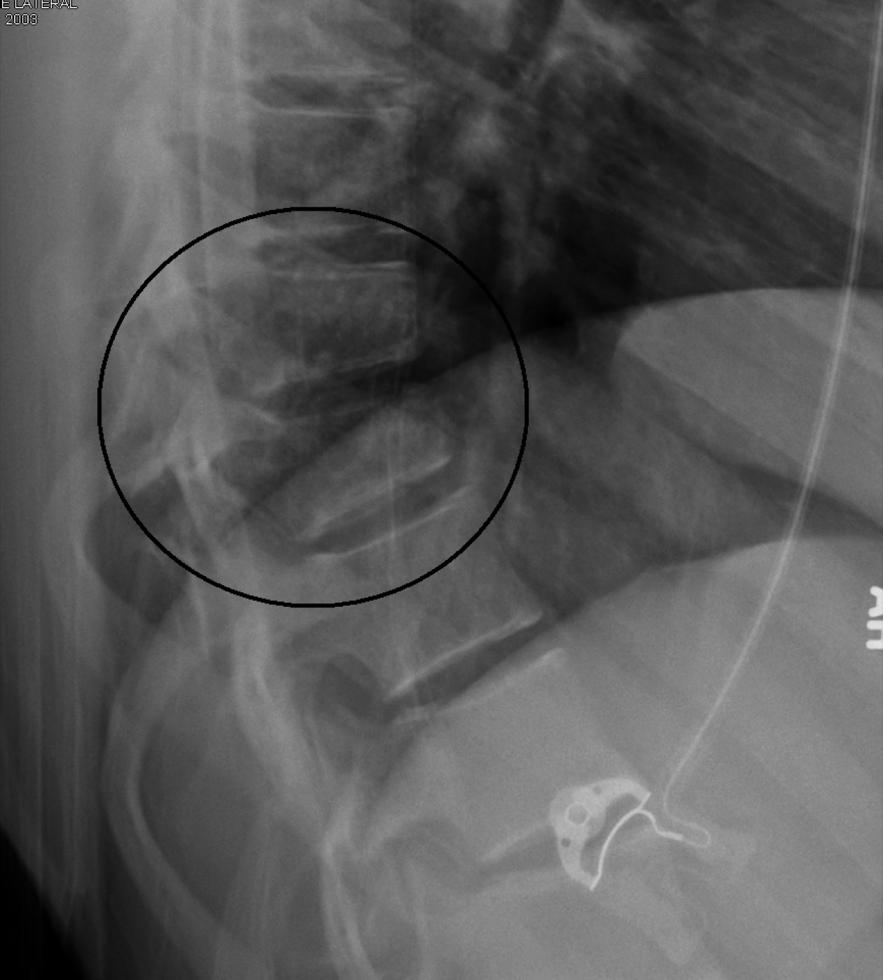
Figure 11. Lateral radiograph of a Chance (flexion-distraction / seat-belt) fracture (circled): a horizontal tension-band injury. The seat-belt sign mandates a search for abdominal injury. Source: James Heilman, MD, via Wikimedia Commons, CC BY-SA 3.0.
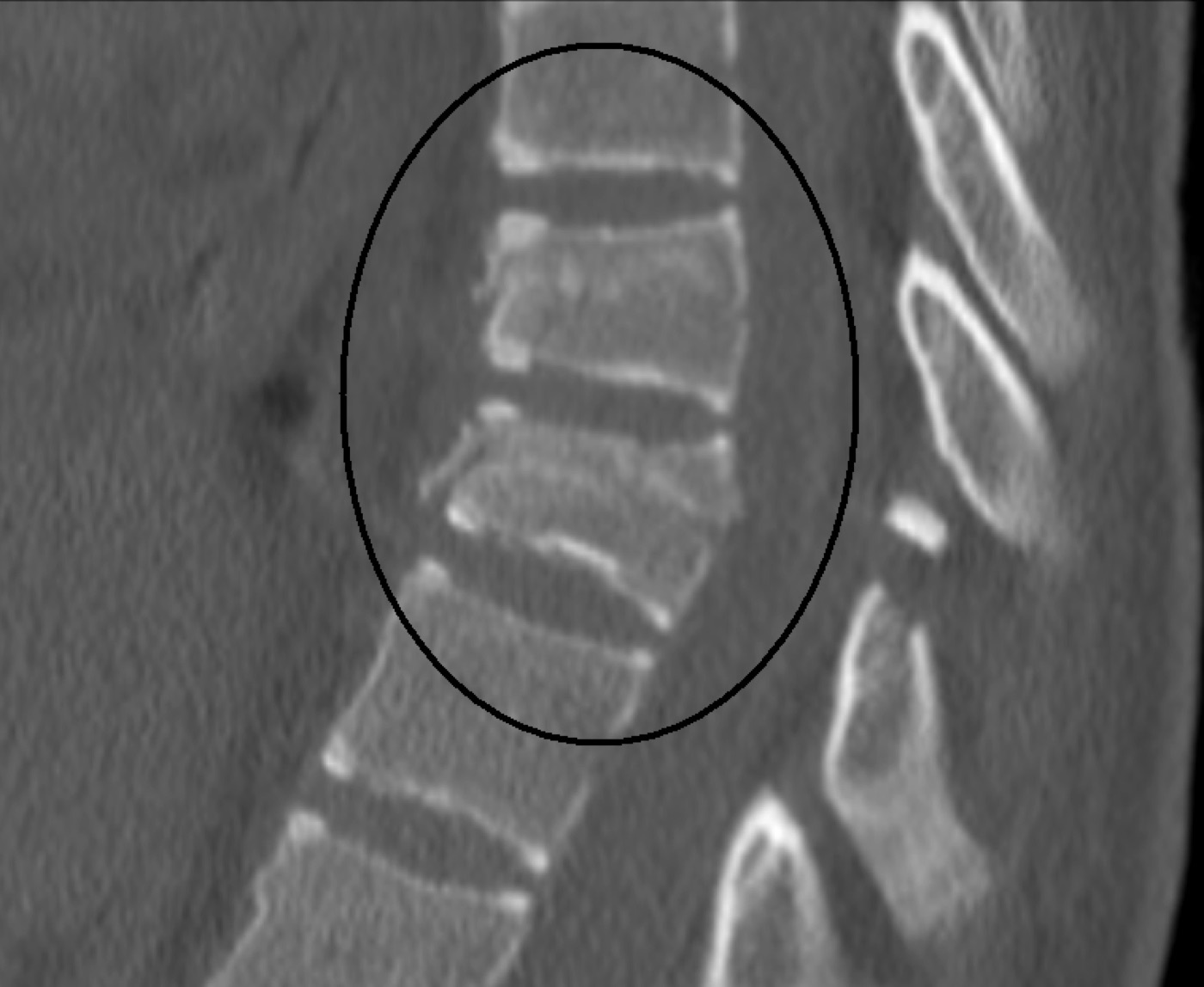
Figure 12. Sagittal CT of the same Chance fracture, showing the horizontal split running through the vertebra. Source: James Heilman, MD, via Wikimedia Commons, CC BY-SA 3.0.
7.6 Osteoporotic vertebral compression fractures
These are the most common osteoporotic fractures, the spine accounting for roughly half of all osteoporosis-related fractures, with over 1.5 million osteoporotic fractures yearly in the United States and a steeply age-dependent incidence.[155] Most are managed conservatively (analgesia, early mobilisation, activity modification, with bracing of uncertain benefit, the authors reserving a Jewett-type hyperextension brace for severe pain).[156] Medication, including bisphosphonates, teriparatide, and calcitonin, is used chiefly for pain, and calcitonin has moderate-strength support for acute pain in the first days.[157] The vertebroplasty/kyphoplasty literature is genuinely conflicted. The 2009 sham-controlled trials (Kallmes; Buchbinder) found vertebroplasty no better than placebo, and the 2018 Cochrane review and AAOS guidelines recommend against vertebroplasty for the neurologically intact osteoporotic fracture, while kyphoplasty retains limited-strength support as an option that may also correct height loss.[158]
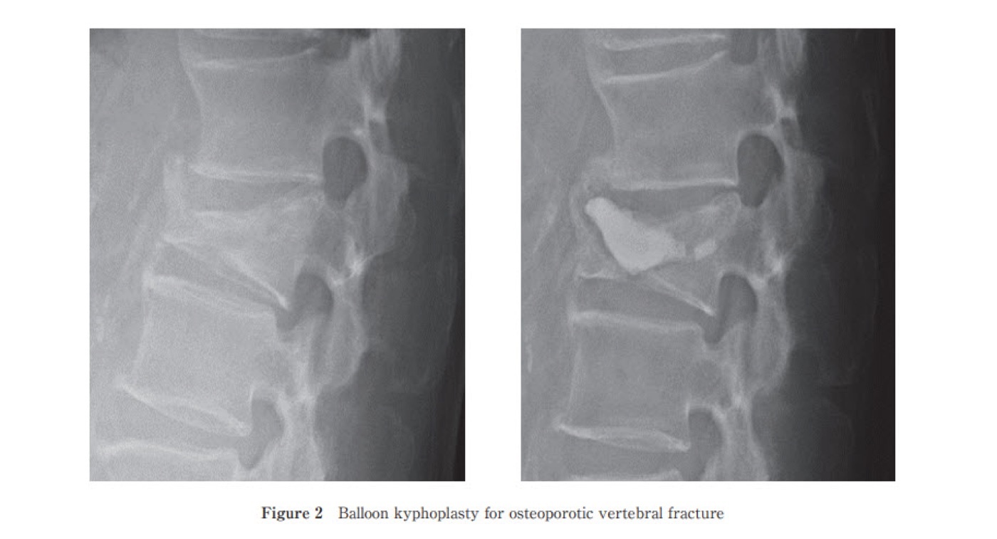
Figure 13. Balloon kyphoplasty for an osteoporotic vertebral compression fracture: before-and-after lateral radiographs with bone cement in the treated body. Current guidelines favour kyphoplasty over vertebroplasty. Source: Rei Momomura, via Wikimedia Commons, CC BY 4.0.
Part VIII - Principles of Treatment
8.1 The non-operative armamentarium: orthoses and the halo
Cervical orthoses work by three-point pressure and reduce but never eliminate motion.[159] The soft collar is the least restrictive (sprains, transverse-process fractures, and as a transitional aid). The rigid collars (Miami J, Philadelphia, Aspen) are more restrictive, with the Miami J favoured for combining rigidity and comfort and even immobilising the upper cervical spine. Pressure ulceration is a real complication, reported up to 38% in severe head injury.[160] Cervicothoracic orthoses (SOMI, Yale, Minerva) outperform collars in every plane, the Minerva restricting some 79-87% of sagittal motion.[161]
The halothoracic vest is the most effective non-operative control of rotational and translational moments, anchoring both the head and the upper thorax, and is used principally for reduction and temporary stabilisation before surgery.[162] It is poor at controlling unstable facet dislocations (the residual subaxial motion called “snaking”). Its pin-related complications (loosening or infection in 6-60%) and its hazards in the elderly (a fourfold mortality above age 65 in one series) have narrowed its use considerably.[163]
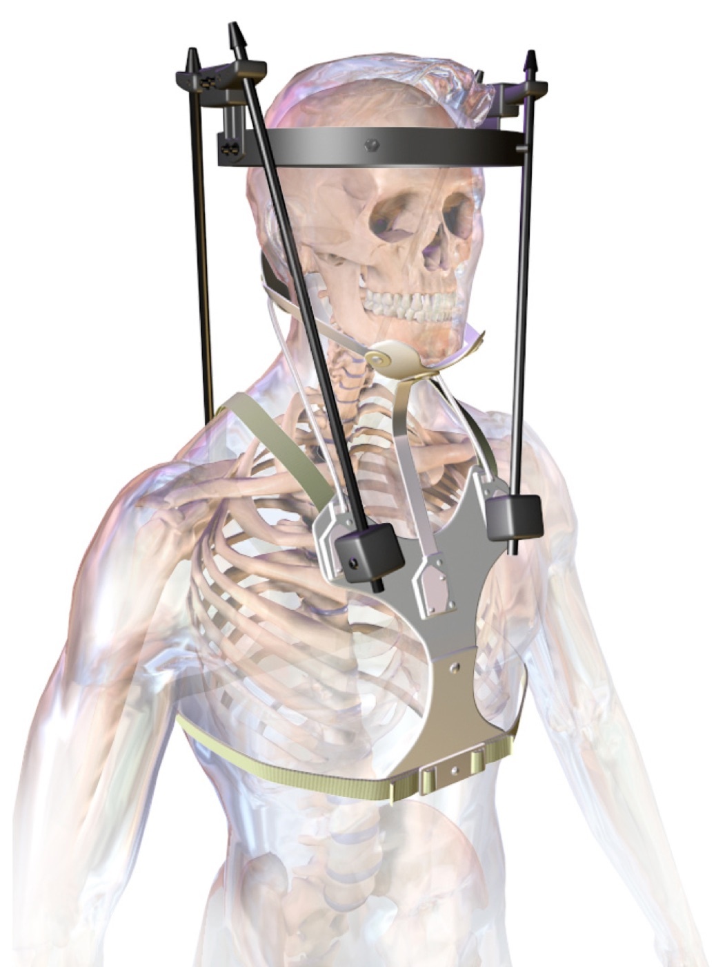
Figure 14. The halo vest (halothoracic orthosis): a cranial ring on skull pins linked by uprights to a thoracic vest, the most effective non-operative control of upper-cervical motion. Source: BruceBlaus, via Wikimedia Commons, CC BY-SA 4.0.
8.2 Skull traction, closed reduction, and the weight rules
Gardner-Wells tongs and the halo ring are the two traction devices. The tongs are quicker but temporary, while the halo ring can transition to a vest.[164] Traction works by progressive distraction and realignment through ligamentotaxis, providing indirect canal decompression. Because it depends on ligamentous continuity, it is contraindicated in occipitocervical dissociation and in type IIa hangman’s fractures.[165] The tong pins are placed about a finger’s breadth above the helix of the ear, in line with the external auditory meatus for a neutral pull (anterior placement adds an extension moment, posterior a flexion moment), after a skull fracture has been excluded.[166]
The reduction protocol for facet dislocations is examinable. One starts at 5-10 lb with a check radiograph to exclude occult occipitocervical instability, then adds 10 lb every 10-15 minutes with serial neurological examinations in an awake patient. Weights up to 140 lb are documented as safe for reducing facet dislocations in the cooperative patient.[167] The reduction is abandoned if the weight exceeds two-thirds of body weight, distraction at the injury site exceeds 10 mm, or the neurological status deteriorates.[168]
8.3 Operative principles: timing, approaches, instrumentation
On timing, the STASCIS study found that decompression within 24 hours of cord injury was safe and associated with a 2.8-fold higher odds of at least a two-grade ASIA improvement, and most surgeons now decompress within 24 hours when possible, earlier still for incomplete injuries.[169] For the amyelic patient timing is governed by stabilisation logistics and overall physiology rather than by cord rescue, but early stabilisation of unstable thoracic injuries reduces pulmonary morbidity and ICU stay.[170]
The anterior approach (in the subaxial cervical spine the Smith-Robinson approach) gives the most direct access to the anterior column for decompression and structural reconstruction (corpectomy with a load-sharing cage or strut graft and a fixed-angle plate). A corpectomy of more than two cervical levels should not be left as a stand-alone anterior construct.[171] The posterior approach is the workhorse for deformity correction and long constructs, with pedicle-screw and rod fixation the biomechanically superior instrumentation in the thoracolumbar spine and lateral-mass screw fixation the current standard in the subaxial cervical spine.[172] Combined approaches are reserved for the severely unstable, since posterior-only fixation tends to fail with severely comminuted bodies and anterior-only fixation tends to fail when the PLC is disrupted (AO B2 and C).[173]
A note on construct length in the thoracolumbar spine: short-segment fixation (one level above and below) of a burst fracture risks higher rates of hardware failure and recurrent kyphosis unless anterior support is added, so it is reserved for younger patients with good bone, while osteoporotic, junctional, and highly comminuted injuries are spanned at least two levels above and below.[174] Ligamentotaxis through a posterior distraction construct can achieve up to 50% canal decompression but fails when the annular attachments are disrupted or the retropulsed fragment has rotated 180°.[175]
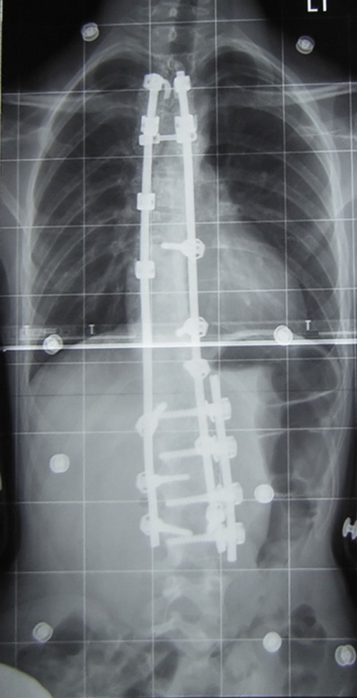
Figure 15. Posterior spinal instrumentation: pedicle screws and dual rods, the biomechanically superior construct for the thoracolumbar spine. (Illustrative post-fusion film of a deformity correction rather than a fracture.) Image by Wikipedia user Silverjonny, public domain, via Wikimedia Commons.
The principal complications to recall are, for the anterior cervical approach, dysphagia (up to 50%, mostly subclinical), recurrent laryngeal nerve palsy, Horner’s syndrome from injury to the sympathetic chain over the longus colli, and the rare but lethal oesophageal perforation; and, for the posterior approach, C5 palsy, durotomy, wound problems, and vertebral artery injury.[176]
Part IX - A Synthesis: How to Reason Through the Amyelic Spine
The candidate who can hold five questions in order will manage almost any spine-trauma case correctly. First, is the patient neurologically intact? Establish ASIA E by a real examination including perianal sensation and rectal tone, and remember that an injury below the conus produces cauda equina signs, not spinal shock. Second, is the column stable? Reach for the three-column concept and, above all, the integrity of the posterior ligamentous complex, the variable that most often decides operative from non-operative care in the intact patient. Third, what is the injury, by name and by classification? Use the treatment-directing scores (SLIC in the neck, TLICS and AO Spine in the thoracolumbar spine) rather than purely mechanistic schemes. Fourth, does the bony or ligamentous pattern carry a hidden lethal partner? Think of the occipitocervical dissociation that looks normal on the first film, the geriatric odontoid that kills out of proportion to its appearance, the seat-belt Chance fracture with a ruptured bowel behind it. Fifth, what does treatment aim to achieve? In the amyelic patient the aim is almost always prevention: to keep an intact cord intact by restoring and protecting a stable column with the least intervention that will reliably do so.
The recurring theme, true from C0 to the sacrum, is that the neurologically intact spine is not the safe spine. It is the spine in which everything is still to be gained or lost, and in which careful classification, honest assessment of stability, and respect for the few injuries that masquerade as benign are what separate a full recovery from a catastrophe.
References
-
Rockwood & Green’s Fractures in Adults, p.2933 (the amyelic, neurologically intact patient corresponds to ASIA grade E, “normal motor and sensory function”).
-
Rockwood & Green’s Fractures in Adults, p.3013 (Bellabarba et al.: a mean two-day diagnostic delay in occipitocervical dissociation caused secondary neurological decline in 29%).
-
Rockwood & Green’s Fractures in Adults, p.2929.
-
Rockwood & Green’s Fractures in Adults, pp.2929-2930.
-
Rockwood & Green’s Fractures in Adults, p.3091.
-
Rockwood & Green’s Fractures in Adults, pp.2929-2930.
-
Rockwood & Green’s Fractures in Adults, p.2930.
-
Rockwood & Green’s Fractures in Adults, p.2930.
-
Rockwood & Green’s Fractures in Adults, p.3092.
-
Rockwood & Green’s Fractures in Adults, p.3092.
-
Rockwood & Green’s Fractures in Adults, p.2930.
-
Rockwood & Green’s Fractures in Adults, p.2929.
-
Rockwood & Green’s Fractures in Adults, p.3091.
-
Rockwood & Green’s Fractures in Adults, p.3091.
-
Rockwood & Green’s Fractures in Adults, p.3091.
-
Rockwood & Green’s Fractures in Adults, pp.3091-3092.
-
Rockwood & Green’s Fractures in Adults, p.2980.
-
Rockwood & Green’s Fractures in Adults, p.3013.
-
Rockwood & Green’s Fractures in Adults, p.2929.
-
Rockwood & Green’s Fractures in Adults, p.2931.
-
Rockwood & Green’s Fractures in Adults, p.2931.
-
Rockwood & Green’s Fractures in Adults, p.2931.
-
Rockwood & Green’s Fractures in Adults, p.2932.
-
Rockwood & Green’s Fractures in Adults, p.2932.
-
Rockwood & Green’s Fractures in Adults, p.2932.
-
Rockwood & Green’s Fractures in Adults, p.2932.
-
Rockwood & Green’s Fractures in Adults, p.2932.
-
Rockwood & Green’s Fractures in Adults, p.2933.
-
Rockwood & Green’s Fractures in Adults, p.2933.
-
Rockwood & Green’s Fractures in Adults, p.2934.
-
Rockwood & Green’s Fractures in Adults, p.2934.
-
Rockwood & Green’s Fractures in Adults, p.2934.
-
Rockwood & Green’s Fractures in Adults, p.3093.
-
Rockwood & Green’s Fractures in Adults, p.3093.
-
Rockwood & Green’s Fractures in Adults, p.2933 (Table 46-1; the scale is graded after resolution of spinal shock and is influenced by the Frankel classification; it is not necessarily reliable within 72 hours of injury).
-
Rockwood & Green’s Fractures in Adults, pp.3094-3095.
-
Rockwood & Green’s Fractures in Adults, p.2933 (Table 46-1; the scale is graded after resolution of spinal shock and is influenced by the Frankel classification; it is not necessarily reliable within 72 hours of injury).
-
Rockwood & Green’s Fractures in Adults, p.2983 (Kang et al.).
-
Rockwood & Green’s Fractures in Adults, p.3111.
-
Rockwood & Green’s Fractures in Adults, p.3111.
-
Rockwood & Green’s Fractures in Adults, p.3111.
-
Rockwood & Green’s Fractures in Adults, p.3111.
-
Rockwood & Green’s Fractures in Adults, p.3111.
-
Rockwood & Green’s Fractures in Adults, p.3091.
-
Rockwood & Green’s Fractures in Adults, p.3091.
-
Rockwood & Green’s Fractures in Adults, p.2982.
-
Rockwood & Green’s Fractures in Adults, p.3096 (upright versus supine films disclose tension-band incompetence as increasing kyphosis under load).
-
Rockwood & Green’s Fractures in Adults, p.2986 (operative indications separate mechanical instability from neurological deficit with canal compromise).
-
Rockwood & Green’s Fractures in Adults, p.2944.
-
Rockwood & Green’s Fractures in Adults, p.2935.
-
Rockwood & Green’s Fractures in Adults, p.2937.
-
Rockwood & Green’s Fractures in Adults, p.2937.
-
Rockwood & Green’s Fractures in Adults, p.2937.
-
Rockwood & Green’s Fractures in Adults, p.2937.
-
Rockwood & Green’s Fractures in Adults, p.2939.
-
The individual CCR factors are standard teaching; the mined Rockwood extract (p.2937) referenced them only as a figure (Fig. 46-3) and did not enumerate them in text. The list given here follows Stiell et al., JAMA 2001, the rule’s derivation study.
-
Rockwood & Green’s Fractures in Adults, pp.2936-2941 (Algorithm 46-1).
-
Rockwood & Green’s Fractures in Adults, pp.2934-2935.
-
Rockwood & Green’s Fractures in Adults, pp.2934-2935.
-
Rockwood & Green’s Fractures in Adults, p.2935.
-
Rockwood & Green’s Fractures in Adults, pp.3103-3104.
-
Rockwood & Green’s Fractures in Adults, p.2935; p.3108.
-
Rockwood & Green’s Fractures in Adults, p.3108.
-
Rockwood & Green’s Fractures in Adults, p.2940 (Resnick et al. versus Ackland et al.).
-
Rockwood & Green’s Fractures in Adults, pp.2940-2941.
-
Rockwood & Green’s Fractures in Adults, p.2941.
-
Rockwood & Green’s Fractures in Adults, p.2940.
-
Rockwood & Green’s Fractures in Adults, pp.3095-3096.
-
Rockwood & Green’s Fractures in Adults, pp.3095-3096.
-
Rockwood & Green’s Fractures in Adults, p.3096 (Fig. 48-8).
-
Rockwood & Green’s Fractures in Adults, p.3096.
-
Rockwood & Green’s Fractures in Adults, p.3013 (the BAI and BDI, “Harris rule of 12,” are named as the most useful craniocervical measures; the source names the rule but does not restate the 12 mm value, which is standard teaching from Harris et al., AJR 1994).
-
Rockwood & Green’s Fractures in Adults, pp.3043, 3048-3049.
-
Rockwood & Green’s Fractures in Adults, p.3013.
-
Rockwood & Green’s Fractures in Adults, p.3013.
-
Rockwood & Green’s Fractures in Adults, p.3013.
-
Rockwood & Green’s Fractures in Adults, p.3013.
-
Rockwood & Green’s Fractures in Adults, pp.3013-3014.
-
Rockwood & Green’s Fractures in Adults, p.3014.
-
Rockwood & Green’s Fractures in Adults, pp.3014-3019.
-
The Powers ratio is standard teaching for anterior atlanto-occipital dislocation (Powers et al., Neurosurgery 1979; a ratio greater than 1.0 is abnormal) but was not found in the mined Rockwood extract, which named the BAI/BDI as the most useful measures. It is included for completeness and not falsely page-cited.
-
Rockwood & Green’s Fractures in Adults, p.3019.
-
Rockwood & Green’s Fractures in Adults, p.3019.
-
Rockwood & Green’s Fractures in Adults, p.3019.
-
Rockwood & Green’s Fractures in Adults, p.3020.
-
Rockwood & Green’s Fractures in Adults, pp.3020-3021.
-
Rockwood & Green’s Fractures in Adults, pp.3020-3021.
-
Rockwood & Green’s Fractures in Adults, p.3022.
-
Rockwood & Green’s Fractures in Adults, p.3022.
-
Rockwood & Green’s Fractures in Adults, p.3022.
-
Rockwood & Green’s Fractures in Adults, p.3022. The mined source gives the threshold as combined overhang “>7 or 8 mm” and does not use the term “rule of Spence” or the figure 6.9 mm; that eponym and value are classic teaching (Spence et al., JBJS 1970). The source explicitly flags contradicting cadaveric data (Woods et al., failure at 3.8 mm; Radcliff et al., no correlation) and recommends MRI as the adjunct.
-
Rockwood & Green’s Fractures in Adults, p.3022. The mined source gives the threshold as combined overhang “>7 or 8 mm” and does not use the term “rule of Spence” or the figure 6.9 mm; that eponym and value are classic teaching (Spence et al., JBJS 1970). The source explicitly flags contradicting cadaveric data (Woods et al., failure at 3.8 mm; Radcliff et al., no correlation) and recommends MRI as the adjunct.
-
Rockwood & Green’s Fractures in Adults, pp.3024-3025.
-
Rockwood & Green’s Fractures in Adults, pp.3022-3025.
-
Rockwood & Green’s Fractures in Adults, p.3025.
-
Rockwood & Green’s Fractures in Adults, p.3025.
-
Rockwood & Green’s Fractures in Adults, pp.3025-3028.
-
The Dickman classification (type I intrasubstance rupture versus type II bony avulsion; Dickman et al., Neurosurgery 1996) is standard teaching and clinically useful because type II injuries may heal with immobilisation whereas type I will not. It was not found in the mined Rockwood extract, which characterised the injury by ADI magnitude and ligamentous involvement rather than by the Dickman types.
-
Rockwood & Green’s Fractures in Adults, p.3028.
-
Rockwood & Green’s Fractures in Adults, p.3028.
-
Rockwood & Green’s Fractures in Adults, pp.3028-3029.
-
Rockwood & Green’s Fractures in Adults, p.3029.
-
Rockwood & Green’s Fractures in Adults, pp.3029-3030.
-
Rockwood & Green’s Fractures in Adults, p.3030.
-
Rockwood & Green’s Fractures in Adults, p.3030.
-
Rockwood & Green’s Fractures in Adults, pp.3030-3032.
-
Rockwood & Green’s Fractures in Adults, pp.3030-3032.
-
Rockwood & Green’s Fractures in Adults, pp.3031-3032 (Tashjian et al.; source disagreement on halo mortality explicitly noted).
-
Rockwood & Green’s Fractures in Adults, pp.3032-3033.
-
Rockwood & Green’s Fractures in Adults, pp.3032-3033.
-
Rockwood & Green’s Fractures in Adults, pp.3033-3034.
-
Rockwood & Green’s Fractures in Adults, pp.3033-3034.
-
Rockwood & Green’s Fractures in Adults, p.3035.
-
Rockwood & Green’s Fractures in Adults, p.3035.
-
Rockwood & Green’s Fractures in Adults, p.3035.
-
Rockwood & Green’s Fractures in Adults, p.3036.
-
Rockwood & Green’s Fractures in Adults, pp.3036-3039.
-
Rockwood & Green’s Fractures in Adults, pp.3039-3040.
-
Rockwood & Green’s Fractures in Adults, pp.3040-3043.
-
Rockwood & Green’s Fractures in Adults, p.3041.
-
Rockwood & Green’s Fractures in Adults, pp.3043-3044.
-
Rockwood & Green’s Fractures in Adults, pp.3043-3044.
-
Rockwood & Green’s Fractures in Adults, p.3044.
-
Rockwood & Green’s Fractures in Adults, p.3044.
-
Rockwood & Green’s Fractures in Adults, pp.3043-3044.
-
Rockwood & Green’s Fractures in Adults, pp.3048-3051.
-
Rockwood & Green’s Fractures in Adults, pp.3051-3054.
-
Rockwood & Green’s Fractures in Adults, pp.3054-3059, 3070.
-
Rockwood & Green’s Fractures in Adults, pp.3054-3059, 3070.
-
Rockwood & Green’s Fractures in Adults, pp.3059-3065.
-
Rockwood & Green’s Fractures in Adults, pp.3059-3065.
-
Rockwood & Green’s Fractures in Adults, pp.3065-3070.
-
Rockwood & Green’s Fractures in Adults, pp.2993-2994, 3065-3070.
-
Rockwood & Green’s Fractures in Adults, pp.2993-2994, 3065-3070.
-
Rockwood & Green’s Fractures in Adults, pp.3065-3070.
-
Rockwood & Green’s Fractures in Adults, p.3076.
-
Rockwood & Green’s Fractures in Adults, pp.3071-3073.
-
Rockwood & Green’s Fractures in Adults, p.3111.
-
Rockwood & Green’s Fractures in Adults, p.3135.
-
Rockwood & Green’s Fractures in Adults, pp.3112-3113 (Table 48-1). (The “burst” morphology carries 2 points in the standard TLICS; the mined report’s table transcription should be read with the published 1-2-3-4 morphology scale.)
-
Rockwood & Green’s Fractures in Adults, p.3112.
-
Rockwood & Green’s Fractures in Adults, p.3112.
-
Rockwood & Green’s Fractures in Adults, pp.3113-3116.
-
Rockwood & Green’s Fractures in Adults, p.3116.
-
Rockwood & Green’s Fractures in Adults, p.3117. The Load-Sharing Classification’s three components (comminution, apposition, kyphosis) are named in the source, but the per-component 1-3 scoring and the commonly taught threshold (a total of 7 or more out of 9 favouring anterior reconstruction; McCormack et al., Spine 1994) are standard teaching not quantified in the mined extract.
-
Rockwood & Green’s Fractures in Adults, pp.3135-3136.
-
Rockwood & Green’s Fractures in Adults, pp.3136-3137.
-
Rockwood & Green’s Fractures in Adults, pp.3136-3137 (Fig. 48-23).
-
Rockwood & Green’s Fractures in Adults, p.3137.
-
Rockwood & Green’s Fractures in Adults, p.3120 (Bailey et al.).
-
Rockwood & Green’s Fractures in Adults, pp.3135-3136.
-
Rockwood & Green’s Fractures in Adults, p.3092, p.3135.
-
Rockwood & Green’s Fractures in Adults, pp.3135, 3137-3148.
-
Rockwood & Green’s Fractures in Adults, p.3148.
-
Rockwood & Green’s Fractures in Adults, p.3149.
-
Rockwood & Green’s Fractures in Adults, p.3150.
-
Rockwood & Green’s Fractures in Adults, pp.3150-3151.
-
Rockwood & Green’s Fractures in Adults, pp.3151-3152.
-
Rockwood & Green’s Fractures in Adults, pp.2986-2987.
-
Rockwood & Green’s Fractures in Adults, pp.2986-2987.
-
Rockwood & Green’s Fractures in Adults, p.2987.
-
Rockwood & Green’s Fractures in Adults, pp.2987-2989.
-
Rockwood & Green’s Fractures in Adults, pp.2987-2989.
-
Rockwood & Green’s Fractures in Adults, pp.2989-2991.
-
Rockwood & Green’s Fractures in Adults, pp.2989-2991.
-
Rockwood & Green’s Fractures in Adults, p.2989.
-
Rockwood & Green’s Fractures in Adults, pp.2991-2992.
-
Rockwood & Green’s Fractures in Adults, pp.2991-2992.
-
Rockwood & Green’s Fractures in Adults, pp.2994-2995, 3120-3121.
-
Rockwood & Green’s Fractures in Adults, pp.2994-2995, 3120-3121.
-
Rockwood & Green’s Fractures in Adults, pp.2996-3000, 3124-3125.
-
Rockwood & Green’s Fractures in Adults, pp.3000-3009, 3121-3124.
-
Rockwood & Green’s Fractures in Adults, pp.3125-3126.
-
Rockwood & Green’s Fractures in Adults, pp.3121-3124.
-
Rockwood & Green’s Fractures in Adults, pp.3121-3124.
-
Rockwood & Green’s Fractures in Adults, pp.3010-3013.
-
Rockwood & Green’s Fractures in Adults, pp.2933, 3013.
-
Rockwood & Green’s Fractures in Adults, p.3111.
-
Rockwood & Green’s Fractures in Adults, p.2932.
-
Rockwood & Green’s Fractures in Adults, p.2937.
-
Rockwood & Green’s Fractures in Adults, pp.2935, 3108.
-
Rockwood & Green’s Fractures in Adults, pp.3030-3032.
-
Rockwood & Green’s Fractures in Adults, pp.3022-3025.
-
Rockwood & Green’s Fractures in Adults, pp.3035-3036.
-
Rockwood & Green’s Fractures in Adults, pp.3120, 3135-3137.
-
Rockwood & Green’s Fractures in Adults, pp.3043-3044, 3112.
-
Rockwood & Green’s Fractures in Adults, p.3092.
-
Rockwood & Green’s Fractures in Adults, pp.2991-2994.
-
Rockwood & Green’s Fractures in Adults, pp.2989, 3035, 3071-3073.
-
Rockwood & Green’s Fractures in Adults, pp.3151-3152.