Contents
- Orientation: Mobility Bought at the Price of Stability
- Part I - Stabilisers of the Glenohumeral Joint
- Part II - The Acute Traumatic Dislocation
- Part III - Reduction and Acute Management
- Part IV - The Pathoanatomy of Recurrence
- Part V - Recurrent (Habitual) Instability: Risk, Dichotomy, and Classification
- Part VI - Surgical Treatment of Anterior Instability
- Part VII - Posterior Instability
- Part VIII - Multidirectional and Voluntary Instability
- Part IX - A Synthesis: How to Reason Through Shoulder Instability
- References
Orientation: Mobility Bought at the Price of Stability
The glenohumeral joint is the most mobile joint in the body, and it pays for that mobility in stability. A large spherical humeral head sits against a small, shallow glenoid, with only a quarter to a third of the head in contact with the socket at any moment, so the bony architecture contributes almost nothing to keeping the joint located.[1] Stability instead depends on a layered system of soft tissues: the labrum that deepens the socket, the capsule and glenohumeral ligaments that tighten at the extremes of motion, the negative intra-articular pressure that seals the joint, and the rotator cuff and scapular muscles that actively centre the head. When a traumatic dislocation tears the anteroinferior corner of this system, and especially when that tear fails to heal, the joint is left prone to dislocate again. That is the through-line of this topic. The acute dislocation is an event, whereas the recurrent (habitual) dislocation is a disease of failed healing, and the two must be understood together.
One conceptual caution frames everything that follows. Laxity is not instability. Normal shoulders are lax, gymnasts and other healthy people show large asymptomatic translations, and laxity is often not even increased in the unstable shoulder.[2] Instability is symptomatic, unwanted translation. The distinction matters because the commonest error in this field is to mistake a lax but stable shoulder, or a voluntary “party trick” dislocator, for a surgical problem and to operate when one should not.
Part I - Stabilisers of the Glenohumeral Joint
1.1 The static stabilisers
The glenoid labrum is a fibrocartilaginous ring around the rim that deepens the anteroposterior socket from about 2.5 to 5.0 mm and increases the superior-inferior concavity to about 9 mm; losing it reduces overall socket depth by up to 50%.[3] Habermeyer’s image is useful. The joint behaves like a piston in a sealed syringe, and the labrum is the valve block that seals the joint against atmospheric pressure, so an anteroinferior labral detachment breaks the seal and abolishes the stabilising negative pressure.[4] Through concavity-compression (the term is Lippitt and Matsen’s), an intact labrum lets the head resist a tangential force up to 60% of the compressive load pressing it into the socket, and the labrum contributes roughly 10% of overall stability by this mechanism.[5]
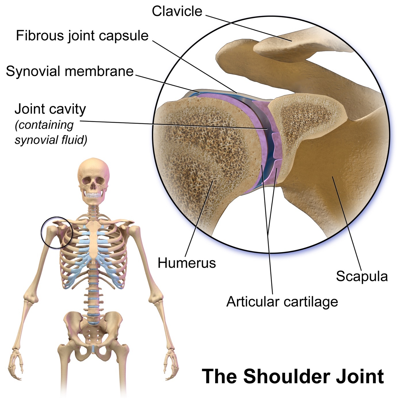
Figure 1. The glenohumeral joint: a large humeral head on a small, shallow glenoid, where most stability comes not from bone but from the capsule, labrum, and ligaments. Source: BruceBlaus / Blausen Medical 2014, via Wikimedia Commons, CC BY 3.0.
The capsule and glenohumeral ligaments are slack in mid-range and tighten at the extremes, each ligament according to arm position. The superior glenohumeral ligament is the primary restraint to inferior translation of the adducted arm; the inferior glenohumeral ligament complex (IGHL), a hammock with a thick anterior band, a thinner posterior band, and an axillary pouch between them, is the key restraint in abduction, the anterior band tightening at 45° and the posterior band at 90° of abduction, the whole complex sliding beneath the head in abduction and external rotation to block anterior translation.[6] The rotator interval (between the upper border of subscapularis and the anterior border of supraspinatus), containing the superior glenohumeral and coracohumeral ligaments, restrains inferior translation with the arm at the side, and its deficiency contributes to multidirectional instability.[7] The middle glenohumeral ligament is the most variable, absent in up to 30% of people.[8]
1.2 The dynamic stabilisers and proprioception
The rotator cuff stabilises in three ways: by compressing the head into the socket, by coordinated contraction that centres the head, and by tightening the ligaments through its attachments; a modelled 50% reduction in cuff force increases anterior displacement by 46%.[9] The long head of biceps and the deltoid become more important as the passive restraints fail, the deltoid’s stabilising contribution actually growing once a Bankart lesion is present.[10] Finally, proprioception, mediated by mechanoreceptors in the capsule, is disrupted in the unstable shoulder and recovers only gradually after stabilisation, which is part of the rationale for rehabilitation.[11]
Part II - The Acute Traumatic Dislocation
2.1 Epidemiology and directions
The shoulder is the most commonly dislocated major joint, accounting for nearly half of all dislocations, and the vast majority, about 85% or more, are anterior.[12] Rowe’s classic series of 500 dislocations found 96% were traumatic.[13] Anterior instability has a population incidence of about 0.08 per 1,000 person-years but reaches 3% per year in young male collision athletes, and it is higher still in military personnel.[14] Posterior dislocations make up only about 2-4%, and inferior dislocations are rare.[15] One background fact is worth holding onto: having dislocated the shoulder makes later glenohumeral arthrosis 10 to 20 times more likely.[16]
Anterior dislocation results from a force applied to the abducted, externally rotated, extended arm, levering the head out anteriorly, most often into the subcoracoid position (the head sits anterior to the glenoid and below the coracoid).[17] The patient holds the arm in slight abduction and resists motion. The classic clinical picture is a squared-off shoulder with the humeral head palpable anteriorly and a hollow beneath the acromion.[18]
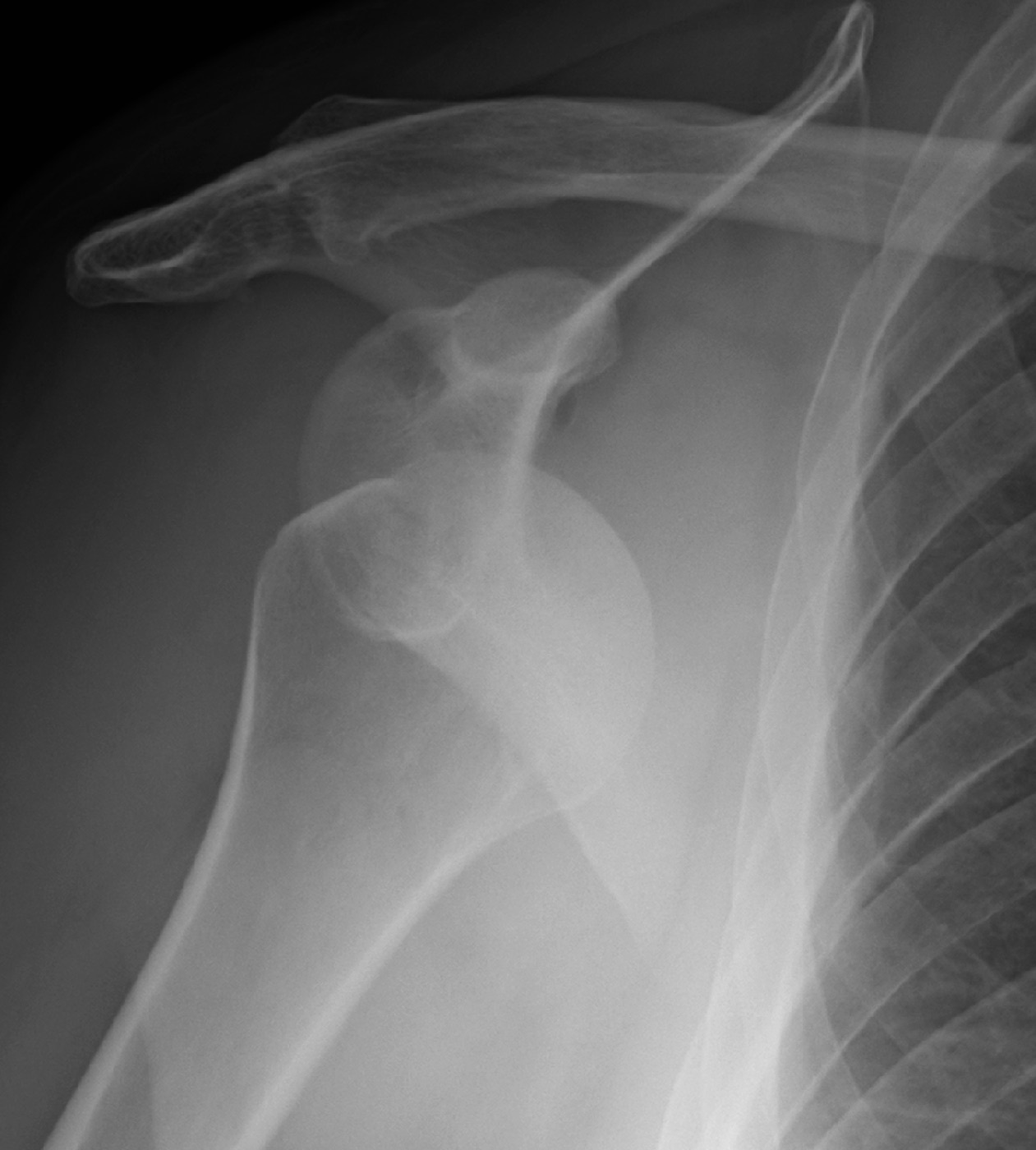
Figure 2. Anterior shoulder dislocation, AP radiograph: the humeral head lies anteroinferior to the glenoid (subglenoid/subcoracoid). Source: James Heilman, MD, via Wikimedia Commons, CC BY-SA 4.0.
Posterior dislocation is the great diagnostic trap. It follows a seizure, an electric shock, or an axial load on the adducted, internally rotated arm, the powerful internal rotators overwhelming the external rotators, and it is missed in more than 60-79% of cases because the arm sits in a deceptively ordinary-looking internally rotated “sling” position and the AP film looks nearly normal.[19] The diagnostic keys are clinical (the arm is locked in internal rotation with external rotation blocked) and radiographic (the axillary or scapular-Y view, or CT).[20] On the AP film the internally rotated head loses its normal asymmetry and looks symmetrical and rounded: the so-called “light-bulb” sign.[21]
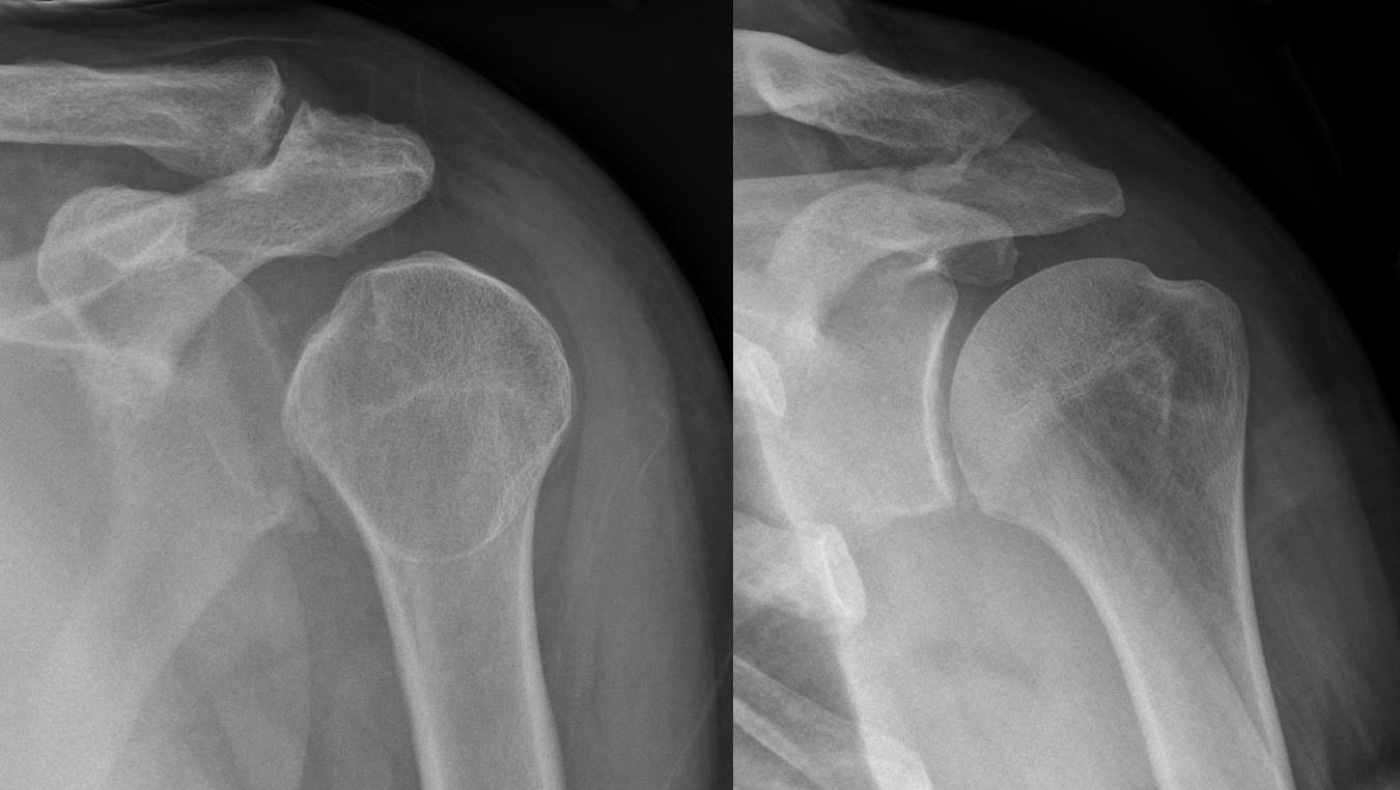
Figure 3. Posterior dislocation: fixed internal rotation makes the head look rounded and symmetrical, the “light-bulb” sign (left), shown with the same shoulder after reduction (right). Source: Hellerhoff, via Wikimedia Commons, CC BY-SA 3.0.
Inferior dislocation, luxatio erecta, follows forced hyperabduction that levers the head out below the glenoid, leaving the arm fixed and locked overhead, an unmistakable presentation.[22] It carries a very high rate of associated injury. In one series of 19 patients, all 19 had a brachial plexus injury and some vascular compromise, the rotator cuff is essentially always detached, and axillary-artery injury is reported.[23]
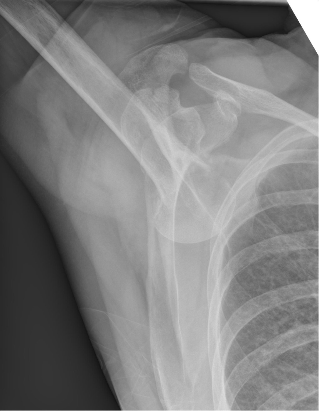
Figure 4. Luxatio erecta (inferior dislocation): the head sits below the glenoid with the arm fixed overhead. Source: Hellerhoff, via Wikimedia Commons, CC BY-SA 3.0.
2.2 Associated injuries
The axillary nerve is injured in about 13.5% of anterior dislocations, vulnerable because it is tethered as it wraps the humeral neck. It is tested by sensation over the lateral deltoid (the “regimental badge” area) and by deltoid contraction, and the lesion is usually a neurapraxia that recovers.[24] Rotator cuff tears accompany dislocation increasingly with age, so that in patients over 40 a cuff tear must be actively excluded, the older patient tending to tear the cuff where the younger patient tears the labrum.[25] Bony injuries, the greater-tuberosity fracture, the bony Bankart (glenoid rim fracture), and the Hill-Sachs impaction of the head, are common and shape later management.[26] A complete neurovascular examination is mandatory before and after reduction.[27]
2.3 Imaging
The trauma series is the true AP (Grashey), scapular-Y, and axillary views, the axillary being the single best view for the head-glenoid relationship.[28] The cardinal rule is to obtain an axillary or scapular-Y view (or a modified axillary such as the Velpeau view if the patient cannot abduct) precisely to avoid missing a posterior dislocation.[29] The workup is completed by special views (the Stryker notch for the Hill-Sachs lesion and the West Point view for the bony Bankart), by CT with three-dimensional reconstruction for quantifying bone loss, and by MR arthrography for the labrum.[30]
Part III - Reduction and Acute Management
3.1 Reduction techniques
Many techniques exist, and the unifying principle of the gentle ones is traction with countertraction and muscle relaxation rather than force. The Hippocratic method (countertraction by the operator’s stockinged foot across the axilla, not into it) and the Kocher leverage method are the oldest. Kocher is now discouraged because levering the head risks fracturing the humeral neck and the historical recurrence rate after it was high (McMurray: 40% recurrence after Kocher versus 12% after gently lifting the head).[31] The Milch technique abducts and externally rotates the arm and pushes the head back with the thumb.[32] The widely used modern methods (the Stimson prone hanging-weight technique, scapular manipulation, external rotation, and the Cunningham massage-and-gentle-adduction method) are gentle, atraumatic, and often need little or no sedation.[33] Reduction is performed under intra-articular local anaesthetic or procedural sedation, and difficult or locked dislocations may need general anaesthesia with muscle relaxation in theatre.[34]
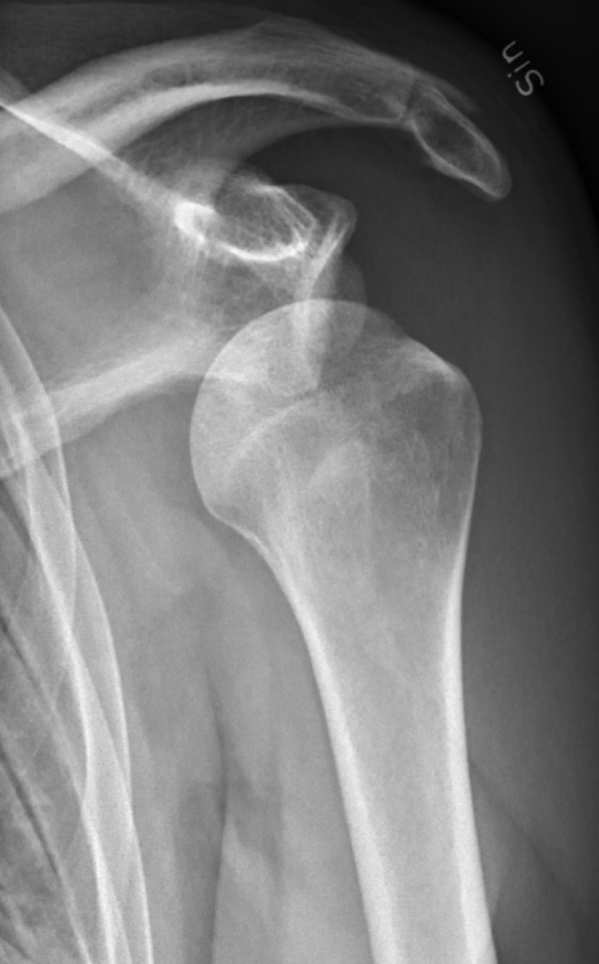
Figure 5. Anterior dislocation before reduction. Source: Mikael Häggström, M.D., via Wikimedia Commons, CC0 1.0.
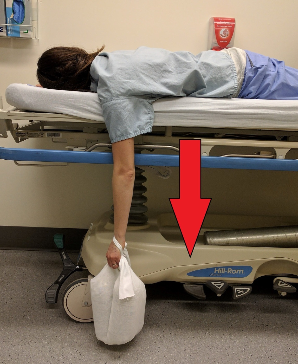
Figure 6. The Stimson reduction technique: the patient lies prone with the arm hanging and a suspended weight applying gentle downward traction. Source: James Heilman, MD, via Wikimedia Commons, CC BY-SA 4.0.
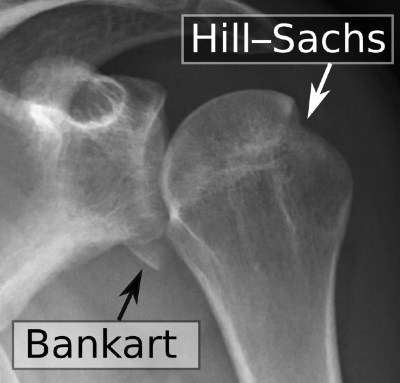
Figure 7. The same shoulder after reduction, with the annotated Bankart (anteroinferior glenoid) and Hill-Sachs (posterolateral head) lesions the dislocation produced. Source: Mikael Häggström, M.D., via Wikimedia Commons, CC0 1.0.
After any reduction, the position is confirmed on a radiograph and the neurovascular status is rechecked.[35]
3.2 Immobilisation and the recurrence problem
The reduced shoulder is immobilised in a sling, traditionally in internal rotation for about three weeks. Itoi proposed external-rotation immobilisation on the rationale that it better coapts the Bankart lesion to the glenoid (and his early data showed lower recurrence), but several subsequent randomised trials and a meta-analysis of 632 patients found no difference in recurrence between internal- and external-rotation immobilisation, so the question remains unsettled.[36] The harder problem lies one step deeper. After a first dislocation the capsule undergoes deformation and disordered collagen healing, beginning the self-perpetuating cycle of recurrence, and the dominant predictor of that recurrence is youth. A first dislocation in the teens or early twenties carries a very high recurrence rate (commonly quoted at 70-90%), which falls steeply with age, so that the older first-time dislocator rarely redislocates.[37]
Part IV - The Pathoanatomy of Recurrence
4.1 The Bankart lesion and its variants
The essential lesion of recurrent traumatic anterior instability is avulsion of the anteroinferior labrum and IGHL from the glenoid rim, the Bankart lesion, and its failure to heal is the major reason the shoulder remains unstable.[38] When the avulsion takes a fragment of glenoid bone with it, it is a bony Bankart.[39] Several variants are recognised and worth naming, although they were not all detailed in the mined sources: the Perthes lesion (the labrum is stripped but remains attached medially by intact periosteum), the ALPSA (anterior labroligamentous periosteal sleeve avulsion, in which the labroligamentous sleeve heals medially on the glenoid neck in a displaced position), the GLAD (glenolabral articular disruption, a labral tear with an articular cartilage flap), and the HAGL (humeral avulsion of the glenohumeral ligaments, in which the capsule fails on the humeral rather than the glenoid side).[40]
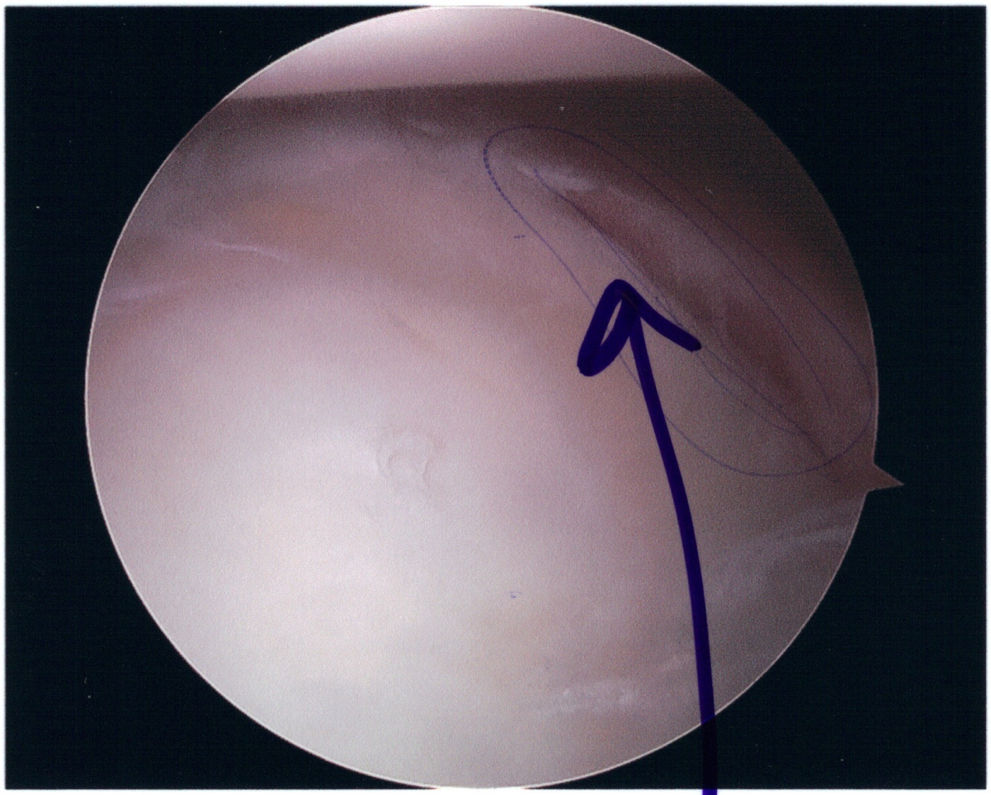
Figure 8. Bankart lesion at arthroscopy: the anteroinferior labrum detached from the glenoid rim (arrow). Source: Rwillia4, via Wikimedia Commons, public domain.
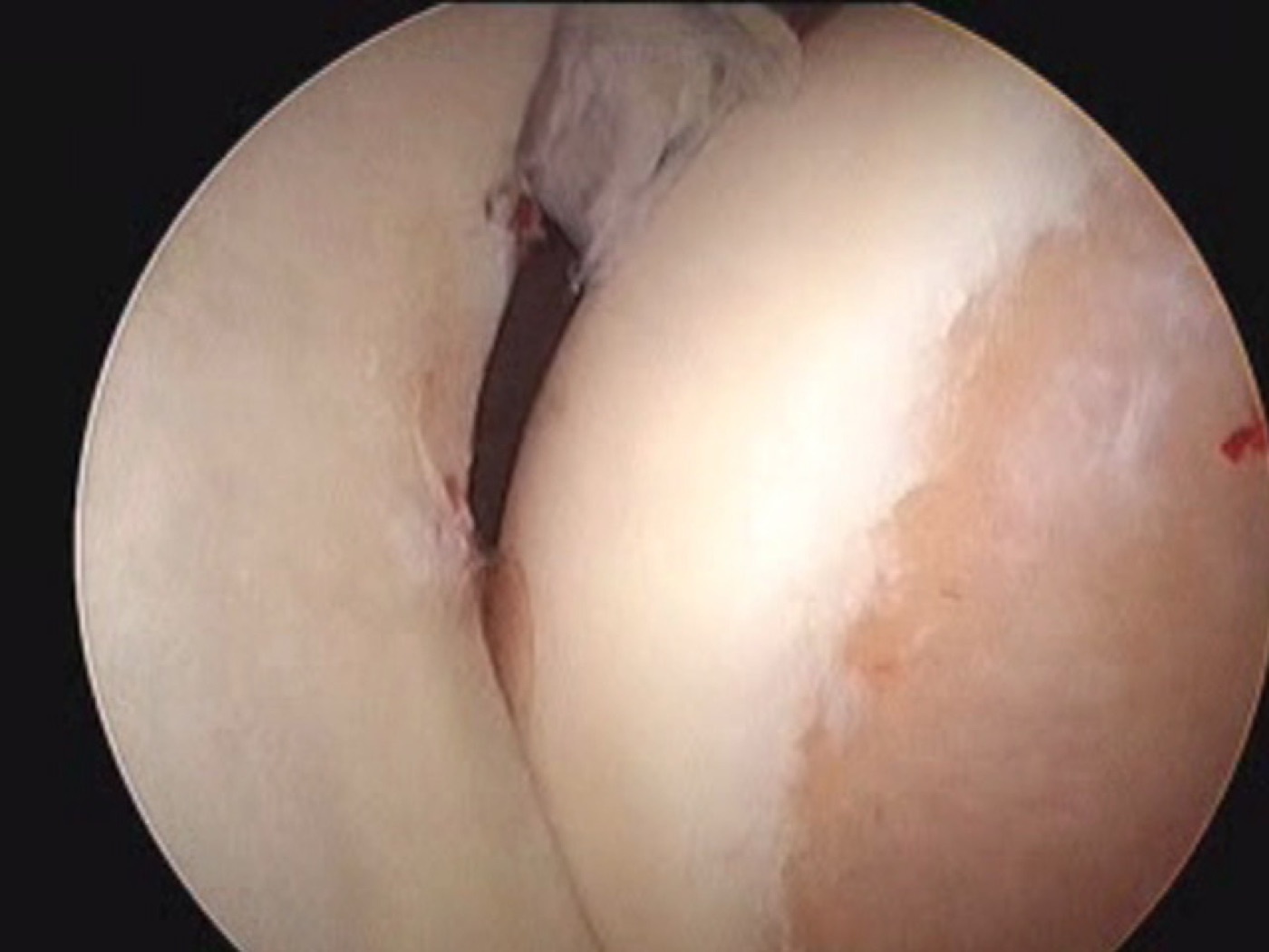
Figure 9. Bankart lesion at arthroscopy: the cleft between the detached anteroinferior labrum and the glenoid rim. Source: Drfrancescsoler, via Wikimedia Commons, CC BY-SA 4.0.
4.2 The Hill-Sachs lesion and the glenoid track
A Hill-Sachs lesion is an impaction fracture of the posterolateral humeral head where it strikes the anterior glenoid rim during an anterior dislocation. It is present in roughly a quarter to a third of acute dislocations and over half of recurrent ones, and it enlarges with repeated dislocation.[41] Its posterior counterpart, the reverse Hill-Sachs or McLaughlin lesion, is an anteromedial head defect from a posterior dislocation.[42]
Whether a Hill-Sachs lesion matters depends on the glenoid track concept. During abduction and external rotation the glenoid contacts a band across the head. If the Hill-Sachs lesion stays within this band it is “on-track” (non-engaging) and is clinically unimportant, whereas if it extends medial to the track it is “off-track” (engaging) and will catch on the glenoid rim and re-dislocate.[43] The track is approximately 83% of the glenoid width (reduced further by any anterior glenoid bone loss), the threshold formalised by Yamamoto, Itoi, and Di Giacomo.[44] In a series of 100 recurrent anterior dislocators, 93% were on-track and treatable by soft-tissue Bankart repair, and only 7% were off-track and needed a bony or filling procedure.[45]
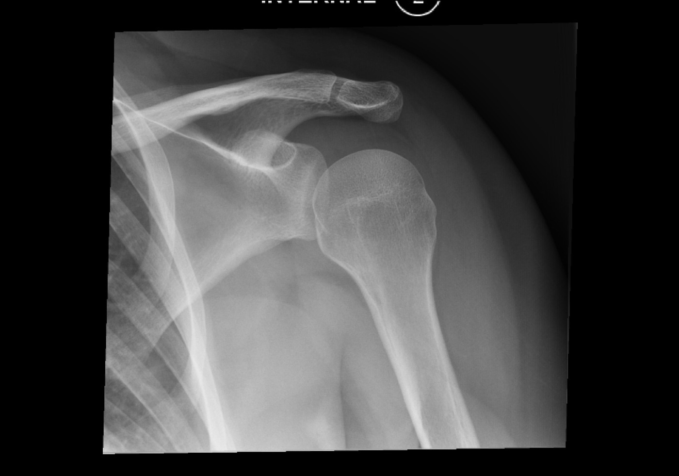
Figure 10. Hill-Sachs lesion on an AP internal-rotation radiograph: the posterolateral humeral-head impaction defect. Source: Andrew, via Wikimedia Commons, CC BY-SA 4.0.
4.3 Glenoid bone loss
Anterior glenoid bone loss progressively flattens the socket (the normal pear-shaped glenoid becoming an “inverted pear” with significant loss) and is one of the strongest predictors of failure of a soft-tissue repair.[46] The critical threshold is debated. Rockwood & Green place it at 13.5-17.3% of glenoid width (Burkhart found 67% recurrence after arthroscopic Bankart with more than 25% loss versus 4% without significant loss, and Shin found 42.9% failure above 17.3% versus 3.7% below), whereas The Shoulder (Itoi, Wirth) places the critical value nearer 25% of the width or 20% of the surface area, leaving a 17-25% “grey zone” decided by patient factors.[47] Bone loss is best measured on an en-face (Grashey or 3D CT) view using a best-fit circle over the inferior glenoid.[48]
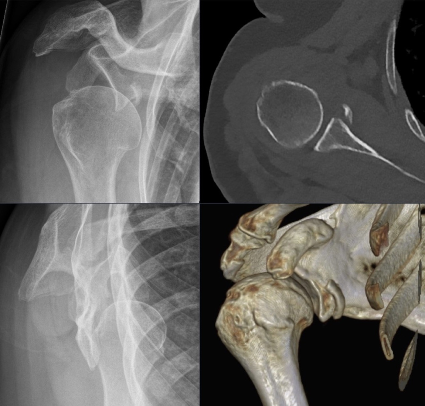
Figure 11. Bony Bankart lesion: an anteroinferior glenoid rim fracture on radiograph, CT, and 3D reconstruction. Source: Hellerhoff, via Wikimedia Commons, CC BY-SA 4.0.
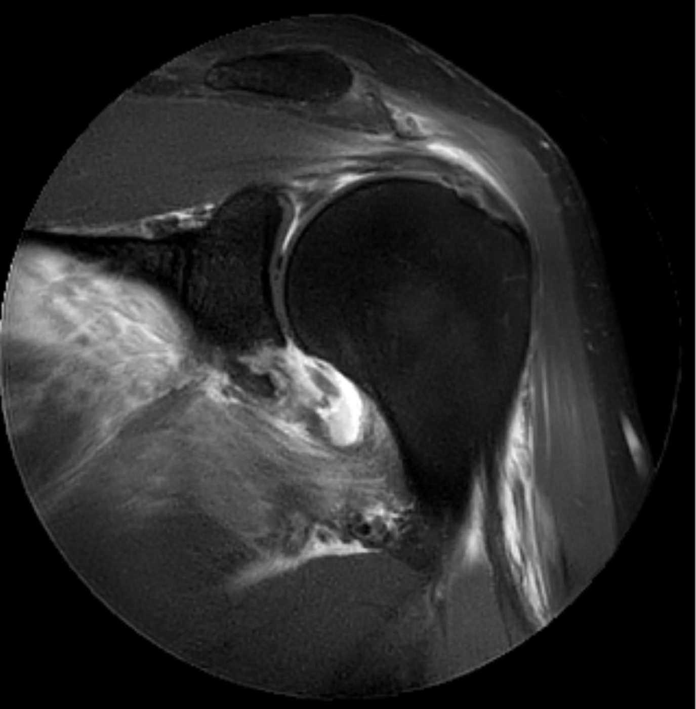
Figure 12. The same bony Bankart lesion on MRI (paracoronal proton-density fat-saturated) after an anteroinferior dislocation. Source: Hellerhoff, via Wikimedia Commons, CC BY-SA 3.0.
Part V - Recurrent (Habitual) Instability: Risk, Dichotomy, and Classification
5.1 Who re-dislocates
Beyond youth, the established risk factors for recurrence are a higher number of prior dislocations, significant glenoid or humeral bone loss, inferior capsular hyperlaxity, male sex, participation in contact or overhead sport, and a longer delay from first dislocation to surgery.[49] The clinical corollary, captured by Itoi, is that operating on every first-time dislocator would mean unnecessary surgery in 60-85% of them (the number needed to treat is 7 for the general population but only 2.6 for high-risk patients). That is the central tension in deciding whom to stabilise early.[50]
5.2 TUBS versus AMBRI
Thomas and Matsen’s dichotomy remains the most useful first cut in classifying instability.[51] TUBS describes the typical young traumatic dislocator: Traumatic, Unidirectional, with a Bankart lesion, usually requiring Surgery. AMBRI (or AMBRII) describes the opposite patient: Atraumatic, Multidirectional, Bilateral laxity, who responds to Rehabilitation and, if surgery is truly needed, an Inferior capsular shift with closure of the rotator Interval. The cardinal practical point is that confusing the two and tightening only the anterior structures of an AMBRI shoulder produces obligate posterior subluxation and capsulorrhaphy arthropathy.[52]
5.3 The Instability Severity Index Score and other classifications
The Instability Severity Index Score (ISIS) of Balg and Boileau predicts failure of an arthroscopic Bankart repair from six preoperative variables: age under 20, competitive sport, contact or forced-overhead sport, shoulder hyperlaxity, a Hill-Sachs lesion visible on the AP radiograph in external rotation, and loss of the normal glenoid contour on the AP film; a score over 6 (of 10) carries an unacceptable ~70% recurrence risk and steers the patient toward a bony (Latarjet) procedure rather than a soft-tissue repair.[53] Several formal classifications coexist, none universally accepted except the validated FEDS system (Frequency, Etiology, Direction, Severity); the others are the direction-based OTA system, the Matsen and Thomas (TUBS/AMBRI) framework, the Stanmore triangle (polar type 1 traumatic-structural, type 2 atraumatic-structural, type 3 muscle-patterning/non-structural, with a continuum between them), and the posterior-instability ABC classification.[54]
Part VI - Surgical Treatment of Anterior Instability
6.1 Soft-tissue repair: the Bankart operation
The foundational operation is repair of the Bankart lesion, reattaching the labrum and capsule to the glenoid rim, first done by Perthes in 1906 and popularised by Bankart.[55] Open Bankart repair was long the gold standard, with recurrence typically under 10%, but at the cost of some lost external rotation. Arthroscopic Bankart repair with suture anchors has now supplanted it for most cases (about 84% of US stabilisations are arthroscopic), giving better range of motion and recurrence rates around 8-13% in suitable patients.[56] Whether open or arthroscopic gives fewer recurrences is genuinely contested. Rockwood & Green’s reading of the meta-analyses favours open repair for recurrence (while arthroscopy gives better motion), whereas The Shoulder finds no convincing difference and its editor concludes the data are insufficient to declare a winner.[57] Capsular plication or an inferior capsular shift is added when there is excess capsular laxity.[58]
6.2 The historical operations
A family of older non-anatomic operations is now largely abandoned, and knowing why is examinable. The Putti-Platt (deliberate shortening of subscapularis and capsule) and the Magnuson-Stack (transfer of the subscapularis insertion laterally) both stabilised the shoulder by restricting external rotation, and both produced loss of external rotation, obligate posterior subluxation, and late capsulorrhaphy arthropathy (a 2.5-cm Putti-Platt subscapularis limits rotation to about 57°).[59] The Bristow procedure transferred the coracoid tip with the conjoined tendon to the glenoid neck, but suffered hardware complications, a roughly 50% coracoid union rate, restricted rotation, and poor return to throwing. Rockwood and Young concluded coracoid transfers should not be used for routine reconstruction.[60]
6.3 Bony procedures: Latarjet, bone block, and remplissage
The Latarjet procedure transfers a larger block of coracoid, with the conjoined tendon attached, flat onto the anterior glenoid neck fixed with two screws.[61] It stabilises by three mechanisms (sometimes called the triple-blocking effect): the bone block that restores glenoid width, the dynamic sling effect of the conjoined tendon across the lower subscapularis, and the repair of the capsule to the coracoacromial stump.[62] It is indicated for significant glenoid bone loss, for failed soft-tissue repair, and for the high-risk contact athlete or the patient with an ISIS over 6. The bone-loss threshold that tips the decision from arthroscopic Bankart to Latarjet is the same disputed 13.5-17.3% (Rockwood & Green) versus 20-25% (The Shoulder) discussed above.[63] Latarjet gives the lowest recurrence of the common operations (around 3-10%, lower than Bankart) but the highest complication rate (about 10-15%, including graft nonunion or osteolysis and injury to the musculocutaneous or axillary nerves).[64]
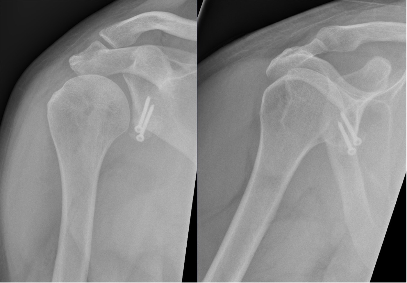
Figure 13. Latarjet procedure: the transferred coracoid bone block fixed to the anterior glenoid rim with two screws (postoperative radiographs). Source: Hellerhoff, via Wikimedia Commons, CC BY-SA 4.0.
For very large defects (over 30% of the glenoid, or a failed Latarjet) an anterior glenoid bone graft is used, an iliac-crest autograft (the Eden-Hybinette operation) or a distal-tibia osteoarticular allograft.[65] For an off-track (engaging) Hill-Sachs lesion with acceptable glenoid bone, remplissage (filling the Hill-Sachs defect with the infraspinatus tendon and posterior capsule) is added to a Bankart repair to stop the lesion engaging, at the cost of about 10° of external rotation; it does not treat glenoid bone loss.[66]
6.4 Putting it together: a decision framework
The modern algorithm integrates bone loss and the glenoid track.[67] A primary dislocator under about 14 years is rehabilitated. The young, active patient with a documented Bankart and minimal bone loss (under 13.5-17.3%) and an on-track lesion has an arthroscopic or open Bankart repair, whereas the same patient with an off-track lesion has a Bankart plus remplissage (or a Latarjet). Bone loss above the critical threshold (roughly 17-25%) calls for a Latarjet, and loss above 30% or a failed Latarjet calls for structural bone grafting. The recurring caution is that recurrence climbs steeply in patients under 20 whatever the operation.[68]
Part VII - Posterior Instability
Posterior instability is less common and historically under-recognised, though increasingly diagnosed.[69] Acutely it follows a seizure, an electric shock, or a posteriorly directed force on the flexed, adducted, internally rotated arm, and it is the dislocation most often missed.[70] The structural lesions are the mirror image of anterior instability: a posterior (reverse) Bankart of the posterior labrum and a reverse Hill-Sachs (McLaughlin) lesion on the anteromedial head.[71] A locked (chronic) posterior dislocation is managed by the size of the anteromedial head defect. A small defect is reduced and held, a defect of 20-40% is filled by transferring the subscapularis (the McLaughlin procedure) or the lesser tuberosity (the modified McLaughlin) into it, and a defect over 40% (or a chronically dislocated head) may need arthroplasty.[72]
Recurrent posterior instability is classified by the ABC system (A first-time, B dynamic/recurrent, C static/chronic) and is treated by activity modification and strengthening first; surgery (arthroscopic posterior labral repair and posterior capsular shift, or a posterior bone-block for bone loss over about 20-25% or excessive retroversion) is reserved for failures, with about 90% return to play in the published series.[73]
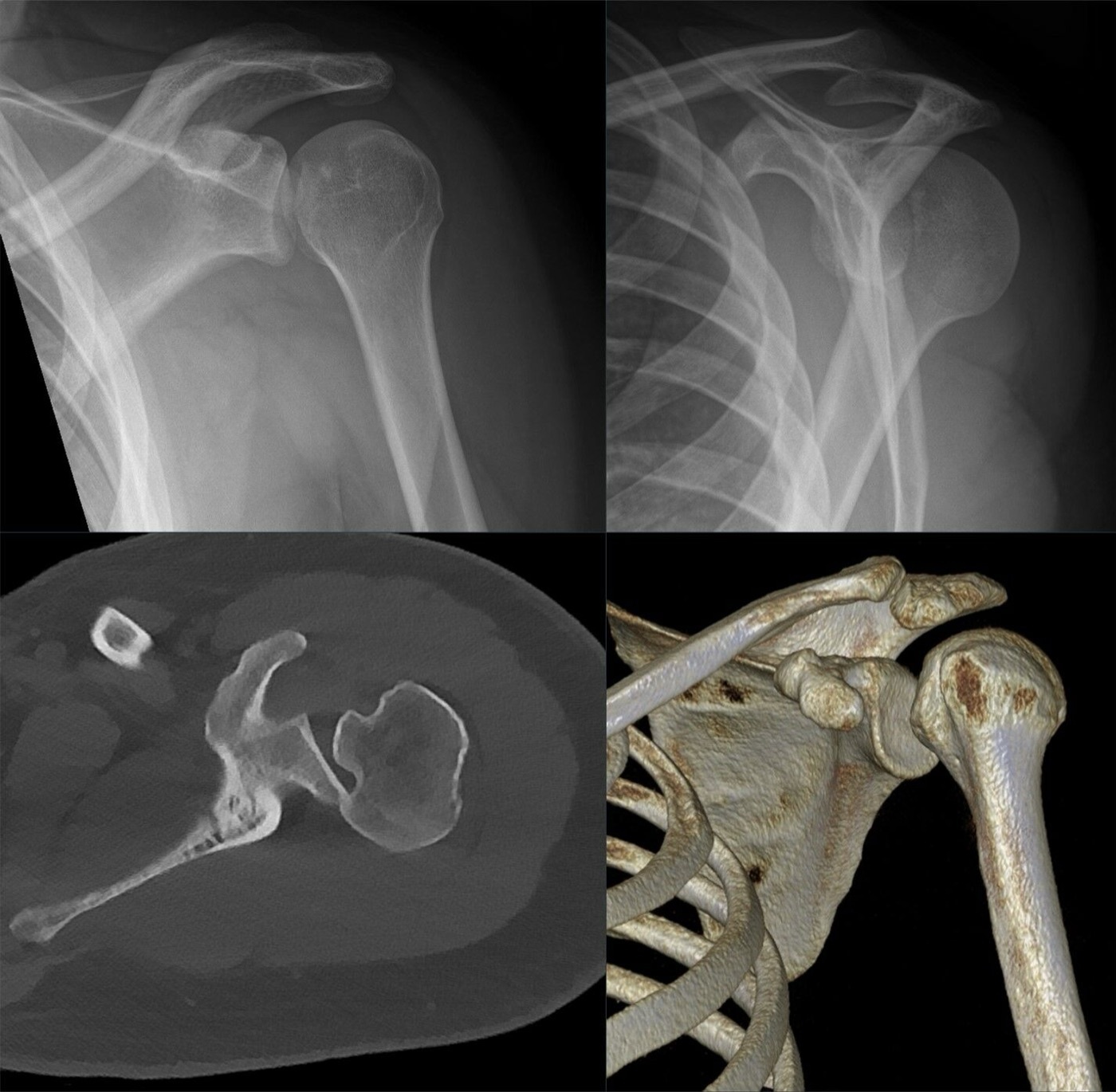
Figure 14. Posterior dislocation on CT: the humeral head displaced behind the glenoid (axial slice and 3D reconstruction), the displacement easily missed on the AP film. Source: Hellerhoff, via Wikimedia Commons, CC BY-SA 4.0.
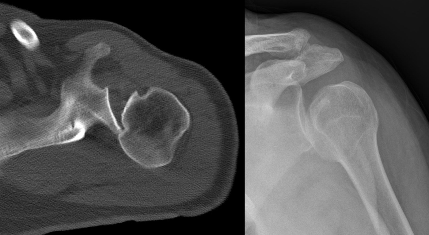
Figure 15. Reverse Hill-Sachs (McLaughlin) lesion: the anteromedial humeral-head impaction defect of a posterior dislocation, on axial CT (left) and radiograph (right). Source: Hellerhoff, via Wikimedia Commons, CC BY-SA 3.0.
Part VIII - Multidirectional and Voluntary Instability
Multidirectional instability (MDI) is symptomatic instability in more than one direction, always including an inferior component, on a background of a patulous, redundant capsule and often generalised ligamentous laxity (assessed by the Beighton score).[74] Its clinical hallmark is the sulcus sign, a hollow appearing below the acromion when the arm is pulled downward. A sulcus that persists in external rotation signifies an incompetent rotator interval and dictates closing the interval at surgery.[75] The first-line treatment is rehabilitation, strengthening the rotator cuff and scapular stabilisers, which succeeds in the majority (Burkhead and Rockwood reported 80% success in atraumatic subluxation), and surgery (an inferior capsular shift, originally described by Neer, or an arthroscopic capsular plication with rotator-interval closure) is offered only after 6-12 months of failed rehabilitation.[76]
The single most important rule in this whole area is negative: surgery is contraindicated in the voluntary or psychogenic dislocator and in the patient with secondary-gain or significant psychological issues, and in the muscle-patterning (Stanmore polar type 3) instability that is driven by abnormal muscle activation rather than structural damage.[77] These patients are managed by rehabilitation and, where appropriate, psychological support, and operating on them reliably fails.[78]
Part IX - A Synthesis: How to Reason Through Shoulder Instability
Five questions order the whole topic. First, which way does it go out, and is this really instability? Anterior is usual and obvious; posterior is the one that is missed, so any seizure or electric shock or any blocked external rotation demands an axillary or scapular-Y view. And remember that laxity is not instability: the voluntary dislocator and the lax-but-asymptomatic shoulder are not surgical. Second, is this a first event or a disease of recurrence? A first traumatic dislocation is reduced gently and immobilised, but the prognosis is dictated by age, with the young athlete carrying a high recurrence rate that justifies considering early stabilisation. Third, what is the structural lesion? The Bankart lesion and its failure to heal drive recurrence, but the two quantities that actually choose the operation are the glenoid bone loss and whether the Hill-Sachs lesion is on-track or off-track. Fourth, what operation fits the lesion? A soft-tissue Bankart repair for the on-track shoulder with little bone loss, remplissage added for an off-track Hill-Sachs, and a Latarjet or bone graft once bone loss passes the critical threshold; the old non-anatomic operations are of historical interest and a warning about capsulorrhaphy arthropathy. Fifth, is this the patient I should not operate on? The AMBRI/multidirectional shoulder responds to rehabilitation, and the voluntary or muscle-patterning dislocator is harmed by surgery.
The unifying lesson is that the shoulder is unstable because it is built to be mobile, that a traumatic dislocation becomes a recurrent one when the anteroinferior soft-tissue and bony restraints fail to heal, and that good treatment matches the intervention to the precise lesion, recognising the patients for whom the right operation is no operation at all.
References
-
Rockwood & Green’s Fractures in Adults, p.1809 (only 25-30% of the humeral head contacts the glenoid; articular geometry contributes minimally to stability).
-
Rockwood & Matsen’s The Shoulder, p.624 (normal shoulders are lax; laxity is often not increased in unstable shoulders).
-
Rockwood & Green’s Fractures in Adults, pp.1809-1810.
-
Rockwood & Green’s Fractures in Adults, p.1810 (Habermeyer: the labrum is the valve block sealing the joint from atmospheric pressure).
-
Rockwood & Green’s Fractures in Adults, pp.1809-1810, 1812 (Lippitt et al. concavity-compression).
-
Rockwood & Green’s Fractures in Adults, pp.1808, 1811.
-
Rockwood & Green’s Fractures in Adults, pp.1808-1811.
-
Rockwood & Green’s Fractures in Adults, pp.1810-1811.
-
Rockwood & Green’s Fractures in Adults, p.1812.
-
Rockwood & Green’s Fractures in Adults, pp.1812-1813.
-
Rockwood & Green’s Fractures in Adults, pp.1813-1814.
-
Rockwood & Matsen’s The Shoulder, p.596 (shoulder dislocations are ~45% of all dislocations; ~85% anterior). The frequently quoted statement that the shoulder is the most commonly dislocated major joint is standard teaching consistent with this figure.
-
Rockwood & Matsen’s The Shoulder, p.593 (Rowe: 96% of 500 dislocations traumatic).
-
Rockwood & Green’s Fractures in Adults, p.1782.
-
Rockwood & Matsen’s The Shoulder, pp.597-598.
-
Rockwood & Matsen’s The Shoulder, p.595.
-
Rockwood & Matsen’s The Shoulder, pp.596-597; Rockwood & Green’s Fractures in Adults, p.1783.
-
Rockwood & Green’s Fractures in Adults, p.1785. The “squared-off shoulder” with an anteriorly palpable head is standard clinical teaching; the mined text describes a posterior sulcus and the arm held in slight abduction.
-
Rockwood & Matsen’s The Shoulder, pp.597-598; Rockwood & Green’s Fractures in Adults, pp.1783-1785, 1793-1794 (Rowe & Zarins 79% missed; Xu et al. 73% missed when only AP/lateral taken).
-
Rockwood & Green’s Fractures in Adults, pp.1785, 1793-1794.
-
The “light-bulb” (or “drumstick”) sign of posterior dislocation is standard radiographic teaching; the mined sources convey the equivalent mechanism (the internally rotated head, only the lesser tuberosity in the glenoid, a deceptively normal AP) but do not use the eponymous sign name.
-
Rockwood & Matsen’s The Shoulder, p.598; Rockwood & Green’s Fractures in Adults, pp.1783-1785.
-
Rockwood & Matsen’s The Shoulder, p.598.
-
Rockwood & Green’s Fractures in Adults, pp.1782, 1785-1786. The term “regimental badge” for the lateral deltoid sensory patch, and the observation that axillary-nerve palsy after dislocation is usually a neurapraxia that recovers, are standard teaching; the mined text specifies testing sensation over the lateral deltoid and gives the 13.5% incidence.
-
Rockwood & Green’s Fractures in Adults, pp.1785-1786; Rockwood & Matsen’s The Shoulder, p.613.
-
Rockwood & Green’s Fractures in Adults, pp.1783-1784.
-
Rockwood & Green’s Fractures in Adults, pp.1782, 1786.
-
Rockwood & Green’s Fractures in Adults, p.1793.
-
Rockwood & Green’s Fractures in Adults, pp.1793-1795.
-
Rockwood & Green’s Fractures in Adults, pp.1796-1799.
-
Rockwood & Matsen’s The Shoulder, pp.567-571.
-
Rockwood & Matsen’s The Shoulder, p.571.
-
Scapular manipulation and the external-rotation method are named in the mined text (Rockwood & Matsen’s The Shoulder, p.571) among the simple post-1975 techniques. The detailed Stimson prone-weight protocol, the Spaso method, and the Cunningham technique are standard emergency-medicine teaching and were not described step-by-step in the mined extract; included for completeness, not falsely page-cited.
-
Rockwood & Green’s Fractures in Adults, pp.1782, 1801. Intra-articular lidocaine versus intravenous sedation is standard ED teaching; the mined text states manual reduction may need conscious sedation or muscle paralysis (often in theatre) but does not compare the two analgesic routes.
-
Confirmation of reduction by post-reduction radiograph and a repeat neurovascular check is standard teaching; the mined instability chapters stress the pre- and post-reduction neurovascular examination and the axillary view for confirming concentric reduction.
-
Rockwood & Green’s Fractures in Adults, pp.1817-1818 (the 632-patient meta-analysis found no difference between external- and internal-rotation immobilisation).
-
Rockwood & Green’s Fractures in Adults, p.1800; Rockwood & Matsen’s The Shoulder, pp.580, 672. Age is repeatedly named as the dominant risk factor for recurrence; the specific age-stratified percentages (very high recurrence under 20-25, low over 40) are standard teaching and were not tabulated numerically in the mined extracts.
-
Rockwood & Matsen’s The Shoulder, pp.609-611 (the capsulolabral detachment typical of traumatic instability; non-healing is the major factor in recurrence).
-
Rockwood & Green’s Fractures in Adults, pp.1815-1816.
-
The Bankart-versus-Perthes “essential lesion” distinction and the named variants ALPSA, Perthes, GLAD, and HAGL (and bony HAGL) are standard instability teaching but were not present in the mined Rockwood/Shoulder extracts, which describe only the generic anteroinferior capsulolabral detachment and the bony Bankart. Included for completeness, not falsely page-cited.
-
Rockwood & Matsen’s The Shoulder, pp.608-611, 629-631; Rockwood & Green’s Fractures in Adults, p.1783.
-
Rockwood & Matsen’s The Shoulder, pp.616-617.
-
Rockwood & Green’s Fractures in Adults, pp.1832-1834; Rockwood & Matsen’s The Shoulder, p.672 (Itoi: on-track versus off-track).
-
The numerical glenoid-track width (~83% of the glenoid diameter) and its attribution to Yamamoto/Itoi and Di Giacomo are standard teaching; the mined extracts describe the on-track/off-track concept and its clinical use but do not state the 83% figure.
-
Rockwood & Matsen’s The Shoulder, p.672 (of 100 recurrent dislocators, 93% on-track, 7% off-track).
-
Rockwood & Green’s Fractures in Adults, pp.1800, 1815. The “inverted-pear” glenoid is standard teaching for advanced anterior bone loss; the mined pathoanatomy extract describes only the normal pear shape.
-
Rockwood & Green’s Fractures in Adults, pp.1800-1801, 1820, 1828; Rockwood & Matsen’s The Shoulder, pp.672-673.
-
The best-fit-circle en-face CT method of quantifying glenoid bone loss is standard teaching; the mined extracts acknowledge bone loss as a risk factor and use CT/3D-CT for it but do not detail the measurement method.
-
Rockwood & Green’s Fractures in Adults, p.1837; Rockwood & Matsen’s The Shoulder, p.654.
-
Rockwood & Matsen’s The Shoulder, p.672 (Itoi: NNT 7.0 general, 2.6 high-risk; unnecessary surgery in 60-85%).
-
Rockwood & Green’s Fractures in Adults, pp.1804-1805.
-
Rockwood & Green’s Fractures in Adults, pp.1804-1805; Rockwood & Matsen’s The Shoulder, p.650.
-
Rockwood & Matsen’s The Shoulder, pp.637-638; Rockwood & Green’s Fractures in Adults, pp.1815-1816. The mined Shoulder text gives the ISIS risk factors and the >6-point/~70%-recurrence threshold but states “2 points for each” rather than reproducing Balg & Boileau’s original weighted 10-point table (age <20 = 2, competition = 2, contact/overhead = 1, hyperlaxity = 1, Hill-Sachs on AP = 2, glenoid loss of contour = 2), which is included here as standard teaching.
-
Rockwood & Green’s Fractures in Adults, pp.1802-1806 (FEDS validated; OTA, Matsen-Thomas, Stanmore, posterior ABC classifications).
-
Rockwood & Matsen’s The Shoulder, p.640.
-
Rockwood & Green’s Fractures in Adults, pp.1818, 1837-1839.
-
Rockwood & Green’s Fractures in Adults, p.1839; Rockwood & Matsen’s The Shoulder, pp.637, 654, 673.
-
Rockwood & Matsen’s The Shoulder, pp.640-641.
-
Rockwood & Matsen’s The Shoulder, pp.644-646.
-
Rockwood & Matsen’s The Shoulder, pp.646-648.
-
Rockwood & Matsen’s The Shoulder, p.648.
-
Rockwood & Matsen’s The Shoulder, pp.646-648. The mined text describes the bone-block and conjoined-tendon “sling effect”; the popular term “triple-blocking” (bone block + sling + capsular/coracoacromial-ligament repair) is standard teaching and is not used verbatim in the source.
-
Rockwood & Green’s Fractures in Adults, pp.1820, 1828, 1833-1834; Rockwood & Matsen’s The Shoulder, pp.638, 662, 672-673.
-
Rockwood & Green’s Fractures in Adults, pp.1841-1843.
-
Rockwood & Green’s Fractures in Adults, pp.1834, 1839-1840; Rockwood & Matsen’s The Shoulder, p.646.
-
Rockwood & Matsen’s The Shoulder, p.656; Rockwood & Green’s Fractures in Adults, p.1834.
-
Rockwood & Green’s Fractures in Adults, pp.1832-1834.
-
Rockwood & Matsen’s The Shoulder, p.648; Rockwood & Green’s Fractures in Adults, p.1837.
-
Rockwood & Green’s Fractures in Adults, p.1820.
-
Rockwood & Matsen’s The Shoulder, pp.597-598. The mined Rockwood chapter names “seizure disorder” only as a surgical contraindication; the full seizure/electrocution/axial-load mechanism triad is given in The Shoulder and is standard teaching.
-
Rockwood & Green’s Fractures in Adults, pp.1822-1823; Rockwood & Matsen’s The Shoulder, pp.616-617.
-
Rockwood & Matsen’s The Shoulder, pp.616-617, 622-623 (reverse-Hill-Sachs size thresholds and the McLaughlin / modified-McLaughlin transfers).
-
Rockwood & Green’s Fractures in Adults, pp.1806, 1820, 1829-1831.
-
Rockwood & Green’s Fractures in Adults, pp.1809, 1815. The Beighton score is referenced in the mined text as a prognostic factor (higher scores predict less improvement after plication) but its scoring criteria are not defined there; the score itself is standard teaching.
-
Rockwood & Green’s Fractures in Adults, pp.1808, 1836-1837, 1839.
-
Rockwood & Green’s Fractures in Adults, pp.1818, 1832-1833; Rockwood & Matsen’s The Shoulder, p.637 (Burkhead & Rockwood rehabilitation results).
-
Rockwood & Green’s Fractures in Adults, pp.1781, 1832, 1840.
-
Rockwood & Green’s Fractures in Adults, pp.1832, 1840.
-
Rockwood & Green’s Fractures in Adults, pp.1809-1813.
-
Rockwood & Matsen’s The Shoulder, pp.596-598; Rockwood & Green’s Fractures in Adults, pp.1783-1785.
-
Rockwood & Matsen’s The Shoulder, pp.597-598; Rockwood & Green’s Fractures in Adults, pp.1793-1794.
-
Rockwood & Green’s Fractures in Adults, pp.1785-1786.
-
Rockwood & Matsen’s The Shoulder, pp.567-571.
-
Rockwood & Green’s Fractures in Adults, pp.1800, 1837; Rockwood & Matsen’s The Shoulder, p.672.
-
Rockwood & Matsen’s The Shoulder, pp.609-611; Rockwood & Green’s Fractures in Adults, pp.1815-1816.
-
Rockwood & Green’s Fractures in Adults, pp.1832-1834; Rockwood & Matsen’s The Shoulder, p.672.
-
Rockwood & Green’s Fractures in Adults, pp.1804-1805; Rockwood & Matsen’s The Shoulder, p.650.
-
Rockwood & Matsen’s The Shoulder, pp.637-638; Rockwood & Green’s Fractures in Adults, pp.1815-1816.
-
Rockwood & Green’s Fractures in Adults, pp.1800, 1820, 1828, 1834; Rockwood & Matsen’s The Shoulder, pp.672-673.
-
Rockwood & Matsen’s The Shoulder, pp.646-648; Rockwood & Green’s Fractures in Adults, pp.1841-1843.
-
Rockwood & Matsen’s The Shoulder, pp.644-648.
-
Rockwood & Green’s Fractures in Adults, pp.1818, 1832-1833, 1840.