Contents
- Part I - The Polytrauma Patient
- Part II - The ATLS Primary Survey
- Part III - The Secondary and Tertiary Surveys
- Part IV - Traumatic and Haemorrhagic Shock
- Part V - Resuscitation
- Part VI - The Principle of Damage Control
- Part VII - Damage-Control Orthopaedics
- Part VIII - Pelvic-Ring Haemorrhage in the Unstable Patient
- Part IX - SIRS, MODS, ARDS and Fat Embolism
- References
Part I - The Polytrauma Patient
Scope and the pattern of death
Injury is the leading cause of death and disability in people under 50, accounting for more than 200,000 deaths a year in the United States alone, with hospital costs exceeding half a trillion dollars.[1] Trauma deaths classically follow a trimodal distribution: an immediate peak (death at the scene from a catastrophic brain, high cord, heart or great-vessel injury), an early peak (within hours, from salvageable bleeding, the “golden hour” of trauma care), and a late peak (days to weeks later, from infection or multiple-organ dysfunction).[2] The model is taught as the classic framework but is now contested, since most trauma deaths occur within 24 hours and the epidemiology is shifting with an ageing population.[3] Haemorrhage is the leading preventable cause of death, responsible for around 40% of trauma fatalities.[4]
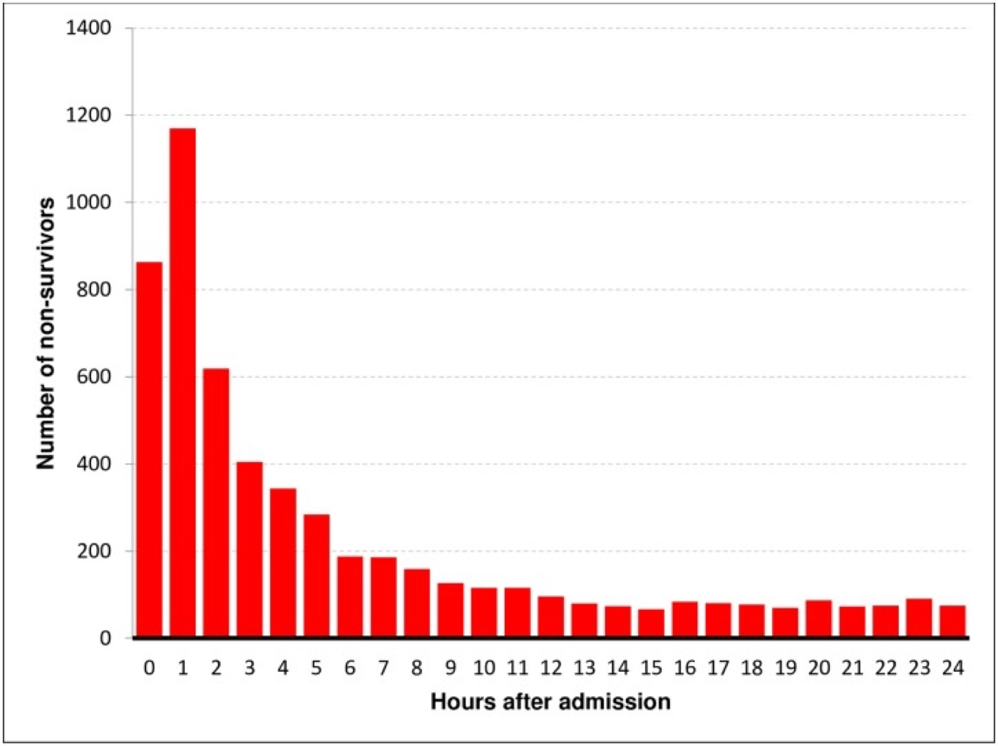
Figure 1. Temporal distribution of in-hospital trauma deaths, showing the dominant early peak (the modern data form of the classic trimodal model). Source: Rauf et al., PLoS ONE 2019;14:e0212095, CC BY 4.0.
There is no single agreed numeric definition of “polytrauma”; the texts use multiply injured, multisystem trauma and polytrauma interchangeably for a patient whose injuries cross more than one body region or system, and the clinical point is that such a patient must be managed by a team under a trauma-team leader, with the systemic consequences of the combined injuries (not any one fracture) driving the priorities.[5]
Injury severity scoring
Severity is quantified to triage, prognosticate and compare outcomes. The building block is the Abbreviated Injury Scale (AIS), which grades the injury in each body region from 1 (minor), 2 (moderate), 3 (severe, non-life-threatening), 4 (severe, life-threatening), 5 (critical, survival uncertain) to 6 (fatal/unsurvivable).[6] The Injury Severity Score (ISS) is the anatomical standard: take the highest AIS in each of the three most severely injured body regions and sum their squares (ISS = A² + B² + C²), giving a range of 0-75.[7] Any single region scored AIS 6 automatically makes the ISS 75, and an ISS above 15 (i.e. ≥ 16, “major trauma”) carries about 10% mortality.[8] Its weakness is that it counts only one injury per region, which the New Injury Severity Score (NISS) corrects by taking the three highest scores regardless of region, and which predicts survival slightly better.[9] Physiological scores (the Glasgow Coma Scale, the Revised Trauma Score) complement the anatomical ones.
Part II - The ATLS Primary Survey
The Advanced Trauma Life Support system, conceived after an orthopaedic surgeon’s own family tragedy, assesses and resuscitates the trauma patient in a single prioritised sequence in which “the abnormality that poses the greatest threat to life is addressed first.”[10] A ten-second look (asking the patient their name and what happened) screens airway, breathing and consciousness at once: a patient who answers clearly has, for that moment, a patent airway, enough breath to speak and an alert brain.[11] The primary survey is the ABCDE:[12]
- A, Airway with cervical-spine protection. Clear and secure the airway (chin-lift or jaw-thrust, suction), assuming a cervical-spine injury in any blunt multisystem or above-the-clavicle injury and maintaining in-line immobilisation. A GCS of 8 or less mandates a definitive (cuffed, secured) airway.
- B, Breathing and ventilation. Expose the chest and find the immediately life-threatening injuries that must be treated now: tension pneumothorax, open (“sucking”) pneumothorax, massive haemothorax, and flail chest with pulmonary contusion. A tension pneumothorax is decompressed immediately (needle, then chest drain); an open chest wound is covered with a dressing taped on three sides as a flutter valve.[13]
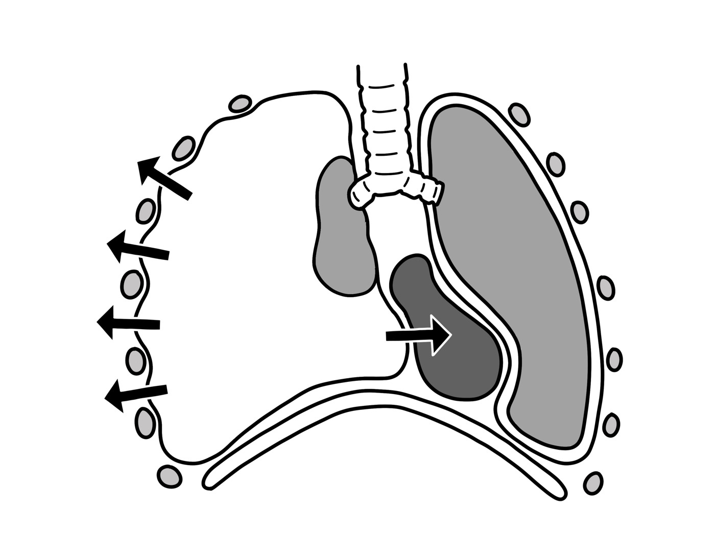
Figure 2. Tension pneumothorax: the collapsed lung and mediastinal shift that must be found and decompressed during the “B” of the primary survey. Source: Baedr-9439, via Wikimedia Commons, CC0 (public domain).
- C, Circulation with haemorrhage control. After excluding tension pneumothorax, hypotension is haemorrhagic until proven otherwise. Control external bleeding by direct pressure (a tourniquet if that fails, a pelvic binder for the pelvis), insert two large-bore (14-16 G) cannulae, and remember the predictable reservoirs of major blood loss: “blood on the floor and four more”: the external floor, the chest, the abdomen, the pelvis/retroperitoneum, and the long bones (thighs).[14]
- D, Disability. A rapid neurological check of conscious level (GCS), pupils and any lateralising or cord signs; altered consciousness is assumed to be hypoperfusion or brain injury until proven otherwise.
- E, Exposure and environment. Fully undress the patient to inspect every surface (a log-roll for the back), then immediately cover and actively warm them: “the patient’s body temperature is more important than the comfort of the health-care providers.”[15]
Adjuncts to the primary survey are added without interrupting resuscitation: ECG, pulse oximetry, urinary and gastric catheters, arterial blood gas and lactate, a portable chest and pelvis radiograph, and the FAST ultrasound scan for free intraperitoneal or pericardial blood.[16]
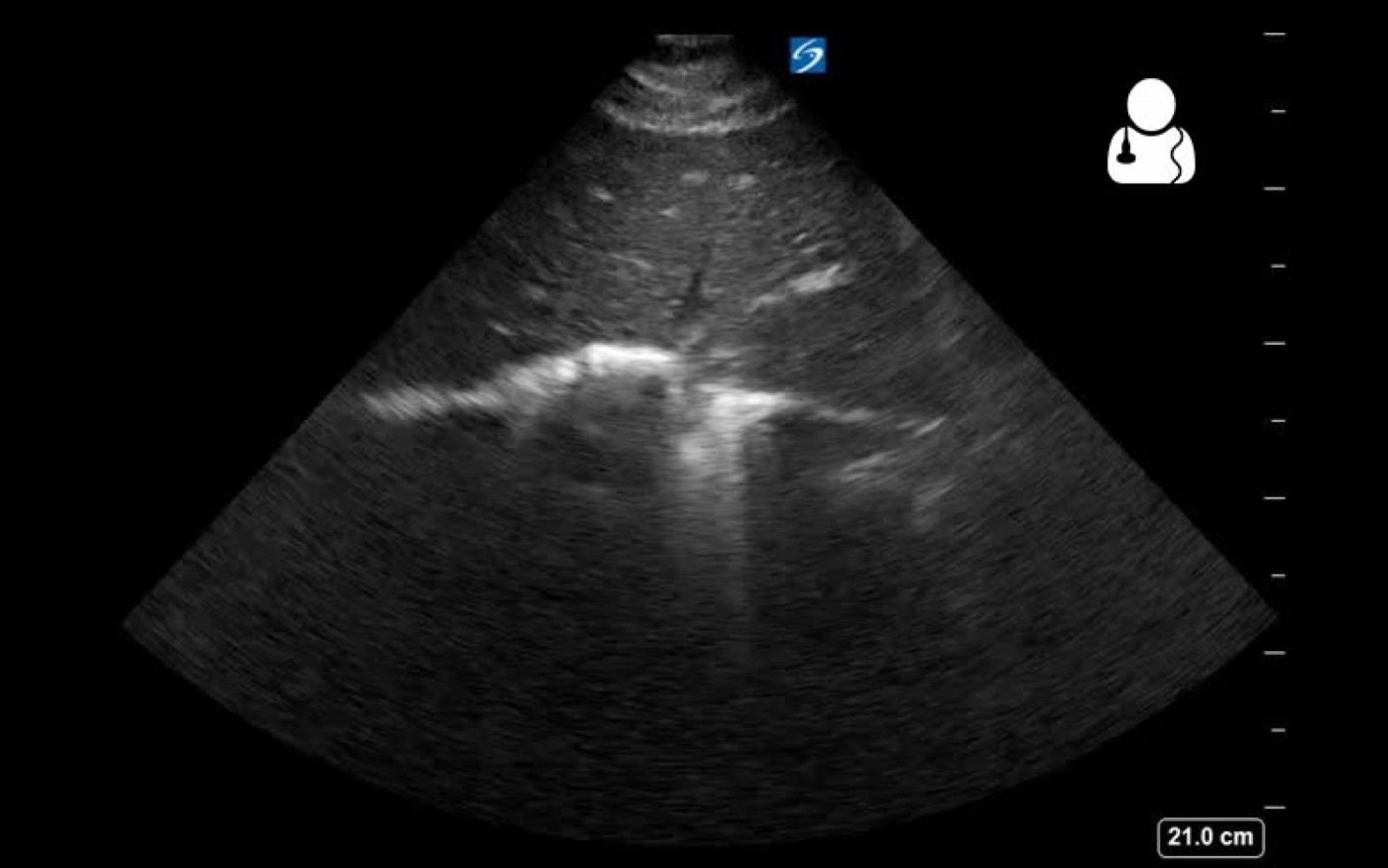
Figure 3. Focused Assessment with Sonography for Trauma (FAST), a bedside adjunct to the primary survey for free intraperitoneal or pericardial blood. Source: Ben Smith / Ultrasound of the Week, via Wikimedia Commons, CC BY-SA 4.0.
Part III - The Secondary and Tertiary Surveys
The secondary survey begins only once the primary survey is complete, resuscitation is under way, and vital signs are normalising.[17] It is a head-to-toe examination with a full set of vitals, a repeat GCS, and an AMPLE history (Allergies, Medications, Past illness/Pregnancy, Last meal, Events of injury).[18] At any sign of deterioration the clinician returns to the primary survey and the ABCs, and if the patient needs transfer to definitive care this takes precedence over completing the secondary survey.[19] A tertiary survey (a repeat head-to-toe examination a day or so later) catches the injuries missed in the resuscitation room, which are common in the obtunded patient and most frequent in those with a head injury needing emergency surgery; a mandatory tertiary survey has been shown to cut missed injuries from 2.4% to 1.5%.[20]
Part IV - Traumatic and Haemorrhagic Shock
Definition and classification
Shock is “an abnormality of the circulatory system that results in inadequate organ perfusion and tissue oxygenation.”[21] In the injured patient it is haemorrhagic until proven otherwise: “any injured patient who is cool and has tachycardia is considered to be in shock until proven otherwise,” and treatment is begun as for hypovolaemia unless there is clear evidence of another cause.[22] The non-haemorrhagic causes to keep in mind are cardiogenic (blunt cardiac injury), obstructive (tension pneumothorax, cardiac tamponade, both reducing venous return), neurogenic (cervical or high-thoracic cord injury, the classic bradycardia-with-hypotension), and, late, septic shock; an isolated brain injury does not by itself cause shock.[23]
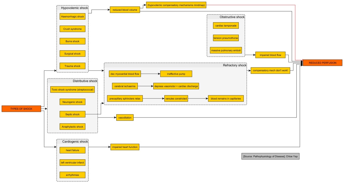
Figure 4. The types of shock and their common end-point of inadequate tissue perfusion. Source: Doyouseewhy7, via Wikimedia Commons, CC BY-SA 4.0.
The cornerstone of recognition is the ATLS classification of haemorrhagic shock, based on a normal blood volume of about 7% of body weight (~5 L in a 70-kg adult; 8-9% in a child):[24]
| Parameter | Class I | Class II | Class III | Class IV |
|---|---|---|---|---|
| Blood loss (mL) | up to 750 | 750-1500 | 1500-2000 | > 2000 |
| Blood loss (% volume) | up to 15% | 15-30% | 30-40% | > 40% |
| Heart rate (/min) | < 100 | 100-120 | 120-140 | > 140 |
| Systolic BP | normal | normal | decreased | decreased |
| Pulse pressure | normal/increased | decreased | decreased | decreased |
| Respiratory rate | 14-20 | 20-30 | 30-40 | > 35 |
| Urine output (mL/h) | > 30 | 20-30 | 5-15 | negligible |
| Mental status | slightly anxious | mildly anxious | anxious, confused | confused, lethargic |
The vital teaching point is that the systolic blood pressure does not fall until 30-40% of the blood volume is lost: “compensatory mechanisms can preclude a measurable fall in systolic pressure until up to 30% of blood volume is lost,” so hypotension is a late sign, and the earlier markers are tachycardia, a narrowed pulse pressure (from catecholamine-driven diastolic rise) and subtle changes in mental status.[25] The corollary: do not wait for a falling pressure before treating, since “bleeding patients need blood.”[26]
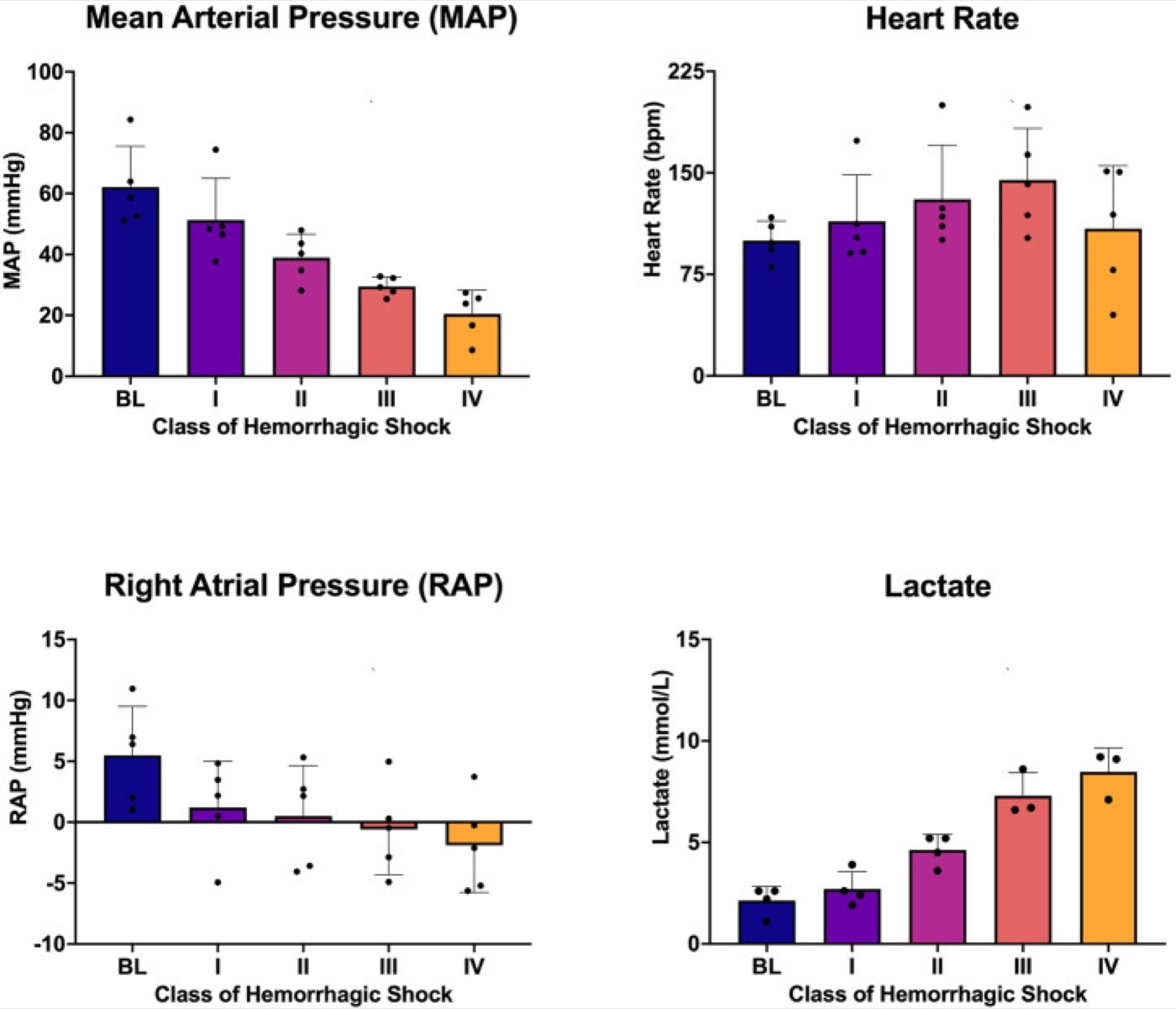
Figure 5. Physiological changes across the four classes of haemorrhagic shock (mean arterial pressure, heart rate, right-atrial pressure and lactate). Source: Elansary et al., Front Physiol 2022;13:1033784, CC BY 4.0.
Pathophysiology and the lethal triad
Cardiac output is heart rate times stroke volume, and stroke volume depends on preload, contractility and afterload.[27] The body answers blood loss with tachycardia and catecholamine-driven vasoconstriction, shunting blood from skin, muscle and gut to brain, heart and kidney, so tachycardia is usually the earliest measurable sign.[28] Under-perfused cells switch to anaerobic metabolism, generating lactic acid and a metabolic acidosis that, with the cold of exposure and the dilution of clotting factors, drives the self-perpetuating lethal triad of hypothermia, acidosis and coagulopathy.[29] Trauma-induced coagulopathy is present in up to 30% of severely injured patients on arrival.[30] Several groups compensate poorly or deceptively: young athletes maintain a normal pressure then crash suddenly; the elderly and the beta-blocked may not mount a tachycardia; and the haematocrit can be near-normal despite massive acute loss.[31]
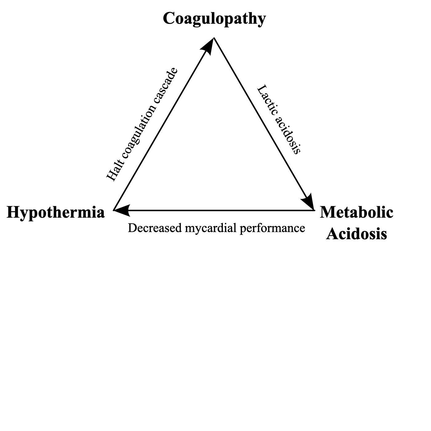
Figure 6. The lethal triad of trauma: hypothermia, acidosis and coagulopathy reinforcing one another. Source: Cburnett, via Wikimedia Commons, CC BY 3.0.
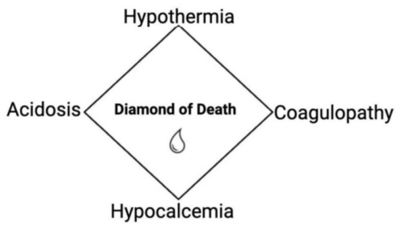
Figure 7. The modern “diamond of death”, adding hypocalcaemia to the classic lethal triad. Source: Klimek et al., J Clin Med 2026;15:2549, CC BY 4.0.
Part V - Resuscitation
Stop the bleeding, restore the volume
The principle is “to stop the bleeding and replace the volume loss.”[32] External haemorrhage is controlled by direct pressure, then a tourniquet for an exsanguinating limb and a pelvic binder for the pelvis; two large-bore cannulae are placed (flow rises with the fourth power of the catheter radius and falls with its length, by Poiseuille’s law, so short and wide beats long and narrow).[33] An initial warmed crystalloid bolus (1-2 L in an adult, 20 mL/kg in a child) is given, but the modern emphasis is firmly away from large-volume crystalloid (ATLS 9e reduced the old 2 L to a 1 L starting bolus) because excessive crystalloid worsens the lethal triad.[34]
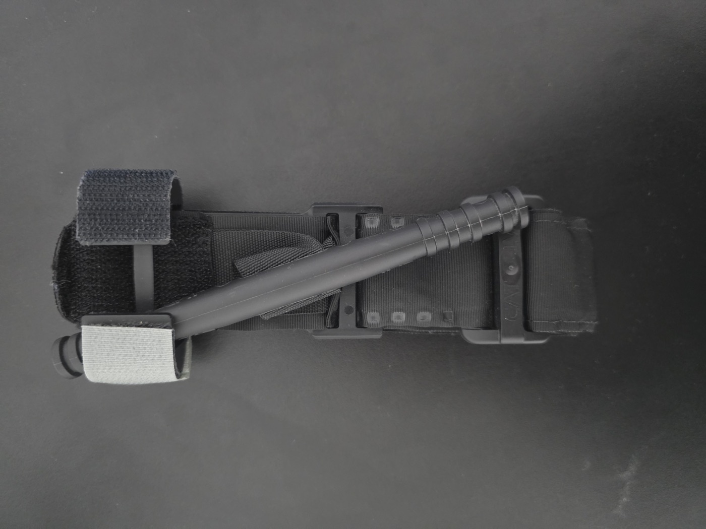
Figure 8. A windlass limb tourniquet (Combat Application Tourniquet) for controlling exsanguinating external limb haemorrhage. Source: Struppig taucher, via Wikimedia Commons, CC BY 4.0.
Two resuscitation strategies define current practice. Permissive (balanced, controlled, hypotensive) resuscitation accepts a lower-than-normal pressure until the bleeding is surgically controlled, because raising the pressure too soon dislodges clot and worsens bleeding: “the goal is the balance, not the hypotension,” and it is a bridge to, not a substitute for, definitive control.[35] It is favoured in penetrating torso haemorrhage but is avoided when there is a traumatic brain injury, where hypotension must be prevented.[36] Damage-control resuscitation means giving blood products early, in a balanced 1:1:1 ratio of plasma to platelets to packed red cells, while minimising crystalloid.[37] A massive transfusion (more than 10 units of red cells in 24 hours) should trigger a pre-set protocol.[38] The antifibrinolytic tranexamic acid improves survival when given within 3 hours of injury (CRASH-2), at a dose of 1 g over 10 minutes then 1 g over 8 hours.[39] Vasopressors are contraindicated in haemorrhagic shock (they worsen tissue perfusion), and sodium bicarbonate is not used for the acidosis of hypovolaemia.[40]
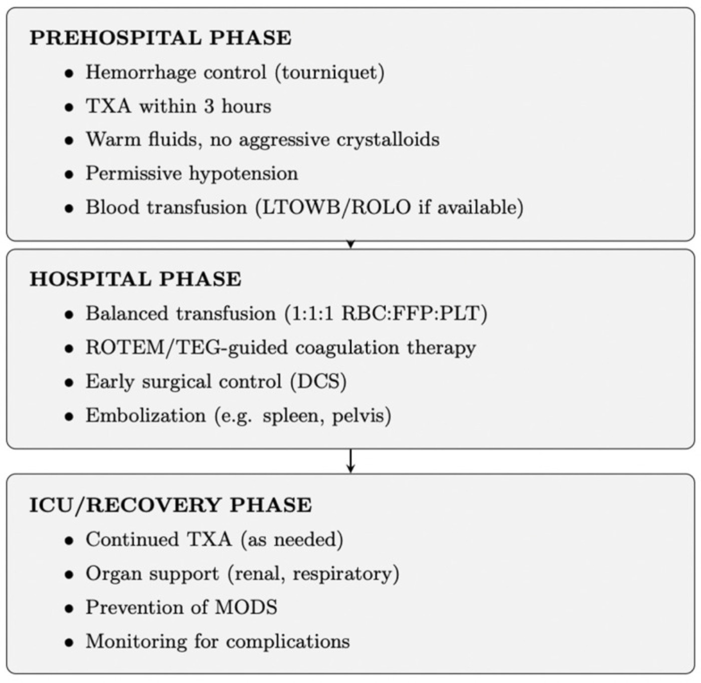
Figure 9. Phased damage-control resuscitation, with the hospital phase delivering a balanced 1:1:1 transfusion, tranexamic acid and permissive hypotension. Source: Klimek et al., J Clin Med 2026;15:2549, CC BY 4.0.
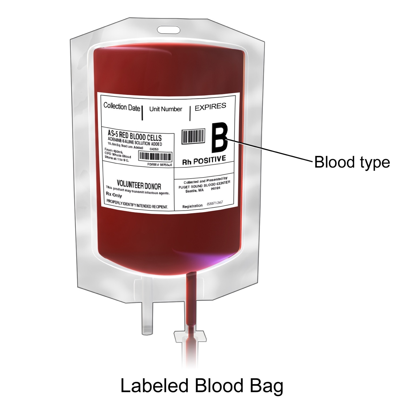
Figure 10. A labelled unit of packed red blood cells for transfusion. Source: BruceBlaus, via Wikimedia Commons, CC BY 3.0.
Endpoints and the response to resuscitation
The aim is organ perfusion, not merely a normal blood pressure; the best single monitor is urine output (≥ 0.5 mL/kg/h in an adult, 1 mL/kg/h in a child), supported by mental status, skin perfusion, and serial base deficit and lactate as measures of shock severity and of resuscitation adequacy.[41] The patient’s response to the initial fluid is diagnostic and triages the next step:[42]
- A rapid responder normalises and stays normal (lost < 20%): no immediate blood needed.
- A transient responder improves then deteriorates (ongoing loss of 20-40%): needs blood and almost certainly operative or angiographic control of bleeding.
- A minimal/non-responder (loss > 40%) needs immediate blood and immediate definitive haemorrhage control.
The commonest cause of a poor response to fluid is an undiagnosed source of continued bleeding, the signal to go to theatre.[43]
Part VI - The Principle of Damage Control
“Damage control” borrows a naval term for keeping a stricken ship afloat by limiting the damage rather than repairing everything at once.[44] Applied by Rotondo in 1993 to the exsanguinating laparotomy, damage-control surgery abandons the attempt to repair every injury in one sitting, which in the badly injured drives “prolonged operative times and persistent bleeding… the lethal triad of coagulopathy, acidosis, and hypothermia, resulting in a mortality of 90%.” It restores normal physiology rather than normal anatomy.[45] It runs in three stages:[46]
- An abbreviated first operation to control haemorrhage and contamination (ligate or shunt, pack, temporary closure), doing “the least to overcome life-threatening conditions.”
- Resuscitation in intensive care to correct the lethal triad: rewarming, correcting the acidosis and the coagulopathy.
- A planned return to theatre, typically at 24-48 hours, for definitive repair once physiology is restored.
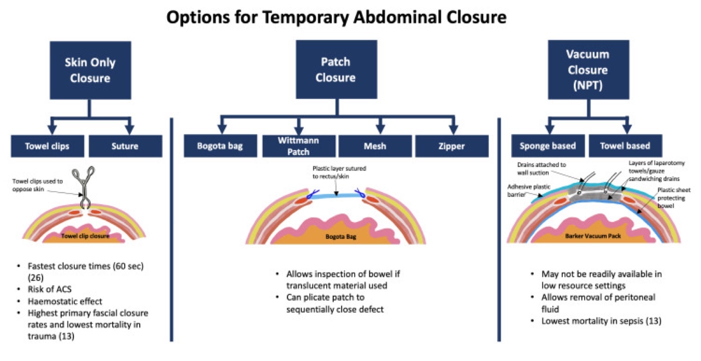
Figure 11. Temporary abdominal closure options for the open abdomen in damage-control surgery (skin-only, patch/Bogotá-bag, and negative-pressure techniques). Source: Milne et al., Cureus 2021;13:e15489, CC BY 3.0.
The rationale is the two-hit model: the injury is the “first hit” that primes a systemic inflammatory response; a long, bloody operation in an under-resuscitated patient is a “second hit” that tips that response into SIRS, ARDS and multiple-organ failure (the lungs fail first, at 48-72 hours).[47] Damage control limits the second hit.
Part VII - Damage-Control Orthopaedics
The pendulum: ETC → DCO → early appropriate care
Orthopaedic practice has swung between extremes. Early in the twentieth century long-bone fractures were left in traction for fear of fat embolism; then in the 1980s the AO group showed that early total care (ETC), definitive fixation within about 24 hours, sharply reduced fat embolism, ARDS and the morbidity of recumbency (fat-embolism syndrome fell from 22% to 4.5% with early fixation; Bone’s 1989 randomised trial confirmed the benefit), and ETC became the standard for the great majority.[48] In the 1990s came the backlash: Pape’s data suggested that in a subset with severe chest injury and instability, immediate reamed femoral nailing could add a pulmonary insult and trigger ARDS, and Scalea coined damage-control orthopaedics (DCO): provisional external fixation as a bridge, with delayed definitive fixation.[49] The modern synthesis, early appropriate care, is physiology-driven rather than dogmatic: if a patient is adequately resuscitated (best judged by clearance of lactate), early definitive fixation of the spine, pelvis, acetabulum and femur within ~36 hours is appropriate for the vast majority, with DCO reserved for those who are not, and will not become, stable.[50] The resuscitation thresholds taken to permit early definitive surgery are a pH ≥ 7.25, base excess ≥ -5.5 mmol/L, or lactate < 4.0 mmol/L.[51]
Who gets damage control: the four grades
Pape’s scheme grades the patient by physiology (shock, temperature, coagulation and the associated injuries) into four categories that map onto the decision:[52]
- Stable: never in shock, minor associated injury → early total care (definitive fixation).
- Borderline: a major fracture plus serious associated trauma (chest, brain) with a propensity to deteriorate → resuscitate and let the injury “declare itself.”
- Unstable: persisting cardiovascular instability → continued resuscitation, traction or external fixation, definitive surgery deferred.
- In extremis: peri-arrest → only life-saving surgery, external fixation as a bridge.
“DCO is generally appropriate for patients who are not stable and will not become stable within 24 hours.”[53] North American centres apply ETC considerably more aggressively than Pape’s criteria suggest, and implementation rates have ranged from 12% to 57% for similar populations, a genuine, acknowledged controversy.[54]
Technique and timing
External fixation is the workhorse of DCO: fast, simple and nearly bloodless, typically two half-pins above and below the fracture, achievable at the bedside in the unstable patient.[55]
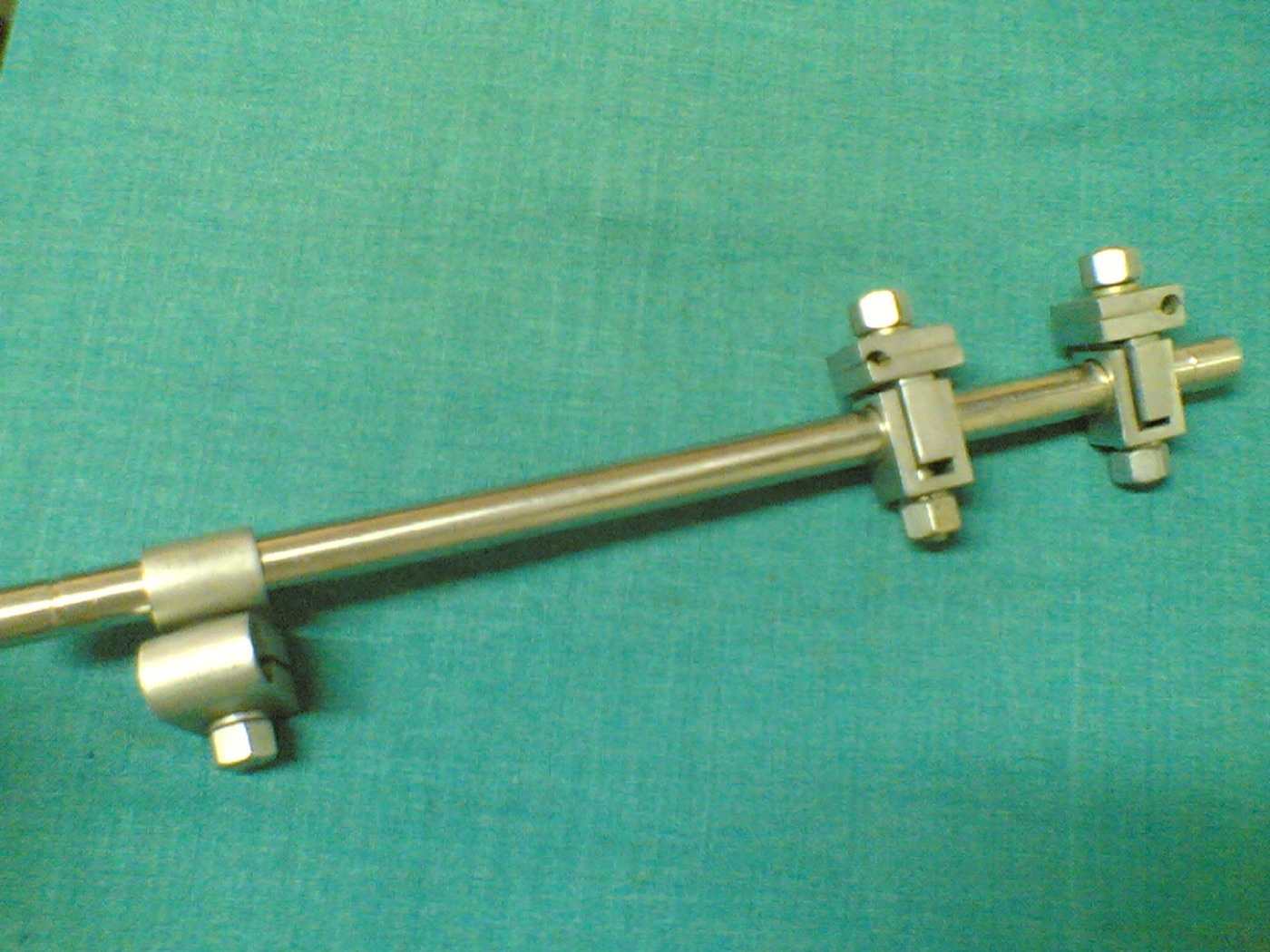
Figure 12. An AO external fixator (connecting rod, clamps and Schanz pin), the apparatus for damage-control spanning fixation. Source: Netha Hussain, via Wikimedia Commons, CC BY-SA 3.0.
The femur is the priority injury (an independent predictor of mortality and ARDS). Conversion to a definitive intramedullary nail is timed to avoid the inflammatory peak of days 2-4 and is best done once the inflammatory response has settled, around days 5-8; conversion is safe and infection rates stay low (~3.6% for the femur) provided the time in the frame is short, whereas prolonged external fixation before nailing drives infection up (markedly so in the tibia).[56] Where an infected pin site is encountered, a short interval with curettage and traction (a “pin holiday”) precedes nailing.[57]
Part VIII - Pelvic-Ring Haemorrhage in the Unstable Patient
The pelvis is a special case of damage control because the unstable pelvic ring can exsanguinate into a retroperitoneum that holds around 4 litres before it tamponades, and roughly 90% of the bleeding is venous (from the presacral plexus and fracture surfaces), with major arterial injury in only about 6-8%.[58] The first manoeuvre is mechanical: a pelvic binder, sheet or C-clamp centred over the greater trochanters reduces the pelvic volume and tamponades the venous bleeding, though it is “not a panacea” and a vertical-shear injury needs skeletal traction.[59] If instability persists despite mechanical stabilisation and resuscitation, the two salvage options are preperitoneal pelvic packing (the European preference, addressing the dominant venous bleeding; one series cut mortality from 46% to 25%) and angiographic embolisation (the North American preference, for the minority with arterial bleeding, best within 3 hours), increasingly used together in a multidisciplinary protocol.[60]
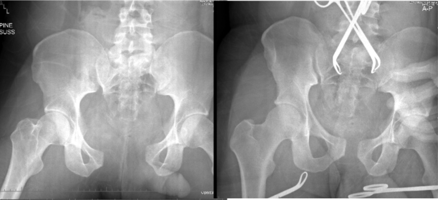
Figure 13. An unstable pelvic ring before (left) and after (right) application of a pelvic circumferential compression binder, reducing the pelvic volume. Source: Vaidya et al., West J Emerg Med 2016;17:766, CC BY 4.0.
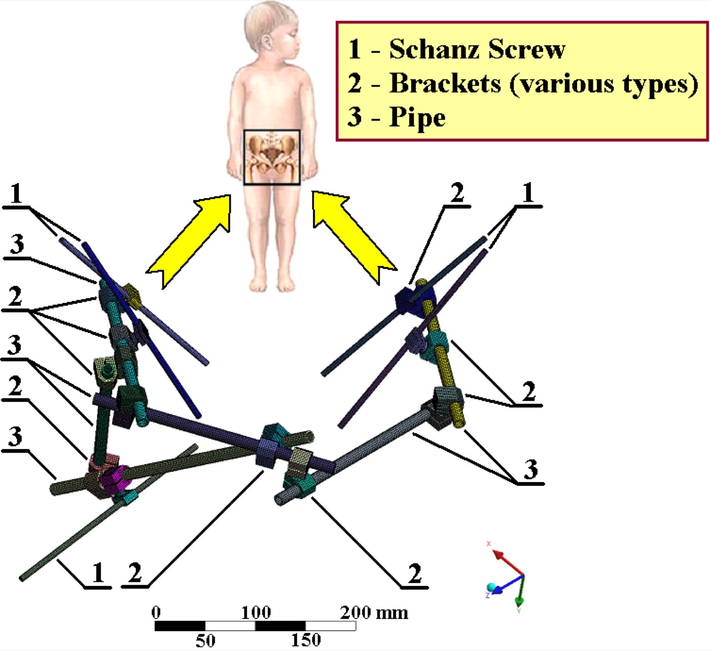
Figure 14. An anterior pelvic external fixator (Schanz screws and connecting frame) for definitive damage control of the pelvic ring. Source: Karel Frydryšek (Fry72), via Wikimedia Commons, CC BY-SA 4.0.
Part IX - SIRS, MODS, ARDS and Fat Embolism
The late deaths of the trimodal distribution are the end-points of a single trauma-induced inflammatory pathway. A massive injury (one hit) or an injury plus an ill-timed surgical insult (two hits) can amplify the systemic inflammatory response syndrome (SIRS) into multiple-organ dysfunction syndrome (MODS): the lungs fail first at 48-72 hours as acute respiratory distress syndrome (ARDS), then the kidneys and liver, with mortality approaching 80% when all three fail.[61] Fat embolism syndrome belongs to the same family: the risk is reduced by early fracture stabilisation and increased by delay, and the mechanism (marrow fat embolising to the lung at the opening of the medullary canal) is the very reason reamed nailing can be a damaging second hit in the under-resuscitated chest-injured patient.[62] The detail of fat embolism syndrome and the thromboembolic complications is taken up in Topic 7.
References
-
Rockwood & Green 9e, ch.13 (Brahmbhatt & Vallier), p.742.
-
Rockwood 9e p.749.
-
Rockwood 9e p.749.
-
Rockwood 9e p.747; ATLS 9e p.40.
-
Rockwood 9e p.742.
-
Rockwood 9e p.750, Table 13-3.
-
Rockwood 9e pp.749-750.
-
Rockwood 9e pp.749-750.
-
Rockwood 9e p.750.
-
ATLS 9e p.37; Rockwood 9e p.742.
-
ATLS 9e p.37.
-
ATLS 9e pp.37-41.
-
Rockwood 9e pp.744, 755.
-
ATLS 9e p.40; Rockwood 9e p.744.
-
ATLS 9e p.41.
-
ATLS 9e pp.42-44; Rockwood 9e p.745.
-
ATLS 9e p.44.
-
ATLS 9e p.46.
-
ATLS 9e p.44.
-
Rockwood 9e pp.746, 774.
-
ATLS 9e p.93.
-
ATLS 9e pp.94-96.
-
ATLS 9e p.94.
-
ATLS 9e p.99, Table 3.1.
-
ATLS 9e p.95; Emergency War Surgery p.106.
-
ATLS 9e p.98.
-
ATLS 9e p.94.
-
ATLS 9e p.95.
-
ATLS 9e pp.95, 102.
-
ATLS 9e p.105.
-
ATLS 9e pp.96, 98, 106.
-
ATLS 9e p.100.
-
ATLS 9e pp.40-42; Rockwood 9e p.744.
-
ATLS 9e p.102; Skeletal Trauma 5e pp.344, 347.
-
ATLS 9e p.102.
-
ATLS 9e p.102.
-
ATLS 9e p.105; Rockwood 9e p.750.
-
ATLS 9e p.105.
-
Skeletal Trauma 5e p.344 establishes the within-3-hour CRASH-2 window; the precise 1 g + 1 g regimen is standard teaching, not stated in the mined extracts.
-
ATLS 9e pp.95, 103.
-
ATLS 9e pp.103, 108.
-
ATLS 9e pp.103-104, Table 3.2.
-
ATLS 9e p.107.
-
Skeletal Trauma 5e p.371; ICRC p.324.
-
Emergency War Surgery pp.162-163; Skeletal Trauma 5e p.371.
-
Emergency War Surgery p.163; ICRC p.324.
-
Skeletal Trauma 5e pp.369, 372.
-
Skeletal Trauma 5e pp.369, 371.
-
Skeletal Trauma 5e pp.369, 373.
-
Skeletal Trauma 5e p.373; Rockwood 9e pp.760-764. The “lactate is the most reliable resuscitation marker” point and the proceed-targets are from Rockwood pp.762-764.
-
Rockwood 9e pp.762, 764.
-
Skeletal Trauma 5e p.374.
-
Skeletal Trauma 5e p.374.
-
Skeletal Trauma 5e pp.373-374.
-
Skeletal Trauma 5e pp.374-375.
-
Skeletal Trauma 5e p.378.
-
The “pin holiday” term is standard teaching; the mined extract describes the practice (interval, curettage, traction) without the label - Skeletal Trauma 5e p.378.
-
Rockwood 9e pp.758, 766.
-
Rockwood 9e pp.745, 758-759; Skeletal Trauma 5e p.382.
-
Skeletal Trauma 5e pp.382-383; Rockwood 9e pp.766-767.
-
Skeletal Trauma 5e pp.369, 372.
-
Skeletal Trauma 5e pp.378-379; Rockwood 9e p.760.
-
Rockwood 9e p.749.
-
Rockwood 9e pp.749-750.
-
ATLS 9e pp.37-44.
-
ATLS 9e p.40; Rockwood 9e pp.744, 753.
-
ATLS 9e pp.95, 99.
-
ATLS 9e p.102; Skeletal Trauma 5e p.348.
-
ATLS 9e pp.102-105; Rockwood 9e p.750.
-
ATLS 9e pp.103-104.
-
Emergency War Surgery p.163; ICRC p.324.
-
Skeletal Trauma 5e pp.369, 373.
-
Skeletal Trauma 5e pp.373-374; Rockwood 9e pp.762-764.
-
Rockwood 9e pp.758-759; Skeletal Trauma 5e pp.382-383.
-
Skeletal Trauma 5e p.378.