Contents
- Orientation: A Ring That Breaks Twice, and a Patient Who Can Bleed to Death
- Part I - Functional Anatomy and Biomechanics
- Part II - Mechanism and Associated Injuries
- Part III - Assessment and Imaging
- Part IV - Classification
- Part V - Initial Management and the Haemorrhage Algorithm
- Part VI - Nonoperative Treatment
- Part VII - Definitive Operative Fixation
- Part VIII - Complications
- Part IX - A Synthesis: How to Reason About the Pelvic Ring
- References
Orientation: A Ring That Breaks Twice, and a Patient Who Can Bleed to Death
Three ideas govern the pelvic ring. The first is geometric: the pelvis is a true ring, so a displaced break in one part of it is almost always accompanied by a second break elsewhere; finding an anterior injury obliges you to search for the posterior one.[1] The second is mechanical: the ring’s stability lives almost entirely in its posterior weight-bearing arch, the sacroiliac joints and their stout posterior ligaments, which act as a tension band; the symphysis contributes only about 15% of the ring’s stability, so the surgeon’s central question is always the integrity of the posterior ring.[2] The third is that the pelvic fracture is, above all, a marker of major trauma and a potential source of fatal haemorrhage: the patient may die not of the fracture but of the bleeding and of the associated head, chest, and abdominal injuries.[3] Everything that follows, the classifications, the binder, the angiogram, the iliosacral screw, serves those three ideas.
Part I - Functional Anatomy and Biomechanics
The pelvis is three bones, two joints, and a sheet of ligaments: the two innominate bones (each a fusion of ilium, ischium, and pubis at the triradiate cartilage of the acetabulum) joined anteriorly at the pubic symphysis and posteriorly to the sacrum through the sacroiliac (SI) joints.[4] The bones themselves have no inherent stability. It is the ligaments that hold the ring, and the dominant ones are posterior. The posterior sacroiliac, sacrotuberous, and sacrospinous ligaments, together with the iliolumbar ligament, form the posterior tension band that carries the load from the spine to the legs, which is why the posterior ring is the cornerstone of stability and the symphysis a minor contributor.[5] The most-injured tissue is the pelvic venous plexus, which lies against the posterior ring and is torn in almost every significant disruption, the anatomical basis of pelvic haemorrhage.[6]
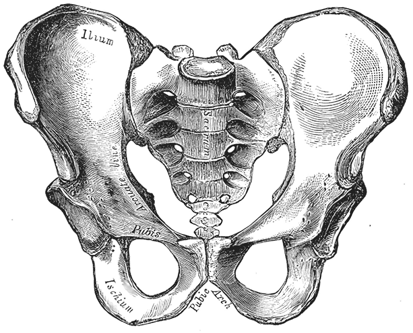
Figure 1. The bony pelvis (anterior view): the two innominate bones (ilium, ischium, pubis) and the sacrum forming the ring. Henry Vandyke Carter, Gray’s Anatomy (1918), public domain, via Wikimedia Commons.
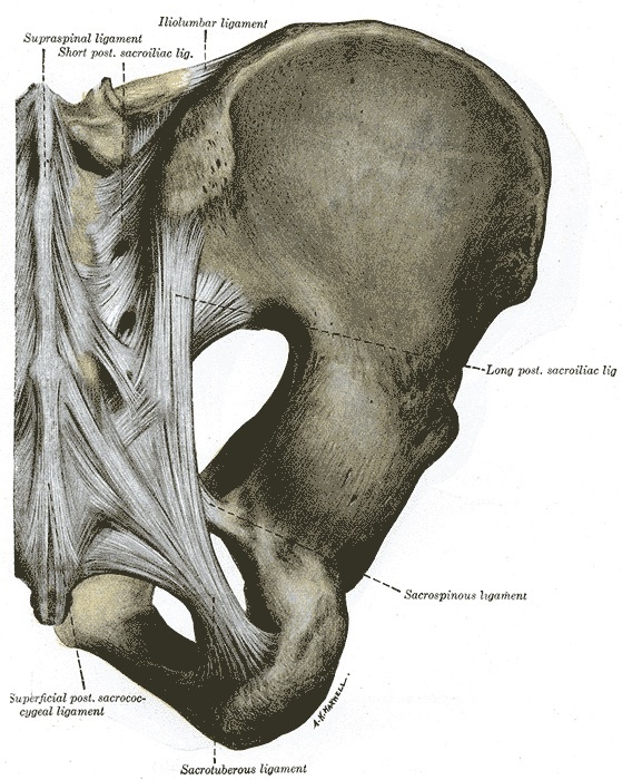
Figure 2. The posterior pelvic ligaments, the posterior tension band: the posterior sacroiliac, sacrotuberous, sacrospinous, and iliolumbar ligaments. Henry Vandyke Carter, Gray’s Anatomy (1918), public domain, via Wikimedia Commons.
The structures at risk around the ring are the examiner’s bread and butter. The lumbosacral plexus runs across the back of the ring, with the L5 nerve root lying only about 1.5 cm from the SI joint on the anterior surface of the sacral ala, where it is endangered by both the injury and the surgeon.[7] The arterial supply is from the internal iliac system, the superior gluteal artery (exiting the greater sciatic notch) being the most frequently injured. The corona mortis, a variable anastomosis between the obturator and external iliac (or inferior epigastric) vessels running about 6 cm from the symphysis on the back of the superior pubic ramus, must be identified and ligated in any anterior approach because it can bleed catastrophically.[8] Anteriorly, the bladder and urethra sit immediately behind the symphysis, and the rectum and vagina lie against the ring, so all are at risk in displaced and open injuries.[9]
Part II - Mechanism and Associated Injuries
Most ring disruptions are high-energy (motorcycle and pedestrian collisions, falls, crush), although the elderly increasingly sustain low-energy fragility fractures of the pelvis from a simple fall.[10] The overriding clinical concern is haemorrhage, the chief cause of early death. The largest share of bleeding is venous (the plexus and the cancellous fracture surfaces), controlled by retroperitoneal tamponade; a minority is arterial (the internal iliac branches, chiefly the superior gluteal), and it is the arterial bleed that overwhelms tamponade and kills.[11] The figures bear this out: a patient who is haemodynamically stable on arrival has about a 3% mortality, but one who is hypotensive has about 38%, and a pelvic haematoma over 500 cm³ on CT carries a nearly fivefold risk of arterial injury.[12]
The associated injuries cluster predictably: chest (about 63%), long-bone fractures (about 50%), head (about 40%), and abdominal/solid-organ injury (about 40%).[13] Two categories deserve separate emphasis. Genitourinary injury occurs in about 6% to 15%: the male posterior urethra is torn by shearing (suspected from blood at the meatus, a high-riding prostate, and a scrotal/perineal haematoma, and investigated by retrograde urethrogram), and the bladder ruptures either extraperitoneally (the commoner) or intraperitoneally.[14] Open pelvic fractures (up to 5%), where the ring communicates with the perineum, rectum, or vagina, are the most lethal of all, with mortality up to 50% from sepsis, and they demand faecal diversion.[15]
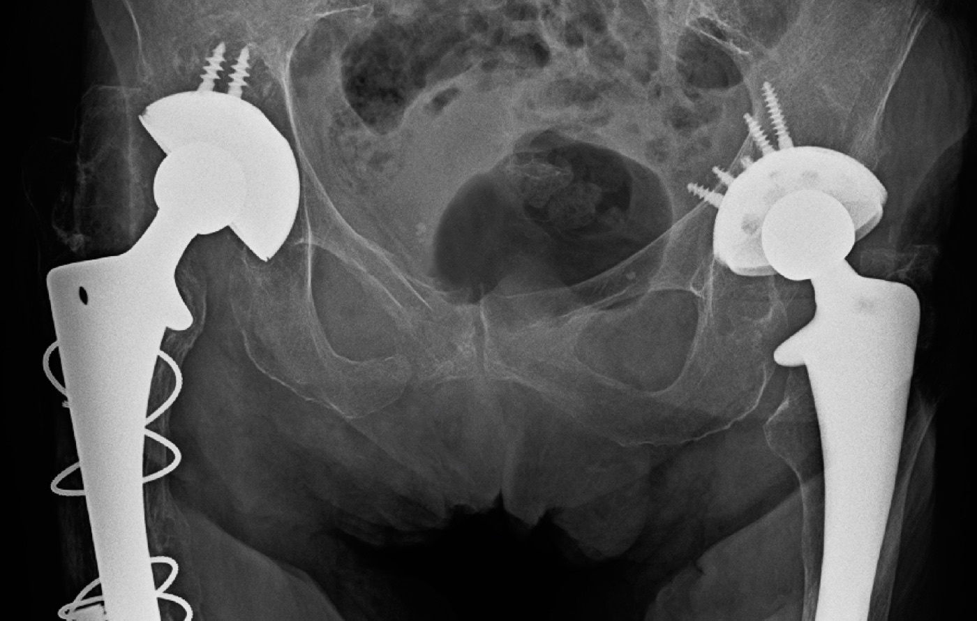
Figure 3. Fractures of the superior and inferior pubic rami on an AP pelvis radiograph, the anterior half of a ring injury. James Heilman, MD, CC BY-SA 4.0, via Wikimedia Commons.
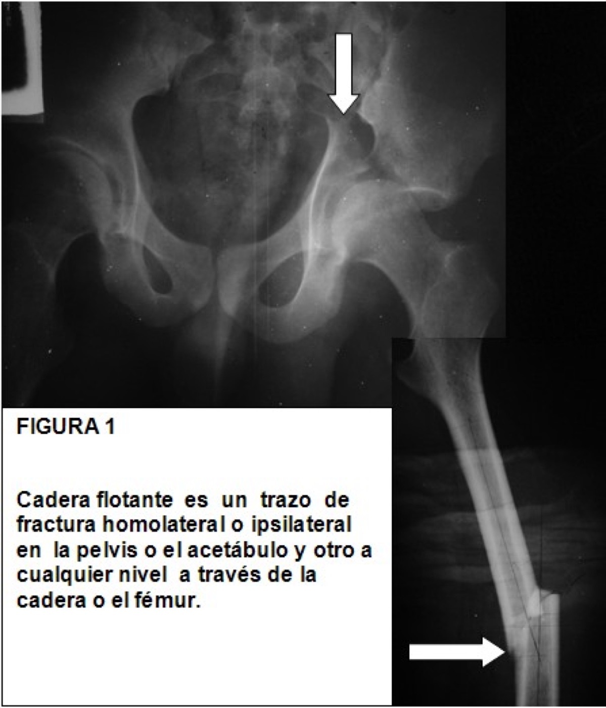
Figure 4. A displaced, unstable ipsilateral pelvic and femoral injury (a floating-hip injury), illustrating the high-energy displaced pattern. DrMedina, CC BY-SA 3.0, via Wikimedia Commons.
Part III - Assessment and Imaging
The primary survey is the ATLS sequence, with the unstable pelvis identified early as a bleeding source.[16] Current practice has moved away from one old reflex: the habit of vigorously “rocking” the pelvis to test stability is abandoned, because it is insensitive and can dislodge clot and restart haemorrhage. Any manual assessment is done once, gently, and formal stress testing is reserved for examination under anaesthesia, which frequently upgrades the classification.[17] The examination also includes inspection for the open wound and the perineal, rectal, and vaginal injury, a careful neurological assessment of the lumbosacral roots, and palpation for a fluctuant Morel-Lavallée closed degloving lesion.[18]
Imaging begins with the AP pelvis radiograph, which alone has high sensitivity for a clinically significant fracture and guides the emergency decision.[19] Detailed assessment then uses two angled views and CT. The inlet view (the beam directed caudally) shows internal/external rotation and anteroposterior translation of the hemipelvis and impaction of the sacral ala; the outlet view (beam cephalad) shows vertical (superior) translation and profiles the sacral foramina.[20] CT is the gold standard for the posterior ring and the sacrum, and a key caution is that a binder applied before imaging can mask an open-book injury by reducing it, so it is important to know whether a film was taken with the binder on.[21] Signs of instability include posterior displacement beyond about 5 mm, SI gapping, and the avulsion of the L5 transverse process (the iliolumbar ligament avulsion, the sentinel sign of a vertical-shear injury).[22]
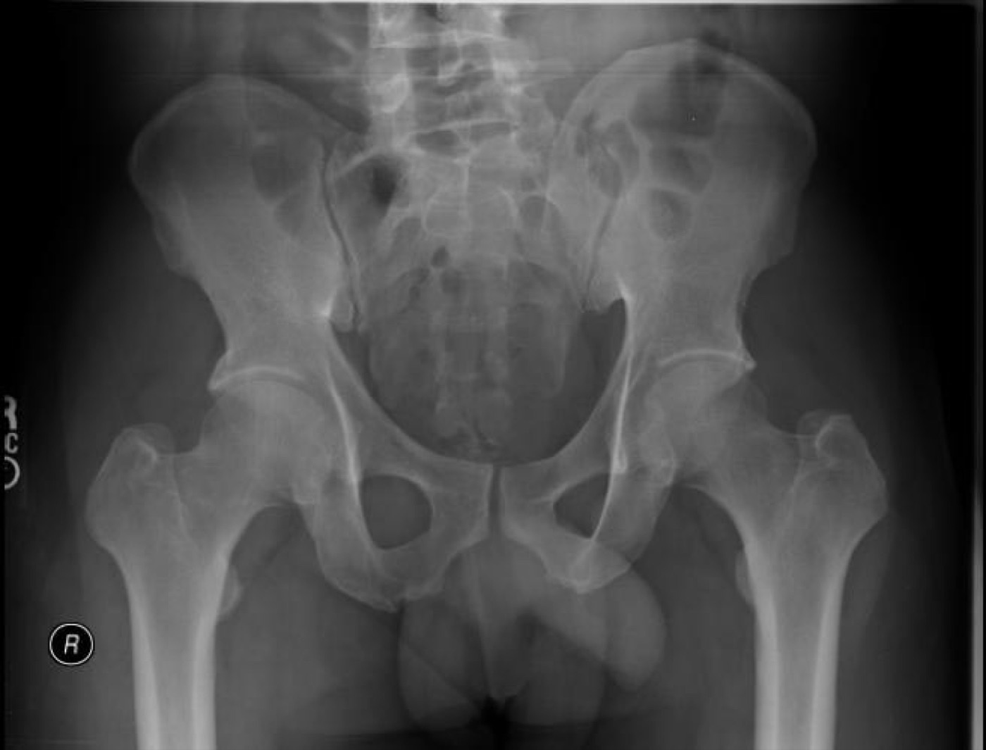
Figure 5. A normal AP pelvis radiograph for comparison, the first and most useful study in the injured patient. RadsWiki, CC BY-SA 3.0, via Wikimedia Commons.
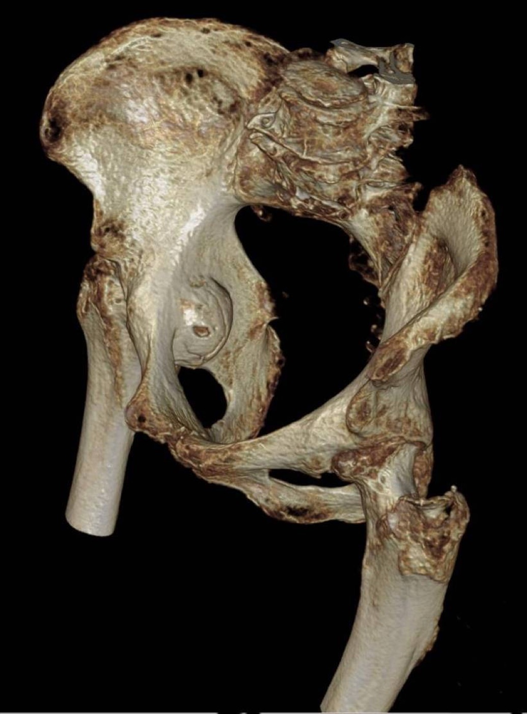
Figure 6. A 3D CT volume rendering of a pelvic and acetabular injury, the kind of reconstruction CT adds to the plain films. Hellerhoff, CC BY-SA 4.0, via Wikimedia Commons.
Part IV - Classification
Two complementary systems are used. The Young-Burgess classification is mechanistic, grouping injuries by the direction of force:[23]
- Lateral compression (LC), the commonest, from a side impact that internally rotates the struck hemipelvis: LC I, a sacral (alar) impaction fracture on the side of impact; LC II, a “crescent” iliac-wing fracture-dislocation; LC III, the “windswept” pelvis, an LC injury on one side with a contralateral open-book.
- Anteroposterior compression (APC, “open book”), from external rotation: APC I, symphyseal widening under 2.5 cm with intact ligaments; APC II, widening over 2.5 cm with disrupted anterior SI ligaments but an intact posterior hinge; APC III, complete SI disruption (and so vertically unstable).
- Vertical shear (VS), from a cephalad force, with vertical displacement of the hemipelvis.
- Combined mechanism (CM).
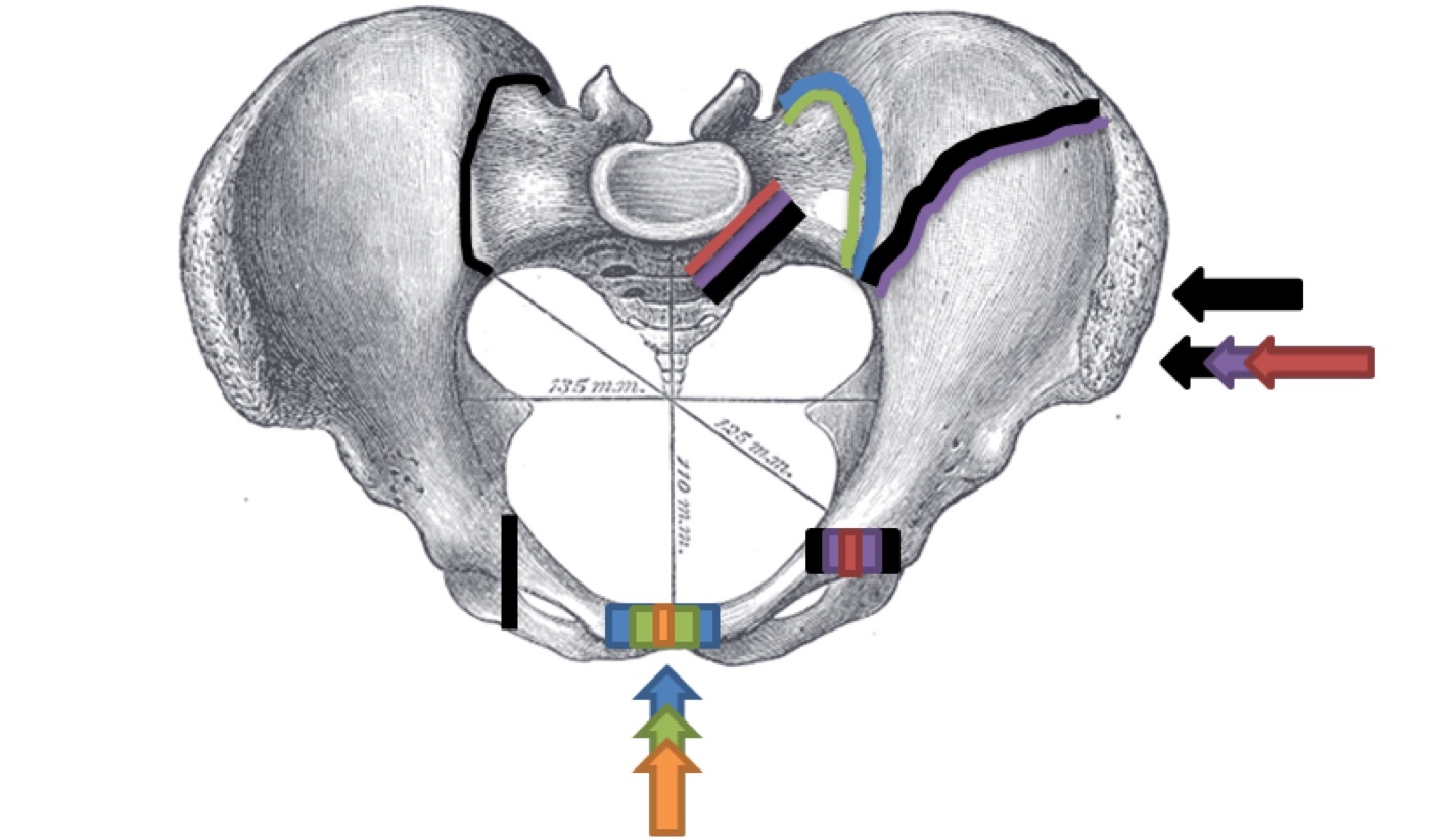
Figure 7. The Young-Burgess mechanisms of pelvic-ring injury (schematic): anteroposterior compression, lateral compression, and vertical shear. Annotation by Kinanat0212 on a Gray’s Anatomy (1918) plate, public domain, via Wikimedia Commons.
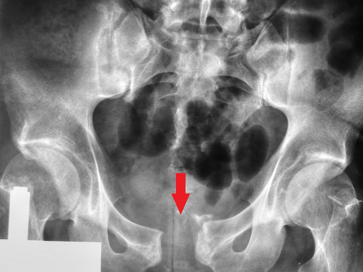
Figure 8. An open-book (anteroposterior-compression) injury: wide diastasis of the pubic symphysis (arrow). Nevit Dilmen, CC BY-SA 3.0, via Wikimedia Commons.
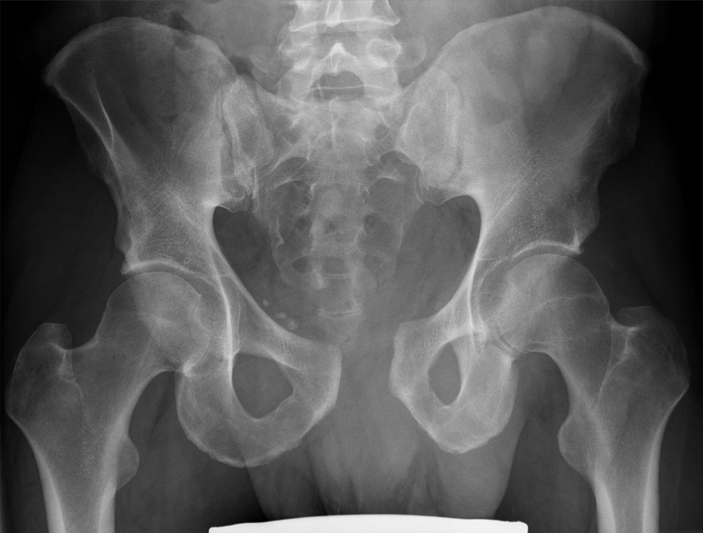
Figure 9. An open-book injury with symphyseal disruption and widening of the sacroiliac joint, the combined anterior and posterior APC disruption. Hellerhoff, CC BY-SA 4.0, via Wikimedia Commons.
The Tile / AO-OTA classification is stability-based and maps onto the AO codes 61A/B/C by the state of the posterior arch: type A is stable (the posterior arch intact, e.g. an avulsion or iliac-wing fracture); type B is rotationally unstable but vertically stable (an incomplete posterior disruption, the open-book B1 and lateral-compression B2); type C is both rotationally and vertically unstable (a complete posterior disruption).[24] Both systems predict severity and transfusion need mainly when collapsed into stable versus unstable groups.[25] The sacral fracture is classified by the Denis zones relative to the foramina: zone 1 (alar, lateral to the foramina), the commonest (about 50%) with a low neurological risk; zone 2 (foraminal), about a third, with a neurological deficit in roughly 30%; and zone 3 (central, into the canal), the least common (about 16%) but with a nerve-injury rate as high as 60%.[26] A transverse sacral fracture with bilateral vertical lines produces the U- or H-shaped pattern of spinopelvic (lumbopelvic) dissociation, a highly unstable injury that separates the spine from the pelvis.[27]
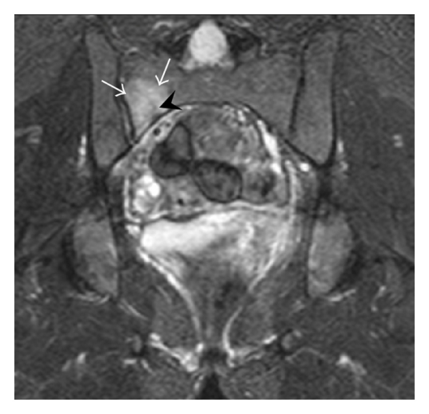
Figure 10. A right sacral alar fracture on coronal STIR MRI (arrows), with surrounding marrow oedema. Jarraya et al., CC BY 3.0, via Wikimedia Commons.
Part V - Initial Management and the Haemorrhage Algorithm
The acute priority in the unstable patient is to control bleeding by stabilising the ring and then choosing between angiographic embolization and pelvic packing.[28] The first manoeuvre is a pelvic binder or circumferential sheet applied at the level of the greater trochanters (not the iliac crests), with the legs internally rotated; it closes an open-book injury, reduces the pelvic volume, and protects the clot (“the initial clot is the most effective clot”). It is a field measure, left in place for up to about 24 hours, but it must not be left too long (skin pressures exceed the threshold for necrosis at about 9.3 kPa), and it should be used cautiously in a lateral-compression pattern, where it can over-reduce and drive rami fragments into the bladder or vessels.[29] Where stabilisation is needed beyond a binder, an anterior external fixator (iliac-crest or supra-acetabular pins) closes an open-book if the posterior hinge is intact, and the pelvic C-clamp, which translates the hemipelvis medially, addresses a posterior disruption; both are emergency, not definitive, tools.[30]
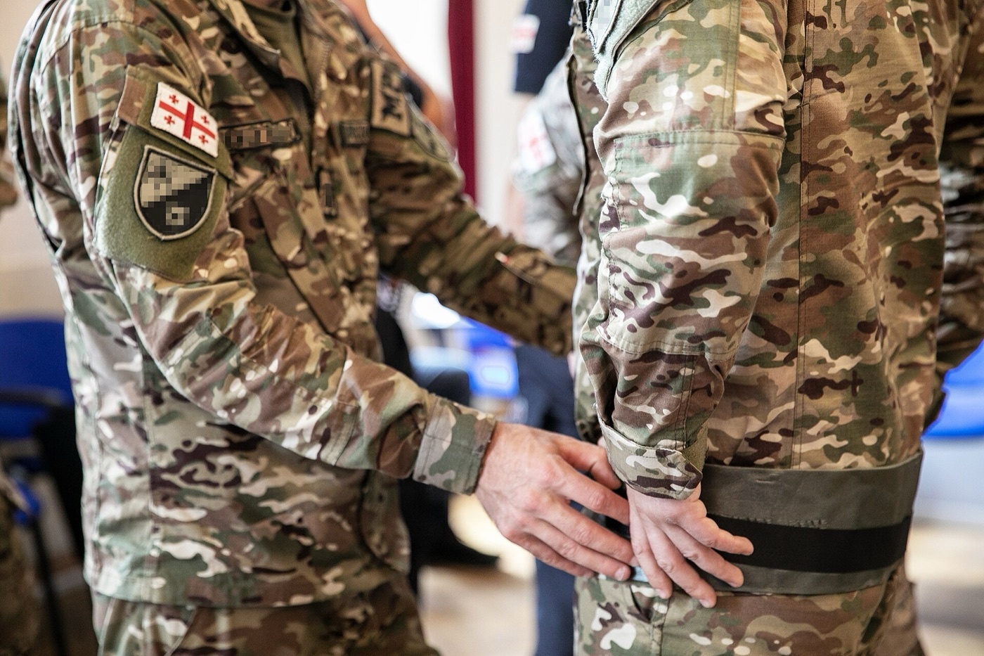
Figure 11. Application of a circumferential pelvic binder around the pelvis. Sgt. Mykaela Martin, U.S. Army, public domain, via Wikimedia Commons.
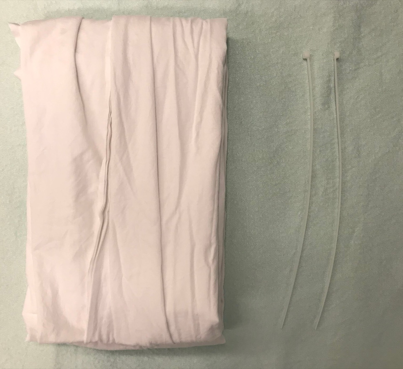
Figure 12. Equipment for an improvised pelvic binder (a folded sheet). James Heilman, MD, CC BY-SA 4.0, via Wikimedia Commons.
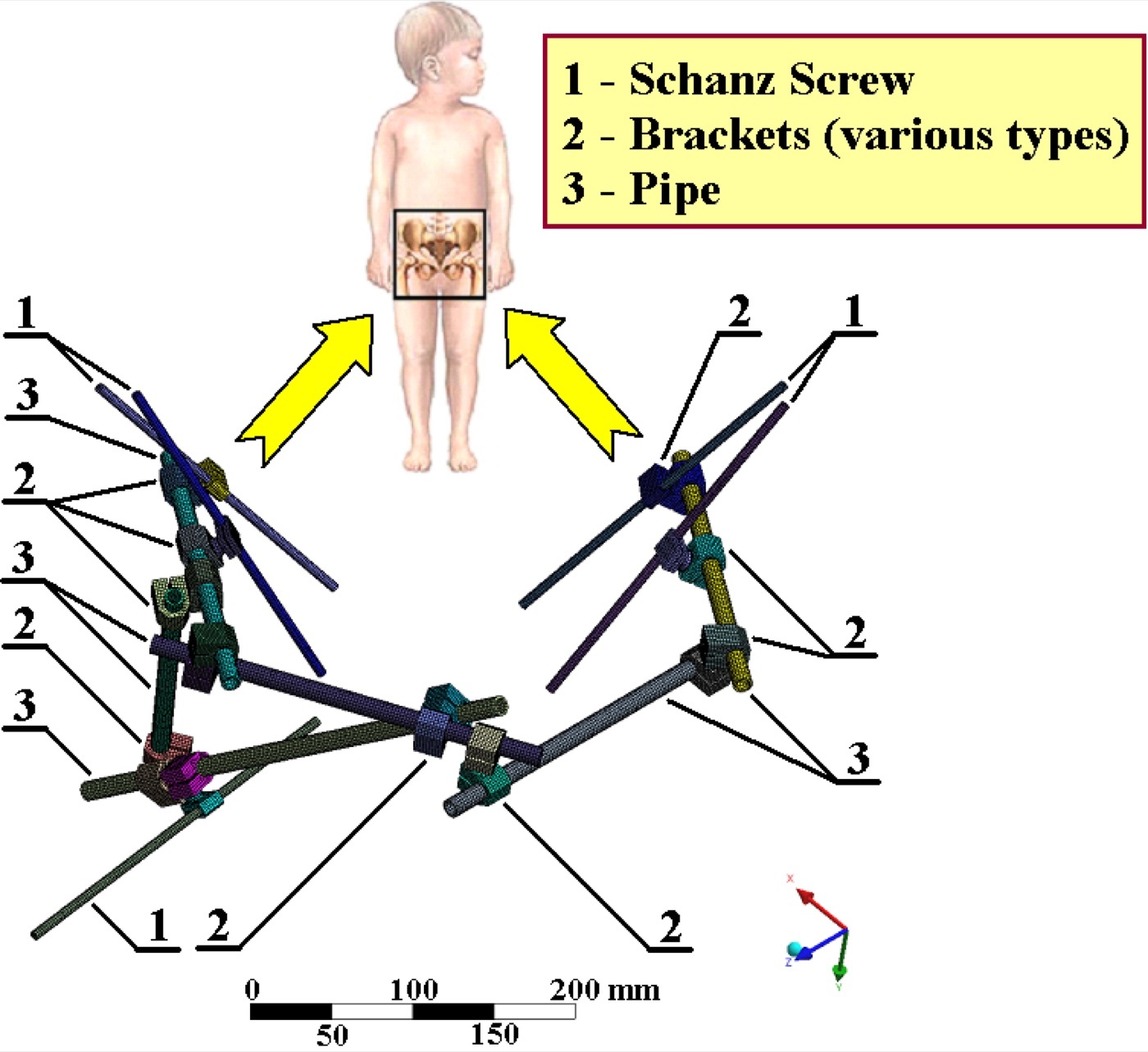
Figure 13. An anterior pelvic external fixator construct (schematic): Schanz screws, brackets, and a connecting rod. Karel Frydrýšek, CC BY-SA 4.0, via Wikimedia Commons.
The choice between angioembolization and preperitoneal pelvic packing is genuinely unsettled and depends on institutional capability.[31] Contrast extravasation on CT signals an arterial bleed and, by the EAST guideline, triggers angiography regardless of haemodynamic status (a Level 1 recommendation); embolization is ideally selective of the bleeding internal-iliac branch (nonselective occlusion risks gluteal and bladder necrosis) and succeeds in over 95% of cases, best within 60 minutes of arrival.[32] Preperitoneal pelvic packing (six laparotomy swabs placed against a posteriorly stabilised ring, which it requires to pack against) is a EAST Level 3 salvage measure used widely in Europe and increasingly in centres with limited rapid angiography; it is quicker and lowers early transfusion. REBOA (resuscitative endovascular balloon occlusion of the aorta) is an emerging temporising measure still under evaluation.[33]
Part VI - Nonoperative Treatment
Nonoperative care suits the stable injury: the APC I, most LC I, and the fragility fracture of the pelvis (Rommens type I), as well as the patient too unwell for surgery.[34] The principle is mobilisation with protected weight-bearing, repeat radiographs after the patient has mobilised to confirm the injury has not displaced, and a follow-up film at about two weeks.[35] The caution is that the LC I is a heterogeneous group: a complete sacral fracture (especially with bilateral rami fractures) can displace, so an apparently stable injury with worrying features is assessed for occult instability with stress views or examination under anaesthesia, which reveals sufficient instability to warrant fixation in a substantial minority.[36] In the elderly fragility fracture, adjuncts such as parathyroid hormone (PTH 1-84) and, for persistent posterior pain, a percutaneous iliosacral screw, have a role.[37]
Part VII - Definitive Operative Fixation
7.1 The governing principle: restore the posterior ring
The cardinal rule of definitive fixation, attributed to Letournel, is that the posterior ring is the weight-bearing arch, so restoring and stabilising it is the primary goal; anterior implants cannot compensate for inadequate posterior fixation.[38] From the biomechanics it follows that a rotationally unstable but vertically stable injury (Tile B / APC II) can often be managed by anterior fixation alone, whereas a vertically unstable injury (Tile C / VS) needs both anterior and posterior fixation.[39]
7.2 Anterior fixation
Symphyseal plating is the standard fixation for the open-book diastasis (symphyseal widening over 2.5 cm warrants stabilisation), performed through a Pfannenstiel approach.[40] An APC injury is adequately held by a four-hole plate acting as a tension band, whereas the higher shear of an LC III symphyseal rupture needs a six-hole or dual plate; in all cases at least two points of fixation are placed on each side (a two-hole plate is inadequate). Kellam showed that reducing the symphysis to within 2 cm gives a return to normal function in essentially all patients when the posterior ring is intact.[41] The alternatives are the definitive external fixator (useful when ORIF is precluded by a bladder rupture or a contaminated laparotomy) and the anterior subcutaneous internal fixator (INFIX), a pair of supra-acetabular pedicle screws joined by a subcutaneous rod; the INFIX spares the soft tissues but endangers the lateral femoral cutaneous nerve, requires the screw heads to sit at least 1.5 cm above the AIIS to avoid compressing the femoral vessels, and must be removed at 8 to 10 weeks before heterotopic bone forms.[42] Individual rami may be fixed with percutaneous antegrade or retrograde ramus screws, watching for the corona mortis.[43]
7.3 Posterior fixation
The workhorse of posterior fixation is the percutaneous iliosacral (SI) screw, a cannulated screw driven across the SI joint into the body of S1 under inlet, outlet, and lateral fluoroscopic views.[44] The danger is neurological: the L5 nerve root on the ala and the sacral roots in the foramina lie within millimetres of the safe corridor, so the screw must stay within it, and a dysmorphic upper sacrum (present in up to about 44% of pelvises) narrows or abolishes the S1 corridor, mandating preoperative CT planning and often a screw at the S2 level instead.[45] A transsacral (transiliac-transsacral) screw crossing into the opposite ilium is used for bilateral injuries and to augment poor fixation, and the U-shaped sacral fracture with spinopelvic dissociation requires lumbopelvic (spinopelvic, triangular) fixation to reconnect the spine to the pelvis.[46] Open reduction of the SI joint or sacrum is performed through anterior or posterior approaches when closed reduction fails, the posterior approach being the route for direct sacral nerve-root decompression.[47]
Timing matters: in the stabilised patient, definitive fixation is best within 2 to 5 days and certainly within 14 days, after which anatomical reduction becomes much harder.[48]
Part VIII - Complications
The complications follow the anatomy.[49] Haemorrhage is the early killer, the superior gluteal artery and corona mortis the surgical hazards (controlled by packing and embolization rather than ligation). Infection and wound dehiscence are particularly associated with the posterior approaches, with rates up to 16%, and with the overlying Morel-Lavallée lesion. Neurological deficit of the L5 and sacral roots may be injury-related or iatrogenic from an errant iliosacral screw. Malunion and nonunion, from inadequate reduction or definitive external fixation, leave a disabling pelvis (chronic pain is reported in up to 97% of malunions, with leg-length discrepancy, sitting imbalance, and dyspareunia).[50]
Two systemic complications complete the picture. The risk of venous thromboembolism is high (a pelvic fracture is an independent risk factor; DVT is reported in about 35% to 61% and PE in about 2% to 10%), so mechanical and chemical prophylaxis is begun early and continued for weeks, with an IVC filter reserved for selected patients.[51] Finally, urogenital and sexual dysfunction is common and under-treated: about 61% of men report sexual dysfunction and 19% persistent impotence, and women may suffer dyspareunia and urinary difficulty; together with chronic posterior pain and the long-term neurological deficits, these explain why good or excellent results are achieved in fewer than 60% of these injuries despite anatomical reconstruction.[52]
Part IX - A Synthesis: How to Reason About the Pelvic Ring
Reason about the pelvis in two clocks. On the resuscitation clock, the pelvic fracture is first a haemorrhage and a marker of major trauma: run the ATLS sequence, look for the bleeding source, and remember that most pelvic bleeding is venous and bony and is controlled by closing and splinting the ring with a binder at the greater trochanters, escalating to embolization (for the arterial bleed flagged by CT extravasation) or preperitoneal packing by your institution’s protocol, while the associated head, chest, and abdominal injuries are what most often determine survival. On the reconstruction clock, think in rings and tension bands: a displaced ring is broken twice, so find the posterior injury; classify it both ways (Young-Burgess to read the mechanism and the bleeding risk, Tile to read the stability); and fix it knowing that the posterior ring is the weight-bearing arch that anterior plates cannot replace. Stabilise the rotationally unstable injury anteriorly and the vertically unstable injury both anteriorly and posteriorly, protect the L5 and sacral nerve roots that hug every posterior screw, and act early, within days, because the reduction and the outcome both decay with time. Through all of it, respect the soft tissues: the bladder behind the symphysis, the corona mortis on the ramus, the venous plexus on the sacrum, and the patient’s later continence, sexual function, and freedom from pain, which are the true measures of a pelvis well treated.
References
-
Rockwood & Green’s Fractures in Adults, pp.3192, 3216 (the pelvis is a ring, so a displaced disruption in one place is accompanied by a second; the posterior structures provide the majority of pelvic stability; the symphyseal ligaments account for only ~15% of the stability of the entire ring); AO Principles of Fracture Management, p.738 (“It is a true ring structure”; “by far the greatest proportion of load goes through the posterior ring structures”).
-
Rockwood & Green’s Fractures in Adults, pp.3192, 3216 (the pelvis is a ring, so a displaced disruption in one place is accompanied by a second; the posterior structures provide the majority of pelvic stability; the symphyseal ligaments account for only ~15% of the stability of the entire ring); AO Principles of Fracture Management, p.738 (“It is a true ring structure”; “by far the greatest proportion of load goes through the posterior ring structures”).
-
Rockwood & Green’s Fractures in Adults, pp.3184, 3219 (the pelvic fracture as a marker of major trauma; mortality is often related to associated head, chest, or abdominal injury rather than the ring fracture itself); AO Principles of Fracture Management, p.737 (“A pelvic injury is an indicator of major trauma and associated injuries must be actively excluded”).
-
Rockwood & Green’s Fractures in Adults, pp.3214-3217 (three bones: sacrum and two innominate bones, each a fusion of ilium/ischium/pubis at the triradiate cartilage; joined anteriorly at the symphysis and posteriorly at the SI joints; the SI joints and their ligaments are the cornerstone of stability, with the sacrotuberous, sacrospinous, posterior sacroiliac, and iliolumbar ligaments; the symphysis ~15% of ring stability); AO Principles of Fracture Management, pp.738-739 (the bones have no inherent stability; the posterior sacroiliac ligaments are key; ligaments listed iliolumbar, posterior/anterior sacroiliac, sacrotuberous, sacrospinous, symphysis).
-
Rockwood & Green’s Fractures in Adults, pp.3214-3217 (three bones: sacrum and two innominate bones, each a fusion of ilium/ischium/pubis at the triradiate cartilage; joined anteriorly at the symphysis and posteriorly at the SI joints; the SI joints and their ligaments are the cornerstone of stability, with the sacrotuberous, sacrospinous, posterior sacroiliac, and iliolumbar ligaments; the symphysis ~15% of ring stability); AO Principles of Fracture Management, pp.738-739 (the bones have no inherent stability; the posterior sacroiliac ligaments are key; ligaments listed iliolumbar, posterior/anterior sacroiliac, sacrotuberous, sacrospinous, symphysis).
-
AO Principles of Fracture Management, p.743 (the pelvic venous plexus is responsible for ~80% of bleeding in unstable pelvic fractures; physiological tamponade does not occur once the ring is disrupted); Rockwood & Green’s Fractures in Adults, p.3217 (the venous plexus is almost always injured to some extent with pelvic disruption).
-
AO Principles of Fracture Management, p.760 (the L5 nerve root is only 1.5 cm from the SI joint, running 10-15 mm across the ala); Rockwood & Green’s Fractures in Adults, pp.3217-3219 (superior gluteal vessels exit the greater sciatic notch and are frequently injured; bladder and urethra behind the symphysis, rectum anterior to the sacrum); Rockwood & Green’s Fractures in Adults, pp.3254, 3263, 3327 (the corona mortis, an obturator-external iliac anastomosis ~6 cm from the symphysis on the inner superior ramus, must be identified and ligated in anterior approaches as it can bleed profusely).
-
AO Principles of Fracture Management, p.760 (the L5 nerve root is only 1.5 cm from the SI joint, running 10-15 mm across the ala); Rockwood & Green’s Fractures in Adults, pp.3217-3219 (superior gluteal vessels exit the greater sciatic notch and are frequently injured; bladder and urethra behind the symphysis, rectum anterior to the sacrum); Rockwood & Green’s Fractures in Adults, pp.3254, 3263, 3327 (the corona mortis, an obturator-external iliac anastomosis ~6 cm from the symphysis on the inner superior ramus, must be identified and ligated in anterior approaches as it can bleed profusely).
-
AO Principles of Fracture Management, p.760 (the L5 nerve root is only 1.5 cm from the SI joint, running 10-15 mm across the ala); Rockwood & Green’s Fractures in Adults, pp.3217-3219 (superior gluteal vessels exit the greater sciatic notch and are frequently injured; bladder and urethra behind the symphysis, rectum anterior to the sacrum); Rockwood & Green’s Fractures in Adults, pp.3254, 3263, 3327 (the corona mortis, an obturator-external iliac anastomosis ~6 cm from the symphysis on the inner superior ramus, must be identified and ligated in anterior approaches as it can bleed profusely).
-
Rockwood & Green’s Fractures in Adults, pp.3184, 3220 (mostly high-energy, motorcycle and pedestrian collisions; elderly fragility fractures; venous bleeding the most frequent source controlled by retroperitoneal tamponade, arterial bleeding from internal iliac branches most commonly the superior gluteal and pudendal arteries overwhelming the tamponade); AO Principles of Fracture Management, p.737 (bimodal distribution, high-energy in the young and low-energy osteoporotic in the elderly).
-
Rockwood & Green’s Fractures in Adults, pp.3184, 3220 (mostly high-energy, motorcycle and pedestrian collisions; elderly fragility fractures; venous bleeding the most frequent source controlled by retroperitoneal tamponade, arterial bleeding from internal iliac branches most commonly the superior gluteal and pudendal arteries overwhelming the tamponade); AO Principles of Fracture Management, p.737 (bimodal distribution, high-energy in the young and low-energy osteoporotic in the elderly).
-
Rockwood & Green’s Fractures in Adults, pp.3189, 3220 (mortality ~3% if haemodynamically stable on presentation, ~38% if hypotensive; a pelvic haematoma >500 cm³ confers a 4.8× increased risk of arterial injury); AO Principles of Fracture Management, p.737 (in-hospital mortality up to 34% with a pelvic fracture plus haemodynamic instability).
-
Rockwood & Green’s Fractures in Adults, pp.3185, 3188 (associated injuries: chest 63%, long-bone 50%, head 40%, solid-organ 40%; GU injury 6-15%, the male posterior urethra torn by shear, signs blood at the meatus / high-riding prostate / scrotal haematoma, retrograde urethrogram; bladder rupture extraperitoneal commoner than intraperitoneal; open fractures up to 5% communicating with rectum/vagina, mortality up to 50% with faecal contamination, diverting colostomy); AO Principles of Fracture Management, pp.744-745 (urethral injury in 80% associated with pelvic fractures, retrograde urethrogram, suprapubic catheter; the open pelvis with perineal involvement needing faecal diversion).
-
Rockwood & Green’s Fractures in Adults, pp.3185, 3188 (associated injuries: chest 63%, long-bone 50%, head 40%, solid-organ 40%; GU injury 6-15%, the male posterior urethra torn by shear, signs blood at the meatus / high-riding prostate / scrotal haematoma, retrograde urethrogram; bladder rupture extraperitoneal commoner than intraperitoneal; open fractures up to 5% communicating with rectum/vagina, mortality up to 50% with faecal contamination, diverting colostomy); AO Principles of Fracture Management, pp.744-745 (urethral injury in 80% associated with pelvic fractures, retrograde urethrogram, suprapubic catheter; the open pelvis with perineal involvement needing faecal diversion).
-
Rockwood & Green’s Fractures in Adults, pp.3185, 3188 (associated injuries: chest 63%, long-bone 50%, head 40%, solid-organ 40%; GU injury 6-15%, the male posterior urethra torn by shear, signs blood at the meatus / high-riding prostate / scrotal haematoma, retrograde urethrogram; bladder rupture extraperitoneal commoner than intraperitoneal; open fractures up to 5% communicating with rectum/vagina, mortality up to 50% with faecal contamination, diverting colostomy); AO Principles of Fracture Management, pp.744-745 (urethral injury in 80% associated with pelvic fractures, retrograde urethrogram, suprapubic catheter; the open pelvis with perineal involvement needing faecal diversion).
-
Rockwood & Green’s Fractures in Adults, pp.3189-3191 (ATLS; manual manipulation done only once because it elicits pain and can dislodge clots and cause persistent bleeding; rectal and vaginal examination; neurological assessment of L5/S1; Morel-Lavallée degloving); AO Principles of Fracture Management, pp.737, 743 (bimanual compression/spring testing has poor sensitivity, may disrupt the clot and reactivate haemorrhage, “no longer recommended” in the ED; stability testing restricted to examination under anaesthesia, which usually upgrades the grade).
-
Rockwood & Green’s Fractures in Adults, pp.3189-3191 (ATLS; manual manipulation done only once because it elicits pain and can dislodge clots and cause persistent bleeding; rectal and vaginal examination; neurological assessment of L5/S1; Morel-Lavallée degloving); AO Principles of Fracture Management, pp.737, 743 (bimanual compression/spring testing has poor sensitivity, may disrupt the clot and reactivate haemorrhage, “no longer recommended” in the ED; stability testing restricted to examination under anaesthesia, which usually upgrades the grade).
-
Rockwood & Green’s Fractures in Adults, pp.3189-3191 (ATLS; manual manipulation done only once because it elicits pain and can dislodge clots and cause persistent bleeding; rectal and vaginal examination; neurological assessment of L5/S1; Morel-Lavallée degloving); AO Principles of Fracture Management, pp.737, 743 (bimanual compression/spring testing has poor sensitivity, may disrupt the clot and reactivate haemorrhage, “no longer recommended” in the ED; stability testing restricted to examination under anaesthesia, which usually upgrades the grade).
-
Rockwood & Green’s Fractures in Adults, pp.3191-3198 (AP pelvis the initial mainstay; inlet view shows internal/external rotation and AP translation and alar impaction, outlet view shows vertical translation and the sacral foramina; CT integral and the gold standard for the posterior ring; a binder can mask the injury; the L5 transverse-process avulsion as the sentinel sign of vertical instability); AO Principles of Fracture Management, pp.737, 743 (a single AP image has high sensitivity/specificity, 45° inlet and outlet views and CT for detailed classification, CT the diagnostic gold standard, repeat the AP with the binder released if injury still suspected).
-
Rockwood & Green’s Fractures in Adults, pp.3191-3198 (AP pelvis the initial mainstay; inlet view shows internal/external rotation and AP translation and alar impaction, outlet view shows vertical translation and the sacral foramina; CT integral and the gold standard for the posterior ring; a binder can mask the injury; the L5 transverse-process avulsion as the sentinel sign of vertical instability); AO Principles of Fracture Management, pp.737, 743 (a single AP image has high sensitivity/specificity, 45° inlet and outlet views and CT for detailed classification, CT the diagnostic gold standard, repeat the AP with the binder released if injury still suspected).
-
Rockwood & Green’s Fractures in Adults, pp.3191-3198 (AP pelvis the initial mainstay; inlet view shows internal/external rotation and AP translation and alar impaction, outlet view shows vertical translation and the sacral foramina; CT integral and the gold standard for the posterior ring; a binder can mask the injury; the L5 transverse-process avulsion as the sentinel sign of vertical instability); AO Principles of Fracture Management, pp.737, 743 (a single AP image has high sensitivity/specificity, 45° inlet and outlet views and CT for detailed classification, CT the diagnostic gold standard, repeat the AP with the binder released if injury still suspected).
-
Rockwood & Green’s Fractures in Adults, pp.3191-3198 (AP pelvis the initial mainstay; inlet view shows internal/external rotation and AP translation and alar impaction, outlet view shows vertical translation and the sacral foramina; CT integral and the gold standard for the posterior ring; a binder can mask the injury; the L5 transverse-process avulsion as the sentinel sign of vertical instability); AO Principles of Fracture Management, pp.737, 743 (a single AP image has high sensitivity/specificity, 45° inlet and outlet views and CT for detailed classification, CT the diagnostic gold standard, repeat the AP with the binder released if injury still suspected).
-
Rockwood & Green’s Fractures in Adults, pp.3200-3208 (Young-Burgess: LC I sacral impaction / II crescent iliac-wing fracture-dislocation / III windswept; APC I diastasis <2.5 cm with intact ligaments / II >2.5 cm with disrupted anterior SI ligaments and intact posterior / III complete SI disruption; VS vertical displacement; CM combined; LC the most common pattern); AO Principles of Fracture Management, p.740 (the same four-vector scheme, with the clinical pearl that a bleeding APC patient is most likely bleeding from the pelvis whereas a bleeding LC patient is most likely bleeding from associated head/chest/abdomen).
-
Rockwood & Green’s Fractures in Adults, pp.3200, 3210 (Tile A stable, B rotationally unstable/vertically stable [B1 open-book, B2 ipsilateral LC], C rotationally and vertically unstable; both systems best discriminate mortality when divided into stable vs unstable); AO Principles of Fracture Management, p.741 (AO/OTA 61A intact posterior arch, 61B incomplete posterior disruption, 61C complete posterior disruption; type A stable, B rotationally unstable/vertically stable, C both unstable).
-
Rockwood & Green’s Fractures in Adults, pp.3200, 3210 (Tile A stable, B rotationally unstable/vertically stable [B1 open-book, B2 ipsilateral LC], C rotationally and vertically unstable; both systems best discriminate mortality when divided into stable vs unstable); AO Principles of Fracture Management, p.741 (AO/OTA 61A intact posterior arch, 61B incomplete posterior disruption, 61C complete posterior disruption; type A stable, B rotationally unstable/vertically stable, C both unstable).
-
Rockwood & Green’s Fractures in Adults, pp.3210-3212 (Denis zone 1 alar, ~50%, low neurological risk; zone 2 foraminal, ~33%, ~30% deficit; zone 3 central/canal, 16%, nerve injury up to 60%; the U-/H-shaped transverse-plus-bilateral-vertical sacral fracture = spinopelvic/lumbopelvic dissociation, highly unstable); AO Principles of Fracture Management, p.762 (U-, H-, and J-pattern sacral fractures needing posterior spinopelvic instrumentation; Denis zones II and III in the LC1 context).
-
Rockwood & Green’s Fractures in Adults, pp.3210-3212 (Denis zone 1 alar, ~50%, low neurological risk; zone 2 foraminal, ~33%, ~30% deficit; zone 3 central/canal, 16%, nerve injury up to 60%; the U-/H-shaped transverse-plus-bilateral-vertical sacral fracture = spinopelvic/lumbopelvic dissociation, highly unstable); AO Principles of Fracture Management, p.762 (U-, H-, and J-pattern sacral fractures needing posterior spinopelvic instrumentation; Denis zones II and III in the LC1 context).
-
Rockwood & Green’s Fractures in Adults, pp.3222-3224 (binder/sheet at the level of the greater trochanters with internal rotation of the legs; the three purposes of external compression; left up to 24 hours; skin pressures exceed 9.3 kPa, the threshold for breakdown; over-reduction risk in LC injuries driving rami spikes into bladder/vessels, a pattern for which binder use is not warranted); AO Principles of Fracture Management, pp.737, 742 (apply the binder over the greater trochanters with internal rotation of the knees, “the initial clot is the most effective clot”; loosen once an LC injury is diagnosed; a correctly placed binder does not impede laparotomy).
-
Rockwood & Green’s Fractures in Adults, pp.3222-3224 (binder/sheet at the level of the greater trochanters with internal rotation of the legs; the three purposes of external compression; left up to 24 hours; skin pressures exceed 9.3 kPa, the threshold for breakdown; over-reduction risk in LC injuries driving rami spikes into bladder/vessels, a pattern for which binder use is not warranted); AO Principles of Fracture Management, pp.737, 742 (apply the binder over the greater trochanters with internal rotation of the knees, “the initial clot is the most effective clot”; loosen once an LC injury is diagnosed; a correctly placed binder does not impede laparotomy).
-
Rockwood & Green’s Fractures in Adults, pp.3225-3226 (emergent anterior external fixator with iliac-crest or supra-acetabular pins, closing the open book with the posterior ring as a hinge; the C-clamp translates the hemipelvis medially, superior for posterior disruption, used emergently and not for definitive treatment); AO Principles of Fracture Management, pp.742-743 (external fixation closes an open book if the posterior complex is intact; the C-clamp for Tile C fractures with uncontrollable haemorrhage).
-
Rockwood & Green’s Fractures in Adults, pp.3220-3229 (the choice of angiography vs packing is institution-specific; contrast extravasation on CT triggers angiography regardless of haemodynamic status, EAST Level 1; selective embolization preferred, nonselective risks gluteal/bladder necrosis, success >95%, best within 60 minutes; preperitoneal packing a EAST Level 3 salvage technique requiring a stabilised posterior ring to pack against; REBOA a temporising measure in early evaluation); AO Principles of Fracture Management, pp.743, 750-751 (embolization vs extraperitoneal packing chosen by local facility, no randomised evidence; nonselective internal iliac embolization controls 85-100% but only as a last resort; six swabs within the true pelvis, tamponade effective only against a stable posterior ring; a 30-minute three-decision emergency algorithm).
-
Rockwood & Green’s Fractures in Adults, pp.3220-3229 (the choice of angiography vs packing is institution-specific; contrast extravasation on CT triggers angiography regardless of haemodynamic status, EAST Level 1; selective embolization preferred, nonselective risks gluteal/bladder necrosis, success >95%, best within 60 minutes; preperitoneal packing a EAST Level 3 salvage technique requiring a stabilised posterior ring to pack against; REBOA a temporising measure in early evaluation); AO Principles of Fracture Management, pp.743, 750-751 (embolization vs extraperitoneal packing chosen by local facility, no randomised evidence; nonselective internal iliac embolization controls 85-100% but only as a last resort; six swabs within the true pelvis, tamponade effective only against a stable posterior ring; a 30-minute three-decision emergency algorithm).
-
Rockwood & Green’s Fractures in Adults, pp.3220-3229 (the choice of angiography vs packing is institution-specific; contrast extravasation on CT triggers angiography regardless of haemodynamic status, EAST Level 1; selective embolization preferred, nonselective risks gluteal/bladder necrosis, success >95%, best within 60 minutes; preperitoneal packing a EAST Level 3 salvage technique requiring a stabilised posterior ring to pack against; REBOA a temporising measure in early evaluation); AO Principles of Fracture Management, pp.743, 750-751 (embolization vs extraperitoneal packing chosen by local facility, no randomised evidence; nonselective internal iliac embolization controls 85-100% but only as a last resort; six swabs within the true pelvis, tamponade effective only against a stable posterior ring; a 30-minute three-decision emergency algorithm).
-
Rockwood & Green’s Fractures in Adults, pp.3229-3231 (nonoperative for stable injuries: APC I, most LC I, FFP type I, and the patient too sick for surgery; mobilise with repeat films after mobilising and at 2 weeks; the LC I a heterogeneous group, complete sacral fractures with bilateral rami fractures may displace, stress/EUA reveals instability warranting fixation in ~41%; parathyroid hormone (PTH 1-84, an RCT showing improved union and pain) and iliosacral screws in the elderly); AO Principles of Fracture Management, p.751 (AP1 and mild LC1 rarely need surgical stabilisation).
-
Rockwood & Green’s Fractures in Adults, pp.3229-3231 (nonoperative for stable injuries: APC I, most LC I, FFP type I, and the patient too sick for surgery; mobilise with repeat films after mobilising and at 2 weeks; the LC I a heterogeneous group, complete sacral fractures with bilateral rami fractures may displace, stress/EUA reveals instability warranting fixation in ~41%; parathyroid hormone (PTH 1-84, an RCT showing improved union and pain) and iliosacral screws in the elderly); AO Principles of Fracture Management, p.751 (AP1 and mild LC1 rarely need surgical stabilisation).
-
Rockwood & Green’s Fractures in Adults, pp.3229-3231 (nonoperative for stable injuries: APC I, most LC I, FFP type I, and the patient too sick for surgery; mobilise with repeat films after mobilising and at 2 weeks; the LC I a heterogeneous group, complete sacral fractures with bilateral rami fractures may displace, stress/EUA reveals instability warranting fixation in ~41%; parathyroid hormone (PTH 1-84, an RCT showing improved union and pain) and iliosacral screws in the elderly); AO Principles of Fracture Management, p.751 (AP1 and mild LC1 rarely need surgical stabilisation).
-
Rockwood & Green’s Fractures in Adults, pp.3229-3231 (nonoperative for stable injuries: APC I, most LC I, FFP type I, and the patient too sick for surgery; mobilise with repeat films after mobilising and at 2 weeks; the LC I a heterogeneous group, complete sacral fractures with bilateral rami fractures may displace, stress/EUA reveals instability warranting fixation in ~41%; parathyroid hormone (PTH 1-84, an RCT showing improved union and pain) and iliosacral screws in the elderly); AO Principles of Fracture Management, p.751 (AP1 and mild LC1 rarely need surgical stabilisation).
-
Rockwood & Green’s Fractures in Adults, pp.3261, 3300 (the posterior ring is the weight-bearing portion and its anatomic reduction is the primary goal, per Letournel; anterior fixation alone is sufficient for rotationally unstable but vertically stable injuries; internal fixation is far better than external for resisting vertical displacement); AO Principles of Fracture Management, pp.751, 763 (AP2 usually needs anterior stabilisation alone; AP2/3, LC2/3, and VS need both anterior and posterior fixation; “additional implants in the anterior pelvic ring will not compensate for inadequate fixation of a posterior injury”).
-
Rockwood & Green’s Fractures in Adults, pp.3261, 3300 (the posterior ring is the weight-bearing portion and its anatomic reduction is the primary goal, per Letournel; anterior fixation alone is sufficient for rotationally unstable but vertically stable injuries; internal fixation is far better than external for resisting vertical displacement); AO Principles of Fracture Management, pp.751, 763 (AP2 usually needs anterior stabilisation alone; AP2/3, LC2/3, and VS need both anterior and posterior fixation; “additional implants in the anterior pelvic ring will not compensate for inadequate fixation of a posterior injury”).
-
Rockwood & Green’s Fractures in Adults, pp.3231, 3258, 3261 (symphyseal stabilisation indicated for diastasis >2.5 cm on static or dynamic imaging; Pfannenstiel approach; at least two points of fixation each side, a two-hole plate not recommended; Kellam’s <2 cm reduction giving 100% return to normal function when no posterior pathology); AO Principles of Fracture Management, pp.751-756 (a four-hole plate as a tension band for an AP mechanism, a six-hole or dual plate for the LC3 shear; screws craniocaudal, usually over 50 mm).
-
Rockwood & Green’s Fractures in Adults, pp.3231, 3258, 3261 (symphyseal stabilisation indicated for diastasis >2.5 cm on static or dynamic imaging; Pfannenstiel approach; at least two points of fixation each side, a two-hole plate not recommended; Kellam’s <2 cm reduction giving 100% return to normal function when no posterior pathology); AO Principles of Fracture Management, pp.751-756 (a four-hole plate as a tension band for an AP mechanism, a six-hole or dual plate for the LC3 shear; screws craniocaudal, usually over 50 mm).
-
Rockwood & Green’s Fractures in Adults, pp.3246-3250, 3263, 3327 (definitive external fixator when ORIF precluded by bladder rupture/contamination; INFIX of supra-acetabular pedicle screws and a subcutaneous rod, LFCN at risk, screw heads ≥1.5 cm above the AIIS to avoid femoral neurovascular compression, removed once healed; antegrade/retrograde ramus screws, the corona mortis ~6 cm from the symphysis at risk); AO Principles of Fracture Management, pp.745-752 (INFIX two supra-acetabular pins under a subcutaneous rod, LFCN protected, removed at 8-10 weeks to avoid heterotopic ossification; ramus fixation with a long screw).
-
Rockwood & Green’s Fractures in Adults, pp.3246-3250, 3263, 3327 (definitive external fixator when ORIF precluded by bladder rupture/contamination; INFIX of supra-acetabular pedicle screws and a subcutaneous rod, LFCN at risk, screw heads ≥1.5 cm above the AIIS to avoid femoral neurovascular compression, removed once healed; antegrade/retrograde ramus screws, the corona mortis ~6 cm from the symphysis at risk); AO Principles of Fracture Management, pp.745-752 (INFIX two supra-acetabular pins under a subcutaneous rod, LFCN protected, removed at 8-10 weeks to avoid heterotopic ossification; ramus fixation with a long screw).
-
Rockwood & Green’s Fractures in Adults, pp.3290-3299 (percutaneous iliosacral screw into the S1 body under inlet/outlet/lateral views; the L5 root and sacral roots within millimetres of the safe corridor; sacral dysmorphism in up to 44%, narrowing or preventing the S1 corridor and favouring an S2 screw; preoperative CT mandatory); AO Principles of Fracture Management, pp.757-759 (7.3 or 8.0 mm cannulated SI screw into the S1 body, the Matta image-intensifier technique, S2 used only if the pedicle is adequate, computer-assisted surgery making it safer).
-
Rockwood & Green’s Fractures in Adults, pp.3290-3299 (percutaneous iliosacral screw into the S1 body under inlet/outlet/lateral views; the L5 root and sacral roots within millimetres of the safe corridor; sacral dysmorphism in up to 44%, narrowing or preventing the S1 corridor and favouring an S2 screw; preoperative CT mandatory); AO Principles of Fracture Management, pp.757-759 (7.3 or 8.0 mm cannulated SI screw into the S1 body, the Matta image-intensifier technique, S2 used only if the pedicle is adequate, computer-assisted surgery making it safer).
-
Rockwood & Green’s Fractures in Adults, pp.3276-3277, 3328 (transsacral/transiliac screw into the contralateral ilium for bilateral injuries and to augment poor fixation; lumbopelvic/spinopelvic constructs for lumbosacral dissociation; the posterior approach for direct sacral nerve-root decompression); AO Principles of Fracture Management, pp.760-763 (transsacral screws for sacral fractures; ilioiliac plating for bilateral sacral fractures; spinopelvic instrumentation for U-/H-/J-pattern sacral fractures with a spine surgeon). The detailed step-by-step technique for spinopelvic/triangular osteosynthesis fell in a page gap of the mined Rockwood operative extract and is drawn here from the AO chapter and the chapter’s annotated references (Sagi; Schroeder).
-
Rockwood & Green’s Fractures in Adults, pp.3276-3277, 3328 (transsacral/transiliac screw into the contralateral ilium for bilateral injuries and to augment poor fixation; lumbopelvic/spinopelvic constructs for lumbosacral dissociation; the posterior approach for direct sacral nerve-root decompression); AO Principles of Fracture Management, pp.760-763 (transsacral screws for sacral fractures; ilioiliac plating for bilateral sacral fractures; spinopelvic instrumentation for U-/H-/J-pattern sacral fractures with a spine surgeon). The detailed step-by-step technique for spinopelvic/triangular osteosynthesis fell in a page gap of the mined Rockwood operative extract and is drawn here from the AO chapter and the chapter’s annotated references (Sagi; Schroeder).
-
AO Principles of Fracture Management, p.752 (definitive surgery ideally within 2-5 days, certainly within 14 days, after which reduction is much more difficult; early stabilisation a key part of early appropriate care); Rockwood & Green’s Fractures in Adults, p.3326 (timing affects outcome through the ease of a better reduction with early fixation).
-
Rockwood & Green’s Fractures in Adults, pp.3326-3330 (the superior gluteal artery and corona mortis as intraoperative haemorrhage hazards controlled by packing/embolization; posterior-approach wound infection/dehiscence up to 16% and the Morel-Lavallée lesion; iatrogenic and injury-related L5/sacral nerve deficits; malunion/nonunion from inadequate reduction or definitive external fixation, 97% pain incidence with dyspareunia and leg-length discrepancy); AO Principles of Fracture Management, pp.763-764 (iatrogenic neurovascular injury, high SI-screw risk; malunion/nonunion rising after 14 days).
-
Rockwood & Green’s Fractures in Adults, pp.3326-3330 (the superior gluteal artery and corona mortis as intraoperative haemorrhage hazards controlled by packing/embolization; posterior-approach wound infection/dehiscence up to 16% and the Morel-Lavallée lesion; iatrogenic and injury-related L5/sacral nerve deficits; malunion/nonunion from inadequate reduction or definitive external fixation, 97% pain incidence with dyspareunia and leg-length discrepancy); AO Principles of Fracture Management, pp.763-764 (iatrogenic neurovascular injury, high SI-screw risk; malunion/nonunion rising after 14 days).
-
AO Principles of Fracture Management, pp.763-764 (DVT 35-61%, PE 2-10%, a 4-week course of LMWH reducing DVT; sexual dysfunction in 61% of men and persistent impotence in 19%, dyspareunia in women; good/excellent results in fewer than 60% driven by neurological and urological deficits); Rockwood & Green’s Fractures in Adults, pp.3330-3332 (the pelvic fracture an independent VTE risk factor, early and prolonged prophylaxis, IVC filters for selected patients).
-
AO Principles of Fracture Management, pp.763-764 (DVT 35-61%, PE 2-10%, a 4-week course of LMWH reducing DVT; sexual dysfunction in 61% of men and persistent impotence in 19%, dyspareunia in women; good/excellent results in fewer than 60% driven by neurological and urological deficits); Rockwood & Green’s Fractures in Adults, pp.3330-3332 (the pelvic fracture an independent VTE risk factor, early and prolonged prophylaxis, IVC filters for selected patients).
-
Rockwood & Green’s Fractures in Adults, p.3192; AO Principles of Fracture Management, p.738.
-
Rockwood & Green’s Fractures in Adults, p.3216; AO Principles of Fracture Management, pp.738-739.
-
AO Principles of Fracture Management, p.743; Rockwood & Green’s Fractures in Adults, p.3220.
-
Rockwood & Green’s Fractures in Adults, pp.3254, 3263, 3327.
-
Rockwood & Green’s Fractures in Adults, pp.3200-3208; AO Principles of Fracture Management, p.740.
-
AO Principles of Fracture Management, p.741; Rockwood & Green’s Fractures in Adults, p.3200.
-
Rockwood & Green’s Fractures in Adults, pp.3210-3212.
-
Rockwood & Green’s Fractures in Adults, pp.3191-3198; AO Principles of Fracture Management, pp.737, 743.
-
Rockwood & Green’s Fractures in Adults, p.3190; AO Principles of Fracture Management, pp.737, 743.
-
Rockwood & Green’s Fractures in Adults, pp.3220-3229; AO Principles of Fracture Management, pp.743, 750-751.
-
Rockwood & Green’s Fractures in Adults, pp.3223-3224; AO Principles of Fracture Management, p.742.
-
Rockwood & Green’s Fractures in Adults, pp.3261, 3300; AO Principles of Fracture Management, pp.751, 763.
-
Rockwood & Green’s Fractures in Adults, pp.3290-3299; AO Principles of Fracture Management, pp.757-762.
-
AO Principles of Fracture Management, pp.763-764; Rockwood & Green’s Fractures in Adults, pp.3326-3332.