Contents
- Orientation: The Fulcrum of the Extensor Mechanism
- Part I - Definition, Epidemiology, and Mechanism
- Part II - Applied Anatomy and the Biomechanics of the Extensor Mechanism
- Part III - Assessment and Imaging
- Part IV - Classification
- Part V - Treatment of Patella Fractures
- Part VI - Extensor-Mechanism Tendon Ruptures
- Part VII - Complications
- Part VIII - Periprosthetic, Paediatric, and Patellar Dislocation
- Part IX - A Synthesis: How to Reason About the Patella Fracture
- References
Orientation: The Fulcrum of the Extensor Mechanism
The patella is the largest sesamoid in the body and the fulcrum of the knee extensor mechanism, and almost everything about its fractures follows from that single mechanical role.[1] By holding the quadriceps tendon away from the knee’s centre of rotation it lengthens the lever arm and increases the force of extension by up to half, providing most of the torque needed for the last fifteen degrees of extension; this is why preserving patellar bone, restoring the articular surface, and re-establishing a competent extensor mechanism are the three goals of treatment, and why a total patellectomy is a poor last resort. The fracture comes in two mechanistic flavours. A direct blow drives the patella against the femoral condyles and shatters it into a comminuted or stellate pattern, often sparing the retinacula so that active extension is preserved, while an indirect, eccentric quadriceps contraction on a flexed knee pulls it apart in a transverse plane and tears the retinacula, abolishing active extension. The single most important examination is therefore whether the patient can perform a straight-leg raise, and the single most important caveat is that the ability to extend the knee does not exclude a patella fracture, because the intact retinacula act as secondary extensors.[2]
Part I - Definition, Epidemiology, and Mechanism
Patella fractures account for about 1 % of all skeletal injuries, and roughly half are nondisplaced with an intact extensor mechanism.[3] The patella is vulnerable because of its subcutaneous position and thin soft-tissue cover, and the incidence rises with age as bone quality declines, superimposed on the bimodal high- and low-energy pattern of most trauma. The mechanism shapes the fracture. A direct mechanism (a dashboard injury or a fall onto the knee) makes the patella fail in compression, producing a comminuted or stellate pattern with chondral damage, and tends to spare the retinacula. An indirect mechanism (a forceful eccentric quadriceps contraction on a flexed knee) makes it fail in tension, producing a transverse fracture or an inferior-pole avulsion that propagates through the retinaculum and abolishes active extension. Most real fractures combine these forces, and the pattern also depends on age, bone quality, and the degree of knee flexion.[4]
Part II - Applied Anatomy and the Biomechanics of the Extensor Mechanism
The patella’s articular surface carries the thickest articular cartilage in the body and is divided by a vertical ridge into a larger lateral and a smaller medial facet, with a separate medial “odd facet”; its distal pole is devoid of cartilage, so most distal-pole fractures are extra-articular.[5] The blood supply is the clinically important feature: an extraosseous anastomotic ring formed by the genicular arteries (with contributions from six arteries in all) enters mainly at the middle third of the anterior surface and the distal pole and perfuses the bone in a distal-to-proximal (retrograde) direction, which is why the proximal pole is at greatest risk of osteonecrosis after a fracture. The extensor mechanism is the quadriceps tendon, the patella, the patellar tendon, and the medial and lateral retinacula; the retinacula insert directly onto the proximal tibia and act as secondary extensors, so a patient with a displaced patella fracture but intact retinacula can still extend the knee. The medial patellofemoral ligament, a condensation off the vastus medialis obliquus, is the major restraint to lateral patellar displacement (50-60 % of the medial restraining force) and is the structure torn in a lateral patellar dislocation.[6]
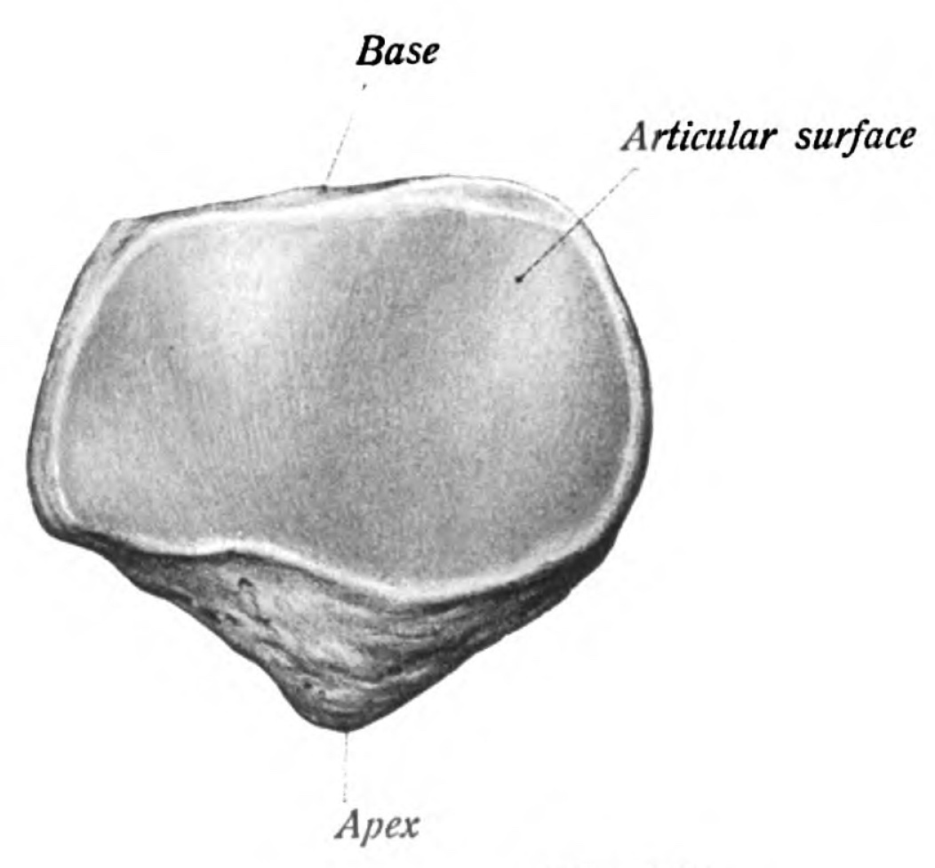
Figure 1. Bony anatomy of the patella, showing the base, articular surface, and apex. Sobotta, Atlas of Human Anatomy (1909), public domain, via Wikimedia Commons.
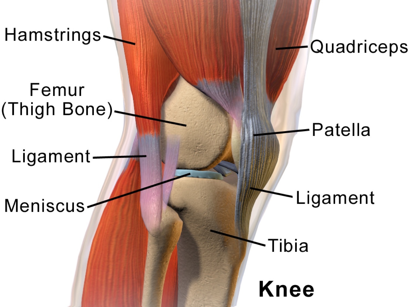
Figure 2. Sagittal overview of the knee extensor mechanism: the quadriceps, patella, and patellar ligament. Illustration by BruceBlaus, CC BY 3.0, via Wikimedia Commons.
The biomechanics explain the goals of fixation.[7] The patella acts as a fulcrum with two lever arms (the quadriceps and patellar tendons), displacing the extensor tendon away from the knee’s axis of rotation and thereby increasing the force of extension by as much as 50 %, supplying the additional ~60 % of torque needed to achieve the last 15° of terminal extension (twice as much torque is needed for that final arc as for the whole range from full flexion to 15°). The patellofemoral joint sees compressive forces of three to seven times body weight during stair climbing and squatting, the highest contact stress of any joint, so any fixation must tolerate enormous load. Removing the patella therefore markedly reduces extension power: a total patellectomy costs about 49 % of extensor strength and roughly 18° of motion, and there is a measurable biomechanical advantage to retaining a fragment of at least three-quarters of the patella, which is why partial is always preferred to total patellectomy.[8]
Part III - Assessment and Imaging
The displaced fracture presents with a hemarthrosis and a palpable gap, and the decisive test is the integrity of the extensor mechanism: ask the patient to perform a straight-leg raise or to extend the partially flexed knee against gravity.[9] A large painful hemarthrosis may prevent this, and aspiration followed by intra-articular local anaesthetic is a useful way to test extension. Two caveats matter: the absence of a large effusion despite a palpable defect suggests the haemarthrosis has decompressed through a torn retinaculum, and, critically, the ability to extend the knee does not rule out a patella fracture because the intact retinacula can maintain extension. A laceration over the patella mandates a saline load test (instil ~150 mL through an 18-gauge needle and watch for egress) to detect a traumatic arthrotomy, which is a surgical emergency.[10]
Imaging is AP, lateral, and axial (skyline) radiographs.[11] The lateral view (best taken at about 30° of flexion) defines the fracture pattern and the displacement and shows the patellar height: a high-riding patella (patella alta, above Blumensaat’s line) points to a patellar tendon rupture, and a low-riding patella (patella baja) to a quadriceps tendon rupture. The axial (Merchant) view (knee flexed 45°, beam angled 30° below horizontal) reveals vertical and osteochondral fractures that are easily missed on the AP. The bipartite patella is the classic mimic: a smooth, sclerotic-edged ossicle in the superolateral corner, usually bilateral, so contralateral films confirm it. CT is not routine for an isolated fracture and tends to over-estimate displacement (a caution against over-indicating surgery), while MRI is reserved for suspected tendon ruptures and the chondral injuries of patellar dislocation.[12]
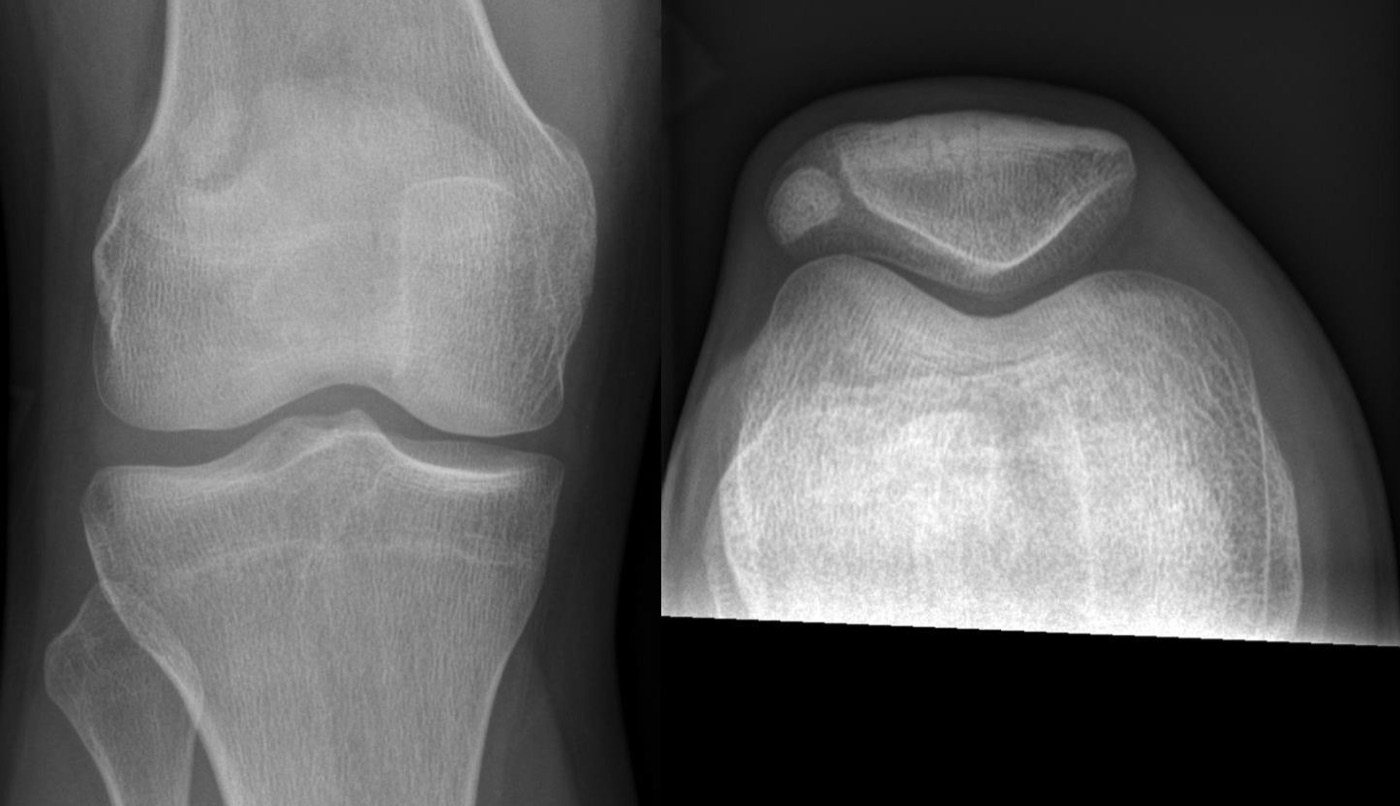
Figure 3. Bipartite patella: a superolateral accessory ossicle on AP and axial (skyline) views, a normal variant that mimics a fracture. Image by Hellerhoff, CC BY-SA 3.0, via Wikimedia Commons.
Part IV - Classification
Patella fractures are classified descriptively by pattern, displacement, and location.[13] The principal patterns are transverse (the commonest, from an indirect tensile mechanism, usually through the middle or lower third), comminuted or stellate (from a direct blow, often nondisplaced with intact retinacula), vertical/longitudinal (12-22 %, usually the lateral facet, the extensor mechanism preserved, easily missed without an axial view), polar (proximal or distal pole avulsions, distal-pole ones usually extra-articular and often with extensor disruption), and osteochondral (after a direct blow or patellar dislocation). A fracture is considered displaced when there is more than about 3 mm of fragment separation or more than 2 mm of articular step (Rockwood and Miller), while the AO uses a stricter threshold of more than 2 mm of gap or step as its surgical indication.[14] The AO/OTA system codes the patella as 34: 34A extra-articular (including avulsions), 34B partial articular in the sagittal plane (B1 lateral, B2 medial), and 34C complete articular in the coronal/frontal plane (C1 simple, C2 wedge, C3 multifragmentary, the stellate fracture); it is not validated but standardises research.[15]
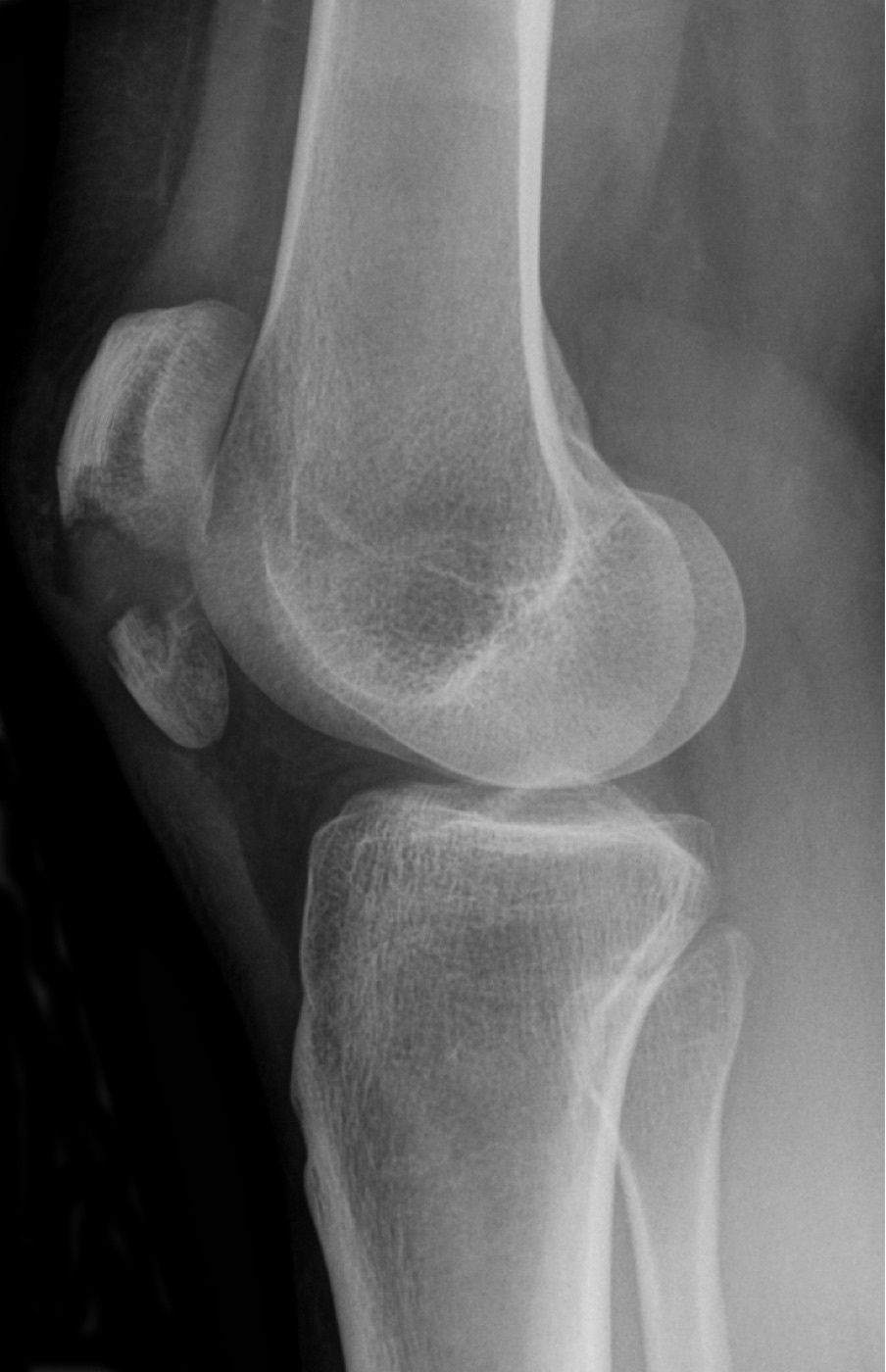
Figure 4. Displaced transverse patella fracture on the lateral radiograph, with a gap between the fragments. Image by Hellerhoff, CC BY-SA 3.0, via Wikimedia Commons.
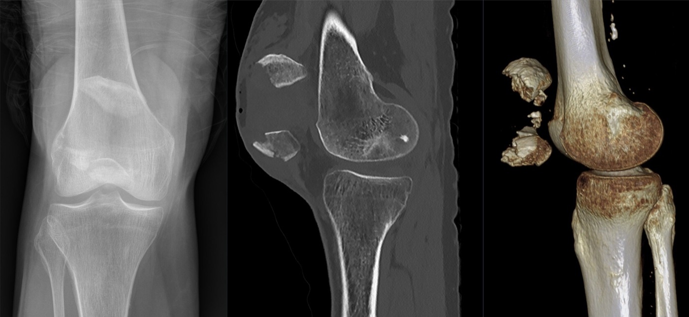
Figure 5. Comminuted patella fracture on AP radiograph, sagittal CT, and 3D volume rendering, with multiple displaced fragments. Image by Hellerhoff, CC BY-SA 4.0, via Wikimedia Commons.
Part V - Treatment of Patella Fractures
5.1 Nonoperative treatment
Nonoperative treatment is appropriate when the extensor mechanism is intact and articular incongruity is below the threshold (Rockwood uses under 3 mm, the AO a stricter 2 mm), and almost any pattern (transverse, stellate, or vertical) qualifies if these criteria are met.[16] The knee is held in extension in a brace or cylinder cast for 4-6 weeks, with early straight-leg raises and isometric quadriceps work and weight bearing as tolerated in extension; range of motion is begun once radiographs show consolidation. The results are excellent: Boström’s series of minimally displaced fractures achieved 98 % good-to-excellent outcomes with no relationship between sub-3-mm incongruity and later arthritis at nearly nine years.[17]
5.2 Operative fixation: tension-band wiring and its alternatives
Operative fixation is indicated by disruption of the extensor mechanism, an articular step beyond the threshold, an open fracture, or osteochondral loose bodies, and the workhorse is the modified anterior tension band.[18] Its principle is to place a wire on the anterior (tension) surface of the patella so that, as the knee flexes, the distracting tensile force of the quadriceps is converted into compression at the articular surface. The classic construct is a figure-of-eight wire over two parallel longitudinal K-wires placed about 5 mm deep to the anterior cortex, the K-wire ends bent 180° and buried to prevent migration. A cannulated-screw tension band (the wire passed through two cannulated lag screws) is biomechanically stronger and, clinically, halves the rate of symptomatic implants, and low-profile plates and braided sutures (in place of stainless-steel wire) are increasingly used to reduce the high rate of hardware irritation. Simple vertical (type B) splits need only lag screws, since the longitudinal integrity of the extensor mechanism is undisturbed and no tension band is required; comminuted (C3) patterns may add a circumferential cerclage or a mesh/basket plate.[19]
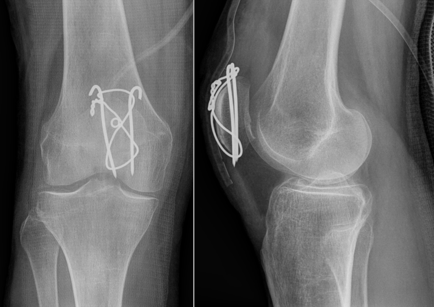
Figure 6. Tension-band osteosynthesis of a transverse patella fracture: two K-wires with an anterior figure-of-eight cerclage wire (AP and lateral). Image by Hellerhoff, CC BY-SA 3.0, via Wikimedia Commons.
5.3 Partial and total patellectomy
When a pole (usually the inferior) is too comminuted or too devascularised to reconstruct, a partial patellectomy is performed: the unsalvageable fragment is excised and the patellar tendon reattached close to the articular surface of the remaining patella through transosseous tunnels with Krackow sutures, taking care not to create patella baja.[20] As much patella as possible is retained, because outcomes deteriorate sharply once more than about 40 % is removed, and in selected cases partial patellectomy gives results equivalent to internal fixation. A total patellectomy is a salvage procedure of last resort, for irreparable comminution, failed fixation, or osteomyelitis, and is best avoided because it costs roughly half the extensor strength; when unavoidable, the redundant extensor tissue is imbricated so that tension is evident at 90° of flexion, and a vastus medialis obliquus advancement improves the result.[21]
Part VI - Extensor-Mechanism Tendon Ruptures
A patient who has lost active knee extension after trauma but has no patella fracture has ruptured a tendon, and the two ruptures separate cleanly by age and patellar height.[22] A quadriceps tendon rupture occurs in patients over 40, usually with a systemic predisposition (diabetes, rheumatoid arthritis, chronic renal failure and dialysis, gout, chronic steroid use or injection, and fluoroquinolones), avulsing from the superior pole; it presents with a suprapatellar gap, an extensor lag, and patella baja on the lateral film. A patellar tendon rupture occurs in patients under 40, typically athletes with pre-existing tendinopathy (“jumper’s knee”), avulsing from the inferior pole; it presents with an infrapatellar gap and patella alta. Patellar height differentiates them, and the standard Insall-Salvati ratio (patellar tendon length over patella length, normally about 1.0) is high (>1.2) with a patellar tendon rupture and low (<0.8) with a quadriceps tendon rupture.[23]
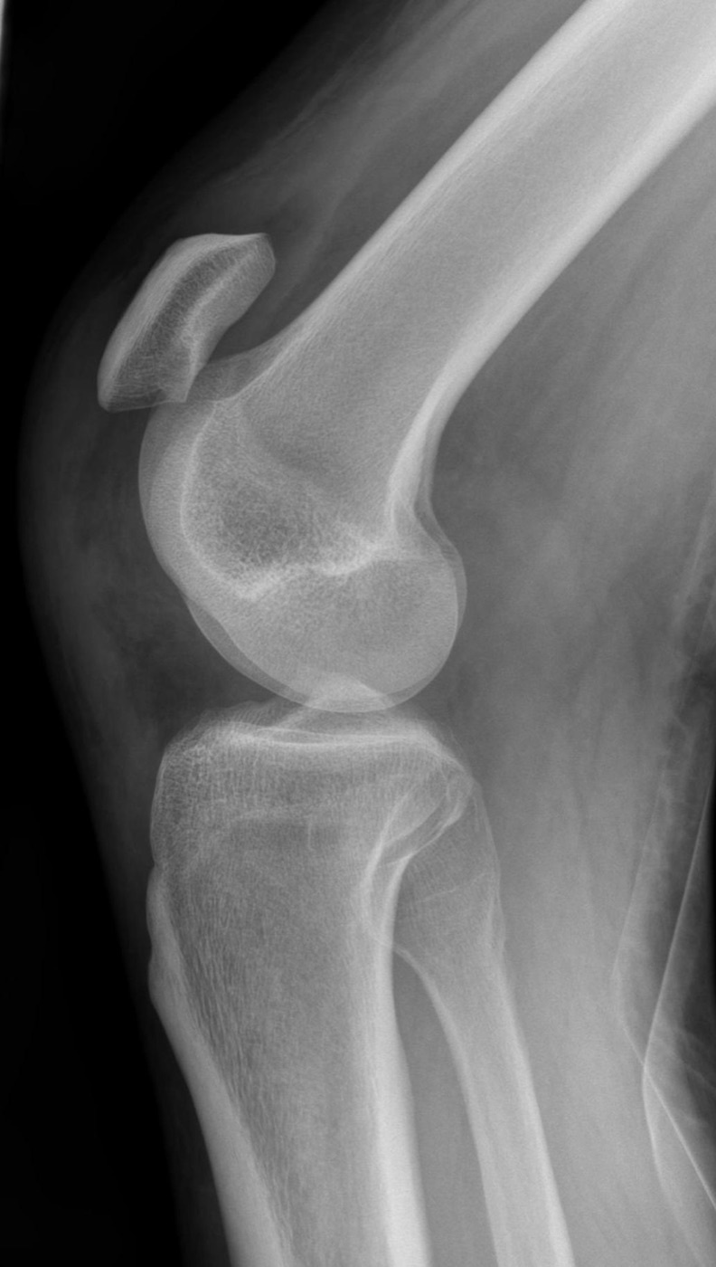
Figure 7. Patella alta from a patellar tendon rupture: the patella sits high above the trochlea on the lateral radiograph. Image by Hellerhoff, CC BY-SA 4.0, via Wikimedia Commons.
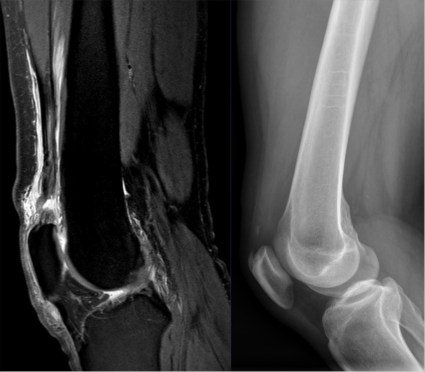
Figure 8. Traumatic quadriceps tendon rupture on sagittal PD-weighted MRI and the lateral radiograph. Image by Hellerhoff, CC BY-SA 4.0, via Wikimedia Commons.
Both ruptures are repaired, and the outcome turns on early surgery.[24] An acute rupture is repaired primarily by reattaching the tendon to its pole through transosseous drill holes (or suture anchors) using locking Krackow sutures, repairing the retinacula, and tying with the knee in extension; a patellar tendon repair is usually protected with a patellotibial cerclage or a hamstring augmentation, and its tension is set against the contralateral patellar height under fluoroscopy. The repair is then protected in a brace with controlled motion, and the single factor that predicts a worse result is a delay beyond 2-3 weeks, after which the muscle retracts (up to 5 cm in the quadriceps) and reconstruction or lengthening (a Codivilla V-Y for the quadriceps, a hamstring graft for the patellar tendon) becomes necessary. Acute repair gives 80-100 % good results for the quadriceps and 70-100 % for the patellar tendon.[25]
Part VII - Complications
The most common complication is symptomatic hardware, a direct consequence of the patella’s subcutaneous position: up to 40 % of operatively treated fractures need implant removal for soft-tissue irritation, and the overall reoperation rate approaches a third, which is why braided sutures are increasingly substituted for stainless-steel wire.[26] Loss of reduction occurs in 0-20 % (about 22 % in Miller), usually from technical error, comminution, or noncompliance, and increasing age predicts fixation failure. Nonunion is uncommon (1-12.5 %, under 5 % in Miller) and, in the young active patient, is revised with rigid fixation and bone graft. Osteonecrosis classically threatens the proximal pole because of its retrograde blood supply. Loss of knee motion and quadriceps weakness are the commonest reasons for a suboptimal result and relate more to the injury than to the surgery, so early motion is pursued, with manipulation considered if 90° of flexion is not regained by eight weeks. Post-traumatic patellofemoral arthritis is common in the long term (radiographic arthritis in 56 % of Nummi’s series and 70 % of Sorensen’s), and infection complicates up to 10.7 % of open fractures.[27]
Part VIII - Periprosthetic, Paediatric, and Patellar Dislocation
The periprosthetic patella fracture about a total knee replacement is the second most common periprosthetic fracture about the knee and is treated almost entirely nonoperatively.[28] It occurs in about 0.7 % of primary and 1.8 % of revision arthroplasties, usually spontaneously within the first two years, with risk factors of patellar resurfacing, excessive bone resection (leaving under 10-15 mm), devascularisation from a lateral retinacular release, maltracking, and osteopenia. The Ortiguera-Berry classification drives treatment: Type I (intact extensor mechanism, well-fixed implant) is managed nonoperatively; Type II (disrupted extensor mechanism) is the usual indication for operative repair, though even here nonoperative treatment is sometimes reasonable; and Type III (loose patellar component, subdivided IIIa with adequate and IIIb with poor bone stock) needs component removal or revision, or a patellectomy when bone stock is poor. The reason for the strongly nonoperative bias is that ORIF of these fractures carries an average nonunion rate around 92 %, so surgery is reserved for a disrupted extensor mechanism or a grossly loose component.[29]
In children, patella fractures are uncommon because the patella is largely cartilaginous and mobile, and the characteristic injury is the sleeve fracture.[30] Seen between 8 and 12 years, it is an avulsion of a cartilaginous sleeve, usually off the distal pole, by a forceful quadriceps contraction, and it is easily missed because the avulsed sleeve is mostly cartilage, so the lateral radiograph may show only patella alta and a tiny fleck of bone, with localised soft-tissue swelling as a useful clue; MRI is diagnostic when in doubt. Treatment mirrors the adult: a nondisplaced fracture with an intact extensor mechanism is cast in extension for 4-6 weeks, and a displaced one is fixed (an AO tension band with suture or wire). Because the patella is a sesamoid that grows by apposition, growth disturbance is uncommon, but a missed displaced sleeve fracture heals with patellar elongation, an extensor lag, and persistent patella alta.[31]
A brief word on the acute patellar (patellofemoral) dislocation, which shares this chapter.[32] The patella almost always dislocates laterally, tearing the medial patellofemoral ligament, and an osteochondral fracture of the medial patellar facet or lateral femoral condyle is highly suggestive of it; articular cartilage injury occurs in up to 95 % of first-time dislocators, so a high-risk knee (a large hemarthrosis) warrants MRI. First-time dislocations are usually treated nonoperatively (a brief period of immobilisation, then a stabilising brace and quadriceps rehabilitation), with recurrence in 15-44 %; a large displaced osteochondral fragment is fixed, and recurrent instability is treated with MPFL reconstruction, a tibial-tubercle osteotomy for malalignment, or a trochleoplasty for trochlear dysplasia.[33]
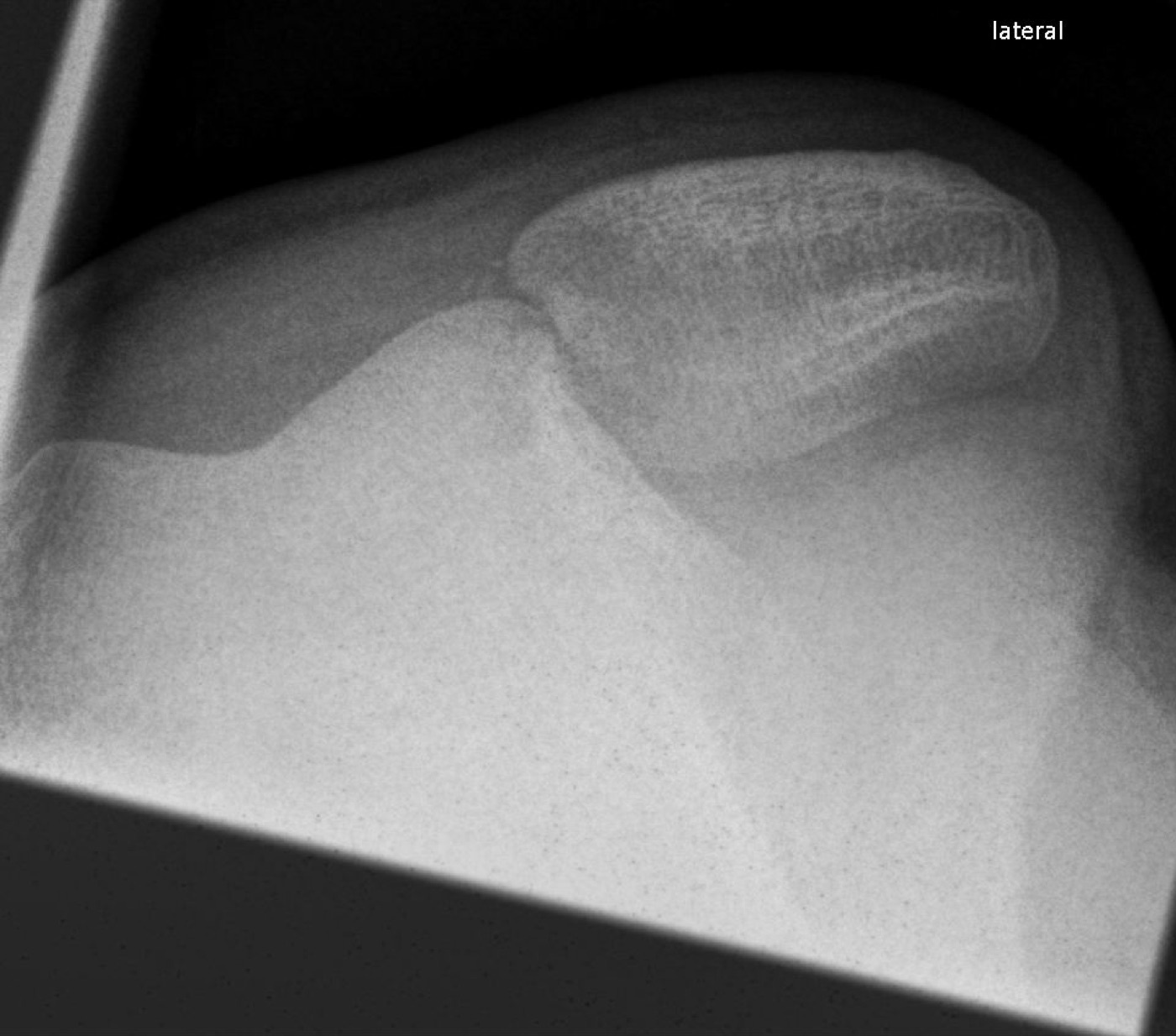
Figure 9. Acute lateral patellar dislocation on the axial (skyline) radiograph, the patella displaced off the trochlea. Image by Hellerhoff, CC BY-SA 3.0, via Wikimedia Commons.
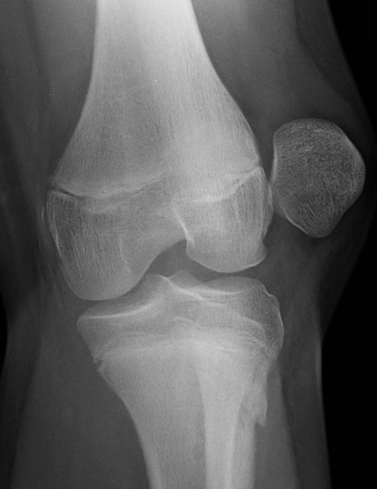
Figure 10. Acute lateral patellar dislocation on the AP radiograph, the patella lying lateral to the femoral condyle. Image by James Heilman, MD, CC BY-SA 4.0, via Wikimedia Commons.
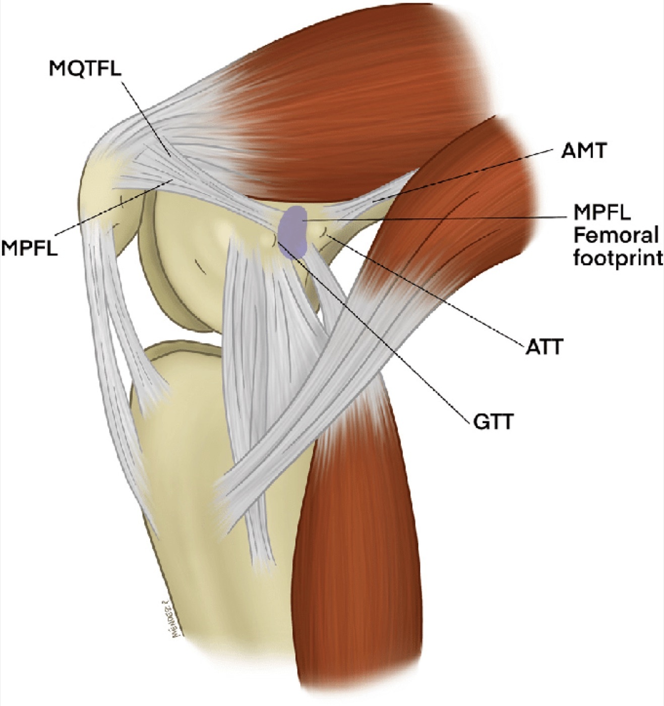
Figure 11. Medial view of a flexed knee showing the medial patellofemoral ligament (MPFL), the main restraint to lateral patellar displacement. Illustration by Negrin et al., CC BY 4.0, via Wikimedia Commons.
Part IX - A Synthesis: How to Reason About the Patella Fracture
Reason about the patella as the fulcrum of the extensor mechanism, and every decision follows. Establish first whether the extensor mechanism is intact by asking for a straight-leg raise, remembering that preserved extension does not exclude a fracture. If the mechanism is intact and the articular surface is congruent within a few millimetres, treat nonoperatively in extension and expect an excellent result. If the mechanism is disrupted or the joint is incongruent, restore the articular surface and re-establish extension with a tension band (a cannulated-screw construct is stronger and less symptomatic), reserving lag screws alone for the vertical split and a partial patellectomy for the unreconstructable pole, always keeping as much patella as possible and avoiding a total patellectomy. Recognise the tendon ruptures by age and patellar height. The over-40 patient with a suprapatellar gap and patella baja has a quadriceps rupture, the under-40 athlete with an infrapatellar gap and patella alta a patellar tendon rupture; repair them early, because delay is what spoils the outcome. Expect that symptomatic hardware will be the commonest complication, that stiffness and weakness track the injury more than the surgery, and that the periprosthetic patella fracture is, almost always, best left alone.
References
-
Rockwood & Green’s Fractures in Adults, pp.4110, 4111, 4118, 4120, 4121 (patella the largest sesamoid and fulcrum of the extensor mechanism; lever-arm increase of extension force up to 50% and the extra ~60% of torque for the last 15°; the three surgical goals; direct vs indirect mechanisms; the straight-leg-raise test and the retinacula as secondary extensors so that preserved extension does not exclude a fracture); AO Principles of Fracture Management, pp.873, 874 (patella the largest sesamoid acting as a fulcrum with two lever arms; preservation of active extension does not rule out a fracture if the auxiliary extensors are intact).
-
Rockwood & Green’s Fractures in Adults, pp.4110, 4111, 4118, 4120, 4121 (patella the largest sesamoid and fulcrum of the extensor mechanism; lever-arm increase of extension force up to 50% and the extra ~60% of torque for the last 15°; the three surgical goals; direct vs indirect mechanisms; the straight-leg-raise test and the retinacula as secondary extensors so that preserved extension does not exclude a fracture); AO Principles of Fracture Management, pp.873, 874 (patella the largest sesamoid acting as a fulcrum with two lever arms; preservation of active extension does not rule out a fracture if the auxiliary extensors are intact).
-
Rockwood & Green’s Fractures in Adults, p.4110 (about 1 % of skeletal fractures; subcutaneous vulnerability; incidence rising with age on a bimodal pattern; direct compression → comminuted/stellate with chondral damage and preserved retinacula, indirect tension → transverse or inferior-pole avulsion with retinacular disruption; combined forces and the role of age/bone quality/flexion); AO Principles of Fracture Management, p.873 (about 1 % of skeletal injuries; about half nondisplaced with an intact extensor mechanism; failure in compression → stellate vs failure in tension → transverse/inferior-pole avulsion).
-
Rockwood & Green’s Fractures in Adults, p.4110 (about 1 % of skeletal fractures; subcutaneous vulnerability; incidence rising with age on a bimodal pattern; direct compression → comminuted/stellate with chondral damage and preserved retinacula, indirect tension → transverse or inferior-pole avulsion with retinacular disruption; combined forces and the role of age/bone quality/flexion); AO Principles of Fracture Management, p.873 (about 1 % of skeletal injuries; about half nondisplaced with an intact extensor mechanism; failure in compression → stellate vs failure in tension → transverse/inferior-pole avulsion).
-
Rockwood & Green’s Fractures in Adults, pp.4118, 4119, 4120 (thickest articular cartilage in the body, the seven-facet architecture with the odd facet, the cartilage-free distal pole making distal-pole fractures extra-articular; six contributing arteries forming an anastomotic ring entering at the mid-anterior body and distal pole with retrograde distal-to-proximal perfusion and proximal-pole osteonecrosis risk; the extensor mechanism components and the retinacula as secondary extensors inserting on the proximal tibia; the MPFL contributing 50-60 % of medial restraint); AO Principles of Fracture Management, pp.874, 875 (largest sesamoid, two facets separated by a vertical ridge, thickest cartilage in the body, the extraosseous genicular arterial ring supplying the patella via mid-patellar and polar vessels).
-
Rockwood & Green’s Fractures in Adults, pp.4118, 4119, 4120 (thickest articular cartilage in the body, the seven-facet architecture with the odd facet, the cartilage-free distal pole making distal-pole fractures extra-articular; six contributing arteries forming an anastomotic ring entering at the mid-anterior body and distal pole with retrograde distal-to-proximal perfusion and proximal-pole osteonecrosis risk; the extensor mechanism components and the retinacula as secondary extensors inserting on the proximal tibia; the MPFL contributing 50-60 % of medial restraint); AO Principles of Fracture Management, pp.874, 875 (largest sesamoid, two facets separated by a vertical ridge, thickest cartilage in the body, the extraosseous genicular arterial ring supplying the patella via mid-patellar and polar vessels).
-
Rockwood & Green’s Fractures in Adults, pp.4121, 4137 (the patella as fulcrum with two lever arms increasing extension force up to 50% and supplying ~60% of the torque for the last 15°, twice the torque of the rest of the arc; patellofemoral compressive forces of 3-7× body weight; total patellectomy reducing extensor strength by 49% with ~18° motion loss, and the advantage of retaining ≥three-quarters of the patella per Albanese); AO Principles of Fracture Management, pp.873, 882 (the lever arm adding ~60% of the force for full extension so extension power is markedly reduced after patellectomy; partial patellectomy preferred to total, with poor outcomes if >40% of the patella is removed).
-
Rockwood & Green’s Fractures in Adults, pp.4121, 4137 (the patella as fulcrum with two lever arms increasing extension force up to 50% and supplying ~60% of the torque for the last 15°, twice the torque of the rest of the arc; patellofemoral compressive forces of 3-7× body weight; total patellectomy reducing extensor strength by 49% with ~18° motion loss, and the advantage of retaining ≥three-quarters of the patella per Albanese); AO Principles of Fracture Management, pp.873, 882 (the lever arm adding ~60% of the force for full extension so extension power is markedly reduced after patellectomy; partial patellectomy preferred to total, with poor outcomes if >40% of the patella is removed).
-
Rockwood & Green’s Fractures in Adults, pp.4111, 4112 (hemarthrosis and palpable gap; the straight-leg-raise / extension-against-gravity test as the key examination; aspiration plus local anaesthetic to enable it; the decompressed-effusion sign of a retinacular tear; preserved extension not excluding a fracture; the saline load test with ~150 mL through an 18-gauge needle for a traumatic arthrotomy); AO Principles of Fracture Management, p.873 (swelling, tenderness, loss of extensor function; preserved extension via intact retinacula not ruling out a fracture).
-
Rockwood & Green’s Fractures in Adults, pp.4111, 4112 (hemarthrosis and palpable gap; the straight-leg-raise / extension-against-gravity test as the key examination; aspiration plus local anaesthetic to enable it; the decompressed-effusion sign of a retinacular tear; preserved extension not excluding a fracture; the saline load test with ~150 mL through an 18-gauge needle for a traumatic arthrotomy); AO Principles of Fracture Management, p.873 (swelling, tenderness, loss of extensor function; preserved extension via intact retinacula not ruling out a fracture).
-
Rockwood & Green’s Fractures in Adults, pp.4112, 4113, 4114, 4115, 4116 (AP/lateral/axial radiographs; the lateral view defining the pattern and patellar height with alta vs baja; the Merchant axial view at 45° flexion and 30° beam angulation for vertical/osteochondral fractures; the superolateral sclerotic bipartite patella, usually bilateral, confirmed on contralateral films; CT over-estimating displacement and the over-indication caution; MRI for tendon and chondral injuries); AO Principles of Fracture Management, p.874 (lateral view at 30° flexion showing true displacement and patellar height; tangential view at 45°; abnormal patellar height indicating tendon rupture).
-
Rockwood & Green’s Fractures in Adults, pp.4112, 4113, 4114, 4115, 4116 (AP/lateral/axial radiographs; the lateral view defining the pattern and patellar height with alta vs baja; the Merchant axial view at 45° flexion and 30° beam angulation for vertical/osteochondral fractures; the superolateral sclerotic bipartite patella, usually bilateral, confirmed on contralateral films; CT over-estimating displacement and the over-indication caution; MRI for tendon and chondral injuries); AO Principles of Fracture Management, p.874 (lateral view at 30° flexion showing true displacement and patellar height; tangential view at 45°; abnormal patellar height indicating tendon rupture).
-
Rockwood & Green’s Fractures in Adults, pp.4115, 4116, 4117, 4118 (descriptive classification by pattern/displacement; transverse the commonest, vertical 12-22 % usually lateral facet and easily missed, comminuted/stellate often nondisplaced, polar fractures with distal pole usually extra-articular, osteochondral after a blow or dislocation; displaced = >3 mm separation or >2 mm step; the unvalidated OTA system); AO Principles of Fracture Management, pp.875, 876 (displaced = step-off or gap >2 mm as the surgical threshold; AO/OTA 34A extra-articular, 34B partial articular sagittal [B1 lateral, B2 medial], 34C complete articular coronal [C1 simple, C2 wedge, C3 multifragmentary]); Miller’s Review of Orthopaedics, p.923 (displaced = 3 mm separation or 2 mm step-off; vertical fractures rarely need surgery).
-
Rockwood & Green’s Fractures in Adults, pp.4115, 4116, 4117, 4118 (descriptive classification by pattern/displacement; transverse the commonest, vertical 12-22 % usually lateral facet and easily missed, comminuted/stellate often nondisplaced, polar fractures with distal pole usually extra-articular, osteochondral after a blow or dislocation; displaced = >3 mm separation or >2 mm step; the unvalidated OTA system); AO Principles of Fracture Management, pp.875, 876 (displaced = step-off or gap >2 mm as the surgical threshold; AO/OTA 34A extra-articular, 34B partial articular sagittal [B1 lateral, B2 medial], 34C complete articular coronal [C1 simple, C2 wedge, C3 multifragmentary]); Miller’s Review of Orthopaedics, p.923 (displaced = 3 mm separation or 2 mm step-off; vertical fractures rarely need surgery).
-
Rockwood & Green’s Fractures in Adults, pp.4115, 4116, 4117, 4118 (descriptive classification by pattern/displacement; transverse the commonest, vertical 12-22 % usually lateral facet and easily missed, comminuted/stellate often nondisplaced, polar fractures with distal pole usually extra-articular, osteochondral after a blow or dislocation; displaced = >3 mm separation or >2 mm step; the unvalidated OTA system); AO Principles of Fracture Management, pp.875, 876 (displaced = step-off or gap >2 mm as the surgical threshold; AO/OTA 34A extra-articular, 34B partial articular sagittal [B1 lateral, B2 medial], 34C complete articular coronal [C1 simple, C2 wedge, C3 multifragmentary]); Miller’s Review of Orthopaedics, p.923 (displaced = 3 mm separation or 2 mm step-off; vertical fractures rarely need surgery).
-
Rockwood & Green’s Fractures in Adults, p.4122 (nonoperative indication of an intact extensor mechanism and <3 mm incongruity, applicable to almost any pattern; 4-6 weeks of extension bracing or cylinder cast with early quadriceps work and weight bearing as tolerated; Boström’s 98 % good-to-excellent with no incongruity-arthritis relationship); AO Principles of Fracture Management, pp.875, 882 (operative threshold of >2 mm gap or step; nonoperative care in a hinged brace locked in extension with active motion from 1-2 weeks and resistance at 6 weeks).
-
Rockwood & Green’s Fractures in Adults, p.4122 (nonoperative indication of an intact extensor mechanism and <3 mm incongruity, applicable to almost any pattern; 4-6 weeks of extension bracing or cylinder cast with early quadriceps work and weight bearing as tolerated; Boström’s 98 % good-to-excellent with no incongruity-arthritis relationship); AO Principles of Fracture Management, pp.875, 882 (operative threshold of >2 mm gap or step; nonoperative care in a hinged brace locked in extension with active motion from 1-2 weeks and resistance at 6 weeks).
-
Rockwood & Green’s Fractures in Adults, pp.4123, 4124, 4125, 4127, 4128, 4129 (operative indications; the tension-band principle of converting quadriceps tension into articular compression with knee flexion; the modified anterior tension band over two K-wires placed 5 mm below the anterior cortex with ends bent 180° and buried; the cannulated-screw tension band biomechanically superior with a 50 % lower symptomatic-implant rate; low-profile plating and braided-suture alternatives; the 33.6 % overall reoperation rate); AO Principles of Fracture Management, pp.877, 878, 880 (figure-of-eight tension band around K-wires or through cannulated screws as the best method for transverse fractures; a 3.5-mm lag screw alone for vertical type B splits without a tension band; circumferential cerclage and plates for C3).
-
Rockwood & Green’s Fractures in Adults, pp.4123, 4124, 4125, 4127, 4128, 4129 (operative indications; the tension-band principle of converting quadriceps tension into articular compression with knee flexion; the modified anterior tension band over two K-wires placed 5 mm below the anterior cortex with ends bent 180° and buried; the cannulated-screw tension band biomechanically superior with a 50 % lower symptomatic-implant rate; low-profile plating and braided-suture alternatives; the 33.6 % overall reoperation rate); AO Principles of Fracture Management, pp.877, 878, 880 (figure-of-eight tension band around K-wires or through cannulated screws as the best method for transverse fractures; a 3.5-mm lag screw alone for vertical type B splits without a tension band; circumferential cerclage and plates for C3).
-
Rockwood & Green’s Fractures in Adults, pp.4133, 4134, 4136, 4137 (partial patellectomy for an unreconstructable or devascularised pole, reattaching the patellar tendon near the articular surface through three drill holes with Krackow sutures and avoiding patella baja, retaining as much patella as possible, with results equivalent to ORIF in selected cases; total patellectomy as a salvage procedure with imbrication so tension is evident at 90° flexion and VMO advancement improving outcome); AO Principles of Fracture Management, p.882 (partial preferred to total patellectomy, poor outcomes if >40 % is removed, the patellar tendon reattached near the anterior aspect; total patellectomy compromising motion and strength).
-
Rockwood & Green’s Fractures in Adults, pp.4133, 4134, 4136, 4137 (partial patellectomy for an unreconstructable or devascularised pole, reattaching the patellar tendon near the articular surface through three drill holes with Krackow sutures and avoiding patella baja, retaining as much patella as possible, with results equivalent to ORIF in selected cases; total patellectomy as a salvage procedure with imbrication so tension is evident at 90° flexion and VMO advancement improving outcome); AO Principles of Fracture Management, p.882 (partial preferred to total patellectomy, poor outcomes if >40 % is removed, the patellar tendon reattached near the anterior aspect; total patellectomy compromising motion and strength).
-
Rockwood & Green’s Fractures in Adults, pp.4143, 4144, 4146, 4147 (quadriceps rupture in patients over 40 with systemic disease, avulsing from the superior pole with a suprapatellar gap and patella baja; patellar tendon rupture under 40 in athletes with tendinopathy, avulsing from the inferior pole with an infrapatellar gap and patella alta; patellar height and Blumensaat’s line differentiating them); Miller’s Review of Orthopaedics, pp.923, 924, 925 (the under-40 vs over-40 split; risk factors including renal failure, rheumatoid arthritis, steroids, diabetes; high-riding patella as the missed-diagnosis clue). The numeric Insall-Salvati cut-offs (normal ~1.0, alta >1.2, baja <0.8) are standard teaching; the source texts differentiate alta from baja by position relative to Blumensaat’s line and the contralateral knee rather than by the numeric ratio.
-
Rockwood & Green’s Fractures in Adults, pp.4143, 4144, 4146, 4147 (quadriceps rupture in patients over 40 with systemic disease, avulsing from the superior pole with a suprapatellar gap and patella baja; patellar tendon rupture under 40 in athletes with tendinopathy, avulsing from the inferior pole with an infrapatellar gap and patella alta; patellar height and Blumensaat’s line differentiating them); Miller’s Review of Orthopaedics, pp.923, 924, 925 (the under-40 vs over-40 split; risk factors including renal failure, rheumatoid arthritis, steroids, diabetes; high-riding patella as the missed-diagnosis clue). The numeric Insall-Salvati cut-offs (normal ~1.0, alta >1.2, baja <0.8) are standard teaching; the source texts differentiate alta from baja by position relative to Blumensaat’s line and the contralateral knee rather than by the numeric ratio.
-
Rockwood & Green’s Fractures in Adults, pp.4146, 4148, 4149, 4151, 4152 (acute primary repair through transosseous tunnels with Krackow sutures, retinacular repair, tying in extension; patellotibial cerclage or hamstring augmentation for the patellar tendon with intraoperative patellar-height check; delay beyond 2-3 weeks the only factor predicting a worse outcome, with up to 5 cm of quadriceps retraction and Codivilla lengthening or graft reconstruction for chronic cases; acute repair 80-100 % good for the quadriceps and 70-100 % for the patellar tendon); Miller’s Review of Orthopaedics, pp.924, 926 (repair through drill holes or suture anchors, repaired acutely; suture anchors with less gap formation and higher strength than transosseous suture in cadaveric testing; chronic cases needing lengthening or allograft).
-
Rockwood & Green’s Fractures in Adults, pp.4146, 4148, 4149, 4151, 4152 (acute primary repair through transosseous tunnels with Krackow sutures, retinacular repair, tying in extension; patellotibial cerclage or hamstring augmentation for the patellar tendon with intraoperative patellar-height check; delay beyond 2-3 weeks the only factor predicting a worse outcome, with up to 5 cm of quadriceps retraction and Codivilla lengthening or graft reconstruction for chronic cases; acute repair 80-100 % good for the quadriceps and 70-100 % for the patellar tendon); Miller’s Review of Orthopaedics, pp.924, 926 (repair through drill holes or suture anchors, repaired acutely; suture anchors with less gap formation and higher strength than transosseous suture in cadaveric testing; chronic cases needing lengthening or allograft).
-
Rockwood & Green’s Fractures in Adults, pp.4156, 4157, 4158 (symptomatic hardware the most common complication, removal in up to 40 %, with braided sutures reducing it; loss of reduction 0-20 %; nonunion 1-12.5 % revised with rigid fixation and graft; loss of motion and quadriceps weakness the commonest reasons for a poor result with manipulation if 90° is not regained by 8 weeks; post-traumatic arthritis in 56 % [Nummi] and 70 % [Sorensen]; infection up to 10.7 % of open fractures); Miller’s Review of Orthopaedics, p.923 (symptomatic hardware very common, loss of reduction 22 %, nonunion <5 %, age predicting fixation failure, K-wire migration prevention). Osteonecrosis of the proximal pole from the retrograde blood supply is standard teaching consistent with the source’s account of the patellar vascular pattern, though the extracts do not discuss patellar-fracture osteonecrosis explicitly.
-
Rockwood & Green’s Fractures in Adults, pp.4156, 4157, 4158 (symptomatic hardware the most common complication, removal in up to 40 %, with braided sutures reducing it; loss of reduction 0-20 %; nonunion 1-12.5 % revised with rigid fixation and graft; loss of motion and quadriceps weakness the commonest reasons for a poor result with manipulation if 90° is not regained by 8 weeks; post-traumatic arthritis in 56 % [Nummi] and 70 % [Sorensen]; infection up to 10.7 % of open fractures); Miller’s Review of Orthopaedics, p.923 (symptomatic hardware very common, loss of reduction 22 %, nonunion <5 %, age predicting fixation failure, K-wire migration prevention). Osteonecrosis of the proximal pole from the retrograde blood supply is standard teaching consistent with the source’s account of the patellar vascular pattern, though the extracts do not discuss patellar-fracture osteonecrosis explicitly.
-
Rockwood & Green’s Fractures in Adults, pp.4078, 4079, 4080, 4081 (the second most common periprosthetic fracture about the knee; 0.7 % primary and 1.8 % revision, usually spontaneous within two years; risk factors of resurfacing, excessive resection, lateral-release devascularisation, maltracking, osteopenia; the Ortiguera-Berry I/II/IIIa/IIIb classification by extensor-mechanism integrity, implant fixation, and bone stock; the ~92 % nonunion rate of ORIF driving a strongly nonoperative approach reserved for a disrupted extensor mechanism or a loose component).
-
Rockwood & Green’s Fractures in Adults, pp.4078, 4079, 4080, 4081 (the second most common periprosthetic fracture about the knee; 0.7 % primary and 1.8 % revision, usually spontaneous within two years; risk factors of resurfacing, excessive resection, lateral-release devascularisation, maltracking, osteopenia; the Ortiguera-Berry I/II/IIIa/IIIb classification by extensor-mechanism integrity, implant fixation, and bone stock; the ~92 % nonunion rate of ORIF driving a strongly nonoperative approach reserved for a disrupted extensor mechanism or a loose component).
-
Lovell & Winter’s Pediatric Orthopaedics, pp.5709, 5710, 5712, 5713 (patella fractures uncommon in children because the patella is largely cartilaginous and mobile; the sleeve fracture at 8-12 years, an avulsion of the distal cartilaginous sleeve, easily missed with only patella alta and a small bony fragment on the lateral film, soft-tissue swelling as a clue, MRI diagnostic; treatment as in adults - nondisplaced cast in extension 4-6 weeks, displaced ORIF with an AO tension band of suture or wire; growth disturbance uncommon as the patella grows by apposition). That a missed displaced sleeve fracture heals with patellar elongation, extensor lag, and persistent alta is standard teaching, the source noting only that growth disturbance is uncommon.
-
Lovell & Winter’s Pediatric Orthopaedics, pp.5709, 5710, 5712, 5713 (patella fractures uncommon in children because the patella is largely cartilaginous and mobile; the sleeve fracture at 8-12 years, an avulsion of the distal cartilaginous sleeve, easily missed with only patella alta and a small bony fragment on the lateral film, soft-tissue swelling as a clue, MRI diagnostic; treatment as in adults - nondisplaced cast in extension 4-6 weeks, displaced ORIF with an AO tension band of suture or wire; growth disturbance uncommon as the patella grows by apposition). That a missed displaced sleeve fracture heals with patellar elongation, extensor lag, and persistent alta is standard teaching, the source noting only that growth disturbance is uncommon.
-
Rockwood & Green’s Fractures in Adults, p.4142 (lateral dislocation the rule, MPFL injury, an osteochondral fracture of the medial patella highly suggestive, cartilage injury in up to 95 % of first-time dislocators, MRI for the high-risk knee, nonoperative treatment with recurrence of 15-44 %, fixation of a large osteochondral fragment). MPFL reconstruction, tibial-tubercle osteotomy, and trochleoplasty for recurrent instability are standard teaching; the extract addresses the acute first-time dislocation, its account of operative comparison being truncated.
-
Rockwood & Green’s Fractures in Adults, p.4142 (lateral dislocation the rule, MPFL injury, an osteochondral fracture of the medial patella highly suggestive, cartilage injury in up to 95 % of first-time dislocators, MRI for the high-risk knee, nonoperative treatment with recurrence of 15-44 %, fixation of a large osteochondral fragment). MPFL reconstruction, tibial-tubercle osteotomy, and trochleoplasty for recurrent instability are standard teaching; the extract addresses the acute first-time dislocation, its account of operative comparison being truncated.
-
Rockwood & Green’s Fractures in Adults, pp.4118, 4121; AO Principles of Fracture Management, p.873.
-
Rockwood & Green’s Fractures in Adults, p.4110; AO Principles of Fracture Management, p.873.
-
Rockwood & Green’s Fractures in Adults, p.4111; AO Principles of Fracture Management, p.873.
-
Rockwood & Green’s Fractures in Adults, pp.4112, 4113, 4114; AO Principles of Fracture Management, p.874.
-
Rockwood & Green’s Fractures in Adults, p.4122; AO Principles of Fracture Management, p.875.
-
Rockwood & Green’s Fractures in Adults, pp.4124, 4125, 4128; AO Principles of Fracture Management, pp.879, 880.
-
Rockwood & Green’s Fractures in Adults, pp.4133, 4137; AO Principles of Fracture Management, p.882.
-
Rockwood & Green’s Fractures in Adults, pp.4143, 4144; Miller’s Review of Orthopaedics, pp.923, 924.
-
Rockwood & Green’s Fractures in Adults, pp.4146, 4148, 4151, 4152; Miller’s Review of Orthopaedics, pp.924, 926.
-
Rockwood & Green’s Fractures in Adults, pp.4156, 4157; Miller’s Review of Orthopaedics, p.923.
-
Rockwood & Green’s Fractures in Adults, pp.4078, 4079, 4081.
-
Lovell & Winter’s Pediatric Orthopaedics, pp.5709, 5710, 5713.