Contents
- Part I - Delayed Union and Non-union: Definitions
- Part II - Why Fractures Fail to Unite
- Part III - Classification of Non-union
- Part IV - Diagnosis and Treatment of Non-union
- Part V - Malunion
- Part VI - Hardware Failure (Débricolage)
- Part VII - Volkmann Ischaemic Contracture
- Part VIII - Complex Regional Pain Syndrome (Sudeck’s Atrophy)
- Part IX - Periprosthetic and Peri-implant Fractures
- References
Part I - Delayed Union and Non-union: Definitions
About 90-95% of fractures heal without difficulty; the complications of this topic concern the small remainder in which “the biological process of fracture repair cannot overcome the local biology and mechanics of the bony injury.”[1] Two terms must be distinguished. A delayed union “has not completely healed in the time expected, but still has the potential to heal without further intervention,” whereas a non-union “has failed to heal… and is not likely to heal without new intervention.” At the cellular level, the reparative process has stopped.[2]
Both definitions are admittedly subjective, with high interobserver variability, and any fixed timeframe is arbitrary.[3] The most-quoted formal criterion is the FDA definition: a fracture at least 9 months old that has shown no signs of healing for 3 consecutive months; Müller’s older definition was failure of a tibia to unite by 8 months.[4] Both are flawed, since a tibia with a 10 cm segmental defect is a non-union from the outset and needs no months of observation, so the practical definition is the treating surgeon’s judgement that the fracture has “zero possibility of healing without further intervention.”[5] The overall non-union rate is about 4.9% (combined impaired healing ~6.9%), rising toward 20% in open fractures with extensive soft-tissue injury, and the quality-of-life burden of a long-bone non-union is severe (a mean utility score below that of type 1 diabetes or stroke).[6]
Part II - Why Fractures Fail to Unite
Healing requires three things, mechanical stability, an adequate blood supply, and bone-to-bone contact, and the loss of any one predisposes to non-union.[7] The unifying principle, used throughout this topic, is that every fracture has both a biological and a mechanical problem: a hypertrophic non-union is a failure of mechanics, an atrophic non-union a failure of biology.[8] The predisposing factors group accordingly:[9]
- Mechanical instability. Inadequate or inappropriate fixation, distraction (“hardware is as capable of holding bone apart as holding it together”), bone loss, poor bone quality.
- Inadequate vascularity. High-energy injury, excessive soft-tissue stripping, vascular injury, and the watershed sites with poor blood supply (the scaphoid waist, femoral neck, talar neck, and the proximal fifth metatarsal), and the relatively avascular tibial diaphysis.
- Poor contact. Soft-tissue interposition, malposition, bone loss, distraction.
Host and treatment factors add to these: smoking (surgeons rank it the single most important; it reduces periosteal BMP expression and makes union about 37% less likely in open tibiae), diabetes, NSAIDs (which blunt the COX-2/prostaglandin inflammatory phase; meta-analysis odds ratio ~3), corticosteroids, malnutrition, and vitamin D deficiency (found in two-thirds of non-union patients, sometimes correctable without surgery).[10] Infection both promotes non-union (loosening implants, osteolysis, sequestra) and complicates its repair.[11]
Part III - Classification of Non-union
The standard scheme is the Weber-Cech classification, which divides non-unions by their radiographic healing effort and vascularity into viable and non-viable types:[12]
- Hypertrophic (viable, hypervascular) non-union. Abundant callus but a persistent un-mineralised cartilage gap; the biology is good and the problem is inadequate stability. Its subtypes are the “elephant-foot” (abundant callus) and “horse-hoof” (less callus) patterns. It is treated by providing stability alone; debridement and bone graft are neither needed nor desired, and it usually unites within about 8 weeks of rigid fixation.[13]
- Oligotrophic (viable) non-union. Viable but with little or no callus, usually from inadequate reduction with fragment displacement; treated by restoring contact and compression (with grafting if surfaces are poor).[14]
- Atrophic (non-viable, avascular) non-union. The most advanced non-viable type, with absent vascularity and a fibrous gap and no osteogenic capacity; treated by restoring the biology, debriding the inert ends to bleeding bone and adding an osteogenic/osteoinductive graft.[15]
Two further categories sit alongside these. A synovial pseudarthrosis (nearthrosis) is a “false joint,” a fluid-filled cavity lined by a synovium-like pseudocapsule with the medullary canals sealed off, betrayed by a “cold cleft” between two hot bone ends on the technetium bone scan; it is treated by resecting the synovium, reopening the canals, grafting and compressing.[16] An infected non-union is classified by its activity (actively draining, active non-draining, or quiescent) and is the most difficult of all to treat.[17]
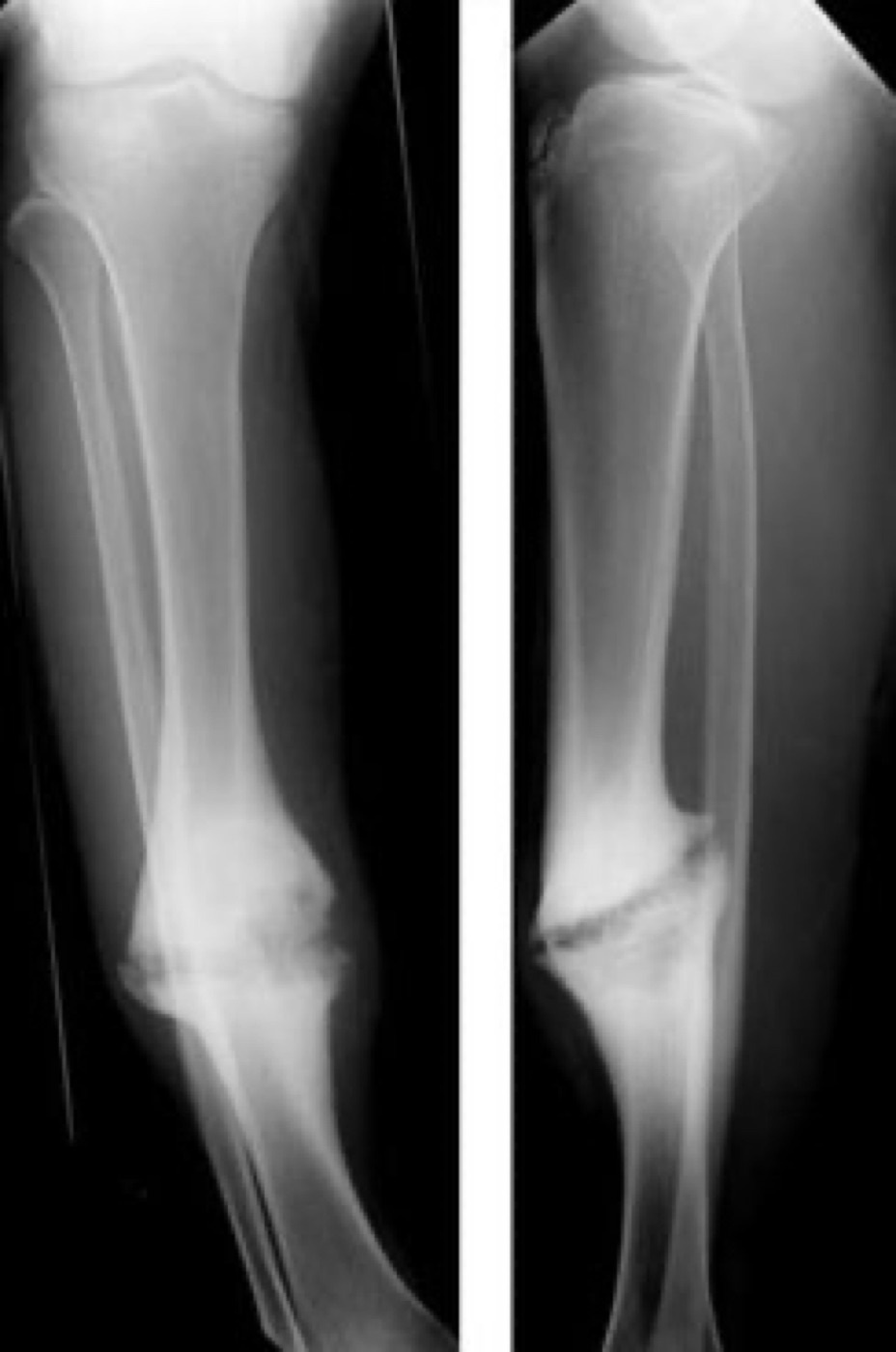
Figure 1. Hypertrophic (“elephant-foot”) non-union of the tibia: abundant callus with a persistent fracture line, a failure of stability rather than biology. Source: Lindsaydavidson, via Wikimedia Commons, CC BY 3.0.
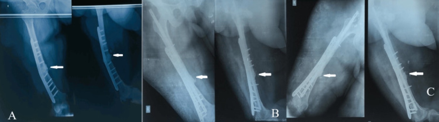
Figure 2. Atrophic non-union of the femur with implant breakage (A), revised with an exchange nail, plate and graft (B) and consolidated at three months (C). From Walia et al. (2024), Cureus, PMC11377906, CC BY 4.0.
Part IV - Diagnosis and Treatment of Non-union
Diagnosis: always exclude infection
A non-union is diagnosed clinically (pain and motion at the fracture site, where gross motion is unequivocal) and radiographically (failure to bridge, with loosened or broken implants as indirect signs: “the race between union and hardware failure has been lost”).[18] The single imperative of the work-up is to exclude infection: inflammatory markers (ESR, CRP, white count) and, where suspicion is real, image-guided aspiration or operative deep-tissue and bone cultures (the gold standard), with antibiotics stopped at least two weeks beforehand.[19] Even apparently aseptic non-unions yield “surprise positive cultures” in around 20%.[20] CT confirms the non-union and quantifies bridging (under 5% of the cross-section in a non-union, over 25% in a healing fracture).[21]
Treatment: correct the deficient element
The governing rule is to identify and correct whichever element is deficient: provide stability for the hypertrophic non-union, restore biology for the atrophic, and do both for the oligotrophic; eradicate any infection first; and correct malalignment.[22] The options are:
- Non-operative. Functional bracing and weight-bearing (Sarmiento’s tibial series healed over 90%), and the bone-stimulation modalities (low-intensity pulsed ultrasound, LIPUS, ~30 mW/cm²; pulsed electromagnetic fields; capacitive coupling), whose evidence is best for hypertrophic non-unions with acceptable alignment and no pseudarthrosis.[23]
- Operative. Exchange nailing (a larger reamed nail that deposits autograft and stimulates healing, best for diaphyseal non-unions without bone loss or malalignment), compression plating, dynamisation, decortication, and bone grafting.[24]
- Bone graft and adjuncts. Autogenous iliac-crest graft remains the gold standard (osteogenic, osteoinductive and osteoconductive; union over 90% in long-bone non-unions, but limited to defects of about 5-7 cm), with the reamer-irrigator-aspirator (RIA) an alternative high-volume source; allograft and ceramics are osteoconductive extenders only; and BMP-7 (OP-1) and BMP-2 are recombinant osteoinductive options.[25] These map onto the components of the “diamond concept” of healing: osteogenic cells, osteoinductive signals, an osteoconductive scaffold, mechanical stability and vascularity.[26]
- Infected non-union. Staged: radical debridement, antibiotic cement beads or an antibiotic-coated nail for dead-space and stability, organism-specific antibiotics, and definitive reconstruction once quiescent.[27]
- Segmental bone defect. The Masquelet induced-membrane technique (a PMMA spacer induces an osteogenic membrane at 4-6 weeks, then is replaced by cancellous graft) and Ilizarov distraction osteogenesis / bone transport (mono-, bi- or trifocal), or a vascularised fibular graft.[28]
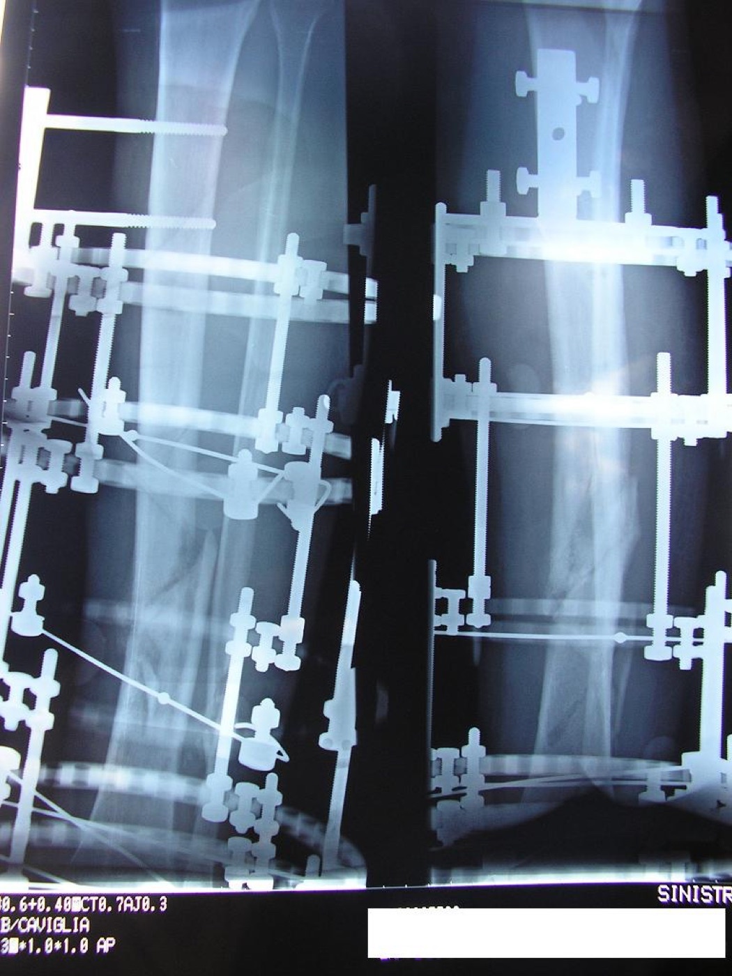
Figure 3. A tibia and fibula in an Ilizarov ring fixator, with callus forming three months on, the workhorse for non-union and bone transport. Source: Viapastrengo, via Wikimedia Commons, CC BY-SA 3.0.
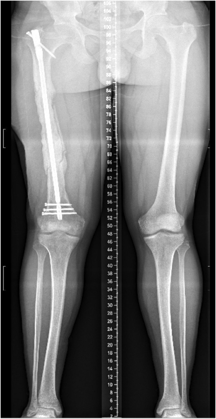
Figure 4. The Masquelet induced-membrane technique: a PMMA cement spacer bridging a large femoral segmental defect (stage 1), later replaced by cancellous graft within the induced membrane. From Kubes et al. (2021), Int J Surg Case Rep, PMC8219760, CC BY 4.0.
Part V - Malunion
A malunion is a fracture that has healed in a non-anatomical position, characterised by abnormalities of length (shortening or overdistraction), angulation, rotation and translation, defined by their location, magnitude and direction.[29] A simple bedside test separates it from a non-union: manual stress of a true malunion is painless, and pain on stressing should raise the suspicion of an ununited fracture.[30] Not every deformity needs correction, only those that impair function, since small translations or sagittal angulations are often well tolerated, and the functional thresholds for shortening are useful (up to 3-4 cm tolerated in the upper limb; up to 2 cm in the leg managed with a shoe-raise, with restoration of length usually warranted beyond 4 cm).[31] A malunion drives symptoms through adjacent-joint overload and the post-traumatic osteoarthritis of altered load transfer (covered in Topic 7).
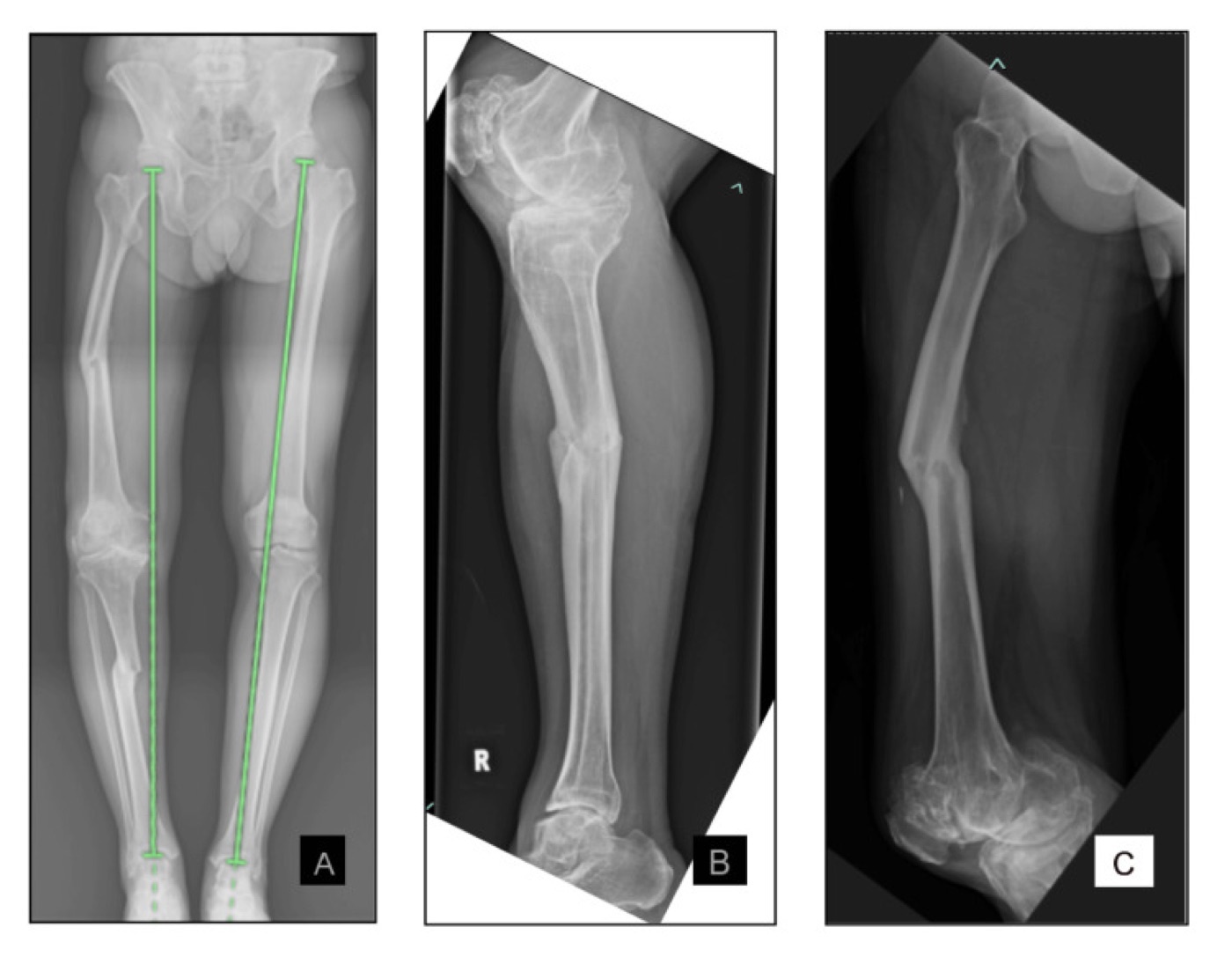
Figure 5. Malunion of a “floating-knee” injury: a standing scanogram with mechanical-axis lines (A) and the malunited tibia (B) and femur (C) with multiplanar angular deformity. From Matheen et al. (2024), Cureus, PMC11446639, CC BY 4.0.
The radiographic analysis rests on whole-limb, weight-bearing alignment films and a small set of concepts:[32]
- The mechanical axis deviation (MAD), the distance from the knee centre to the hip-to-ankle line (normally 1-15 mm medial; greater is varus, any lateral deviation is valgus).
- The CORA (centre of rotation of angulation), the intersection of the proximal and distal axes of the bone, the point about which the deformity is corrected; the angle there equals the angular deformity.
- The bisector rule: correcting the angulation about a point on the line bisecting the CORA angle realigns the axes without introducing an iatrogenic translation.
A clinically vital principle is the compensatory adjacent-joint deformity: a periarticular malunion may be balanced by a fixed deformity at the neighbouring joint, and “correction of the malunion without addressing a compensatory joint deformity results in a straight bone with a maloriented joint.”[33] Treatment is by corrective osteotomy (opening-, closing- or neutral-wedge, a wedge through the CORA for diaphyseal deformities, dome, transverse or oblique/derotation cuts), fixed acutely (plate, intramedullary nail) or corrected gradually with an Ilizarov frame, a hexapod (Taylor Spatial Frame) or a lengthening nail when there is shortening or when the neurovascular structures on the concave side would not tolerate acute correction.[34]
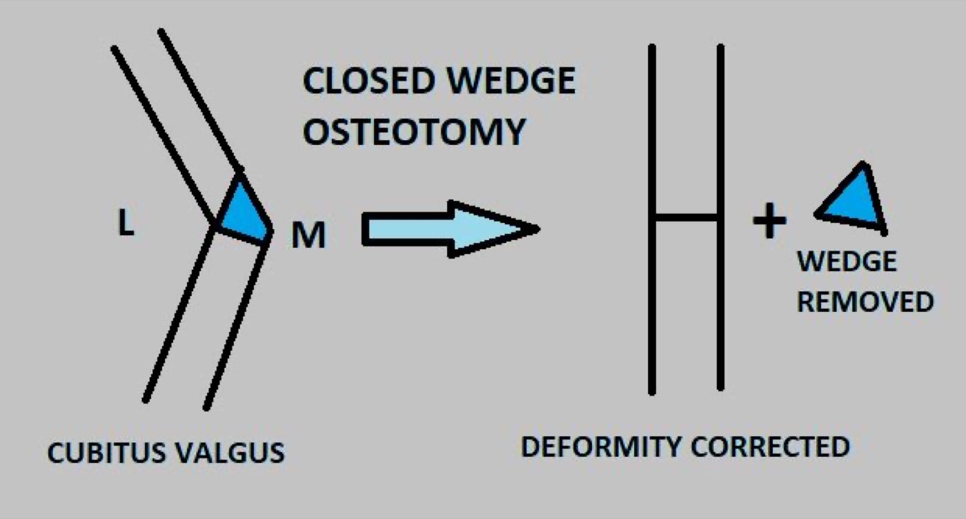
Figure 6. A closing-wedge corrective osteotomy: a bone wedge is removed to realign the deformity (here a cubitus valgus). Source: Dr. Vijayachandar, via Wikimedia Commons, CC BY-SA 4.0.
Part VI - Hardware Failure (Débricolage)
Implant failure, what the older European literature calls “débricolage” (“coming unstuck”), is the mechanical counterpart of non-union, and the two are intertwined: every fixation is a race between fracture union and implant fatigue.[35] An implant carrying the whole load fails by fatigue after a finite number of cycles if union is delayed (the biomechanics are detailed in Topic 4): plates break, screws loosen or back out, and nails or interlocking screws fracture. The chief culprits are stress risers (an empty screw hole over the fracture, the end of a plate, a cortical perforation, or the junction between two implants) together with the failure of construct stiffness to match the biology: “although stiff constructs can prevent failure of fixation, excessive stiffness is also associated with poor rates of fracture union.”[36] Management of a failed construct is to diagnose any infection, then revise the fixation (often with a longer, load-sharing construct or a change from plate to nail), add bone graft to restore biology, and span the stress riser, for example by overlapping a plate beyond an adjacent stem to prevent the next peri-implant fracture.[37]
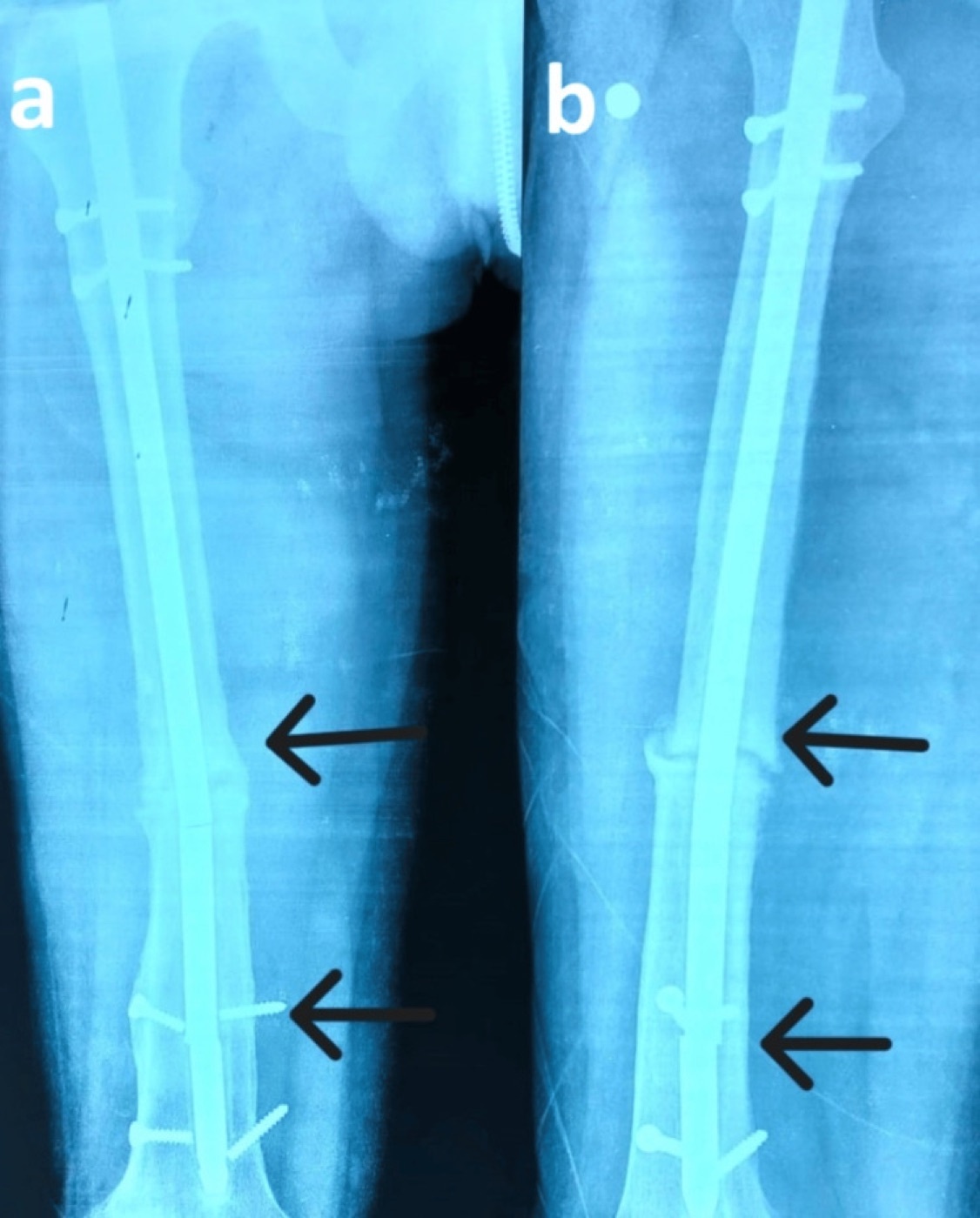
Figure 7. Hardware failure (“débricolage”): a femoral intramedullary nail fractured at two points over a non-union. From Durrani et al. (2025), Cureus, PMC12461672, CC BY 4.0.
Part VII - Volkmann Ischaemic Contracture
Volkmann ischaemic contracture is the established sequel of an untreated acute compartment syndrome (or an arterial injury), in which infarcted muscle is replaced by inelastic fibrous tissue that contracts into a fixed deformity.[38] The prevention is everything: an acute compartment syndrome is recognised by pain out of proportion and pain on passive stretch (the “5 Ps” are late and unreliable), and the threshold for emergency fasciotomy is a differential pressure (ΔP = diastolic blood pressure - compartment pressure) below 30 mmHg; muscle tolerates only a few hours of ischaemia before the damage becomes irreversible.[39]
The established contracture classically affects the deep flexor compartment of the forearm (flexor digitorum profundus and flexor pollicis longus), producing the clawed hand (wrist flexion, finger flexion, forearm pronation and thumb adduction), with the deformity worsening as the wrist is extended (a tenodesis effect), and frequently a combined median (more than ulnar) nerve deficit. It is graded by severity (the Tsuge classification: mild, affecting part of the deep flexors; moderate, most flexors with some nerve involvement; severe, all flexors and extensors with marked nerve involvement). Treatment is by severity: splinting and therapy for mild cases, a flexor-pronator muscle slide (Max Page) with tendon transfers for moderate cases, and excision of infarcted muscle with neurolysis and a free functioning muscle transfer (gracilis) for severe cases.
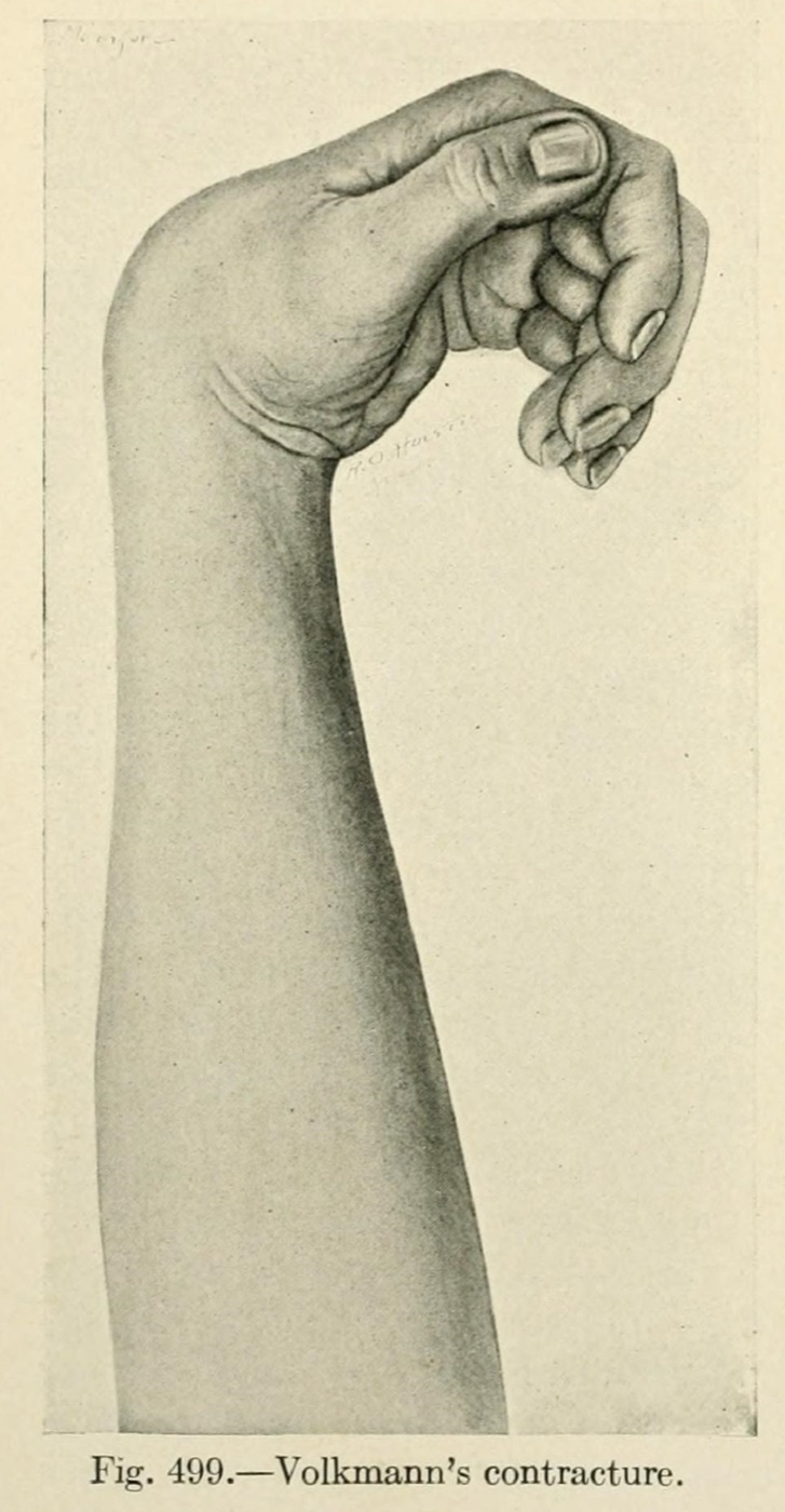
Figure 8. Volkmann ischaemic contracture of the forearm and hand: the flexed wrist and clawed fingers of established flexor-compartment infarction. From J. G. Mumford, The Practice of Surgery (1910), public domain, via Internet Archive / Wikimedia Commons.
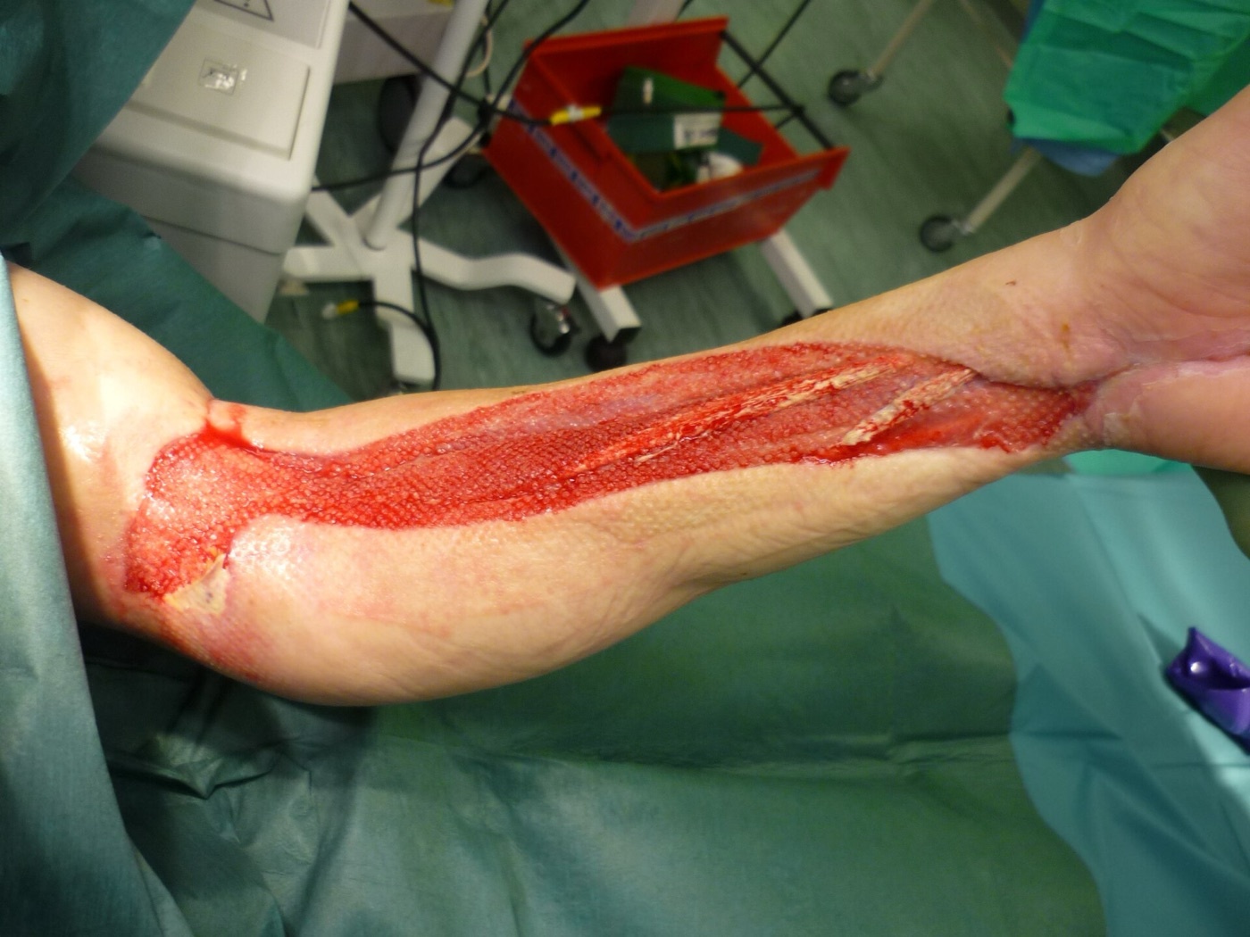
Figure 9. Forearm fasciotomy, the timely decompression that prevents Volkmann contracture (here dressed with a skin graft before closure). Source: Guyprocter, via Wikimedia Commons, CC0 1.0.
Part VIII - Complex Regional Pain Syndrome (Sudeck’s Atrophy)
Formerly called reflex sympathetic dystrophy or Sudeck’s atrophy, complex regional pain syndrome (CRPS) is divided into type I (no identifiable nerve injury, the old RSD) and type II (causalgia, with a documented nerve injury).[40] It is characterised by continuing pain with allodynia or hyperalgesia disproportionate to the inciting event, accompanied by oedema and changes in skin blood flow and sweating, and is diagnosed clinically (the modern Budapest criteria require symptoms and signs across the sensory, vasomotor, sudomotor/oedema and motor/trophic categories, with no better explanation).[41] Trauma is the commonest cause, and it follows about 1-5% of nerve injuries, 28% of Colles fractures and 30% of tibial fractures, classically evolving through three stages: an acute (warm, swollen, painful) stage, a dystrophic stage, and an atrophic (cold, dry, stiff) stage.[42] Treatment is multimodal and early (physiotherapy and desensitisation, analgesics and neuropathic agents such as gabapentin, sympathetic blocks, and bisphosphonates), and vitamin C has evidence for prevention (after foot and ankle surgery, and after distal radial fracture).[43]
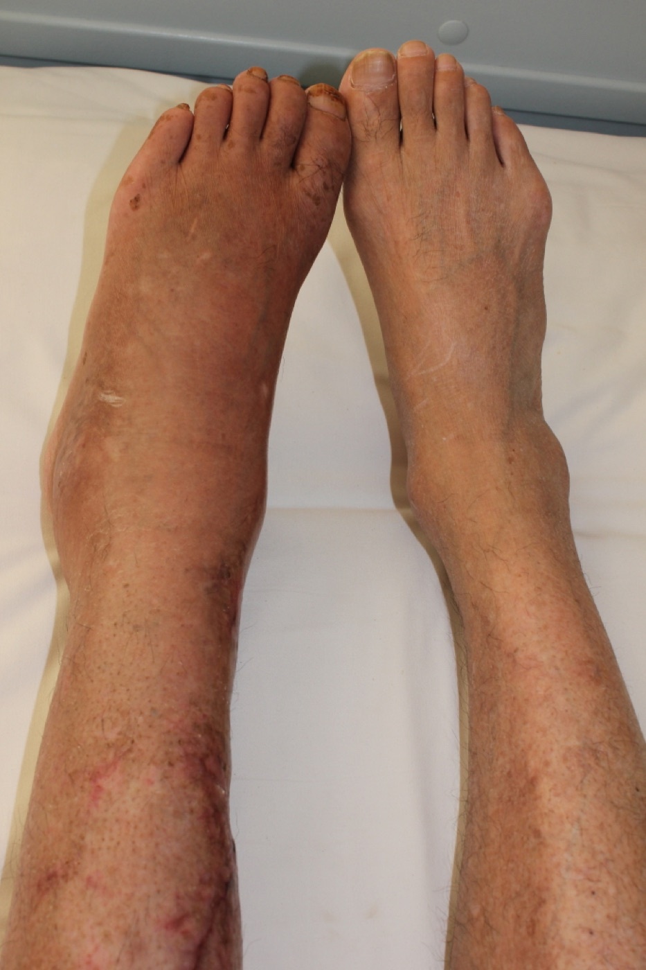
Figure 10. Type-I CRPS (Sudeck) of the left lower limb after a tibial fracture: swelling and colour change versus the normal side. Source: Timsong311, via Wikimedia Commons, CC BY-SA 3.0.
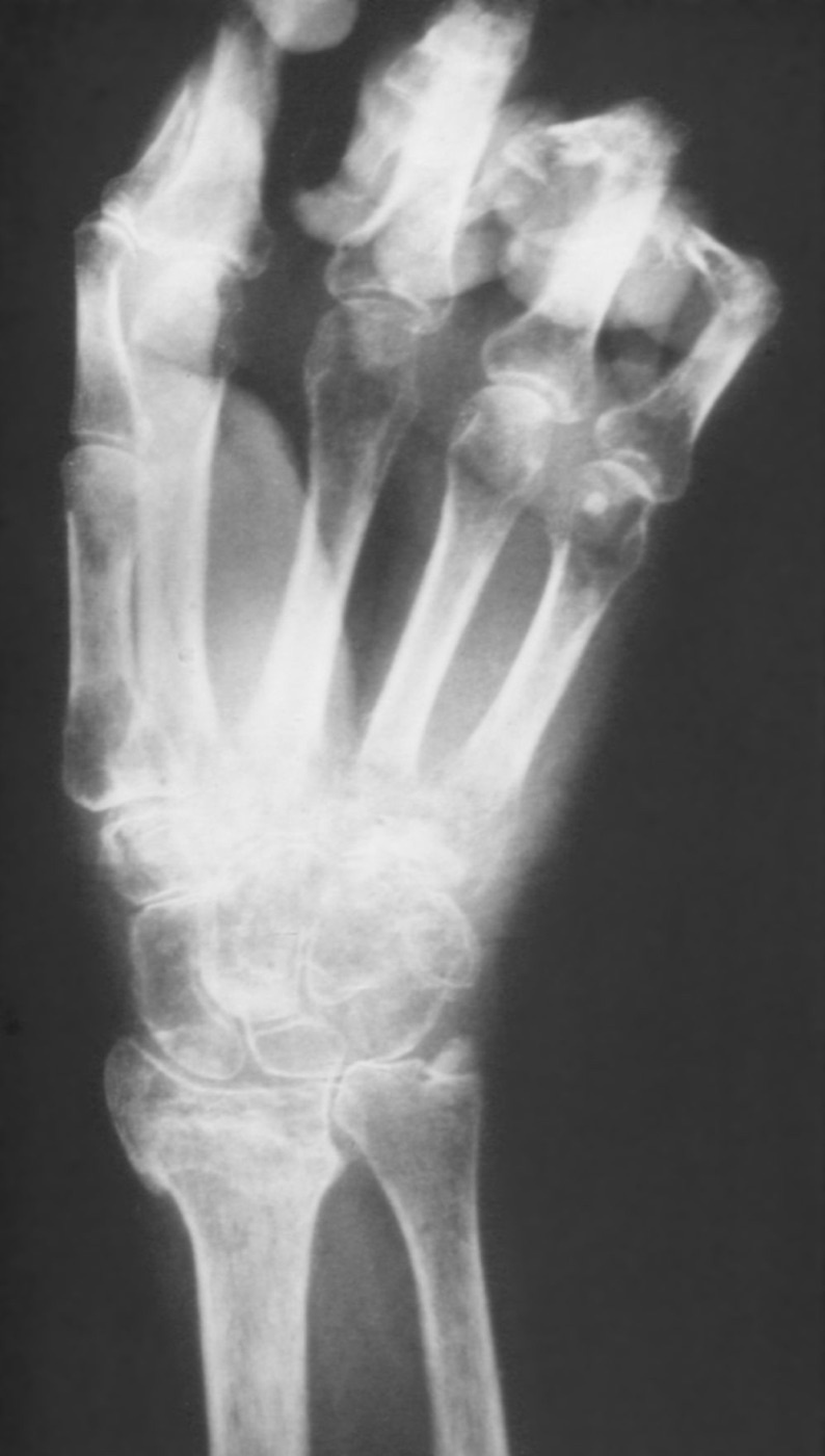
Figure 11. Sudeck’s atrophy: patchy juxta-articular osteopenia of the hand on radiograph. Source: Mehlauge, via Wikimedia Commons, CC BY-SA 3.0.
Part IX - Periprosthetic and Peri-implant Fractures
As arthroplasty volumes and the elderly population grow, fractures around implants are an increasing burden (one model projects a rise of about 4.6% per decade), and they carry a one-year mortality approaching that of a hip fracture (around 18-25%).[44] They occur intraoperatively (more common with uncemented and revision components) or postoperatively (usually a low-energy fall), and the dominant risk factors are osteoporosis, osteolysis and implant loosening, stress risers, and revision surgery.[45]
The key classification is the Vancouver classification of periprosthetic femoral fractures about a hip replacement, built on three things, fracture site, implant stability, and bone stock:[46]
- Type A. Trochanteric (AG greater, AL lesser trochanter); usually treated non-operatively or with fixation if displaced.
- Type B1. Around or just below the stem, with a well-fixed stem → open reduction and internal fixation (a lateral locking plate, often with a cable/strut).
- Type B2. Around the stem, with a loose stem but adequate bone → revise the stem to a long uncemented stem.
- Type B3. A loose stem with deficient bone stock → revise the stem with allograft (or a proximal femoral/tumour replacement).
- Type C. Well below the stem tip, treated independently of the prosthesis with a plate that spans up to overlap the stem.
The pivotal decision is therefore whether the stem is loose, because a B1 fracture mistaken for a stable stem and plated has a high failure rate (radiographically “stable” stems are loose in up to 20%), so stem stability is tested intraoperatively.[47] For the periprosthetic distal femur above a knee replacement, the Lewis-Rorabeck classification guides treatment: type I (undisplaced, prosthesis intact) non-operatively, type II (displaced, prosthesis well-fixed) by internal fixation (a distal femoral locking plate or a retrograde nail), and type III (loose or failing prosthesis) by revision arthroplasty, while the Su classification adds the fracture’s level relative to the component.[48] Anterior femoral cortical notching is the much-discussed (though clinically debated) stress-riser risk factor for these supracondylar fractures.[49] The femur fractured between a hip and a knee replacement, the interprosthetic fracture, is managed by a long plate spanning and protecting the whole femur.[50] (The Unified Classification System of Duncan and Haddad generalises the Vancouver A-B-C logic to any bone and implant, but is not detailed in the mined sources.[51])
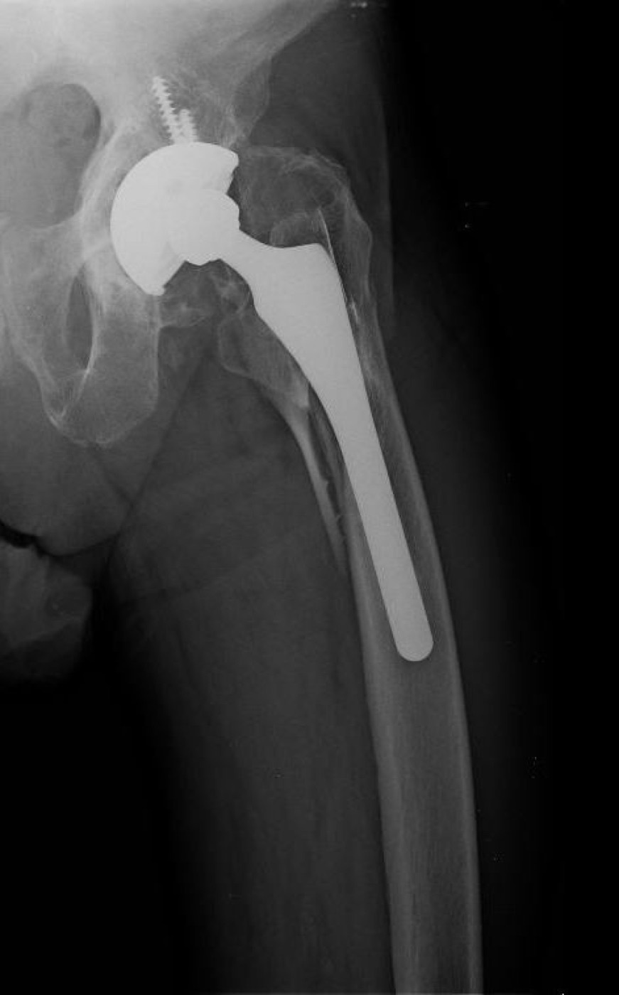
Figure 12. A periprosthetic femoral fracture about a hip stem (Vancouver type B). Source: Jmarchn, via Wikimedia Commons, CC BY-SA 3.0.
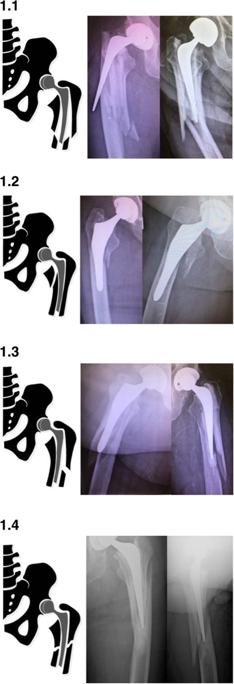
Figure 13. Vancouver type B2 sub-patterns of periprosthetic femoral fracture (schematics with matched radiographs); the loose stem is revised to a long stem. From Karam et al. (2020), J Orthop Surg Res, PMC7063746, CC BY 4.0.
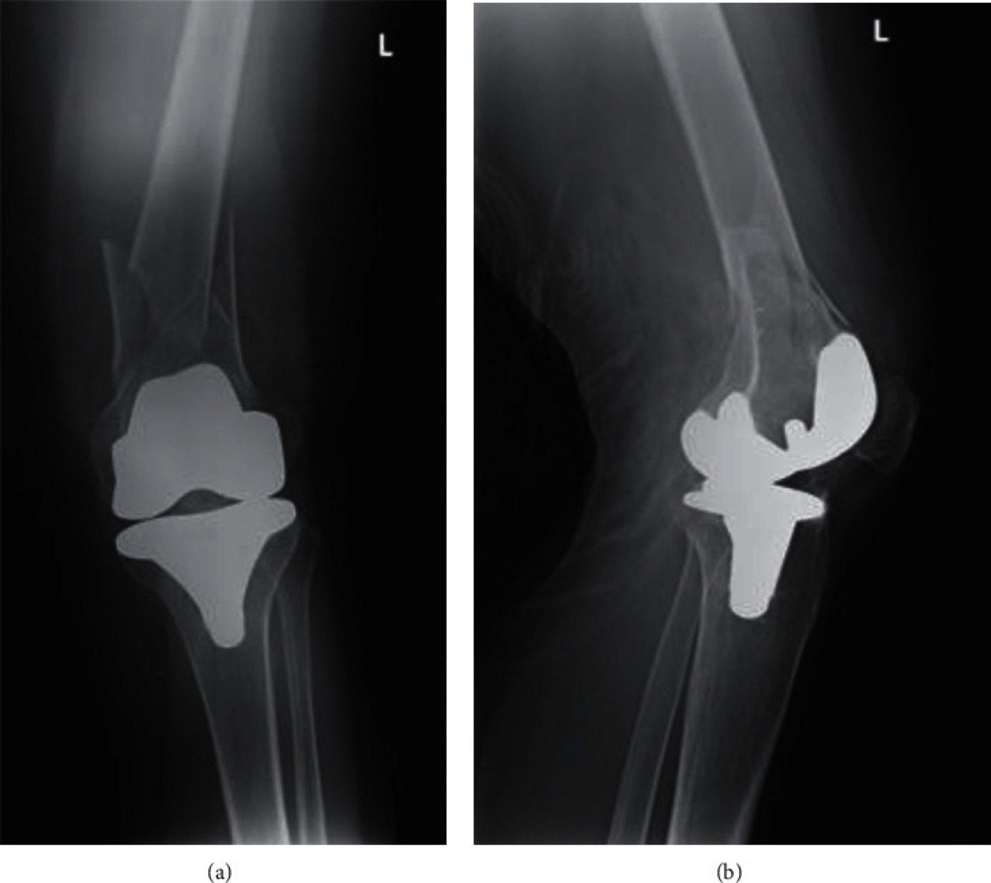
Figure 14. A comminuted periprosthetic distal-femoral fracture above a total knee replacement (Lewis-Rorabeck type II). From Moukarzel et al. (2021), Case Reports in Orthopedics, PMC8612792, CC BY 4.0.
References
-
Skeletal Trauma 5e, ch.25 (Brinker & O’Connor), p.731.
-
Rockwood & Green 9e, ch.29 (Ricci), pp.1410-1411.
-
Rockwood 9e p.1410; Skeletal Trauma 5e p.731.
-
Skeletal Trauma 5e p.731.
-
Skeletal Trauma 5e p.731.
-
Rockwood 9e pp.1410-1411.
-
Skeletal Trauma 5e p.732.
-
Rockwood 9e p.1418; Skeletal Trauma 5e p.732.
-
Skeletal Trauma 5e pp.732-736.
-
Rockwood 9e pp.1412-1415, 1432-1433.
-
Rockwood 9e p.1417; Skeletal Trauma 5e p.732.
-
Skeletal Trauma 5e p.740; Rockwood 9e pp.1418-1420.
-
Skeletal Trauma 5e p.740; Rockwood 9e pp.1418-1419.
-
Rockwood 9e pp.1419-1420.
-
Skeletal Trauma 5e p.740; Rockwood 9e p.1418.
-
Skeletal Trauma 5e pp.743, 750, 755.
-
Skeletal Trauma 5e pp.750-751.
-
Rockwood 9e pp.1422-1426; Skeletal Trauma 5e p.738.
-
Rockwood 9e pp.1461-1462; Skeletal Trauma 5e p.744.
-
Rockwood 9e p.1461.
-
Skeletal Trauma 5e p.748.
-
Rockwood 9e pp.1428-1430; Skeletal Trauma 5e p.751.
-
Rockwood 9e pp.1432-1435.
-
Rockwood 9e pp.1437-1442.
-
Rockwood 9e pp.1451-1456.
-
The “diamond concept” term is standard teaching; the mined extracts discuss all its component elements (Rockwood 9e pp.1451-1457) without using the phrase.
-
Rockwood 9e pp.1461-1464; Skeletal Trauma 5e pp.753-755.
-
Rockwood 9e pp.1458-1461.
-
Rockwood 9e p.1491. The chapter characterises rather than formally defines malunion; the one-line definition is standard teaching.
-
Rockwood 9e p.1491.
-
Rockwood 9e pp.1492, 1514.
-
Rockwood 9e pp.1493-1501.
-
Rockwood 9e p.1491.
-
Rockwood 9e pp.1509-1526.
-
Synthesised from the dispersed hardware-failure material in Skeletal Trauma 5e p.2688 and Rockwood 9e; the term “débricolage” is standard exam terminology not used verbatim in the mined extracts.
-
Skeletal Trauma 5e p.2688.
-
Skeletal Trauma 5e pp.2688, 2692; Rockwood 9e pp.4027-4032.
-
The established-contracture detail (definition, von Volkmann, the elliptical infarct, the Tsuge grading and the surgical treatments below) is standard teaching; the mined Rockwood/Skeletal Trauma compartment extract covers acute compartment syndrome but not the established contracture. The acute-compartment thresholds are from Rockwood 9e p.937 and Skeletal Trauma 5e p.514.
-
Rockwood 9e p.937; Skeletal Trauma 5e p.514.
-
Skeletal Trauma 5e pp.684-685, Table 23-12.
-
Skeletal Trauma 5e p.685; the Budapest criteria are the standard modern refinement of the IASP criteria given in the source.
-
Skeletal Trauma 5e pp.684-686.
-
Skeletal Trauma 5e pp.687-689; the distal-radius evidence is the Zollinger RCTs cited there.
-
Rockwood 9e pp.4022, 4024, 4056.
-
Skeletal Trauma 5e pp.2680-2681.
-
Rockwood 9e pp.4018-4020; Skeletal Trauma 5e pp.2682-2683.
-
Rockwood 9e pp.4018, 4032; Skeletal Trauma 5e p.2687.
-
Rockwood 9e p.4053; Skeletal Trauma 5e pp.2693-2694.
-
Rockwood 9e p.4055; Skeletal Trauma 5e p.2681.
-
Rockwood 9e p.4058.
-
The Unified Classification System (UCS/Duncan-Haddad 2014) is standard teaching; it does not appear in the mined Rockwood/Skeletal Trauma extracts.
-
Rockwood 9e pp.1410-1411; Skeletal Trauma 5e p.731.
-
Skeletal Trauma 5e p.740; Rockwood 9e pp.1418-1420.
-
Rockwood 9e pp.1461-1462; Skeletal Trauma 5e p.744.
-
Rockwood 9e pp.1452, 1458-1461.
-
Rockwood 9e pp.1500-1501.
-
Rockwood 9e p.1491.
-
Standard teaching; Skeletal Trauma 5e pp.2688, 2692.
-
Rockwood 9e p.937; Skeletal Trauma 5e p.514; established-contracture detail is standard teaching.
-
Skeletal Trauma 5e pp.684-689.
-
Rockwood 9e pp.4018-4020, 4032.
-
Rockwood 9e pp.4053-4055.