Contents
- Part I - The Place of Non-operative Treatment
- Part II - Closed Reduction: The Maneuvers
- Part III - Maintaining Reduction I: Casts and Splints
- Part IV - Maintaining Reduction II: Traction
- Part V - Functional Bracing (Sarmiento) and Cast-Bracing
- Part VI - Acceptable Alignment and Remodelling
- Part VII - Non-operative Management Region by Region
- Part VIII - Complications of Cast and Closed Treatment
- Part IX - When to Convert to Operative Treatment
- References
Part I - The Place of Non-operative Treatment
A craft that predates the operating theatre
For almost the whole history of medicine, a broken bone was set with the hands and held with whatever the era could fashion into a splint. Operative fixation is recent. Manipulation, traction and external splintage were essentially the only methods available until around 1750, and the surgical treatment of fractures only accelerated after the Second World War, once anaesthesia, asepsis and antibiotics had matured enough to make opening a fracture reasonably safe.[1] Working in a modern trauma centre surrounded by plates and nails, it is easy to assume that surgery is the default and the cast an afterthought. The numbers say otherwise.
Across an entire population of fractures, non-operative treatment remains the single most common method of fracture management.[2] In the Edinburgh epidemiological series of 6,996 adult fractures, only 27.6% were treated surgically; 72.4% were treated without an operation.[3] When proximal femoral fractures (which almost always need fixation in the elderly) are set aside, the non-operative share climbs to 81.8% of all adult fractures, 82.4% in those over 65, and 90.1% in those over 80.[4] Some fracture groups are treated non-operatively more than nine times out of ten: toe phalanges (97.2%), metatarsals (96.8%), the proximal radius (96.5%), the pelvis (95.8%), the fibula (95.3%), metacarpals (93.3%), the proximal humerus (92.9%), finger phalanges (92.0%) and the clavicle (91.4%).[5]
The textbooks are candid that this skill is quietly being lost. Skeletal Trauma warns of “an increasing tendency to treat surgically those fractures that might equally effectively be treated without ORIF,” driven by convenience, the surgeon’s own enjoyment of operating, implant-company marketing, and the way healthcare systems reimburse procedures rather than plaster.[6] Randomised trials have repeatedly shown surgery to be no more effective than non-operative care, at greater cost, for distal radial, humeral shaft, proximal humeral and surgical-neck fractures, acromioclavicular dislocations, calcaneal fractures and some ankle fractures.[7] The same text is blunt about the demands of the conservative path: closed management is “technically demanding, time consuming, and requires more diligence than surgical treatment,” and the skill “atrophies by lack of use and training” until it risks being lost altogether.[8] In much of the world, where an operating theatre is not reliably available, this craft is not a historical curiosity but the standard of care.[9]
What non-operative treatment is trying to achieve
Three principles govern the management of any fracture, operative or not:[10]
- Reduction: restoring the fragments as close to normal anatomy as possible, correcting alignment, length, rotation and lateral shift. The best position should be obtained at the first manipulation.
- Maintenance of that reduction throughout the period of healing.
- Promotion of functional recovery: restoring the injured part to useful function as quickly as possible, with the best achievable cosmetic result.
A limb is a functioning unit, not a row of bones: the skeleton is only the framework, and it depends on mobile joints, working muscles and nerves, and an adequate blood supply.[11] This is why the goal is never simply a healed radiograph. Skeletal Trauma describes three overlapping phases of treatment: a primary phase (reduction and provisional holding by plaster, traction or fixation, followed by rest and elevation, with early movement encouraged to limit wasting and adhesions); a definitive phase (the definitive cast or fracture brace, usually applied between ten days and four weeks once swelling has settled, allowing rehabilitation and active use); and a rehabilitation phase, which after prolonged plaster or traction can require “a fairly long period of physiotherapy” to mobilise stiff joints and rebuild wasted muscle and osteopenic bone.[12]
When non-operative treatment is the right choice
Modern non-operative technique is used principally to treat stable fractures, rather than to reduce and hold unstable ones, a deliberate shift from the era when traction held a femoral shaft for weeks.[13] The candidates are, broadly:
- Undisplaced or minimally displaced fractures, where the bone is already in an acceptable position and simply needs protection; and
- Patients in whom surgery carries disproportionate risk: the elderly, the frail, and those with significant medical or social comorbidity.[14]
The choice is “frequently subjective and influenced by the patient’s age, physical condition, mental status, and degree of pre-fracture mobility,” and rests not on the fracture alone but on whether it can be reduced and held, and on whether the patient could realistically mobilise after an operation.[15] Skeletal Trauma puts the balance plainly: do not advocate non-operative treatment for fractures best treated surgically, but do advocate it for the large group of fractures that “do well when treated without surgery providing the treatment is appropriate and done correctly.”[16]
The trade-offs are real. Advantages: there is no surgical wound and therefore no implant infection, and none of the complications of fixation, such as nonunion (the fracture’s biology being undisturbed), hardware failure, or implant-related pain. Casts are cheap, quick to apply, and (being awkward to remove) are genuinely useful in unreliable patients who might otherwise discard a brace.[17] Disadvantages: the constant threat of malunion if the position drifts, joint stiffness and muscle wasting from immobilisation, and a heavy surveillance burden. As Rockwood notes, “cast management of unstable fractures is very labor-intensive. Follow-up must be assiduous until callus starts to stabilize the fracture, as it is easy to miss secondary fracture displacement.”[18]
Part II - Closed Reduction: The Maneuvers
Read the fracture before you touch it
“It is essential to understand the pathologic anatomy of the fracture” before any attempt at reduction.[19] Displacement only happens when the periosteum is torn and/or the bone is comminuted; the pattern of the fracture predicts how it will behave in your hands:[20]
- Transverse fractures (a force applied at right angles to the shaft) can be stable once reduced, because the broad bone ends abut and resist shortening. A transverse fracture that has overridden, however, has usually stripped periosteum, and that overriding must be undone.
- Oblique and spiral fractures (a rotational force along the limb’s axis) tend to spike through soft tissue and may trap muscle between the fragments; once disimpacted they are inherently unstable and slide back to length.
- Comminuted fractures are often the easiest to reduce but the least stable, because the fragments have no interlocking geometry to hold them.
The Charnley principle: recreate the deformity, then reverse it
The single most important idea in closed reduction is counter-intuitive: you often cannot pull a displaced transverse fracture out to length by straight longitudinal traction, because an intact strip of periosteum on one side acts as a tether (the periosteal hinge), and the harder you pull, the more it resists.[21] Charnley’s solution, still taught verbatim, is to reproduce and even exaggerate the original angular deformity to disengage the fragment ends, hook the cortices together, and then reverse the deformity back to neutral, using the intact periosteal hinge on the concave side as a stabiliser that snaps the bone into place and then holds it there.[22] The reduced position is then maintained by three-point fixation within the cast (see Part III). The classic verbal formula for a Colles fracture captures the same sequence, “replicate the deformity, apply traction, and then manipulate it into the appropriate position,” followed immediately by a well-moulded, three-point cast.[23]
The complementary force is traction and counter-traction. For a wrist, finger traps on the thumb, index and middle fingers provide traction while a counterweight of about 2.3 kg (5 lb) slung above the elbow provides counter-traction; this fatigues the deforming muscles, disimpacts the fragments, and conveniently frees the surgeon’s hands to apply plaster without an assistant.[24] In a long bone, counter-traction is supplied by an assistant, by a perineal post, or, in continuous traction, by tilting the whole bed (see Part IV).
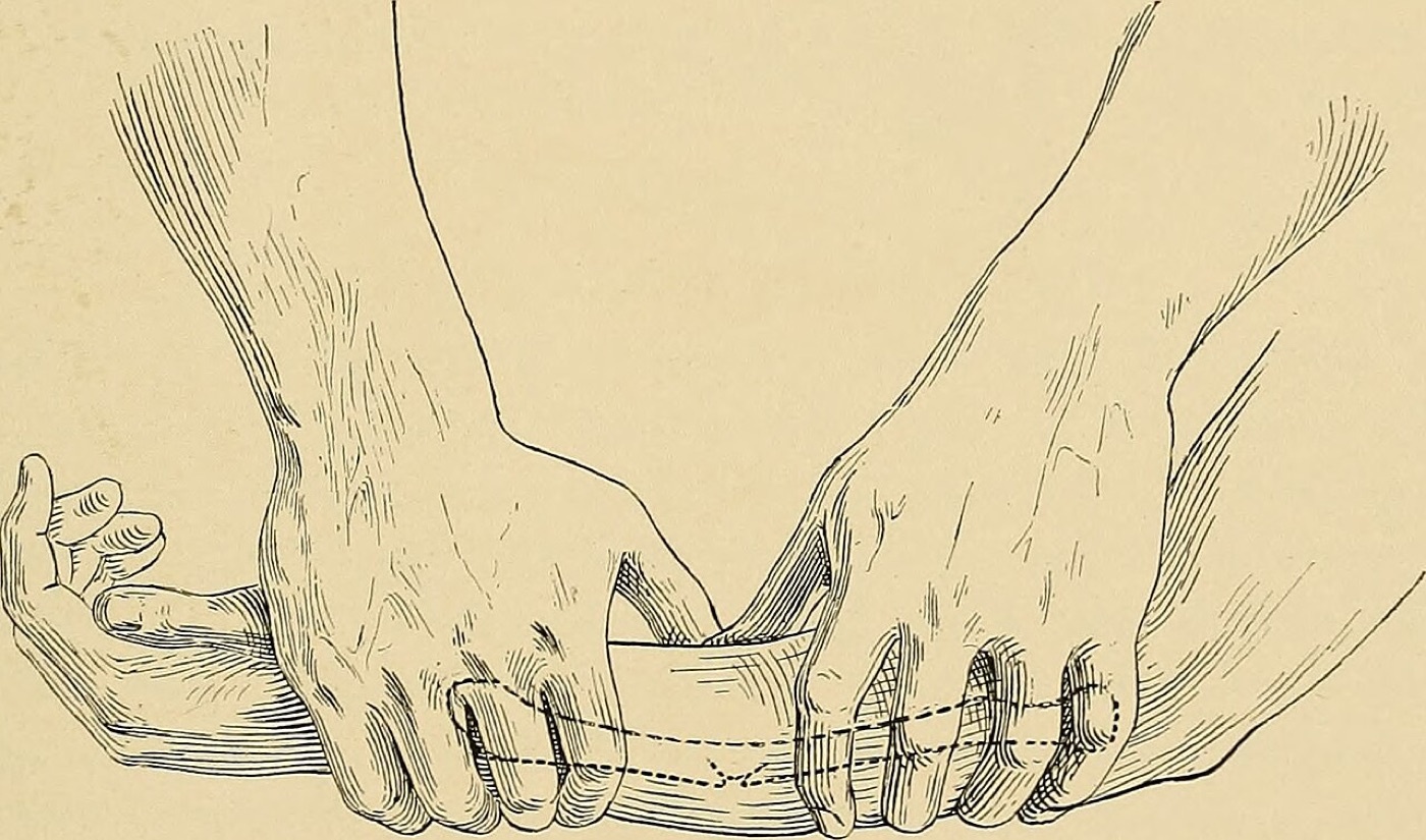
Figure 1. Closed reduction of a forearm fracture by manual manipulation - traction and moulding pressure applied by the surgeon’s hands. From C. L. Scudder, The Treatment of Fractures (1901). Public domain, via Wikimedia Commons.
Pain relief is not optional
The body’s response to a fracture is protective muscle spasm, and the stimulus driving that spasm is pain; until the pain is abolished the muscles will fight every attempt at reduction. Skeletal Trauma states it flatly: “In general, it is wrong to attempt manipulative reduction of the fracture or dislocation without anesthesia.”[25] A cautionary case in the same text describes an inexperienced attempt at reducing a shoulder without proper anaesthesia that ended in an open reduction, a permanently stiff shoulder, and an unrecovered brachial plexus injury.[26] The options, from most to least involved:[27]
- General anaesthesia: the anaesthetic of choice in a properly equipped hospital, giving complete relaxation and a still field.
- Regional blocks: a brachial plexus block (analgesia from the mid-upper arm down, more effective the more distal the injury) or a femoral nerve block (local anaesthetic around the femoral nerve beneath the inguinal ligament), which gives excellent analgesia and muscle relaxation for reducing a femoral fracture.
- Intravenous regional anaesthesia (Bier block): effective, but it fell into disrepute after deaths with lignocaine; prilocaine (Citanest), with its low systemic toxicity, is now the agent recommended, and only with strict precautions (fasting, secure venous access in the opposite arm, a checked and constantly observed tourniquet, full resuscitation equipment to hand).
- Haematoma block: local anaesthetic injected directly into the fracture haematoma, the workhorse for a Colles fracture in a frail elderly woman, with the warning that “dangerously high levels of local anesthetic may appear in the general circulation.”
- Sedation and adjuncts: midazolam is “probably the most useful,” being short-acting, safe and reliably amnesic; ketamine provides no muscle relaxation; intravenous diazepam is unpredictable; and inhaled Entonox is convenient but often unsatisfactory for anything more than a dislocation or minor fracture.
Timing, and the few who do not need reducing
“Reduction within a few hours after injury should be the aim.”[28] Delay allows the haematoma to clot firmly and the inflammatory oedema to set in, and “the longer the delay in reduction, the more difficult it will be to obtain satisfactory reduction.”[29] There are two important qualifications. First, an emergency exists, regardless of the operating list, when a displaced fragment or dislocation is tenting and threatening the skin or compromising the distal circulation: the classic ankle fracture-dislocation blanching the skin over the medial malleolus, or a knee dislocation with an ischaemic foot, must be realigned immediately with whatever analgesia is to hand.[30] Second, some fractures cannot be reduced acutely because of swelling: the severely displaced supracondylar fracture in a child is the textbook example, where traction and elevation are used for a day or two until the tense fracture haematoma subsides and reduction becomes possible.[31]
Not every fracture needs a manipulation, and in the very old this is increasingly recognised. For low-demand elderly distal radial fractures, Beumer and McQueen found that 53 of 60 wrists lost their reduction anyway, concluding that closed reduction “is of minimal value in the very old and frail, dependent, or demented patient”; other series report similar functional outcomes whether or not the wrist was reduced.[32] The authors stop short of recommending that reduction be abandoned, pending stronger evidence, but the principle that a manipulation must earn its place is sound.
Two safety rules close the loop. A check radiograph is mandatory after manipulation and before the patient is woken, since it is “safer to be kept asleep a few more minutes” than to wake the patient, find a poor position, and need a second anaesthetic.[33] And the films must be a good AP and lateral that always include the joint above and below the fracture.[34]
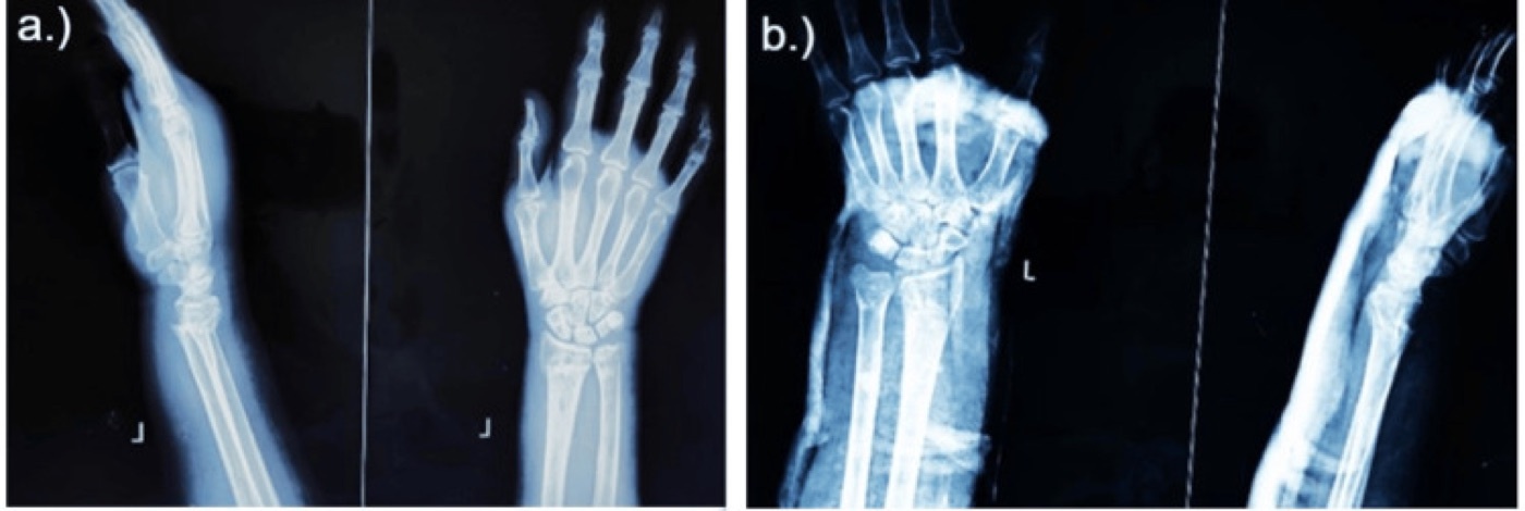
Figure 2. Distal-radius (Colles-type) fracture before (a) and after (b) closed reduction and cast application - the documented goal of a successful manipulation. From Kaushik et al. (2025), PMC12739557, CC BY 4.0.
Part III - Maintaining Reduction I: Casts and Splints
Plaster of Paris and its modern rivals
Plaster of Paris (POP) is calcined gypsum impregnated into a bandage; on contact with water it recrystallises and hardens, and its strength lies entirely in the crystal lattice that grows as it sets. This is why moulding must be finished while the plaster is still creamy, since disturbing a half-set cast fractures the forming lattice and leaves it weak.[35] Its qualities (ease of application, mouldability, conformability, absorbency and cheapness) “have not been bettered by any of the modern bandages,” and for this reason Skeletal Trauma takes a firm line: “the modern synthetic materials have no place in the management of acute fractures, and plaster of Paris remains the material of choice.”[36] The history is worth a footnote of its own: the technique reaches back to ancient Egypt, India and Arabia; the name derives from the gypsum quarried at Montmartre in Paris; Mathijsen introduced plaster-smeared bandages in Europe in 1852; and Samuel St John in the United States advocated padding the skin with cotton wool and splitting the cast in acute fractures.[37]
Fiberglass (water-activated polyurethane) bandages are stronger, lighter, more durable and more radiolucent, which makes them ideal for definitive casts and braces once swelling has settled; a below-knee cast needs only about two rolls of fiberglass against four to six of plaster.[38] Thermoplastics (Sansplint, Orthoplast, Hexalite) soften in a water bath and are moulded directly to the limb, but need extra training.[39] Rockwood is more relaxed than Skeletal Trauma about using fiberglass for acute injuries, noting it is “now more frequently used” and that casts are applied in the same way whatever the material, a genuine difference in editorial emphasis worth knowing for a viva.[40]
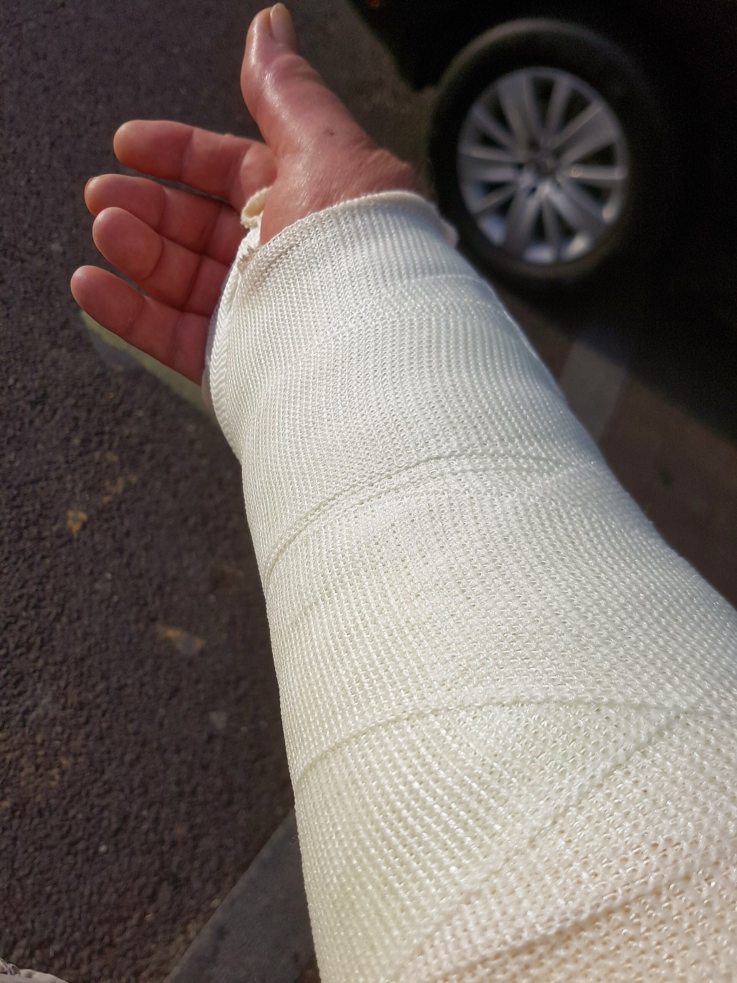
Figure 3. Synthetic (resin/fiberglass) short-arm cast for a wrist fracture - lighter and more radiolucent than plaster, favoured for definitive casts. Source: Acabashi, via Wikimedia Commons, CC BY-SA 4.0.
Applying a cast: padding, and never too tight
Padding is a balancing act. Too much padding lets the fracture displace inside a loose cast; too little causes skin pressure problems and raises the risk of compartment syndrome. Rockwood’s rule of thumb is two layers of wool with 50% overlap, with extra layers over bony prominences, and minimal padding when the cast must actually control a reduced fracture.[41] Skeletal Trauma scales the padding to the injury: one layer for an acute fracture with little swelling, three or more layers for a crush injury, and, critically, when there is any concern about the circulation, the cast (and the wool beneath it) is split instantly from top to bottom so the skin is visible.[42] Blood-soaked wool that dries becomes rigid and produces a tourniquet effect, so a bleeding wound under plaster is dangerous.[43] Extra padding always goes over the malleoli, the heel, the base of the foot and the head and neck of the fibula, the last to protect the common peroneal nerve as it winds around the fibular neck.[44] The roll is started at the narrowest part of the limb so the bandage does not drift, and the plaster is smoothed and laminated with the flat of the hand while soft.[45]
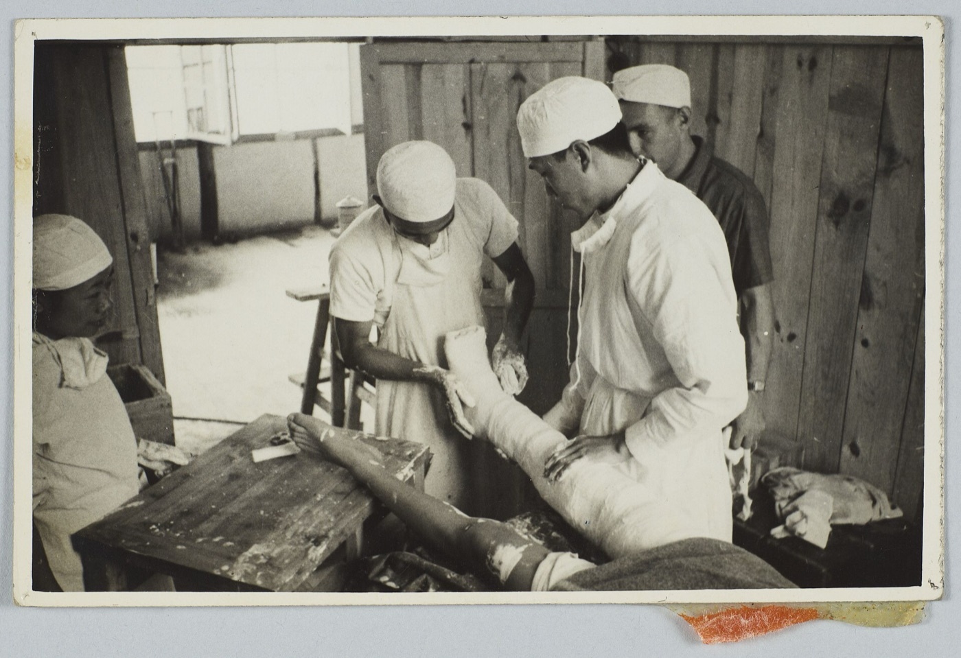
Figure 4. Application and moulding of a plaster-of-Paris cast to the leg over padding. IU Indianapolis University Library, via Wikimedia Commons. Public domain.
Three-point fixation: “a curved cast makes a straight bone”
The principle that allows a rigid tube to hold a fracture reduced is three-point fixation. A fracture with an intact periosteal hinge on its concave side will spring back toward deformity as the muscles pull and the swelling subsides; three points of moulded pressure (one over the apex of the original deformity and two at the ends of the cast on the opposite side) generate a continuous corrective force that opposes that tendency.[46] The technique is to build the loading into the cast with the flat of the hand while the plaster is still creamy, avoiding focal pressure over bony points, “so that as the swelling subsides, sufficient loading remains to maintain the fracture position.” This is proven for the Colles fracture while still leaving the wrist free to move.[47]
Rockwood frames the holding of an unstable fracture in a cast as resting on three pillars:[48]
- Use of the intact soft tissues: the soft-tissue and periosteal hinge that resists the deforming force;
- Three-point fixation, as above; and
- Hydrostatic pressure: the incompressible muscle and bone, once encased in a complete cast, behave as a rigid cylinder that maintains position (an explanation Rockwood concedes is “somewhat simplistic,” since it ignores active muscle contraction).
The crucial caveat is that the soft-tissue hinge fails when there is no good hinge to use: it works for a transverse diaphyseal fracture in a young patient, but is far less reliable in spiral, butterfly, segmental or comminuted fractures, after high-energy injury, and in older patients, whose thin, friable periosteum tears easily. The young distal radius with an intact hinge holds in plaster, while the elderly comminuted distal radius does not.[49] This single principle explains most of the cases where a closed reduction “won’t hold” and surgery is needed.
The cast index - judging whether a cast actually grips
A well-moulded forearm cast is oval, not round, hugging the limb in the plane of correction. The cast index quantifies this: it is the ratio of the cast’s internal sagittal (dorsal-to-volar) diameter to its coronal (radial-to-ulnar) diameter at the fracture level, and a value of ≤0.8 indicates an adequately moulded cast, while a higher (more circular) index predicts loss of reduction. The metric is used especially in paediatric distal radial and forearm fractures.[50] The mined texts describe the same idea qualitatively (an oval, three-point-moulded cast holds; a loose round one does not) but do not give the numeric threshold.
The casts you must be able to name
Upper limb:[51]
- Long-arm (above-elbow) cast: axilla to just proximal to the MCP joints, thumb free, elbow at 90° and wrist in about 30° dorsiflexion; for forearm and elbow fractures.
- Forearm (Colles) cast: below the elbow to just proximal to the metacarpal necks, never crossing the distal palmar crease so the MCP joints can flex; the most widely used upper-limb cast, for distal radial, distal ulnar and some carpal injuries, often preceded by a dorsal slab until swelling settles.
- Scaphoid cast: wrist in slight dorsiflexion, thumb abducted and slightly flexed “as if holding a glass between index and thumb,” extending to just short of the thumb IP joint.
- Burkhalter cast: for metacarpal and phalangeal fractures, wrist in 40° dorsiflexion and MCP joints in 70-90° flexion, using the intact dorsal hood as a tension band.
- James cast: the fingers held in the position of function (wrist 40°, MCP joints 70-90° flexed, IP joints in full extension), which keeps the collateral ligaments at maximal stretch and prevents the stiff, clawed hand.
- Bruner cast: a short extended-scaphoid cast cut back to release the wrist, for thumb MCP ligament injuries.
- Hanging cast / U-slab (sugar-tong splint): for the humeral shaft, the weight of the arm, slung from the neck with the elbow at 90°, applies gravity traction; a shorter cuff produces more varus, and it is usually changed to a functional brace at two to four weeks.
Lower limb:[52]
- Below-knee cast: from just below the fibular neck to the metatarsal heads, ankle at 90° and foot plantigrade; the commonest lower-limb cast.
- Long-leg cast: a below-knee cast extended to the groin with the knee in 10-15° flexion, for an unstable tibial fracture before conversion to a brace.
- Patellar-tendon-bearing (PTB) cast: a below-knee cast carried up to the lower pole of the patella and moulded around the patellar tendon for rotational control, with explicit care not to press on the common peroneal nerve at the fibular neck.
A walking cast must hold the foot truly plantigrade (the sole at 90° to the front of the leg) or the gait will be abnormal; the heel is reinforced and buffered, and a simple walking shoe such as the Aberdeen boot is ideal.[53]
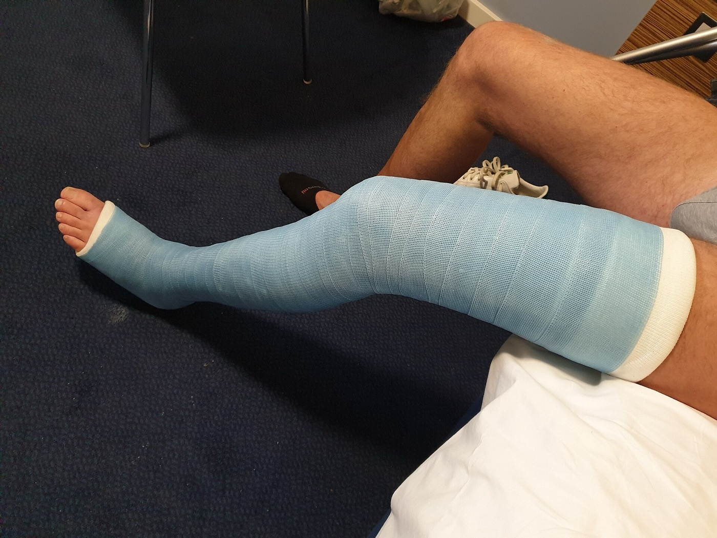
Figure 5. Long-leg fiberglass cast for a tibial fracture, with the knee in slight flexion and the foot plantigrade. Source: Jonuscumgi, via Wikimedia Commons, CC BY-SA 4.0.
Slabs, splints, slings and strapping
A full circumferential cast is rarely applied immediately after injury, precisely because a swelling limb in a rigid tube risks compartment syndrome; instead a slab (a backslab posteriorly for the leg, a U-slab for the humerus, a dorsal slab for the wrist) gives support while leaving room to swell.[54] The governing maxim: “if swelling is anticipated, use a slab; if there is swelling, the cast should be cut.”[55] For lighter injuries the armamentarium runs from the sling (clavicle, proximal humerus, radial head; roughly two weeks then mobilise) and the figure-of-eight bandage (for the clavicle, though it “loosens quickly and is no better than a sling”), to buddy strapping for stable finger and toe fractures, the mallet-finger splint (DIP joint in full extension for six weeks), and tubular elastic supports for minor sprains.[56]
Wedging and windowing
A cast need not always be removed to correct a drifting angulation. In cast wedging, the cast is divided around about two-thirds of its circumference, leaving a 2-3 cm hinge on the appropriate side, and is then opened (for a valgus deformity, a medial hinge is left and a varus force opens the lateral gap) under image guidance, the wedge being filled with fresh plaster.[57] Timing matters: wedging too early risks displacing an unstable fracture, so it is safest at about two to three weeks when early organisation of the haematoma has begun.[58] A window can be cut to inspect a wound or relieve a pressure point, but the cut-out plaster should be replaced and bandaged back to prevent soft tissue herniating (“window oedema”). Rotational malalignment is theoretically correctable by cutting the cast at the fracture, but the position is easily lost, and it is often better to remove and reapply the cast altogether.[59]
Part IV - Maintaining Reduction II: Traction
What traction does, and how it is balanced
Continuous traction overcomes the shortening pull of muscle spasm and restores and maintains length and alignment, principally for fractures of the femoral shaft.[60] The limb is supported on a splint with a ring seated in the groin, and the pull is applied either through the skin or through a transosseous pin in the distal femur or proximal tibia.[61] Two arrangements are distinguished: in fixed traction the pin is tied to the distal end of the splint, so the splint frame provides the counter-force; in balanced (sliding) traction the splint is suspended on pulleys and a second pulley applies the actual traction weight, with counter-traction supplied by tilting the patient head-down and raising the foot of the bed.[62] Once set up, the alignment is checked radiologically and pads are inserted to coax the femur straight: a posterior pad beneath the distal femur is almost always needed to lift the distal fragment against gravitational sag.[63]
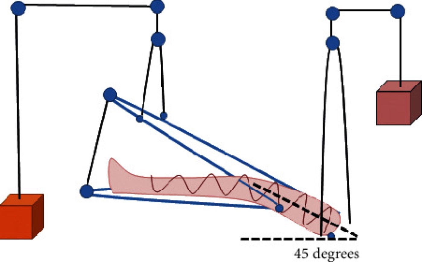
Figure 6. Balanced suspension of the lower limb using a Thomas splint with a Pearson knee attachment, pulleys and weights. From Yuenyongviwat et al. (2020), PMC7396022, CC BY 4.0.
Skin versus skeletal traction
Skin traction transmits the pull through adhesive strapping and foam applied to the skin and is limited to light loads, conventionally no more than about 4.5 kg (10 lb), beyond which the skin blisters and shears; it is therefore a temporary, temporising measure, classically used to settle a femoral fracture before surgery.[64] Skeletal traction runs a pin through bone and tolerates much heavier loads, suitable for definitive non-operative management. The pin is most often a stiff Steinmann pin or a thinner tensioned Kirschner wire; the Denham pin carries a central threaded segment that engages the near cortex and resists sliding.[65] Common insertion sites are the distal femur and proximal tibia (for femoral fractures), the calcaneus (historically for tibial traction) and the olecranon (for the elbow).[66] Inserting a tibial pin requires local anaesthetic into skin, periosteum and muscle.[67]
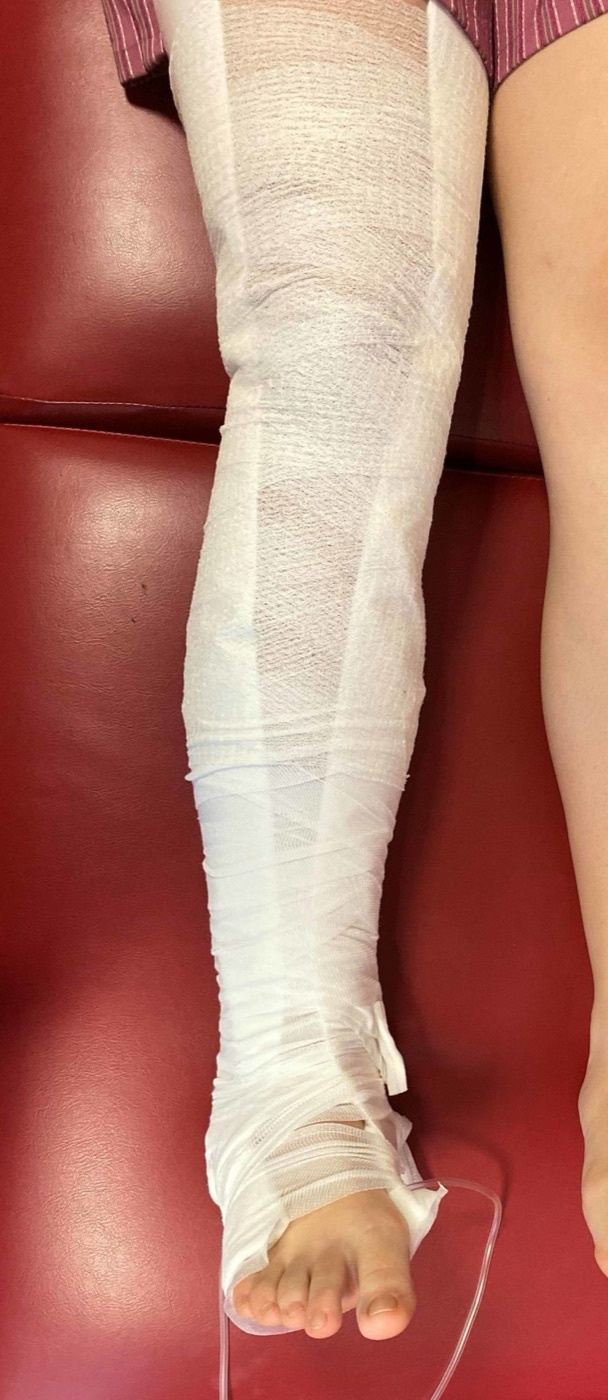
Figure 7. Skin (Buck’s-type) traction applied to the leg with felt and a crepe bandage - a light-load, temporary measure. From Mc Carthy et al. (2020), PMC7100621, CC BY 4.0.
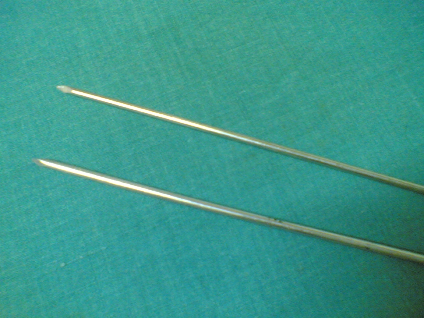
Figure 8. Steinmann pins - the stiff trans-osseous pins through which skeletal traction (or external fixation) is applied. Source: Netha Hussain, via Wikimedia Commons, CC BY-SA 3.0.
The named traction systems
The classic systems, as illustrated in Rockwood’s Figure 9-4, are worth knowing by name:[68]
- Thomas splint with a Pearson knee-flexion piece: the workhorse of balanced femoral traction; the knee piece is added at four to six weeks to allow knee movement.
- Braun traction: a simple weight-and-pulley frame giving longitudinal pull only; fragment control is poor, but the skin-traction version is still used as temporary pre-operative traction.
- Hamilton-Russell traction: a single sling under the knee with a pulley arrangement that, by doubling the longitudinal pull against the upward pull, produces a resultant force at about 30° to the horizontal, roughly in line with the femur.
- Perkins traction: a straight pull through a proximal tibial pin with no splint, on a split bed, allowing early knee mobilisation; alignment control is poor and malunion common.
- Fisk traction: a short Thomas splint with a hinged knee piece, letting the patient actively flex the hip and knee.
- 90-90 traction: hip and knee both flexed to 90°, so that gravity no longer sags the femoral fragments posteriorly; useful for proximal femoral shaft fractures (where the proximal fragment is flexed by iliopsoas) and still used for paediatric femoral fractures.
In small children, gallows (Bryant) traction (both legs suspended vertically with the hips flexed to 90° so the buttocks just clear the bed, the child’s body weight providing counter-traction) is the traditional method for femoral shaft fractures, restricted to infants under roughly two years and under about 12-16 kg because of the danger of vascular compromise and Volkmann ischaemia in larger children.[69]
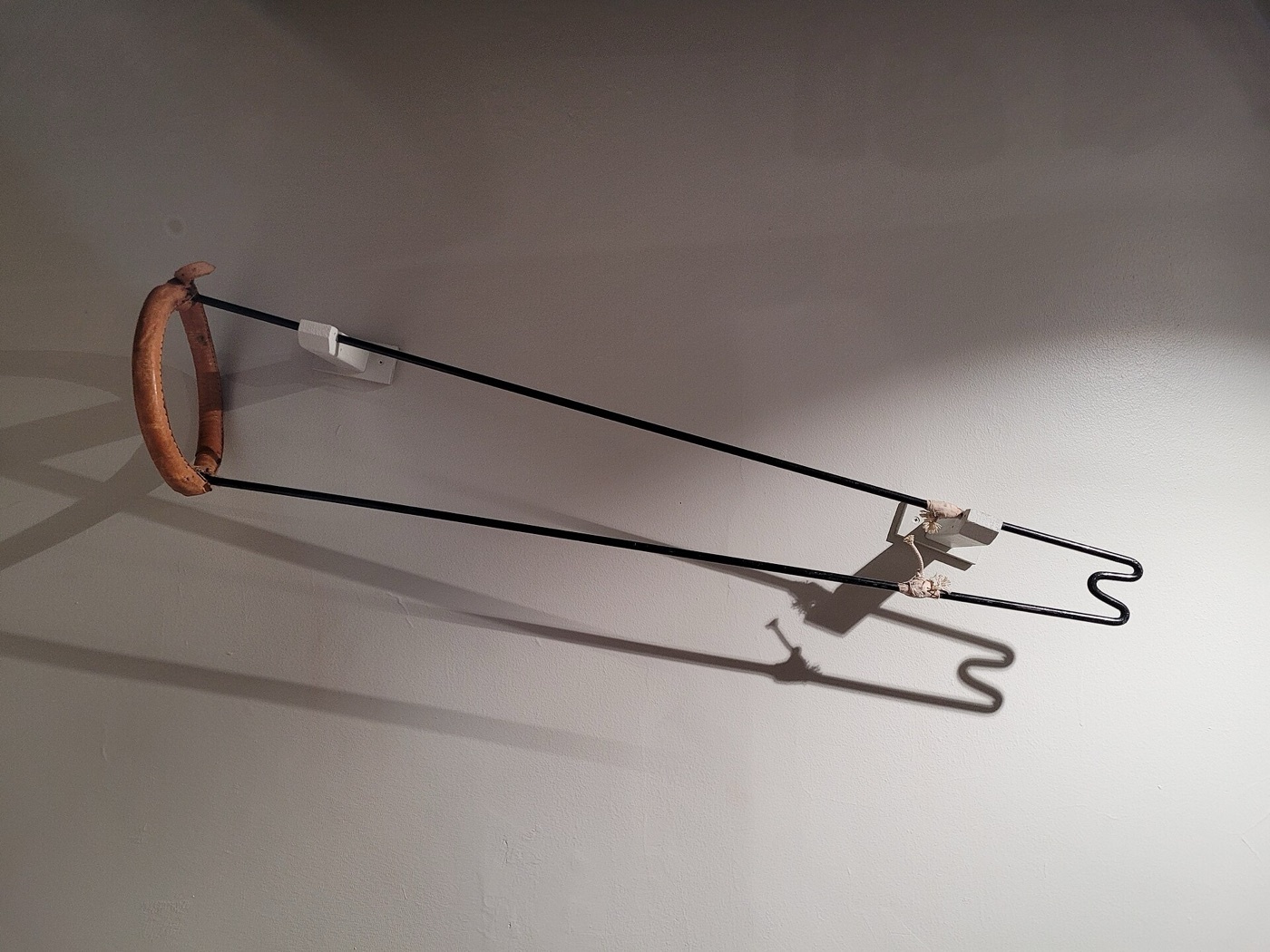
Figure 9. A Thomas leg-traction splint (padded ring and side bars) - the classic frame for balanced lower-limb traction. Source: Museumjack / Thackray Museum of Medicine, via Wikimedia Commons, CC BY 4.0.
Traction has largely left the limbs
Intramedullary nailing superseded traction for femoral fractures in the 1970s and 1980s, and its role today is “extremely limited and essentially confined to situations when internal and external fixation techniques are unavailable,” a holding or resource-limited measure, not a definitive treatment.[70] The morbidity explains why. Reviewing femoral traction, Connolly reported malunion or nonunion needing surgery in 11-29%, shortening over 2 cm in 14-30%, refracture in 4-17%, and knee stiffness in 30-50% (the last being the most significant problem), on top of the medical hazards of prolonged bed rest: pressure sores, thromboembolism, deconditioning, lost employment and psychological strain.[71] Tibial traction is now actively condemned: “there is now no indication for tibial traction unless appropriate internal or external fixation techniques are unavailable,” not least because excessive calcaneal-pin traction raises intracompartmental pressure and risks compartment syndrome.[72] The cast-brace (a long-leg cast with knee hinges, used to transition off traction) reduced these complications (one Connolly series reported only 0.7% nonunion and 5.4% loss of knee motion) but has itself “essentially disappeared” in favour of nailing.[73]
Spinal traction: the one place it still thrives
By contrast, traction of the cervical spine remains in widespread use to reduce a fracture or dislocation, decompress the neural elements, and provide provisional stability, though it is usually a bridge to a halo-vest or to surgery rather than a definitive treatment.[74] Two devices dominate:[75]
- Cranial (Gardner-Wells) tongs: two spring-loaded pins set into the outer table of the skull about 1 cm above the pinna and 1 cm behind the external auditory meatus, below the skull’s widest diameter so the pull is slightly upward; inserted to 6-8 inch-pounds of torque. Reduction begins at about 10 lb with roughly 5 lb added per spinal level (so about 40 lb for a C5-6 injury), always with serial imaging and vigilance for over-distraction and neural injury.
- Halo ring: fixed with four pins (two anterior, two posterior), the anterior pair about 1 cm above the lateral third of the orbital rim (safely lateral to the supraorbital and supratrochlear nerves) and the posterior pair above the helix of the ear, tightened in opposing pairs and re-tightened at 24-48 hours. It tolerates higher loads than tongs and can be built into a halo-body vest (originally the Perry-Nickel device) for definitive treatment.
Even a halo-vest permits up to about 31% of normal cervical motion, and roughly 10% of patients lose reduction, so serial radiographs are essential; pin-track sepsis occurs in up to 20% and pin loosening in 36-60%.[76]
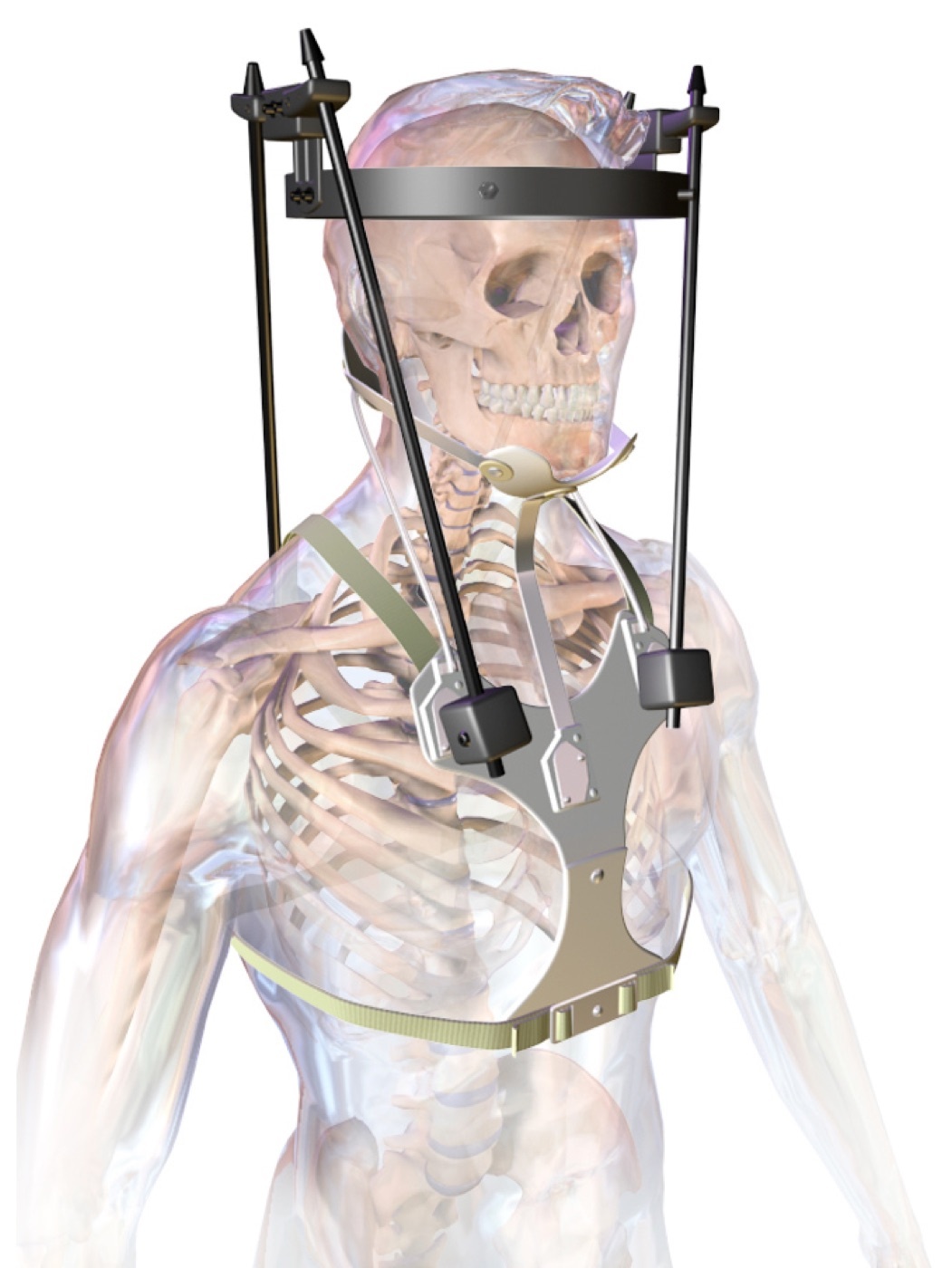
Figure 10. Halo vest for cervical-spine traction and immobilisation - skull pins fixed to a rigid vest by anterior and posterior rods. Source: BruceBlaus, via Wikimedia Commons, CC BY-SA 4.0.
Thoracolumbar traction is not used definitively, but a short period of traction on a Cotrel frame is occasionally used to reduce a burst fracture before casting, and prolonged bed rest may require a rotating (Stryker) frame for skin care.[77]
Part V - Functional Bracing (Sarmiento) and Cast-Bracing
The idea: motion is not the enemy of union
The orthodoxy that a fracture must be rigidly immobilised to heal is, for many diaphyseal fractures, simply wrong. Augusto Sarmiento, the leading modern advocate of conservative care, showed that a well-moulded brace that compresses the soft tissues while allowing the adjacent joints to move could let a tibial, and later a humeral, fracture heal with excellent function.[78] He introduced functional bracing to the humeral shaft in 1977 after good results in the tibia, femur, forearm and wrist, and humeral functional bracing is now regarded as the gold standard for that fracture, having displaced every other form of conservative humeral treatment.[79]
Three biomechanical principles underlie it:[80]
- Active muscle contraction self-corrects alignment. As the muscles around the fracture contract, their fibres recoil and pull the fragments back into rotation and alignment; the small amount of motion permitted at the fracture site actually promotes osteogenesis through increased vascularity, piezoelectric potentials and local metabolic changes.
- The “hydraulic effect.” Circumferential compression of the muscle compartment by the brace turns the incompressible soft tissue into an “inner splint” that aligns and supports the fragments.
- The beneficial effect of gravity on a dependent limb.
These map neatly onto Rockwood’s three cast principles (soft-tissue hinge, three-point fixation, hydrostatic pressure): the brace simply trades some rigidity for the freedom of the joints.[81]
The braces in practice
The humeral functional brace is two prefabricated polypropylene sleeves clamped together by adjustable Velcro straps, wider on the lateral side to cradle the humerus.[82] It is not applied at once: the arm is first rested for 5-10 days in a hanging cast, coaptation splint, Velpeau dressing or sling-and-swathe until the acute pain and swelling settle, and only then is the brace fitted, with a collar-and-cuff for comfort and active elbow movement encouraged from about four weeks.[83] Sarmiento’s large follow-up, 620 of 922 fractures, reported nonunion under 2% in closed fractures (6% in open), full shoulder range in about 60% and full elbow range in about 76% at brace removal, with roughly 90% healing within 16° of angular deformity.[84] The technique has limits: it struggles in morbid obesity or with large pendulous breasts (which drive the fracture into varus), and contemporary patient-reported-outcome studies challenge the old assumption of uniformly good results, with some authors now arguing that surgery should be the standard for displaced shaft fractures, a genuine and live controversy.[85]
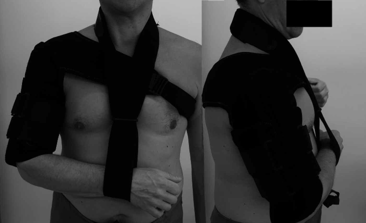
Figure 11. Humeral functional (clamshell) brace with a support sling for conservative treatment of a humeral-shaft fracture - the Sarmiento principle in practice. From Arealis et al. (2021), PMC8174392, CC BY 4.0.
For the tibia, the patellar-tendon-bearing (PTB) brace carries the same below-knee shell up to the patellar tendon but adds an ankle hinge and heel cup so the ankle can move, worn inside a shoe; the usual sequence is a long-leg cast for four to six weeks followed by a PTB cast or brace.[86] Its great advantage over operative care is the absence of infection and of fixation-related problems (hardware failure, anterior knee pain), and union rates are high in selected fractures, but the evidence is honestly mixed. Sarmiento’s excellent results came from selected, ambulatory patients who excluded those with excessive initial shortening or progressive angulation; an RCT of functional bracing against intramedullary nailing (Hooper) was stopped early because nailing did better, and IM nailing is now the treatment of choice for the tibial shaft.[87] Comparable distal forearm and metacarpal braces exist; comparative trials of cast versus brace at the wrist show no clear advantage to either.[88]
Part VI - Acceptable Alignment and Remodelling
What “acceptable” means
Because it is genuinely difficult to hold an unstable fracture in a cast, surgeons long ago defined how much malposition could be tolerated without functional penalty (the “acceptable” malunion) and the corollary rule: “if the fracture position is not maintained by the cast, consideration should be given to operative treatment.”[89] The thresholds differ by bone and by plane, but one general principle holds across the skeleton: angulation, shortening and displacement have tolerance limits, but rotational malalignment is essentially never accepted, because the body cannot remodel rotation and a malrotated limb is functionally and cosmetically obvious.[90]
The numbers most worth carrying into an exam:
- Humeral shaft (which tolerates wide malunion thanks to the mobile shoulder and hinged elbow): acceptable up to < 3 cm of shortening (bayonet apposition), < 20° anterior angulation, < 30° varus angulation and < 30° rotation.[91]
- Distal radius (adult): the AAOS guideline accepts radial shortening < 5 mm versus the other side, radial inclination > 15° on the PA film, sagittal tilt between 15° dorsal and 20° volar, and an intra-articular step or gap < 2 mm.[92] The biomechanical rationale is striking: shortening of as little as 2.5 mm raises the load on the distal ulna by 18-42%, and dorsal tilt shifts the ulnar load from 21% (at 10° volar tilt) to 67% (at 45° dorsal tilt), with half the load on the ulna at just 30° dorsal tilt.[93] Lafontaine’s predictors of losing a good reduction (dorsal tilt > 20°, comminution, intra-articular extension, an associated ulnar fracture and age > 60) flag the wrist that will need surgery.[94]
- Scaphoid: a fracture is “displaced,” and so unstable, with a four-fold higher nonunion rate in cast, if there is a gap ≥ 1 mm, an intrascaphoid angle > 45°, or a height-to-length ratio > 0.65 (humpback collapse).[95]
Why children are different
The growing skeleton forgives far more, because an open physis and an active periosteum remodel deformity over time, most reliably in the plane of joint motion, least reliably for rotation, and more powerfully the younger the child and the closer the fracture to the physis. Noonan’s widely used guide for the distal radius captures the scale of it: under 9 years, complete displacement, 15° of angulation and 45° of malrotation may all be acceptable; at 9 years and over, up to 30° of malrotation and complete bayonet apposition can still be accepted provided angulation is ≤ 20° and at least two years of growth remain.[96] This is why a both-bones forearm fracture that would demand open reduction in an adult is often treated in a simple cast in a child. Buckle (torus) fractures, being inherently stable, increasingly warrant a removable splint rather than a cast.[97]
Part VII - Non-operative Management Region by Region
The principles above translate into a fairly consistent regional practice. The following condenses Rockwood’s Tables 9-9 and 9-10, distinguishing where non-operative care is the genuine treatment of choice from where it is only a last resort when surgery is unavailable.[98]
Shoulder girdle and arm. The clavicle, scapula, proximal humerus and radial head are nearly all treated with a sling for about two weeks followed by mobilisation, the treatment of choice for the great majority, with surgery reserved for completely displaced midshaft clavicles in active adults and for selected displaced proximal humeral fractures (where Cochrane-level evidence shows surgery is not superior at one to two years).[99] The figure-of-eight clavicle bandage offers no advantage over a sling and causes more discomfort.[100] The humeral shaft is the home of functional bracing: a U-slab or sugar-tong for about two weeks, then a brace for 8-12 weeks.[101]
Forearm and hand. The distal radius is treated by a cast or brace for six weeks if stable, or by closed reduction (replicate the deformity, traction on finger-traps, three-point mould) and a cast if displaced, with a check film at 7-10 days; in the frail elderly, reduction is often of minimal value.[102] The scaphoid needs a scaphoid or short-arm cast for 6-12 weeks (slow union), the treatment of choice for undisplaced fractures, with surgery for displaced or proximal-pole fractures and high-demand patients.[103] For the metacarpals, the boxer’s fracture of the fifth neck is the commonest, 96% non-operative; stable fractures get buddy strapping, unstable ones a Burkhalter or James cast.[104] The phalanges take buddy strapping or an aluminium splint for stable fractures, a mallet splint (DIP extended six weeks) for the bony mallet.[105]
Both-bone forearm and the diaphyseal long bones tell the opposite story. The adult both-bone forearm, the femoral shaft and the tibial shaft are predominantly operative: the forearm because it is a “two-bone joint” demanding anatomical restoration of length and rotation, the femur and tibia because intramedullary nailing is decisively better. Non-operative treatment here is reserved for narrow stable subsets (an isolated minimally displaced ulnar shaft; a stable transverse tibia with an intact fibula) or for when no operative facility exists.[106]
Hip and lower limb. Proximal femoral and femoral shaft fractures are explicitly NOT recommended for non-operative care (the morbidity of prolonged bed rest, nonunion and mortality is too high), with the narrow exceptions of an undisplaced greater-trochanter or stress fracture.[107] Around the knee, undisplaced distal femoral, patellar and proximal tibial fractures are managed in a hinged knee brace or cylinder cast for 6-8 weeks.[108] The ankle is the lower-limb workhorse for conservative care: a below-knee cast or brace for six weeks for stable fractures (only ~9% of infrasyndesmotic type-A injuries need surgery), and close-contact casting performs as well as surgery in elderly unstable fractures.[109] Talar, calcaneal, midfoot, metatarsal and toe fractures are mostly non-operative (the displaced intra-articular calcaneus being a genuine ORIF-versus-cast controversy), and the pelvis is overwhelmingly non-operative in the elderly low-energy population, with restricted weight-bearing guided by pain for stable APC-1 and LC-1 patterns.[110]
Spine. The great majority of spinal fractures are elderly osteoporotic compression or insufficiency fractures, all managed non-operatively; bracing ranges from soft and hard collars (which still allow ~80% of cervical motion and act mainly as proprioceptive reminders) and the Philadelphia collar through to the Jewett brace (three-point, permitting extension but not flexion, for flexion-unstable T6-L3 injuries) and the TLSO, though Kim et al. found no advantage of a rigid over a soft or even no brace after an osteoporotic compression fracture.[111]
Part VIII - Complications of Cast and Closed Treatment
Acute compartment syndrome - the emergency that must never be missed
The most dangerous complication of a circumferential cast is acute compartment syndrome. “A full cast is rarely applied immediately after injury because of the potential for swelling… to lead to compartment syndrome if the limb is encased in a rigid cast,” and the principal complication of casts generally is elevated intracompartmental pressure, which in turn causes nerve injury, muscle necrosis and pressure sores.[112] The cardinal early sign is pain out of proportion to the injury and pain on passive stretch of the muscles in the compartment; the classically taught “5 Ps” (pain, paraesthesia, pallor, pulselessness, paralysis) are late and unreliable, and a palpable pulse never excludes the diagnosis.[113] The management of a threatened limb in plaster is immediate and mechanical: split the cast and all the underlying wool down to skin along its entire length (a univalved cast still raises pressure; bivalving and spreading releases more), elevate to the level of the heart (not above), and if symptoms persist, measure compartment pressures and proceed to fasciotomy.[114] Blood-soaked wool that dries into a rigid sheath produces a literal tourniquet effect, so any bleeding wound under plaster is a warning.[115] Every fresh cast mandates circulation checks in the first 24-48 hours, and the patient is told to return at once for increasing pain, numbness or swelling.[116]
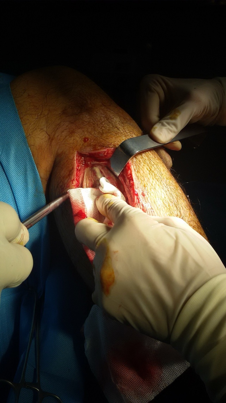
Figure 12. Fasciotomy for acute compartment syndrome - the limb-threatening end-point of an unrelieved tight cast. Source: Armin, via Wikimedia Commons, CC0 1.0.
Pressure sores, thermal injury and skin problems
Plaster (pressure) sores arise where a fold or tuck in the wool, or focal finger-pressure during moulding, creates a high-pressure point over a bony prominence; they are prevented by extra padding over the malleoli, heel, fibular head and sacrum and by moulding with the flat of the hand.[117] Thermal injury is a real and under-appreciated hazard: plaster of Paris sets by an exothermic reaction, and a thick cast, a fast-setting plaster, or dip-water that is too hot can raise the temperature against the skin enough to cause burns. Dip water should be cool-to-tepid, and a thick cast should not be rested on a pillow while it cures, which traps the heat.[118] Skin maceration and contact dermatitis occur under both casts and braces (the humeral brace in particular demands daily skin care), and patients are routinely warned to keep the cast dry and to put nothing down inside it.[119]
Fracture (cast) disease and joint stiffness
Prolonged immobilisation exacts a price that the older literature called “fracture disease” (or “cast disease”): the triad of joint stiffness, muscle wasting and disuse osteopenia, sometimes compounded by complex regional pain syndrome (CRPS).[120] Skeletal Trauma, which deliberately avoids the word “immobilisation,” warns that prolonged plaster or traction leads to “gross muscle wasting,” stiff joints and osteoporotic bone needing a long course of physiotherapy.[121] The figures are sobering: long-leg casts cause knee stiffness in up to a quarter to two-thirds of patients, and femoral traction produces knee stiffness in 30-50%.[122] This is the central argument for functional bracing and against needlessly long, joint-spanning immobilisation: every joint that can safely be left free, should be.
Loss of reduction, thromboembolism and nerve injury
Loss of reduction leading to malunion is the cardinal failure of closed treatment: “it is difficult to maintain the position of an unstable fracture in a cast,” and secondary displacement is easy to miss, which is why follow-up must be assiduous and any drift corrected early before soft-tissue contracture makes re-reduction impossible.[123] Venous thromboembolism is a genuine risk of lower-limb cast immobilisation and of the bed rest that traction imposes (a femoral cast-brace series reported 3% pulmonary emboli), and risk-stratified thromboprophylaxis should be considered for any patient immobilised in a lower-limb cast.[124] Finally, direct nerve compression under a cast is preventable: the common peroneal nerve at the fibular neck is the one to protect in any below-knee or PTB cast, and metacarpal braces have caused skin necrosis from local pressure.[125]
Part IX - When to Convert to Operative Treatment
Non-operative treatment is a decision that must be revisited, not made once and forgotten. The triggers to abandon the cast for surgery follow directly from the principles above:
- An unacceptable or unmaintainable reduction: if the fracture cannot be brought into acceptable alignment, or will not stay there as swelling resolves, “consideration should be given to operative treatment.”[126] A wrist that re-displaces despite a good cast (the Lafontaine high-risk pattern) is the everyday example.
- An inherently unstable pattern with no usable soft-tissue hinge: spiral, comminuted, segmental or high-energy fractures, and fractures in the elderly with friable periosteum, where three-point fixation cannot work.[127]
- An intra-articular fracture exceeding the joint’s congruity tolerance: a step or gap beyond the regional threshold (e.g. > 2 mm at the distal radius).[128]
- A threatened limb: open fracture, neurovascular injury, impending or established compartment syndrome, or skin in jeopardy.[129]
- A fracture for which the evidence simply favours surgery: the displaced femoral and tibial shaft (intramedullary nailing), the adult both-bone forearm, the displaced femoral neck.[130]
- The patient who cannot tolerate the conservative regimen: those who cannot endure prolonged recumbency or comply with a demanding bracing programme.
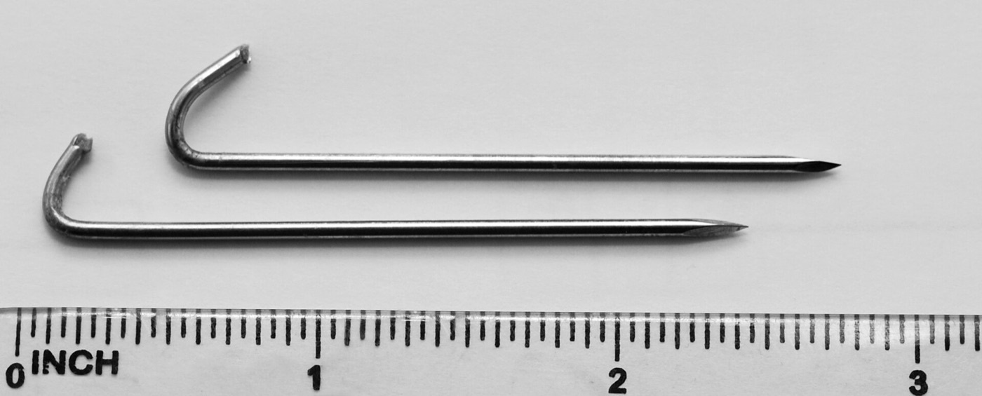
Figure 13. Kirschner wires, used for percutaneous pin fixation after closed reduction of a distal-radius fracture - the common minimally invasive step beyond a cast when a reduction will not hold. Source: Ard0, via Wikimedia Commons. Public domain.
The reverse is equally true: surgery is not automatically superior. For a large and growing list of fractures (the proximal humerus, the humeral shaft, many ankle fractures, the displaced midshaft clavicle, the calcaneus) high-quality trials show that careful conservative treatment matches operative treatment at lower cost and risk.[131] The skilled traumatologist is the one who can do both, and who knows which fracture is which.
References
-
Rockwood & Green, Fractures in Adults 9e, ch.9 (Court-Brown & Davidson), p.444.
-
Rockwood 9e p.444.
-
Rockwood 9e pp.449, 457 (Table 9-7).
-
Rockwood 9e p.457, Table 9-7.
-
Rockwood 9e p.457.
-
Skeletal Trauma: Basic Science, Management, and Reconstruction 5e, ch.6 “Closed Fracture Management” (Waddell, Wardlaw et al.), p.158.
-
Skeletal Trauma 5e p.158.
-
Skeletal Trauma 5e pp.158-159.
-
Skeletal Trauma 5e p.158.
-
Skeletal Trauma 5e p.159.
-
Skeletal Trauma 5e p.160.
-
Skeletal Trauma 5e p.160.
-
Rockwood 9e p.466.
-
Rockwood 9e p.466.
-
Rockwood 9e pp.449, 463.
-
Skeletal Trauma 5e p.158.
-
Rockwood 9e pp.466, 486; Skeletal Trauma 5e p.158.
-
Rockwood 9e p.467.
-
Skeletal Trauma 5e p.161.
-
Skeletal Trauma 5e p.161.
-
Skeletal Trauma 5e p.161.
-
Skeletal Trauma 5e p.161.
-
Skeletal Trauma 5e p.174.
-
Skeletal Trauma 5e p.175, Fig 6D-1.
-
Skeletal Trauma 5e p.161.
-
Skeletal Trauma 5e p.162.
-
Skeletal Trauma 5e pp.161-162.
-
Skeletal Trauma 5e p.162.
-
Skeletal Trauma 5e p.162.
-
Skeletal Trauma 5e p.161.
-
Skeletal Trauma 5e p.162.
-
Skeletal Trauma 5e p.174.
-
Skeletal Trauma 5e p.162.
-
Skeletal Trauma 5e p.162.
-
Skeletal Trauma 5e p.163.
-
Skeletal Trauma 5e p.163.
-
Skeletal Trauma 5e p.163.
-
Skeletal Trauma 5e p.163; Rockwood 9e p.466.
-
Skeletal Trauma 5e p.163.
-
Rockwood 9e p.466.
-
Rockwood 9e pp.466-467, Table 9-8.
-
Skeletal Trauma 5e p.163.
-
Skeletal Trauma 5e p.163.
-
Skeletal Trauma 5e p.164.
-
Skeletal Trauma 5e p.163.
-
Skeletal Trauma 5e pp.163-164, Fig 6B-4; Rockwood 9e p.463.
-
Skeletal Trauma 5e pp.163-164.
-
Rockwood 9e pp.463-465.
-
Rockwood 9e p.464.
-
Cast index (Chess et al.) is standard orthopaedic teaching; the numeric value is not stated in the mined Rockwood/Skeletal Trauma extracts and is supplied here from established practice.
-
Rockwood 9e pp.468-472; Skeletal Trauma 5e p.164.
-
Rockwood 9e pp.472-474; Skeletal Trauma 5e p.164.
-
Skeletal Trauma 5e pp.165-167.
-
Rockwood 9e pp.465-466.
-
Rockwood 9e p.467, Table 9-8.
-
Rockwood 9e pp.486-489.
-
Skeletal Trauma 5e p.166; Rockwood 9e pp.467-468, Fig 9-13.
-
Skeletal Trauma 5e p.166.
-
Rockwood 9e p.467.
-
Rockwood 9e p.457.
-
Rockwood 9e p.457.
-
Rockwood 9e pp.457-458.
-
Rockwood 9e p.458.
-
Rockwood 9e p.458. The numeric skin-traction load limit (~4.5 kg/10 lb) is standard teaching; it is not stated in the mined extracts.
-
Pin nomenclature (Steinmann, Kirschner, Denham) is standard orthopaedic teaching; the mined extracts name pin sites but not these devices.
-
The extracts name the distal femur, proximal tibia and calcaneus as pin sites (Rockwood 9e pp.457, 460); the olecranon site is standard teaching.
-
Skeletal Trauma 5e p.161.
-
Rockwood 9e pp.458-459.
-
Bryant/gallows traction is standard paediatric teaching; the mined Rockwood extract notes only that 90-90 traction is “still used for pediatric femoral fractures” and does not name Bryant traction.
-
Rockwood 9e pp.457-459.
-
Rockwood 9e p.459.
-
Rockwood 9e pp.460, 508.
-
Rockwood 9e pp.459-460.
-
Rockwood 9e p.460.
-
Rockwood 9e pp.460-462.
-
Rockwood 9e p.462.
-
Rockwood 9e pp.462-463.
-
Rockwood 9e p.445; Skeletal Trauma 5e p.179.
-
Skeletal Trauma 5e pp.177, 179.
-
Skeletal Trauma 5e p.179.
-
Rockwood 9e pp.463-465.
-
Rockwood 9e p.476; Skeletal Trauma 5e pp.177-179.
-
Skeletal Trauma 5e pp.177-179; Rockwood 9e p.497.
-
Skeletal Trauma 5e pp.180-181.
-
Skeletal Trauma 5e pp.177, 181; Rockwood 9e p.496.
-
Rockwood 9e pp.474, 479, 508.
-
Rockwood 9e pp.483-484.
-
Rockwood 9e pp.476-477, 484-485.
-
Rockwood 9e p.467.
-
Rockwood 9e p.467; Skeletal Trauma 5e p.159.
-
Skeletal Trauma 5e p.179.
-
Skeletal Trauma 5e p.174.
-
Skeletal Trauma 5e pp.173-174.
-
Skeletal Trauma 5e p.174.
-
Skeletal Trauma 5e pp.169-170.
-
Skeletal Trauma 5e pp.173-174.
-
Skeletal Trauma 5e p.174.
-
Rockwood 9e pp.489-490 (Table 9-9), 504-505 (Table 9-10), and the regional text pp.490-518.
-
Rockwood 9e pp.490-495.
-
Rockwood 9e pp.486, 491.
-
Rockwood 9e pp.495-497.
-
Rockwood 9e pp.500-501; Skeletal Trauma 5e p.174.
-
Rockwood 9e p.502.
-
Rockwood 9e p.503.
-
Rockwood 9e pp.503-504, 489.
-
Rockwood 9e pp.499-500, 505-508.
-
Rockwood 9e pp.505-506.
-
Rockwood 9e pp.506-507.
-
Rockwood 9e pp.509-510.
-
Rockwood 9e pp.510-514.
-
Rockwood 9e pp.480-483, 514-515.
-
Rockwood 9e pp.465, 467.
-
The “pain out of proportion / pain on passive stretch / 5 Ps” formulation is standard teaching; the mined extracts name compartment syndrome and stress prevention but do not list these clinical signs.
-
Skeletal Trauma 5e p.163; Rockwood 9e p.467.
-
Skeletal Trauma 5e p.163.
-
Skeletal Trauma 5e p.164.
-
Skeletal Trauma 5e p.163.
-
Exothermic-set thermal injury is standard teaching; the mined extracts describe the crystal-lattice setting reaction but do not flag burns.
-
Rockwood 9e p.467; Skeletal Trauma 5e pp.164, 177.
-
The terms “fracture/cast disease” and “CRPS” are standard teaching; the mined extracts describe the component features (stiffness, wasting, osteopenia) without using these labels.
-
Skeletal Trauma 5e pp.159-160.
-
Rockwood 9e pp.460, 484.
-
Rockwood 9e p.467.
-
Rockwood 9e p.459. Routine VTE risk-assessment for lower-limb cast immobilisation is standard current practice; dedicated prophylaxis guidance is not given in the mined extracts.
-
Rockwood 9e pp.474, 477; Skeletal Trauma 5e p.164.
-
Rockwood 9e p.467.
-
Rockwood 9e p.464.
-
Skeletal Trauma 5e p.174.
-
Skeletal Trauma 5e pp.160-161.
-
Rockwood 9e pp.499-508.
-
Skeletal Trauma 5e p.158.
-
Skeletal Trauma 5e p.159.
-
Skeletal Trauma 5e p.161.
-
Skeletal Trauma 5e pp.163-164; Rockwood 9e pp.463-465.
-
Rockwood 9e p.464.
-
Standard teaching (Chess et al.); not numerically stated in the mined extracts.
-
Rockwood 9e pp.465, 467.
-
Rockwood 9e pp.457-458; pin nomenclature and load figure are standard teaching.
-
Rockwood 9e pp.458-459; Bryant traction is standard paediatric teaching.
-
Rockwood 9e pp.457-460.
-
Skeletal Trauma 5e p.179.
-
Skeletal Trauma 5e pp.173-174.
-
Rockwood 9e p.467; Skeletal Trauma 5e p.163; clinical signs are standard teaching.
-
Skeletal Trauma 5e pp.159-160; term is standard teaching.
-
Rockwood 9e pp.464, 467, 499-508; Skeletal Trauma 5e pp.160-161, 174.
-
Skeletal Trauma 5e p.158; Rockwood 9e pp.494-496.