Contents
- Orientation: A Limb-Threatening Multiligament Injury
- Part I - The Knee Dislocation: Definition, Epidemiology, and Mechanism
- Part II - The Vascular and Neurological Emergency
- Part III - Assessment, the Dimple Sign, and Imaging
- Part IV - Classification
- Part V - Ligament Anatomy and Function
- Part VI - Clinical Examination of the Knee Ligaments
- Part VII - Associated Avulsion Fractures
- Part VIII - Treatment of the Multiligament Knee / Dislocation
- Part IX - Isolated Ligament Injuries
- Part X - Complications
- Part XI - A Synthesis: How to Reason About the Knee Dislocation
- References
Orientation: A Limb-Threatening Multiligament Injury
The knee (tibiofemoral) dislocation is, above all, a vascular emergency, and that fact dominates its management.[1] By definition it disrupts at least two of the four major ligaments and almost always tears at least one cruciate, so the knee is grossly unstable; but the danger that must be excluded first is injury to the popliteal artery, which is tethered behind the knee and is torn or contused in a substantial minority of dislocations, with a limb-threatening window of only a few hours. The second trap is that the knee often spontaneously reduces before the patient is seen, so a normal-looking, even effusion-free knee can hide a dislocation that has already endangered the artery and the common peroneal nerve. The clinician therefore approaches the multiligament knee with a fixed sequence: reduce it, exclude a vascular injury with a serial examination and the ankle-brachial index, recognise the irreducible (dimple-sign) dislocation that must go straight to theatre, classify the ligament injury by the Schenck system, and then plan a reconstruction that restores stability while permitting the early motion that prevents the stiffness, which is the commonest long-term complication.[2]
Part I - The Knee Dislocation: Definition, Epidemiology, and Mechanism
A knee dislocation is a displacement of the tibia on the femur with disruption of two or more major ligaments; it must tear at least one cruciate unless there is an associated fracture, and in most dislocations both cruciates are torn.[3] It is uncommon but under-reported, because so many dislocations spontaneously reduce before assessment and so many occur in the polytrauma patient. The mechanisms are bimodal: high-energy trauma (motor-vehicle crashes, contact sport) and, increasingly, ultra-low-velocity dislocations in the morbidly obese from a simple fall, which paradoxically carry a high rate of neurovascular complications. The unifying mechanical element is hyperextension (which tears the cruciates) combined with varus, valgus, or rotatory force; an anterior dislocation follows hyperextension and is the commonest, a posterior dislocation follows a dashboard blow to the anterior tibia, and the posterolateral rotatory dislocation is the classic irreducible pattern.[4]
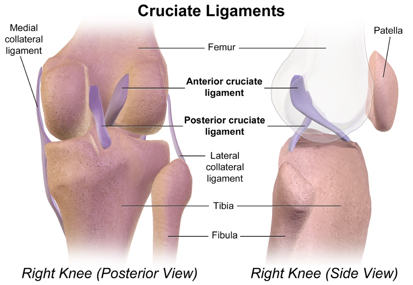
Figure 1. Posterior and lateral views of the knee showing the anterior and posterior cruciate ligaments and the medial and lateral collateral ligaments. Illustration by BruceBlaus, CC BY-SA 4.0, via Wikimedia Commons.
Part II - The Vascular and Neurological Emergency
The popliteal artery is the structure that makes a knee dislocation an emergency.[5] It is tethered at two points, proximally at the adductor hiatus (Hunter’s canal) and distally beneath the soleal arch, so it cannot move out of the way and is injured by traction in anterior (hyperextension) dislocations (it tears at about 50° of hyperextension) and by contusion or intimal damage in posterior dislocations. Reported incidences vary widely: classic series quote up to 40-50 % (detection bias and older data), a systematic review found 25 %, and contemporary literature reports flow-limiting injury in 5-15 %. The critical clinical points are that palpable pulses do not exclude a major arterial injury, that an asymptomatic intimal tear can thrombose late and cost the limb, and that revascularisation after about 6 hours gives very poor results, with delayed reperfusion carrying up to an 86 % amputation rate.[6]
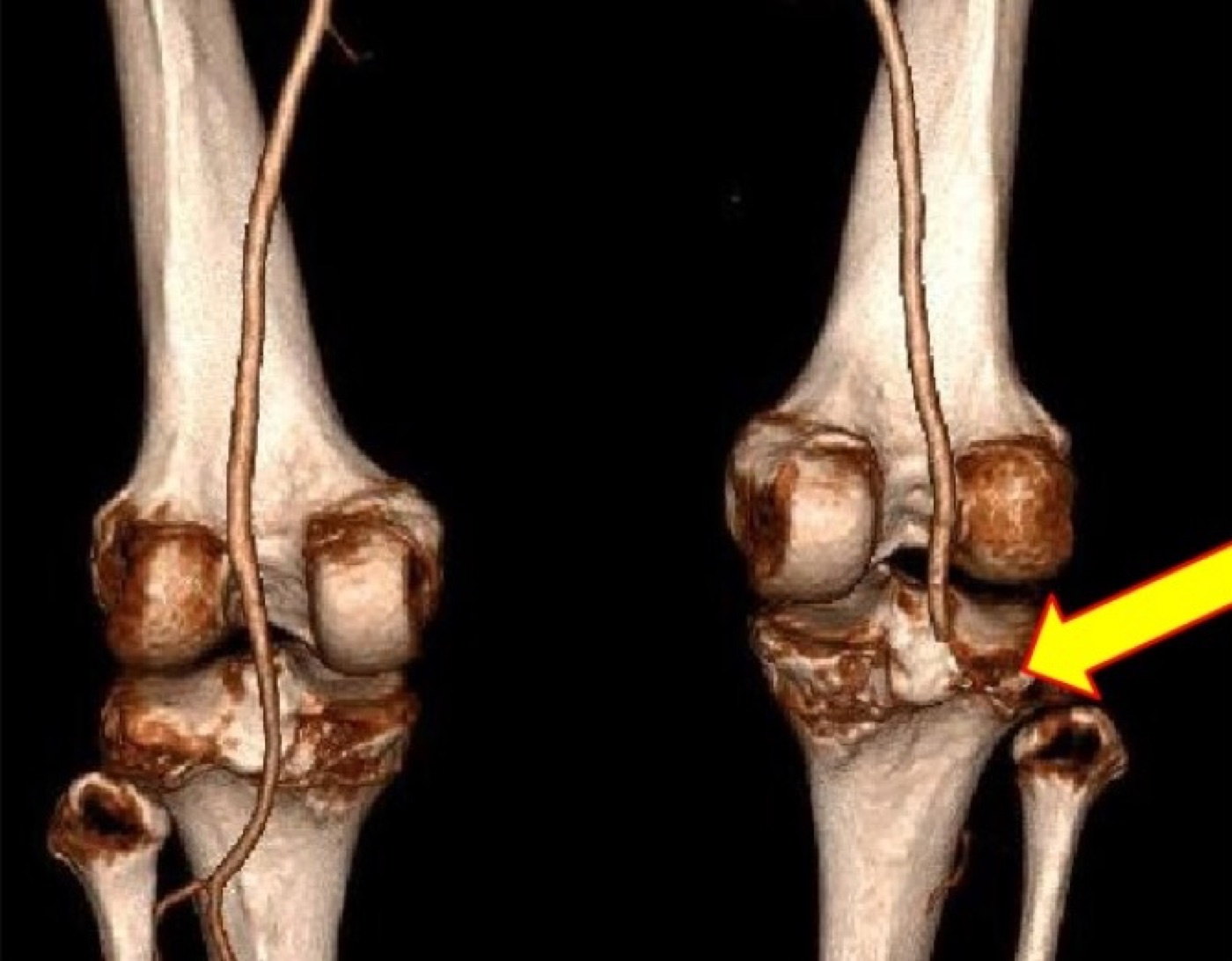
Figure 2. 3D CT angiogram (posterior view) showing occlusion of the right popliteal artery (arrow) with normal vasculature on the left, the vascular complication that makes a knee dislocation a surgical emergency. Image by Godfrey et al., CC BY 4.0, via Wikimedia Commons.
The vascular work-up is therefore systematic.[7] Every documented or presumed dislocation has a serial vascular examination and an ankle-brachial index (ABI): a normal ABI is 0.91-1.3, and a value above 0.9 has a 100 % negative predictive value for a major arterial lesion (Mills), so an ABI above 0.9 is followed by serial examinations (every 2-4 hours for 48 hours), while an ABI below 0.9 mandates CT angiography or formal arteriography. Hard signs of ischaemia in a known zone of injury go straight to surgical exploration; an absent distal pulse is an emergency. Vascular injury is repaired on the day of injury, usually with a vein graft, the limb first stabilised with a spanning external fixator (and sometimes a temporary arterial shunt to shorten ischaemic time) so the repair is not disrupted by an unstable knee. The other neurological casualty is the common peroneal nerve, injured in roughly a quarter to a third of dislocations (especially with posterolateral/varus injuries that stretch it over the lateral femoral condyle); its prognosis is poor, with only about half recovering and complete foot-drop palsies recovering in only about 38 % (versus 83 % of partial palsies), the unrecovered foot drop managed with an ankle-foot orthosis or a tendon transfer (a tibialis posterior tendon transfer, or a posterior tibial nerve transfer).[8]
Part III - Assessment, the Dimple Sign, and Imaging
Because most dislocations have spontaneously reduced, diagnosis rests on a high index of suspicion: a grossly unstable multiligament knee is a dislocation even if reduced on arrival, the effusion may be absent (the capsule has torn and decompressed), and skin clues (anterior-tibial “dashboard” abrasions) help.[9] The knee is reduced immediately by in-line traction and immobilised in extension. The crucial exception is the dimple sign: a fixed pucker of skin on the medial side indicates a posterolateral rotatory dislocation in which the medial femoral condyle has buttonholed through the capsule with the MCL incarcerated in the joint; this is irreducible and a contraindication to closed reduction, mandating open reduction. An open dislocation (5-17 %) is reduced and debrided in theatre. Imaging is AP and lateral radiographs (looking for subtle subluxation and the avulsion signs), with MRI the gold standard for mapping the ligaments, and examination under anaesthesia the most accurate assessment of instability. The AO regards EUA as the diagnostic gold standard, more accurate than MRI for rotational instability, which can over-diagnose tears.[10]
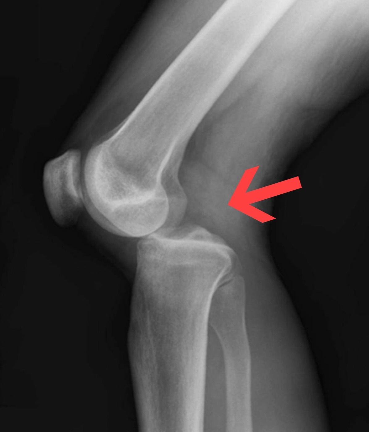
Figure 3. Lateral radiograph demonstrating a posterior tibiofemoral (knee) dislocation, the tibia displaced posteriorly (arrow). Image by Duprey & Lin, CC BY 4.0, via Wikimedia Commons.
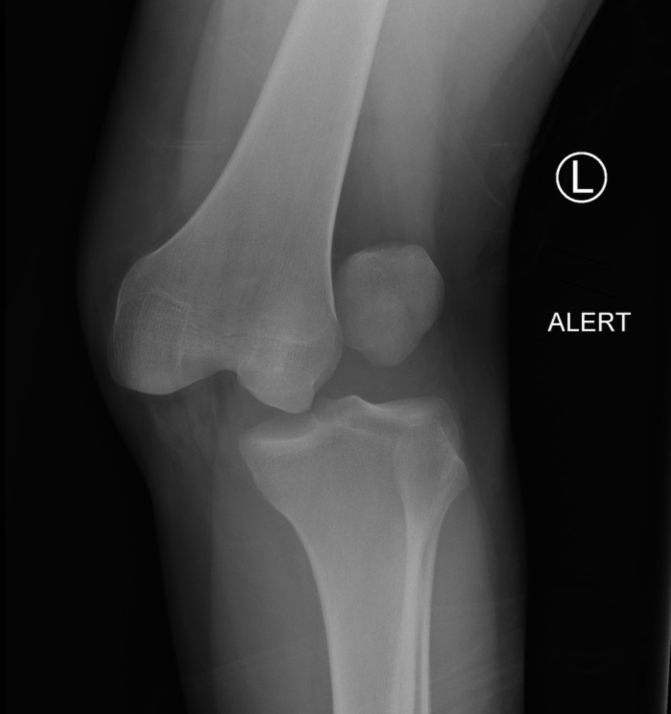
Figure 4. Radiograph of a lateral knee dislocation. Image by Andrew Murphy, CC BY-SA 4.0, via Wikimedia Commons.
Part IV - Classification
The standard scheme is the Schenck anatomic knee-dislocation (KD) classification, which groups injuries by the structures torn (best confirmed by examination under anaesthesia and MRI).[11] KD-I has one cruciate intact; KD-II has both cruciates torn with both collaterals intact (uncommon); KD-III has both cruciates plus one collateral, subdivided into KD-IIIM (medial-sided, the posteromedial corner) and KD-IIIL (lateral-sided, the posterolateral corner); KD-IV has both cruciates and both collaterals; and KD-V is a fracture-dislocation. The suffixes C (circulatory/arterial injury) and N (neurological injury) are appended, so a KD-IIIM with a popliteal injury is “KD-IIIM-C.” The older Kennedy positional classification describes the direction of tibial displacement: anterior (hyperextension, commonest, with traction arterial injury), posterior (dashboard, with arterial contusion and extensor disruption), medial, lateral, and rotatory (the posterolateral subtype being the classic irreducible dislocation with the dimple sign). It applies poorly, however, to the many knees that reduce spontaneously.[12]
Part V - Ligament Anatomy and Function
The four ligaments have distinct primary roles that the surgeon must restore.[13] The anterior cruciate ligament, with its anteromedial and posterolateral bundles, is the primary restraint to anterior tibial translation (its in-situ force is highest at about 30° of flexion). The posterior cruciate ligament is the largest and strongest intracapsular ligament (its cross-section is 120-150 % that of the ACL and the dominant anterolateral bundle, with a tensile strength of about 1620 N, is the major contributor), and it is the primary restraint to posterior tibial translation, providing about 95 % of posterior stability between 30° and 90°. The medial collateral ligament complex (superficial and deep MCL plus the posteromedial corner’s posterior oblique ligament) is the primary restraint to valgus (clinically tested at 30°), with the posteromedial corner contributing about a third of valgus restraint in extension. The fibular (lateral) collateral ligament is the primary restraint to varus at all angles (especially 0-30°), and the posterolateral corner (the FCL, popliteus tendon, and popliteofibular ligament together) is the primary restraint to varus, external rotation, and posterolateral rotation; this is why an unrecognised posterolateral corner injury is a classic cause of cruciate graft failure.[14]
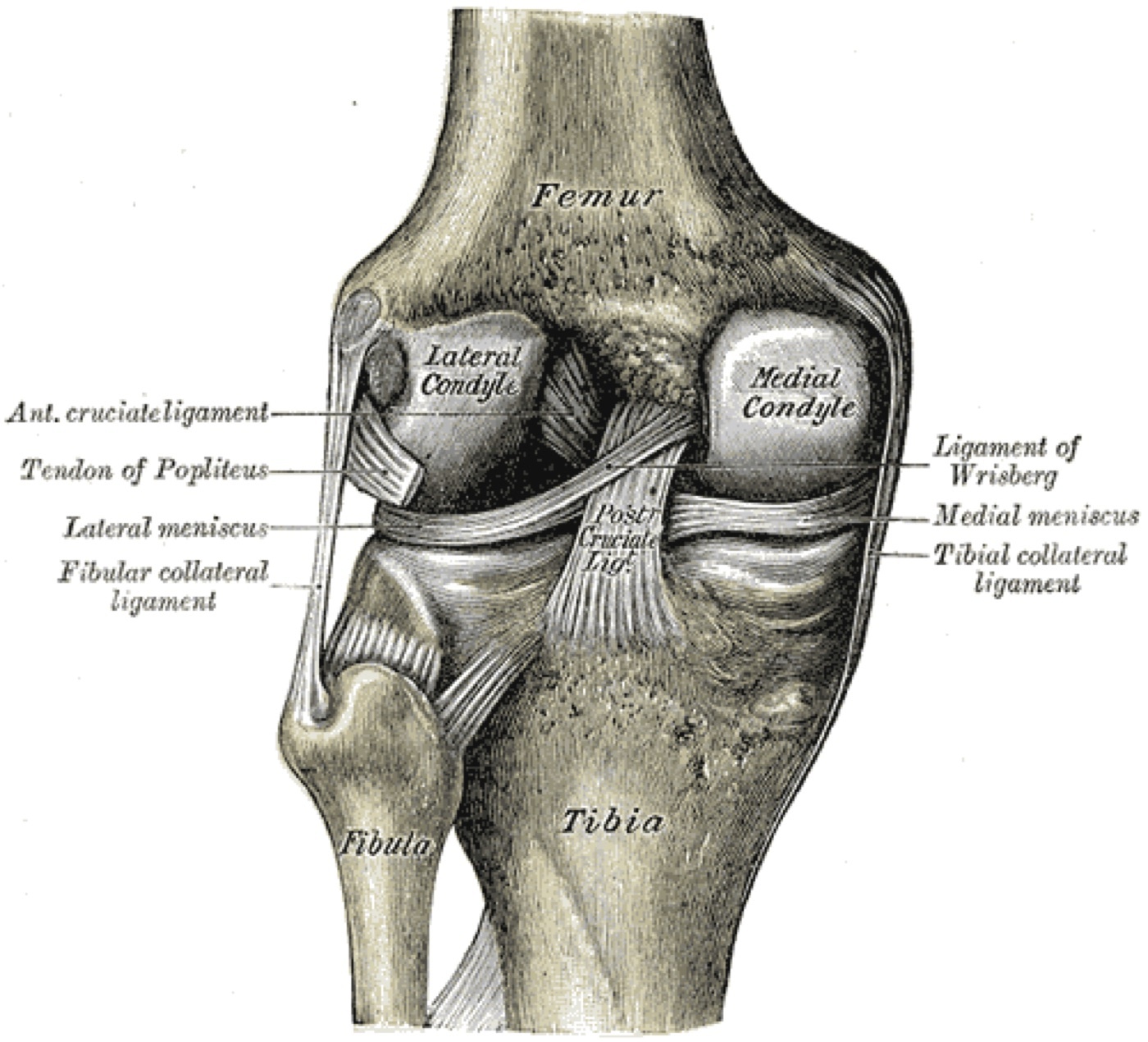
Figure 5. Posterior view of the right knee with the femur lifted, showing the cruciate (ACL, PCL) and collateral (MCL, LCL) ligaments, the menisci, and the popliteus tendon. Gray’s Anatomy (Plate 348), public domain, via Wikimedia Commons.
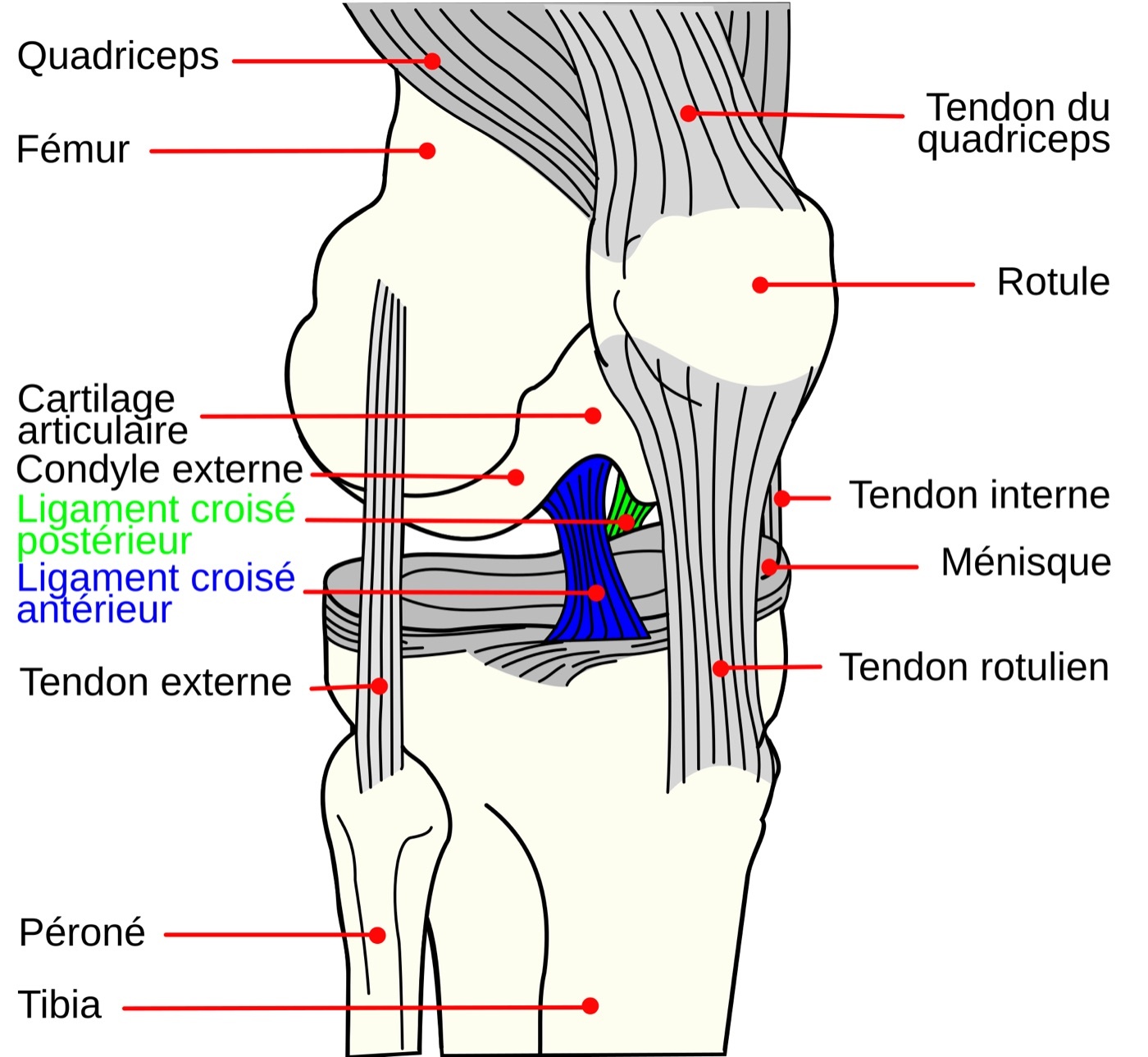
Figure 6. Sagittal schematic of the knee showing the anterior (blue) and posterior (green) cruciate ligaments among the surrounding structures. Public domain, via Wikimedia Commons.
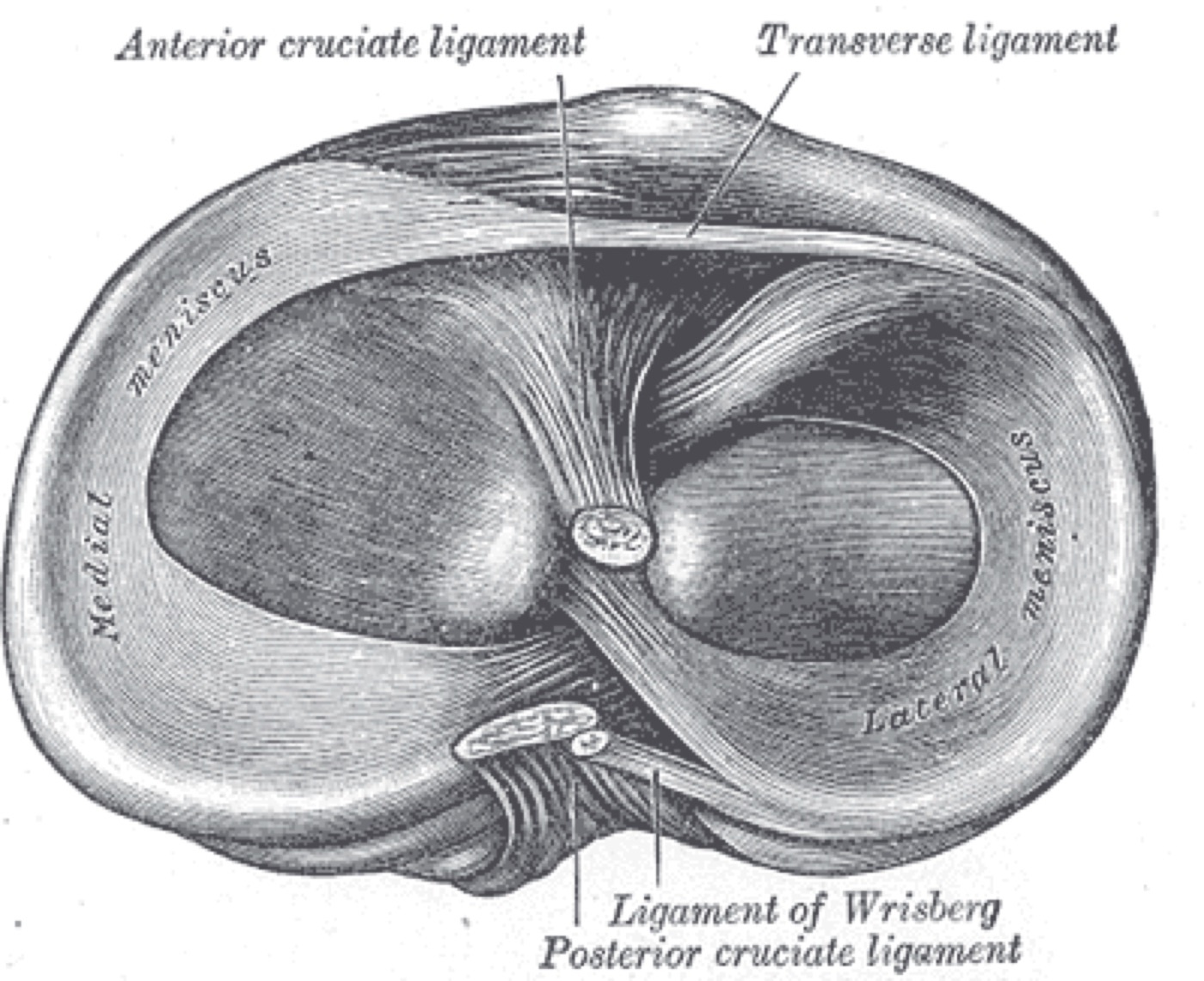
Figure 7. Head of the right tibia seen from above, showing the menisci and the tibial attachments of the cruciate ligaments. Gray’s Anatomy (Plate 349), public domain, via Wikimedia Commons.
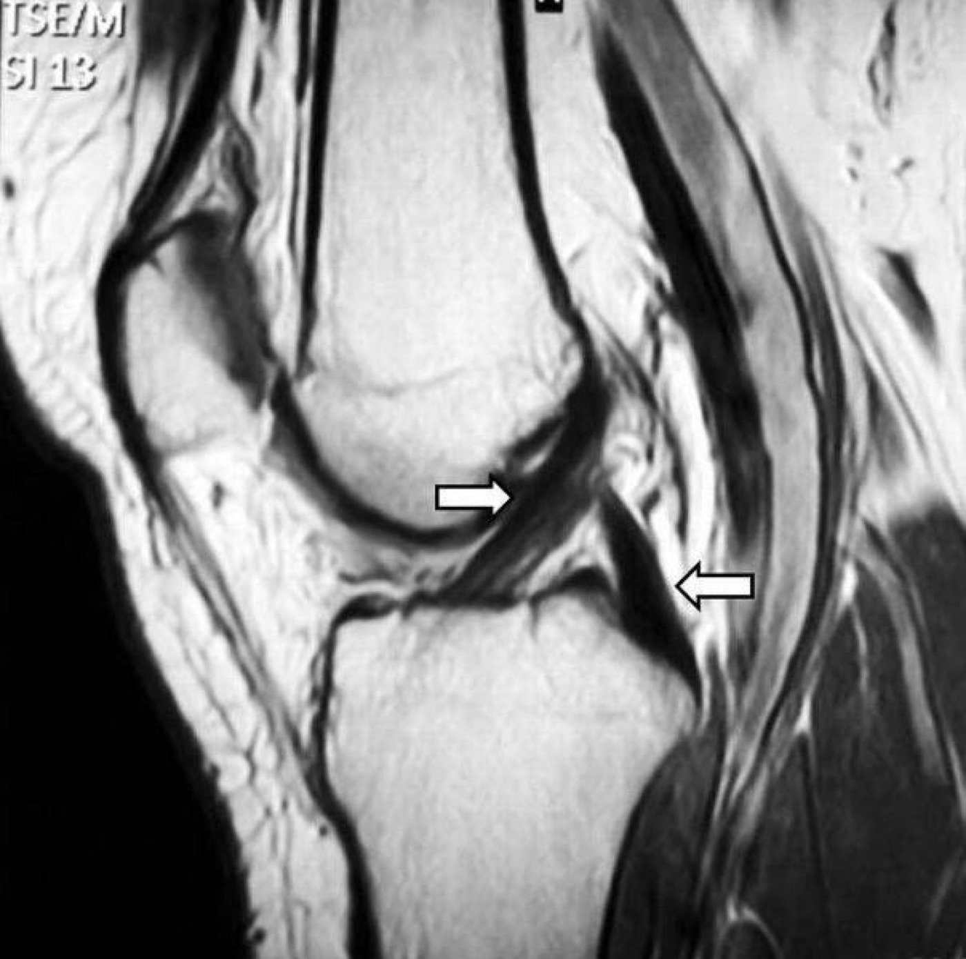
Figure 8. Sagittal knee MRI showing the normal anterior and posterior cruciate ligaments (arrows), the reference appearance against which tears are read. Image by Lykissas et al., CC BY-SA 2.0, via Wikimedia Commons.
Part VI - Clinical Examination of the Knee Ligaments
Each ligament has a defining test.[15] The ACL is assessed by the Lachman test (the most sensitive), the anterior drawer, and the pivot shift (whose grade correlates best with the outcome of reconstruction). The PCL is assessed by the posterior drawer, graded by the tibial step-off: grade I leaves the tibia anterior to the femoral condyles, grade II flush, and grade III posterior (grade III usually means an associated ACL or posterolateral corner injury); the posterior sag (Godfrey) and quadriceps-active tests are supporting signs (standard teaching). The MCL is tested by valgus stress at 30° (laxity that also opens in full extension implies a concurrent cruciate injury), with laxity graded 3-5 mm (I), 6-10 mm (II), and over 10 mm (III). The LCL is tested by varus stress at 30°, and the posterolateral corner by the dial test: increased external rotation at 30° only indicates an isolated posterolateral corner injury, whereas an increase at both 30° and 90° indicates a combined posterolateral corner and PCL injury; the external-rotation recurvatum and reverse-pivot-shift tests support it.[16]
Part VII - Associated Avulsion Fractures
A handful of avulsion fractures are radiographic signatures of specific ligament injuries.[17] The Segond fracture, a small avulsion of the lateral proximal tibia (the lateral capsule / anterolateral ligament), is strongly associated with an ACL injury. Its mirror, the reverse Segond (a medial proximal-tibial avulsion at the deep MCL attachment), is associated with a PCL injury and a medial meniscal tear (standard teaching). The arcuate sign, an avulsion of the fibular styloid by the popliteofibular ligament, is essentially pathognomonic of a posterolateral corner injury, and in such cases an associated cruciate injury is present in about 89 %. A tibial eminence (spine) avulsion is the bony equivalent of an ACL tear (common in children), and the Pellegrini-Stieda lesion is calcification at the femoral MCL origin of a chronic medial injury.[18]
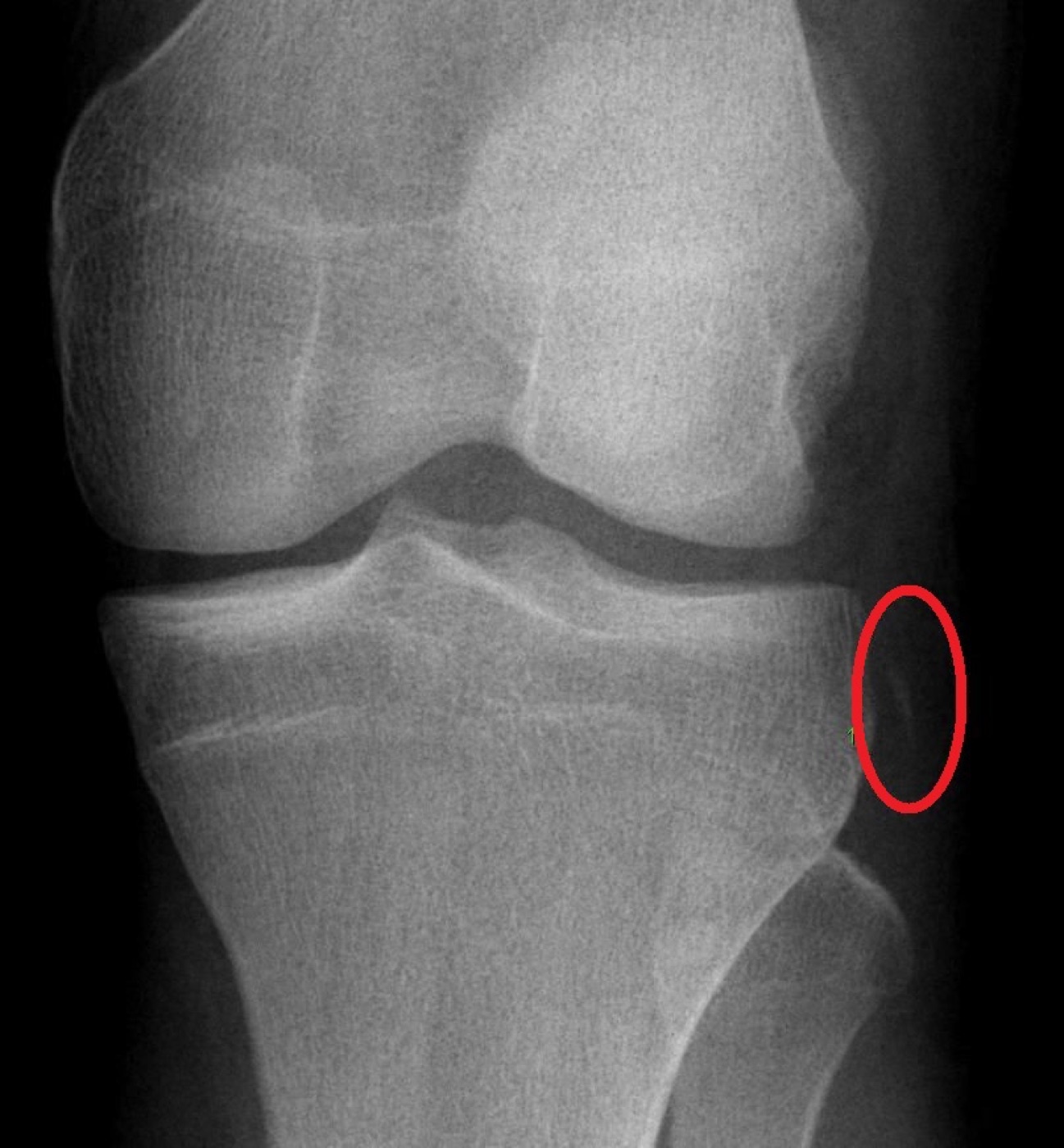
Figure 9. AP knee radiograph showing a Segond fracture, a small avulsion off the lateral tibial plateau (circled), highly associated with an ACL tear. Public domain, via Wikimedia Commons.
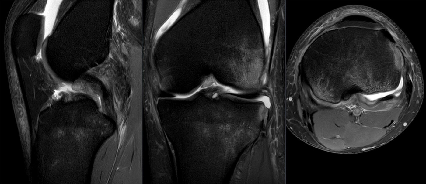
Figure 10. Fat-suppressed knee MRI (sagittal, coronal, axial) of a Segond avulsion with an associated anterior cruciate ligament tear. Image by Hellerhoff, CC BY-SA 4.0, via Wikimedia Commons.
Part VIII - Treatment of the Multiligament Knee / Dislocation
Initial management is reduction, vascular surveillance, and stabilisation.[19] The reduced knee is immobilised in extension (a posterior tibial pad protects a high-grade PCL injury), and a knee-spanning external fixator is used when there is a vascular repair to protect, an open injury, gross instability, or morbid obesity (it is not applied routinely, since pin-site infection threatens later reconstruction). The emergencies are addressed in theatre: vascular injury, open dislocation, irreducible (dimple-sign) dislocation, and compartment syndrome, with the fixator usually applied before the vascular repair so the graft heals undisturbed. Definitive ligament treatment is, for most patients, operative, because operative management gives consistently better stability and function than nonoperative care (nonoperative treatment is reserved for the patient who cannot survive surgery); historical prolonged immobilisation produced a stiff knee and is abandoned.[20]
The reconstruction itself turns on four decisions, each with a clear modern tendency.[21] Timing: early surgery (within about 3 weeks, optimally 10-21 days, while structures are still identifiable) tends to give better function, though very early surgery raises the risk of arthrofibrosis, so a staged approach is often used and a delay beyond 6-8 weeks gives less favourable results. Repair versus reconstruction: the cruciates are reconstructed (the ACL is always reconstructed; the PCL is usually reconstructed and done first as the cornerstone), and the corners are increasingly reconstructed rather than repaired: repair of the posterolateral corner fails in about 37-40 % versus 6-9 % for reconstruction, and even medial-side repair fails more than reconstruction, though a bony avulsion is a good indication for primary repair. Graft source: autograft is favoured in the young and high-demand patient (often from the uninjured contralateral knee), while allograft avoids donor-site morbidity and allows many structures to be reconstructed at once. Rehabilitation: the construct must be strong enough to allow early motion, because arthrofibrosis is the commonest complication, and a hinged external fixator after reconstruction reduces the failure rate (21 % with a brace versus 7 % in a randomised trial).[22]
Part IX - Isolated Ligament Injuries
Away from the dislocation, the isolated ligament injuries have characteristic treatments.[23] An isolated ACL rupture is reconstructed in the active patient (it is not repaired, as the stump will not heal), using a BPTB, hamstring, quadriceps-tendon, or allograft graft, with the key principle of regaining full motion before surgery to avoid arthrofibrosis. An isolated PCL injury of grade I or II is usually treated nonoperatively with quadriceps strengthening and extension bracing, reserving reconstruction for high-grade or combined injuries. An isolated MCL injury, even grade III, heals well nonoperatively in a hinged brace with early motion, the femoral-side injury healing better than the tibial-side. The exception is the posterolateral corner, which does not heal nonoperatively: it requires early repair or reconstruction (isolated repair failing in up to 40 %, so a hybrid of repair plus reconstruction is favoured), and the highest-yield teaching of all is that a missed posterolateral corner injury is a leading cause of ACL and PCL graft failure.[24]
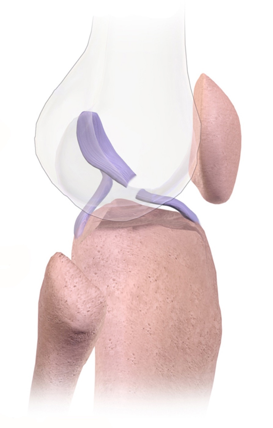
Figure 11. Illustration of an anterior cruciate ligament (ACL) tear. Illustration by BruceBlaus, CC BY-SA 4.0, via Wikimedia Commons.
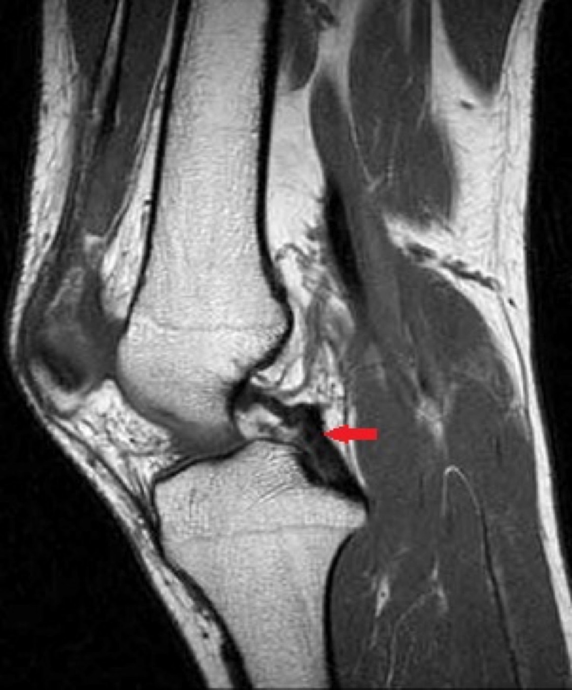
Figure 12. Sagittal T1 knee MRI showing the posterior cruciate ligament (arrow). Image by SCiardullo, CC BY-SA 4.0, via Wikimedia Commons.
Part X - Complications
The commonest complication of the knee dislocation is stiffness / arthrofibrosis: pooling many studies, about 30 % require surgery for arthrofibrosis, and the mean arc of motion has improved from 106° (older series) to 123° with anatomical reconstruction and early motion.[25] Residual laxity is also common (about 42 % have at least one unstable ligament after surgery, and nonoperative treatment leaves essentially universal instability). The feared vascular complication is the missed popliteal injury, with up to an 86 % amputation rate when perfusion is not restored within 6-8 hours, and an iatrogenic injury is a risk during PCL tunnel reaming (drill with the knee flexed to 90° so the neurovascular bundle falls away). Peroneal nerve palsy often does not recover. Heterotopic ossification occurs in about a third, and post-traumatic arthritis is common (about a quarter at ten years). Functional recovery is sobering: long-term pain in 25-68 %, return to some work in 93 % (but a third to a less demanding job), and return to the pre-injury level of sport in only about 39 %.[26]
Part XI - A Synthesis: How to Reason About the Knee Dislocation
Treat the knee dislocation as a vascular emergency first and a ligament problem second. Reduce it immediately (unless the dimple sign says it is an irreducible posterolateral dislocation that must go to theatre), then exclude a popliteal artery injury with a serial examination and the ankle-brachial index, remembering that palpable pulses do not exclude an arterial injury and that an intimal tear can thrombose late; an ABI below 0.9 means CT angiography, and a confirmed arterial injury is repaired the same day over a spanning external fixator. Recognise the common peroneal nerve palsy, which usually does not recover. Classify the ligament injury by the Schenck KD system with its C and N modifiers, map it with MRI and examination under anaesthesia, and plan a definitive reconstruction (the ACL always, the PCL first, the corners reconstructed rather than repaired) timed early but not so early as to provoke arthrofibrosis, delivered through a construct strong enough to allow the early motion that prevents stiffness. Among the isolated injuries, hold the rule that the MCL heals and the posterolateral corner does not, and that a missed posterolateral corner is what makes cruciate grafts fail.
References
-
AO Principles of Fracture Management, pp.885, 888, 889 (a knee dislocation is an abnormal tibiofemoral displacement with injury to two or more major ligaments, almost always at least one cruciate; the popliteal artery emergency; spontaneous reduction making the diagnosis easy to miss); Rockwood & Green’s Fractures in Adults, pp.4170, 4172, 4173, 4199 (the limb-threatening potential, the tethered popliteal artery, the spontaneously-reduced caveat, and the modern shift to early reconstruction with motion).
-
AO Principles of Fracture Management, pp.885, 888, 889 (a knee dislocation is an abnormal tibiofemoral displacement with injury to two or more major ligaments, almost always at least one cruciate; the popliteal artery emergency; spontaneous reduction making the diagnosis easy to miss); Rockwood & Green’s Fractures in Adults, pp.4170, 4172, 4173, 4199 (the limb-threatening potential, the tethered popliteal artery, the spontaneously-reduced caveat, and the modern shift to early reconstruction with motion).
-
AO Principles of Fracture Management, pp.885, 886 (definition as ≥2 major ligaments with at least one cruciate, both cruciates usually torn; under-reporting from spontaneous reduction and polytrauma; motor-vehicle and contact-sport mechanisms, the morbidly-obese ultra-low-velocity dislocation, hyperextension tearing the ACL, the posterolateral irreducible pattern); Rockwood & Green’s Fractures in Adults, pp.4170, 4171 (rarity and under-reporting, high-energy vs ultra-low-velocity obese mechanisms with high neurovascular complication rates, hyperextension plus varus/valgus/rotatory forces).
-
AO Principles of Fracture Management, pp.885, 886 (definition as ≥2 major ligaments with at least one cruciate, both cruciates usually torn; under-reporting from spontaneous reduction and polytrauma; motor-vehicle and contact-sport mechanisms, the morbidly-obese ultra-low-velocity dislocation, hyperextension tearing the ACL, the posterolateral irreducible pattern); Rockwood & Green’s Fractures in Adults, pp.4170, 4171 (rarity and under-reporting, high-energy vs ultra-low-velocity obese mechanisms with high neurovascular complication rates, hyperextension plus varus/valgus/rotatory forces).
-
Rockwood & Green’s Fractures in Adults, pp.4172, 4179, 4180, 4182, 4235 (popliteal artery tethered at the adductor hiatus and soleal arch; traction injury in anterior and contusion/intimal in posterior dislocations; incidence up to 40-50 %; palpable pulses not excluding injury; up to 86 % amputation if perfusion not restored within 6-8 hours); AO Principles of Fracture Management, pp.885, 888, 889 (popliteal artery tears at 50° hyperextension; systematic-review 25 % and contemporary flow-limiting 5-15 %; late thrombosis from an asymptomatic intimal tear; revascularisation after 6 hours very poor). The “6-8 hour” warm-ischaemia window is standard teaching; the AO states results after 6 hours are very poor without using that exact phrase.
-
Rockwood & Green’s Fractures in Adults, pp.4172, 4179, 4180, 4182, 4235 (popliteal artery tethered at the adductor hiatus and soleal arch; traction injury in anterior and contusion/intimal in posterior dislocations; incidence up to 40-50 %; palpable pulses not excluding injury; up to 86 % amputation if perfusion not restored within 6-8 hours); AO Principles of Fracture Management, pp.885, 888, 889 (popliteal artery tears at 50° hyperextension; systematic-review 25 % and contemporary flow-limiting 5-15 %; late thrombosis from an asymptomatic intimal tear; revascularisation after 6 hours very poor). The “6-8 hour” warm-ischaemia window is standard teaching; the AO states results after 6 hours are very poor without using that exact phrase.
-
Rockwood & Green’s Fractures in Adults, pp.4172, 4173, 4180, 4182 (serial vascular examination and ABI in all dislocations; normal ABI 0.91-1.3, >0.9 = 100 % NPV [Mills], <0.9 → CT angiography, serial exams every 2-4 hours for 48 hours; peroneal palsy ~25 %, complete-palsy recovery 38 % vs 83 % partial, posterior tibial transfer); AO Principles of Fracture Management, pp.887, 888, 889, 890 (absent distal pulse → emergent exploration, ABPI <0.9 → urgent imaging/surgery, vascular repair on the day of injury with a vein graft, spanning external fixator and temporary shunt; peroneal injury in one-third, 50 % or fewer recovering, late tibialis posterior transfer).
-
Rockwood & Green’s Fractures in Adults, pp.4172, 4173, 4180, 4182 (serial vascular examination and ABI in all dislocations; normal ABI 0.91-1.3, >0.9 = 100 % NPV [Mills], <0.9 → CT angiography, serial exams every 2-4 hours for 48 hours; peroneal palsy ~25 %, complete-palsy recovery 38 % vs 83 % partial, posterior tibial transfer); AO Principles of Fracture Management, pp.887, 888, 889, 890 (absent distal pulse → emergent exploration, ABPI <0.9 → urgent imaging/surgery, vascular repair on the day of injury with a vein graft, spanning external fixator and temporary shunt; peroneal injury in one-third, 50 % or fewer recovering, late tibialis posterior transfer).
-
Rockwood & Green’s Fractures in Adults, pp.4173, 4174, 4177 (high suspicion, the absent effusion from capsular disruption, skin clues, immediate reduction and extension immobilisation; the medial dimple sign of an irreducible posterolateral dislocation as a contraindication to closed reduction; open dislocation 5-17 %; MRI the ligament gold standard); AO Principles of Fracture Management, pp.885, 888, 889, 891 (spontaneous reduction with no deformity, anterior-tibial contusions raising suspicion; the buttonholed medial condyle and dimple sign; open dislocation reduced and debrided in theatre; examination under anaesthesia the diagnostic gold standard, more accurate than MRI for rotational instability).
-
Rockwood & Green’s Fractures in Adults, pp.4173, 4174, 4177 (high suspicion, the absent effusion from capsular disruption, skin clues, immediate reduction and extension immobilisation; the medial dimple sign of an irreducible posterolateral dislocation as a contraindication to closed reduction; open dislocation 5-17 %; MRI the ligament gold standard); AO Principles of Fracture Management, pp.885, 888, 889, 891 (spontaneous reduction with no deformity, anterior-tibial contusions raising suspicion; the buttonholed medial condyle and dimple sign; open dislocation reduced and debrided in theatre; examination under anaesthesia the diagnostic gold standard, more accurate than MRI for rotational instability).
-
Rockwood & Green’s Fractures in Adults, pp.4183, 4184 (the Schenck KD-I to KD-V classification with KD-IIIM/KD-IIIL subtypes and the C/N modifiers; the Kennedy directional classification of anterior/posterior/medial/lateral/rotatory and its limitation in spontaneously reduced knees); AO Principles of Fracture Management, pp.885, 886 (KD-I one cruciate intact, KD-II both cruciates with collaterals intact, KD-III both cruciates plus one corner, KD-IV both cruciates and both corners, KD-V fracture-dislocation; anterior hyperextension and posterior dashboard mechanisms; posterolateral rotatory the irreducible dislocation).
-
Rockwood & Green’s Fractures in Adults, pp.4183, 4184 (the Schenck KD-I to KD-V classification with KD-IIIM/KD-IIIL subtypes and the C/N modifiers; the Kennedy directional classification of anterior/posterior/medial/lateral/rotatory and its limitation in spontaneously reduced knees); AO Principles of Fracture Management, pp.885, 886 (KD-I one cruciate intact, KD-II both cruciates with collaterals intact, KD-III both cruciates plus one corner, KD-IV both cruciates and both corners, KD-V fracture-dislocation; anterior hyperextension and posterior dashboard mechanisms; posterolateral rotatory the irreducible dislocation).
-
Rockwood & Green’s Fractures in Adults, pp.4188, 4190, 4191, 4192, 4193, 4196, 4198 (ACL the primary anterior-translation restraint with AM/PL bundles; PCL the largest intracapsular ligament, cross-section 120-150 % of the ACL, AL bundle 85 % of cross-section and 1620 N, primary posterior restraint giving 95 % of posterior stability at 30-90°; superficial MCL the primary valgus restraint with POL and the posteromedial corner giving a third of valgus restraint in extension; FCL the primary varus restraint at 0-30° and the posterolateral corner the primary restraint to varus and external rotation, a missed PLC causing cruciate graft failure); Miller’s Review of Orthopaedics, p.350 (ACL in-situ force highest at 30°, AM bundle to anterior translation and PL bundle to rotation). That the PCL is the “strongest” knee ligament is standard teaching consistent with these size and strength figures.
-
Rockwood & Green’s Fractures in Adults, pp.4188, 4190, 4191, 4192, 4193, 4196, 4198 (ACL the primary anterior-translation restraint with AM/PL bundles; PCL the largest intracapsular ligament, cross-section 120-150 % of the ACL, AL bundle 85 % of cross-section and 1620 N, primary posterior restraint giving 95 % of posterior stability at 30-90°; superficial MCL the primary valgus restraint with POL and the posteromedial corner giving a third of valgus restraint in extension; FCL the primary varus restraint at 0-30° and the posterolateral corner the primary restraint to varus and external rotation, a missed PLC causing cruciate graft failure); Miller’s Review of Orthopaedics, p.350 (ACL in-situ force highest at 30°, AM bundle to anterior translation and PL bundle to rotation). That the PCL is the “strongest” knee ligament is standard teaching consistent with these size and strength figures.
-
Miller’s Review of Orthopaedics, pp.350, 353, 354, 355 (Lachman the most sensitive ACL test and the pivot shift correlating with outcome; the posterior drawer with grade I/II/III tibial step-off for the PCL; valgus stress at 30° for the MCL with laxity at 0° implying cruciate injury, MCL grading 3-5/6-10/>10 mm; varus stress at 30° for the LCL; the dial test distinguishing isolated PLC [external rotation at 30° only] from combined PLC+PCL [at 30° and 90°], with external-rotation recurvatum and reverse pivot shift); Rockwood & Green’s Fractures in Adults, p.4192 (the biomechanical basis of the dial test). The posterior sag/Godfrey and quadriceps-active tests are standard teaching, the source emphasising the posterior drawer and step-off.
-
Miller’s Review of Orthopaedics, pp.350, 353, 354, 355 (Lachman the most sensitive ACL test and the pivot shift correlating with outcome; the posterior drawer with grade I/II/III tibial step-off for the PCL; valgus stress at 30° for the MCL with laxity at 0° implying cruciate injury, MCL grading 3-5/6-10/>10 mm; varus stress at 30° for the LCL; the dial test distinguishing isolated PLC [external rotation at 30° only] from combined PLC+PCL [at 30° and 90°], with external-rotation recurvatum and reverse pivot shift); Rockwood & Green’s Fractures in Adults, p.4192 (the biomechanical basis of the dial test). The posterior sag/Godfrey and quadriceps-active tests are standard teaching, the source emphasising the posterior drawer and step-off.
-
Rockwood & Green’s Fractures in Adults, pp.4190, 4192, 4193 (the Segond fracture from the meniscotibial lateral capsule / anterior arm of biceps associated with ACL injury; the arcuate sign as a fibular-styloid avulsion by the popliteofibular ligament pathognomonic of PLC injury, with an associated cruciate injury in 89 %); Miller’s Review of Orthopaedics, pp.350, 355, 367 (Segond as an avulsion of the anterolateral ligament/lateral capsule with ACL injury; Pellegrini-Stieda calcification in chronic MCL injury; the tibial eminence avulsion as the bony ACL). The reverse Segond fracture and its PCL association are standard teaching, named in neither extract.
-
Rockwood & Green’s Fractures in Adults, pp.4190, 4192, 4193 (the Segond fracture from the meniscotibial lateral capsule / anterior arm of biceps associated with ACL injury; the arcuate sign as a fibular-styloid avulsion by the popliteofibular ligament pathognomonic of PLC injury, with an associated cruciate injury in 89 %); Miller’s Review of Orthopaedics, pp.350, 355, 367 (Segond as an avulsion of the anterolateral ligament/lateral capsule with ACL injury; Pellegrini-Stieda calcification in chronic MCL injury; the tibial eminence avulsion as the bony ACL). The reverse Segond fracture and its PCL association are standard teaching, named in neither extract.
-
Rockwood & Green’s Fractures in Adults, pp.4199, 4200, 4201, 4202, 4203 (reduction and extension immobilisation with a posterior tibial pad; the spanning external fixator for vascular repair, open injury, gross instability, and morbid obesity, not used routinely because of pin-site infection; the fixator applied before vascular repair; operative management superior to nonoperative, the latter for those unable to tolerate surgery; abandonment of prolonged immobilisation); AO Principles of Fracture Management, pp.890, 894 (most patients require surgery with poor nonoperative outcomes; spanning external fixator for open dislocation, severe soft-tissue injury, vascular injury, and morbid obesity; surgical reconstruction giving better function than nonoperative treatment).
-
Rockwood & Green’s Fractures in Adults, pp.4199, 4200, 4201, 4202, 4203 (reduction and extension immobilisation with a posterior tibial pad; the spanning external fixator for vascular repair, open injury, gross instability, and morbid obesity, not used routinely because of pin-site infection; the fixator applied before vascular repair; operative management superior to nonoperative, the latter for those unable to tolerate surgery; abandonment of prolonged immobilisation); AO Principles of Fracture Management, pp.890, 894 (most patients require surgery with poor nonoperative outcomes; spanning external fixator for open dislocation, severe soft-tissue injury, vascular injury, and morbid obesity; surgical reconstruction giving better function than nonoperative treatment).
-
Rockwood & Green’s Fractures in Adults, pp.4203, 4205, 4206, 4218, 4239, 4240 (early surgery within 3 weeks, optimal window 10-21 days, with a staged approach and higher arthrofibrosis with very early surgery; the ACL always reconstructed and reconstruction of the corners favoured, PLC repair failing 37-40 % vs 6-9 % reconstruction and medial repair 20 % vs 4 %; central-pivot reconstruction first; autograft vs allograft trade-offs); AO Principles of Fracture Management, pp.890, 891, 892, 893 (delay beyond 6-8 weeks less favourable; the ACL always reconstructed and the PCL reconstructed first; PLC and PMC reconstruction favoured over repair, repair best for bony avulsions; autograft for the young, allograft to reconstruct many structures; early motion and the hinged external fixator cutting failure from 21 % to 7 %).
-
Rockwood & Green’s Fractures in Adults, pp.4203, 4205, 4206, 4218, 4239, 4240 (early surgery within 3 weeks, optimal window 10-21 days, with a staged approach and higher arthrofibrosis with very early surgery; the ACL always reconstructed and reconstruction of the corners favoured, PLC repair failing 37-40 % vs 6-9 % reconstruction and medial repair 20 % vs 4 %; central-pivot reconstruction first; autograft vs allograft trade-offs); AO Principles of Fracture Management, pp.890, 891, 892, 893 (delay beyond 6-8 weeks less favourable; the ACL always reconstructed and the PCL reconstructed first; PLC and PMC reconstruction favoured over repair, repair best for bony avulsions; autograft for the young, allograft to reconstruct many structures; early motion and the hinged external fixator cutting failure from 21 % to 7 %).
-
Miller’s Review of Orthopaedics, pp.350, 351, 353, 354, 355, 356 (ACL reconstructed in active patients, not repaired, graft choices and full ROM before surgery to avoid arthrofibrosis; isolated grade I/II PCL treated nonoperatively with quadriceps strengthening and extension; isolated MCL even grade III healing nonoperatively in a brace, femoral better than tibial; the PLC not healing nonoperatively, isolated repair failing up to 40 % so a hybrid repair-plus-reconstruction is used, and a missed PLC causing cruciate graft failure).
-
Miller’s Review of Orthopaedics, pp.350, 351, 353, 354, 355, 356 (ACL reconstructed in active patients, not repaired, graft choices and full ROM before surgery to avoid arthrofibrosis; isolated grade I/II PCL treated nonoperatively with quadriceps strengthening and extension; isolated MCL even grade III healing nonoperatively in a brace, femoral better than tibial; the PLC not healing nonoperatively, isolated repair failing up to 40 % so a hybrid repair-plus-reconstruction is used, and a missed PLC causing cruciate graft failure).
-
AO Principles of Fracture Management, p.894 (arthrofibrosis requiring surgery in 30 %, motion 106° → 123°; residual instability in 42 % after surgery and near-universal after nonoperative treatment; long-term pain 25-68 %, return to work 93 % with a third to a less demanding job, and return to pre-injury sport in 39 %); Rockwood & Green’s Fractures in Adults, pp.4205, 4234, 4235, 4236 (up to 86 % amputation if perfusion not restored within 6-8 hours; iatrogenic vascular injury during PCL reaming prevented by 90° flexion; heterotopic ossification in 34 %; post-traumatic arthritis in ~25 % at 10 years).
-
AO Principles of Fracture Management, p.894 (arthrofibrosis requiring surgery in 30 %, motion 106° → 123°; residual instability in 42 % after surgery and near-universal after nonoperative treatment; long-term pain 25-68 %, return to work 93 % with a third to a less demanding job, and return to pre-injury sport in 39 %); Rockwood & Green’s Fractures in Adults, pp.4205, 4234, 4235, 4236 (up to 86 % amputation if perfusion not restored within 6-8 hours; iatrogenic vascular injury during PCL reaming prevented by 90° flexion; heterotopic ossification in 34 %; post-traumatic arthritis in ~25 % at 10 years).
-
Rockwood & Green’s Fractures in Adults, pp.4172, 4180; AO Principles of Fracture Management, pp.888, 889.
-
Rockwood & Green’s Fractures in Adults, pp.4180, 4182, 4235; AO Principles of Fracture Management, pp.889, 890.
-
Rockwood & Green’s Fractures in Adults, p.4173; AO Principles of Fracture Management, pp.885, 889.
-
Rockwood & Green’s Fractures in Adults, pp.4183, 4184; AO Principles of Fracture Management, pp.885, 886.
-
Rockwood & Green’s Fractures in Adults, pp.4188, 4192, 4193, 4196, 4198; Miller’s Review of Orthopaedics, p.350.
-
Miller’s Review of Orthopaedics, pp.350, 353, 354, 355; Rockwood & Green’s Fractures in Adults, p.4192.
-
Rockwood & Green’s Fractures in Adults, pp.4192, 4193; Miller’s Review of Orthopaedics, pp.350, 355.
-
Rockwood & Green’s Fractures in Adults, pp.4201, 4202; AO Principles of Fracture Management, p.890.
-
Rockwood & Green’s Fractures in Adults, pp.4205, 4206, 4240; AO Principles of Fracture Management, pp.890, 891.
-
Miller’s Review of Orthopaedics, pp.350, 351, 353, 354, 356.
-
AO Principles of Fracture Management, pp.893, 894; Rockwood & Green’s Fractures in Adults, pp.4229, 4238.
-
Miller’s Review of Orthopaedics, p.350; Rockwood & Green’s Fractures in Adults, p.4192.