Contents
- Part I - The Central Problem: Biofilm
- Part II - Osteomyelitis: Definitions and Terminology
- Part III - Classification of Osteomyelitis
- Part IV - Pathogenesis and Microbiology
- Part V - Diagnosis and Treatment of Osteomyelitis
- Part VI - Septic Arthritis
- Part VII - Implant-Related Infection: FRI and PJI
- Part VIII - Anaerobic Infection
- References
Part I - The Central Problem: Biofilm
Musculoskeletal infection is dominated by one idea: biofilm. Bacteria that adhere to dead bone or an implant secrete a polysaccharide-protein matrix (glycocalyx) and switch from a free-floating (“planktonic”) to a sessile, slow-growing state in which they are far more tolerant of antibiotics (up to 1000-fold) and shielded from host defences.[1] A foreign body lowers the infecting dose dramatically. Elek and Conen’s classic experiment showed that 100 staphylococci caused a stitch abscess, whereas 10,000-fold more were needed without a suture, because granulocytes around the implant suffer a local “frustrated phagocytosis” defect.[2] Two consequences follow and recur throughout this topic. Dead bone (a sequestrum) and hardware behave as inert biofilm substrata that the immune system and antibiotics cannot clear, so cure usually requires surgical removal of the dead bone or implant; and only two antibiotic classes meaningfully penetrate biofilm: rifampicin against staphylococci and fluoroquinolones against gram-negative bacilli.[3]
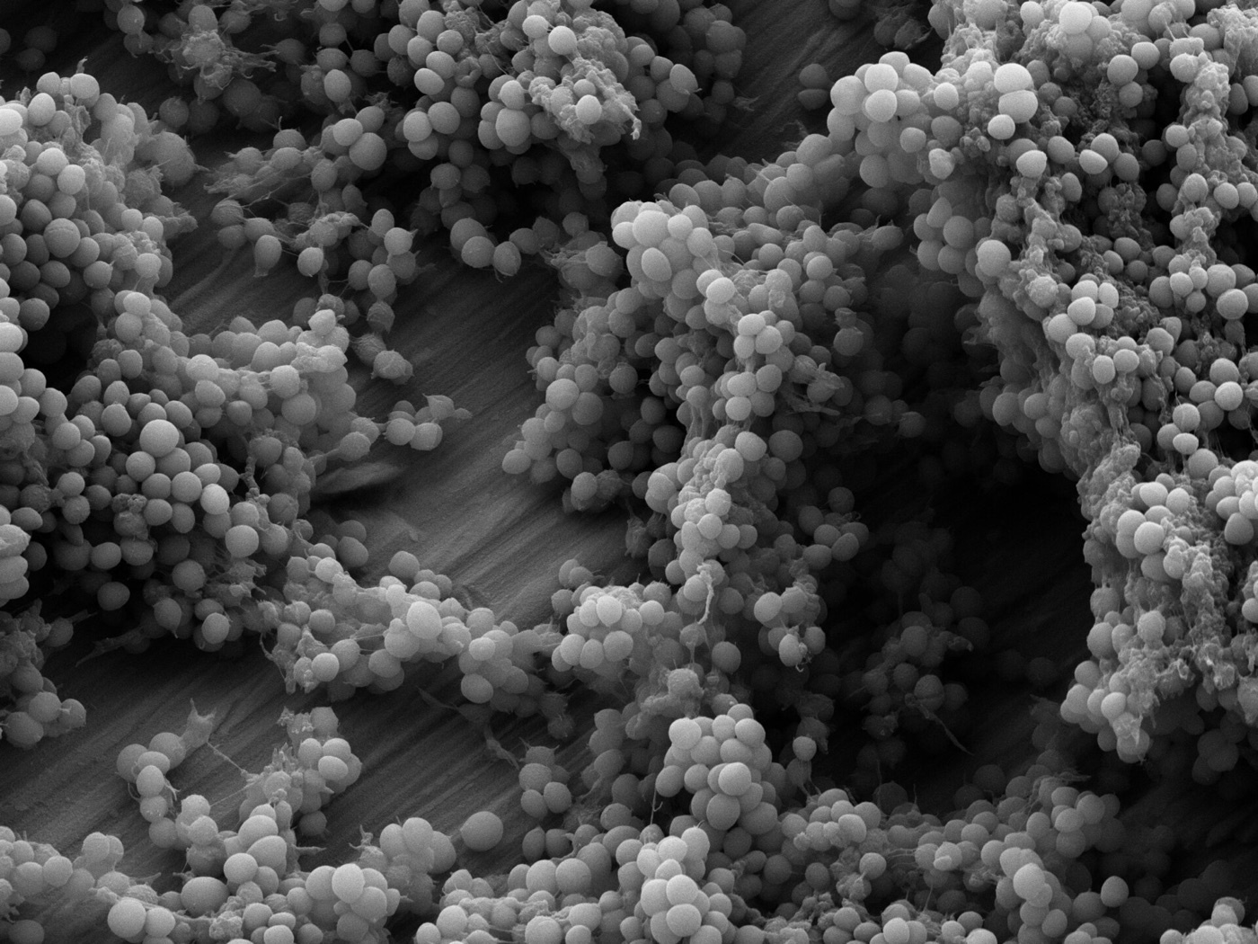
Figure 1. Scanning electron micrograph of a Staphylococcus epidermidis biofilm on a titanium implant surface: clustered cocci embedded in extracellular matrix, the structural basis of antibiotic tolerance. Source: Shahfa84, via Wikimedia Commons, CC BY-SA 4.0.
Part II - Osteomyelitis: Definitions and Terminology
Osteomyelitis is “a progressive inflammatory process… involving the bone, bone marrow, periosteum and surrounding soft tissues,” causing bone destruction, necrosis and new-bone apposition.[4] Strictly, osteitis implies cortical infection and osteomyelitis infection beginning in the marrow, but the two are clinically inseparable and the terms are used interchangeably.[5] The morphological vocabulary is essential:[6]
- Sequestrum: a fragment of dead, devascularised cortical bone detached from living bone (forming within about eight days of infection); it behaves like an implanted foreign body, colonised by biofilm.
- Involucrum: the sleeve of new living bone laid down by the periosteum around the infected dead bone.
- Cloaca: an opening in the involucrum through which pus and sequestra discharge.
- Sinus tract (fistula): the chronic drainage channel to the skin; a discharging sinus is a strong marker of chronic osteomyelitis.
- Brodie abscess: a walled-off subacute intraosseous abscess, classically a well-defined metaphyseal lucency; and Garré’s sclerosing osteomyelitis, a chronic sclerosing form.[7]
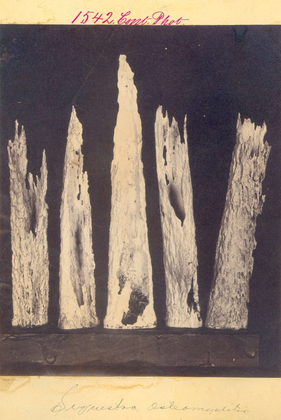
Figure 2. Gross-pathology specimen of bony sequestra: fragments of dead, detached cortical bone that behave as biofilm-colonised foreign bodies. Source: Otis Historical Archives, National Museum of Health & Medicine, via Wikimedia Commons, CC BY 2.0.
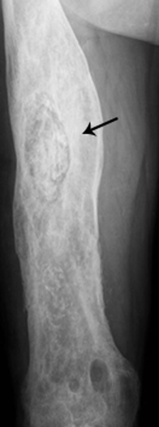
Figure 3. Femoral radiograph in chronic osteomyelitis: a dense detached sequestrum within a thickened, sclerotic cortex (involucrum), with distal lytic destruction. From Desimpel, Posadzy & Vanhoenacker (2017), PMC6032807, CC BY 4.0.
Part III - Classification of Osteomyelitis
Three complementary classifications are used. By pathogenesis (Waldvogel/Lew-Waldvogel), osteomyelitis is haematogenous (blood-borne: the metaphysis of long bones in children, the vertebral bodies in adults over 60), contiguous-focus / exogenous (from open fracture, surgery or an adjacent infection, the commonest form, and overwhelmingly post-traumatic), or secondary to vascular insufficiency (with neuropathy), the diabetic foot.[8] By duration there is no firm time limit, but acute osteomyelitis (presenting at about two weeks, treatable by antibiotics alone in haematogenous childhood disease) is distinguished from subacute (the smouldering Brodie abscess, or tuberculous/brucellar vertebral disease) and chronic osteomyelitis, which is defined by the presence of dead bone and “is still considered a surgical disease.”[9]
The most-used surgical scheme is the Cierny-Mader classification, which combines four anatomical types with three physiological host classes to give 12 stages:[10]
- Anatomical type I, medullary (endosteal); II, superficial (cortical surface under a soft-tissue defect); III, localised (full-thickness sequestrum, mechanically stable); IV, diffuse (through-and-through, mechanically unstable, needing reconstruction).
- Host class A, normal; B, compromised, locally (BL) or systemically (BS) (smoking, diabetes, malnutrition, vascular disease, immunosuppression); C, the patient in whom the treatment would be worse than the disease (suppression or no treatment).
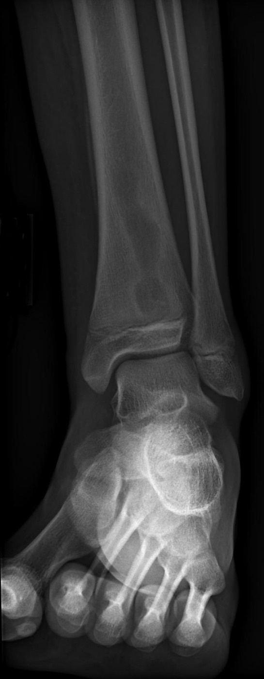
Figure 4. Radiograph of a Brodie abscess: a well-defined metaphyseal lucency in the distal tibia, the plain-film correlate of subacute osteomyelitis. Source: Jto410, via Wikimedia Commons, CC BY-SA 3.0.
Part IV - Pathogenesis and Microbiology
Healthy bone resists infection; it takes trauma, a large inoculum or a virulent organism to establish disease.[11] In childhood haematogenous osteomyelitis, organisms lodge in the metaphysis, where the sluggish blood flow of the capillary loops favours seeding; suppuration raises intraosseous pressure, thromboses vessels and devitalises bone into sequestra, while the periosteum lays down involucrum.[12] Staphylococcus aureus is virulent here because its adhesins bind bone matrix and it is internalised by osteoblasts (explaining long latent persistence), and because it can survive as antibiotic-tolerant small-colony variants that revert years later to cause recurrence.[13]
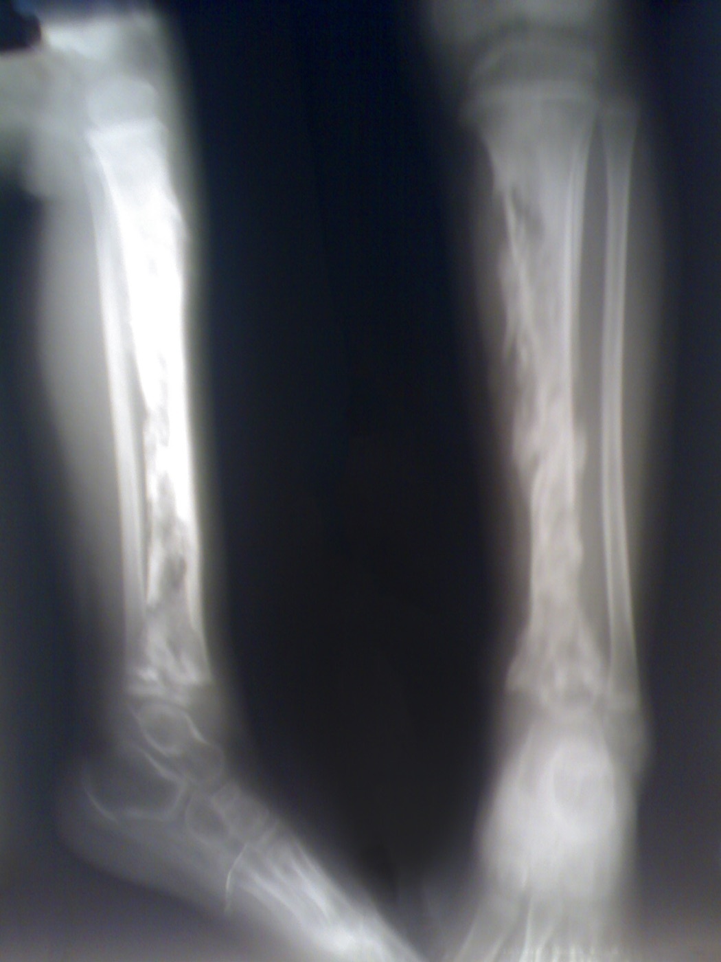
Figure 5. Acute haematogenous osteomyelitis of the tibia in a child: multiple radiolucent abscesses and metaphyseal bony destruction, reflecting metaphyseal seeding. Source: sarindam7, via Wikimedia Commons, CC BY-SA 4.0.
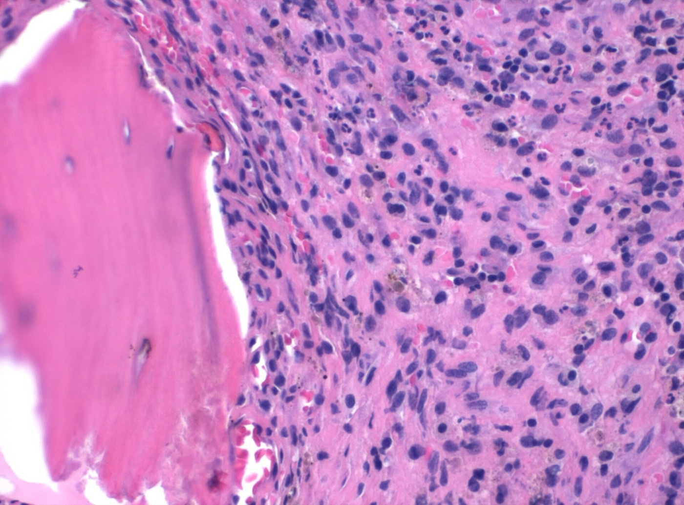
Figure 6. Histopathology of acute osteomyelitis (H\&E): a bony trabecula adjacent to a dense neutrophilic inflammatory infiltrate; cultures grew Staphylococcus aureus. Source: Mikael Häggström, M.D. (CC0 1.0, public domain).
Staphylococcus aureus (including MRSA) is the predominant cause of all types of osteomyelitis, isolated in 30-60%.[14] Haematogenous osteomyelitis is usually monomicrobial; contiguous/post-traumatic disease is frequently polymicrobial (up to ~29%) and adds gram-negatives (Pseudomonas, Enterobacter, E. coli) and coagulase-negative staphylococci.[15] Special hosts have characteristic organisms: sickle-cell disease → Salmonella; intravenous drug use → Pseudomonas at unusual sites (sternoclavicular, sacroiliac); the diabetic foot → polymicrobial; and subacute vertebral disease → Mycobacterium tuberculosis or Brucella.[16] Up to a third of post-traumatic and 41% of vertebral cases are culture-negative, often after empirical antibiotics or because biofilm bacteria evade conventional culture.[17]
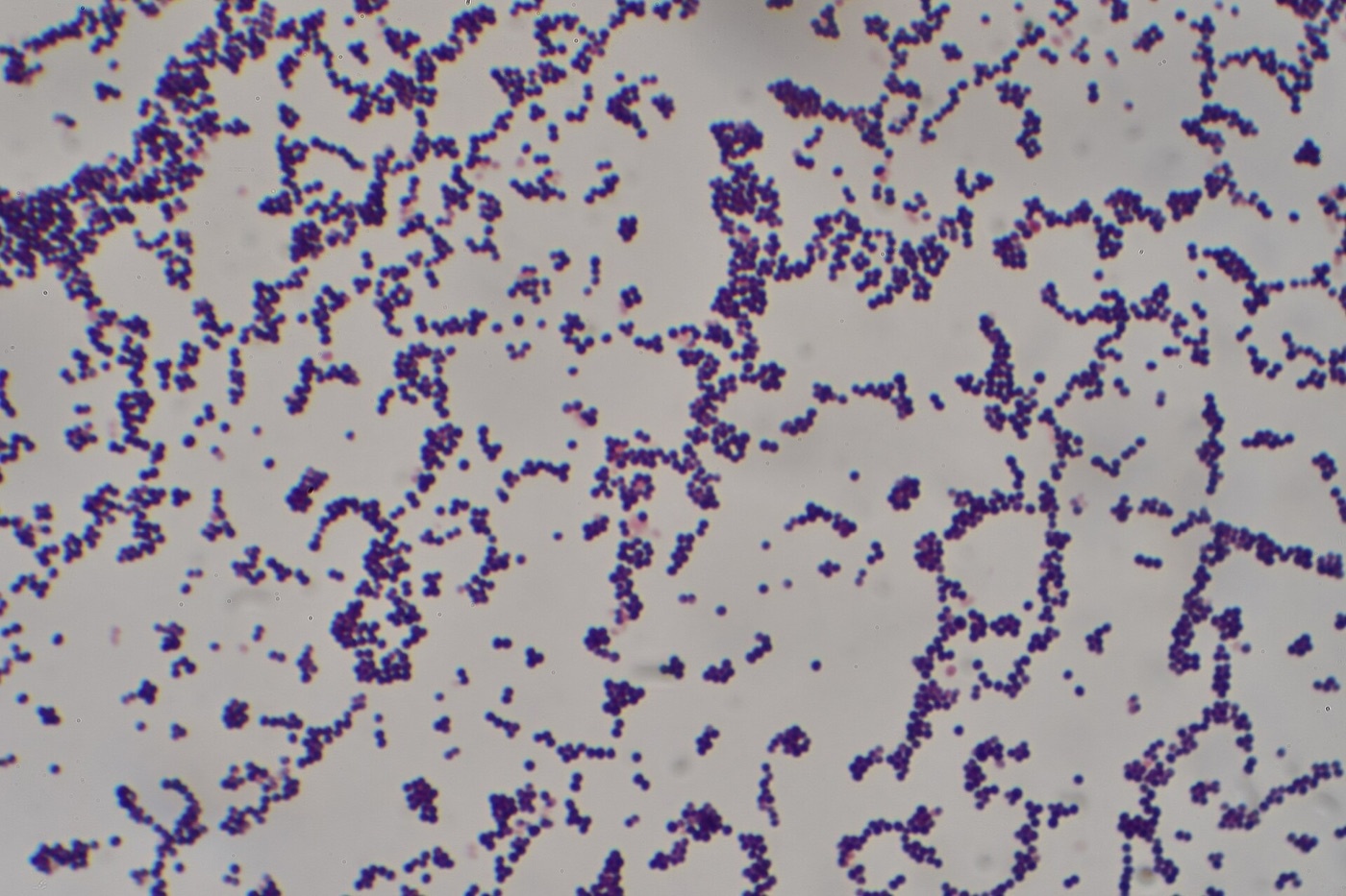
Figure 7. Gram stain of Staphylococcus aureus: gram-positive cocci in grape-like clusters, the predominant cause of all forms of osteomyelitis. Source: Dr Graham Beards, via Wikimedia Commons, CC BY-SA 4.0.
Part V - Diagnosis and Treatment of Osteomyelitis
Diagnosis
The inflammatory markers, ESR and CRP (CRP the better one for monitoring, falling quickly with response), are sensitive but not specific, and the white count is often normal; a persistently normal CRP and ESR make osteomyelitis unlikely.[18] The gold standard is bone biopsy for culture and histopathology, taking several deep samples (not superficial sinus swabs, which mislead), ideally after an “antibiotic-free window” of one to two weeks, with more than five neutrophils per high-power field confirming infection.[19] Imaging runs from the plain radiograph (lytic destruction, periosteal reaction, sequestrum, but normal for the first 1-3 weeks until 30-50% of mineral is lost) to MRI, the most sensitive test, where a normal study virtually excludes osteomyelitis, and labelled-leucocyte or FDG-PET scanning for difficult cases.[20] In the diabetic foot the bedside probe-to-bone test is of high value.[21]
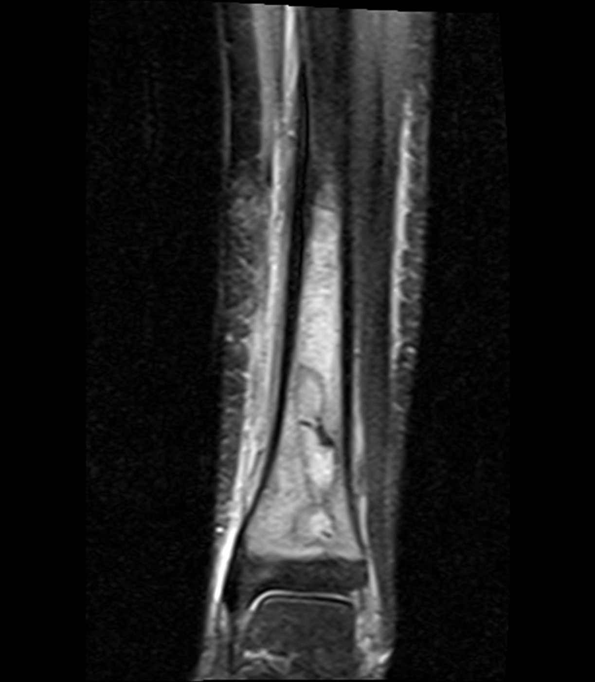
Figure 8. Coronal STIR MRI of a Brodie abscess in the distal tibia: an intraosseous focus surrounded by high-signal marrow oedema. MRI is the most sensitive test, and a normal study virtually excludes osteomyelitis. Source: Jto410, via Wikimedia Commons, CC BY-SA 3.0.
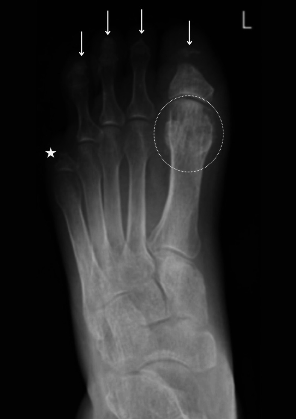
Figure 9. Diabetic-foot osteomyelitis: osteolytic destruction of the phalanges with surrounding soft-tissue swelling. From Ahmed et al. (2025), PMC12108555, CC BY 4.0.
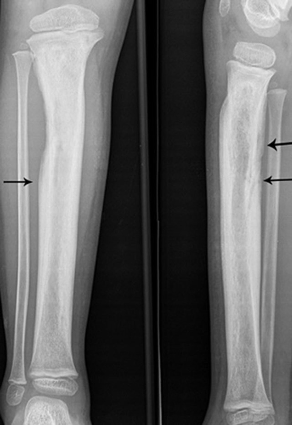
Figure 10. Tibial radiographs showing periosteal new-bone (involucrum) formation in chronic osteomyelitis. From Desimpel, Posadzy & Vanhoenacker (2017), PMC6032807, CC BY 4.0.
Treatment
The principle is that “antibiotic treatment cannot replace surgical debridement, which remains the most effective method of reducing pathogen load,” and “a long-lasting cure is impossible as long as the underlying reason… is not eliminated.”[22] The pillars are:[23]
- Radical debridement: excise all dead and infected bone (sequestrectomy) and necrotic soft tissue to a bleeding margin; the adequacy of debridement is the strongest predictor of success.
- Dead-space management and local antibiotics: antibiotic-impregnated PMMA beads/cement (non-degradable, must be removed) or biodegradable calcium-sulphate/bioglass carriers; the Masquelet induced-membrane technique and Ilizarov bone transport for resulting defects.
- Soft-tissue cover: muscle or free flaps (“orthoplastic” care) to bring vascularity.
- Culture-directed antibiotics, classically about six weeks, though the evidence is weak and an oral switch (with high-bioavailability agents such as fluoroquinolones, clindamycin or rifampicin) is now accepted as equivalent to intravenous therapy (the OVIVA result: 13.2% vs 14.6% failure); rifampicin combinations are first-line for staphylococcal biofilm.[24]
- Suppressive antibiotics for the Cierny-Mader C host, and amputation as salvage (up to 50% in diabetics).[25]
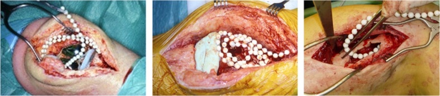
Figure 11. Strings of antibiotic-loaded PMMA beads placed in the surgical field for local antibiotic delivery and dead-space management after debridement. From van Vugt, Arts & Geurts (2019), PMC6671866, CC BY 4.0.
The cardinal complications are recurrence (5-15%, decades later) and the Marjolin ulcer, a squamous-cell carcinoma arising in a chronic sinus after a 27-30 year latency.[26]
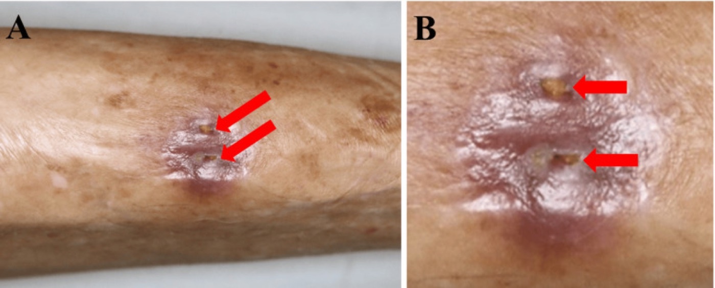
Figure 12. Chronic tibial osteomyelitis with two cutaneous draining sinus-tract openings: a discharging sinus is a strong clinical marker of chronicity. From Ishihara et al. (2025), PMC12459229, CC BY 4.0.
Part VI - Septic Arthritis
Septic arthritis of a native joint is a surgical emergency, because “rapid damage of the joint cartilage by enzymes liberated from microorganisms or granulocytes” can destroy the joint within days.[27] It is most often haematogenous (the synovium is highly vascular and lacks a limiting basement membrane), and the commonest joint is the knee in adults and the hip in children.[28] Staphylococcus aureus causes 50-60%; streptococci and gram-negatives follow; Neisseria gonorrhoeae causes the migratory arthritis-tenosynovitis-dermatitis syndrome of the young sexually active adult; and Kingella kingae is the leading cause in children under four.[29]
The key investigation is joint aspiration: a turbid effusion is infected until proven otherwise, and a synovial white-cell count above 50,000/µL with more than 90% neutrophils suggests sepsis (lower with gonococcus, Kingella and in the immunosuppressed), alongside Gram stain (positive in only ~37% of adult cases), culture and crystal examination to exclude gout.[30] Synovial glucose and lactate are not discriminatory.[31] In a child with an irritable hip, the Kocher criteria (non-weight-bearing, fever > 38.5 °C, ESR > 40, WBC > 12,000) stratify the probability of septic arthritis versus transient synovitis.[32] Treatment rests on two pillars: urgent joint drainage (repeated aspiration for accessible joints, but arthrotomy or arthroscopy for the hip and shoulder, where the deep joint and the femoral head’s single blood supply are at risk, and the Gächter arthroscopic stages I-IV guide the surgical approach) plus empirical then culture-directed antibiotics covering S. aureus.[33] Even with treatment, mechanical sequelae (secondary osteoarthritis, stiffness) occur in up to 30%.[34]
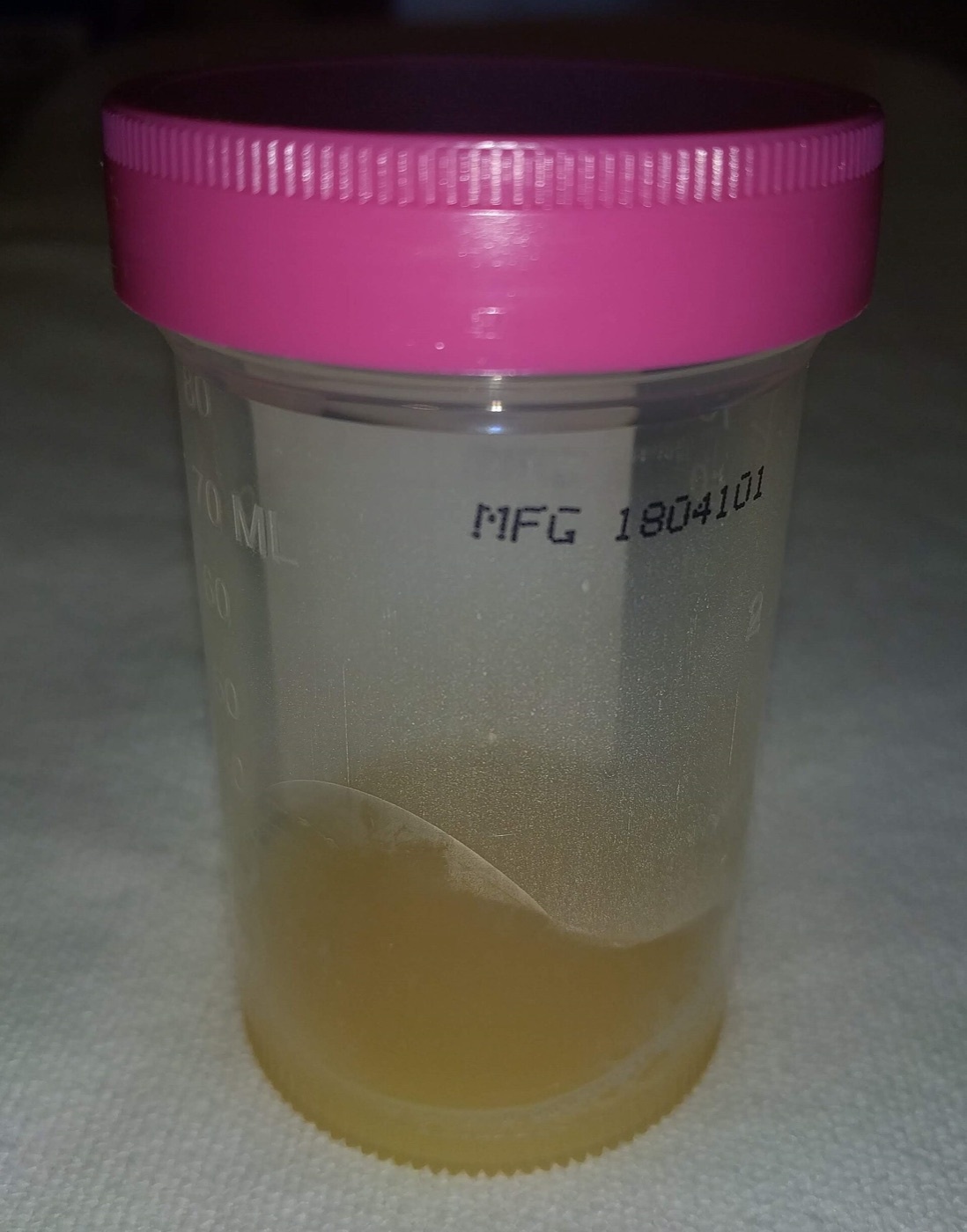
Figure 13. Turbid, purulent synovial fluid aspirated from a septic knee: an effusion this cloudy is infected until proven otherwise. Source: James Heilman, MD, via Wikimedia Commons, CC BY-SA 4.0.
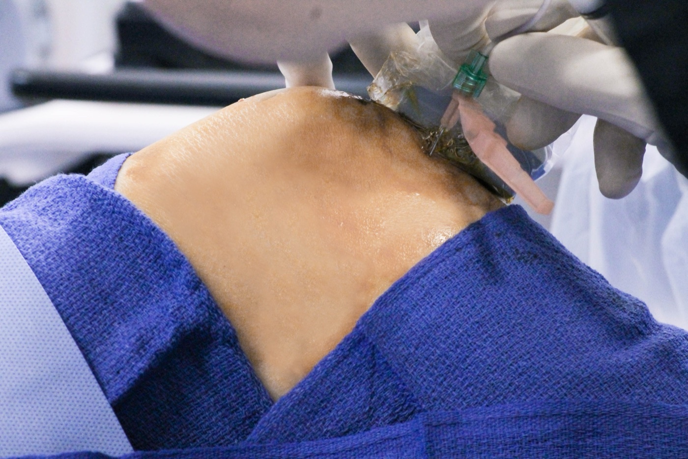
Figure 14. Knee arthrocentesis: aspiration of joint fluid is the key investigation in suspected septic arthritis. Source: PainDoctorUSA, via Wikimedia Commons, CC BY-SA 4.0.
Part VII - Implant-Related Infection: FRI and PJI
Fracture-related infection (FRI)
FRI (post-traumatic osteomyelitis) is defined by the 2018 consensus, which separates confirmatory criteria (a fistula/sinus or wound breakdown communicating with bone or implant; purulent drainage or pus at surgery; phenotypically identical organisms in ≥ 2 deep specimens; or microorganisms on deep-tissue histopathology) from weaker suggestive criteria (clinical inflammation, radiographic signs, a single positive culture).[35] It is classified by timing, early (< 2 weeks), delayed (3-10 weeks), late (> 10 weeks), which tracks biofilm maturity, organism virulence and treatment.[36] Deep-infection rates rise with the Gustilo grade (1.8% in grade I to 16% in IIIC).[37] The pivotal management principle is fracture stability: a stable construct is retained, even over an infected but uniting fracture, with biofilm-active antibiotic suppression until the fracture has united, after which the hardware is removed; an unstable infected fracture must be re-stabilised (often with an external fixator), never left mobile.[38]
Periprosthetic joint infection (PJI)
PJI has no single gold-standard definition; the MSIS/ICM (2018) and EBJIS criteria combine a sinus tract or two positive cultures of the same organism with raised CRP/ESR, a raised synovial leucocyte count and neutrophil percentage, positive leucocyte esterase or alpha-defensin, and histopathology.[39] Crucially, the synovial thresholds are far lower than for native septic arthritis: for a chronic knee prosthesis a synovial count of only about **1,700/µL with
65% neutrophils**, and for a hip about 4,200/µL with > 80% (though the threshold within the first six weeks after surgery is much higher).[40] PJI is classified by route and timing into acute haematogenous (≤ 3 weeks of symptoms), early post-operative (< 1 month after surgery), and chronic (> 3 weeks), which dictates surgery.[41] The four curative strategies, each with a cure rate over 80% when correctly selected, are:[42]
- DAIR (debridement, antibiotics and implant retention), only for an acute infection (early post-operative or acute haematogenous) with a stable implant, a biofilm-susceptible organism and intact soft tissues, with exchange of the mobile parts.
- One-stage exchange, for chronic infection with good soft tissues and a susceptible organism.
- Two-stage exchange (short or long interval, with an antibiotic spacer), the most widely used, and the route for difficult-to-treat organisms.
The antibiotic regimen for staphylococcal implant infection pairs rifampicin with a fluoroquinolone companion for a typical total of about three months; rifampicin must never be used as monotherapy (rapid resistance).[43]
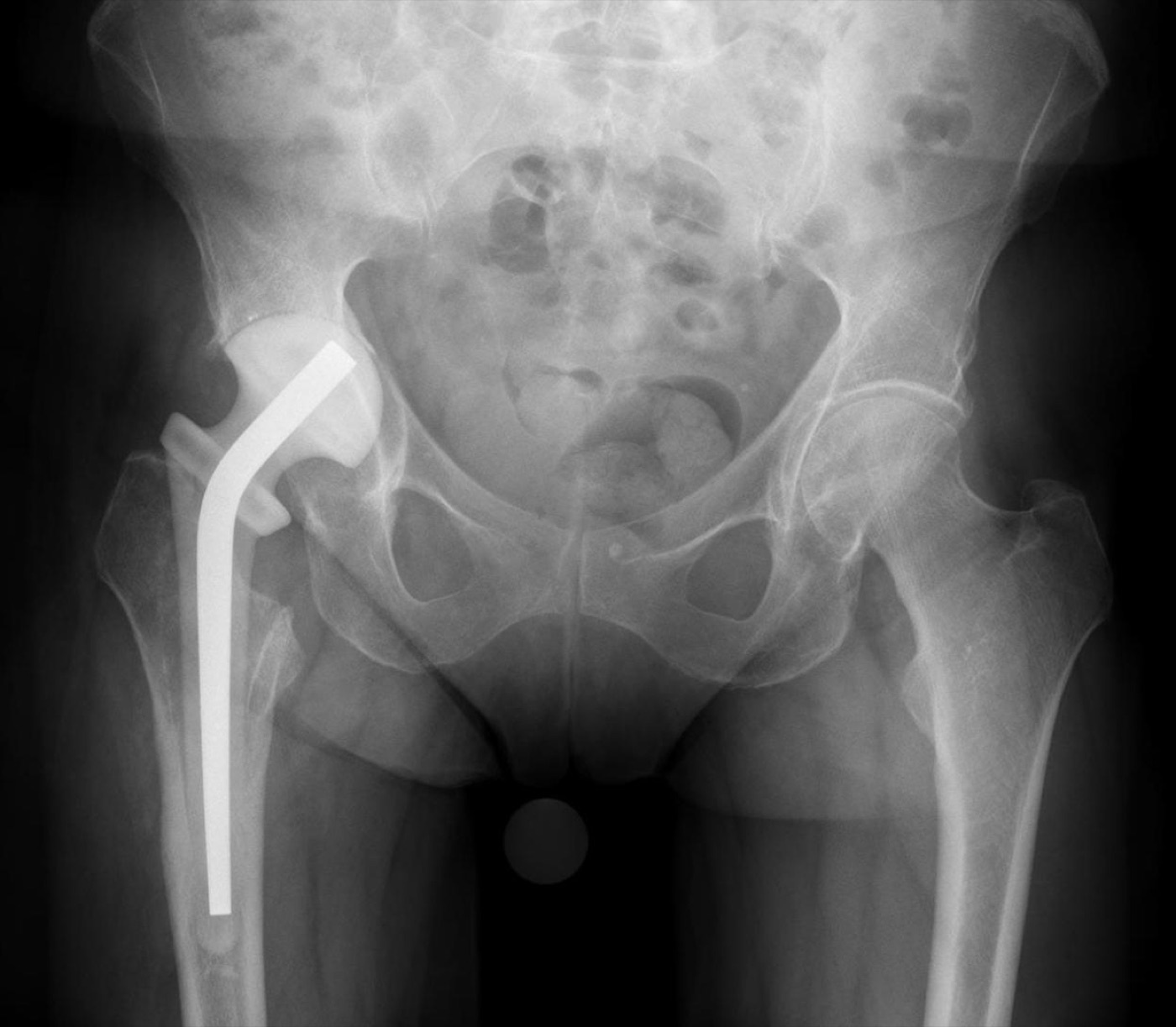
Figure 15. AP pelvis radiograph showing an antibiotic-loaded cement hip spacer placed for periprosthetic joint infection during a staged (two-stage) exchange. Source: Hellerhoff, via Wikimedia Commons, CC BY-SA 4.0.
Part VIII - Anaerobic Infection
Anaerobes are uncommon but important orthopaedic pathogens. Cutibacterium acnes (formerly Propionibacterium acnes), a low-virulence skin anaerobe, is the classic indolent organism of shoulder prosthetic infection and of fixation after proximal humeral fracture; its presentation is subtle, its CRP often normal, and its diagnosis frequently delayed by months because it requires prolonged anaerobic culture (up to 14 days).[44] Gram-negative anaerobes such as Bacteroides are treated with metronidazole, and a range of clostridia (C. difficile, C. perfringens, C. septicum, the last associated with intestinal malignancy) cause indolent prosthetic infection.[45] These indolent anaerobic infections are quite distinct from the fulminant clostridial gas gangrene (clostridial myonecrosis) of the contaminated wound, which is covered with the war and crush injuries of Topic 5.[46] The general lesson of anaerobic and other fastidious infection is to alert the microbiology laboratory, take multiple deep cultures, and incubate them for longer than usual.
References
-
Zimmerli, Bone and Joint Infections 2e, p.197 (the 1000-fold figure); Rockwood & Green 9e, ch.28 (Panteli & Giannoudis), pp.1359-1360 gives ~800-fold for the biofilm concept.
-
Zimmerli 2e pp.194-196.
-
Zimmerli 2e pp.197, 201.
-
Rockwood 9e p.1354.
-
Rockwood 9e p.1354; Zimmerli 2e p.347.
-
Zimmerli 2e pp.312, 348, 350; Rockwood 9e p.1359.
-
Brodie abscess and Garré sclerosing osteomyelitis are standard teaching; the eponyms are not used in the mined Rockwood/Zimmerli extracts (Rockwood notes an “intraosseous abscess” radiographic appearance without the eponym).
-
Rockwood 9e p.1356; Zimmerli 2e pp.287-288.
-
Zimmerli 2e pp.289-290, 311, 351; Rockwood 9e p.1355.
-
Rockwood 9e p.1356; Zimmerli 2e p.291.
-
Rockwood 9e p.1358.
-
Zimmerli 2e pp.288, 312, 348.
-
Zimmerli 2e pp.312, 348; Rockwood 9e p.1360.
-
Rockwood 9e p.1361; Zimmerli 2e p.312.
-
Rockwood 9e pp.1360-1361.
-
Zimmerli 2e pp.288-289, 311, 354.
-
Rockwood 9e pp.1361-1362.
-
Rockwood 9e p.1373; Zimmerli 2e p.319.
-
Rockwood 9e pp.1373-1375; Zimmerli 2e p.349.
-
Rockwood 9e pp.1363-1373; Zimmerli 2e pp.321, 350.
-
Rockwood 9e p.1362.
-
Rockwood 9e p.1378; Zimmerli 2e p.351.
-
Rockwood 9e pp.1376-1393; Zimmerli 2e pp.351-354.
-
Rockwood 9e p.1378; Zimmerli 2e pp.324, 353.
-
Rockwood 9e pp.1380, 1393; Zimmerli 2e p.351.
-
Rockwood 9e p.1394; Zimmerli 2e p.355.
-
Zimmerli 2e p.161.
-
Zimmerli 2e pp.144, 162.
-
Zimmerli 2e pp.140, 162-163.
-
Zimmerli 2e pp.147, 164-165.
-
Zimmerli 2e p.148.
-
The Kocher criteria are standard teaching; the mined Zimmerli text distinguishes septic arthritis from transient synovitis by MRI (p.150) rather than the Kocher score.
-
Zimmerli 2e pp.150-151, 166-167; the Gächter staging is standard teaching, named but not detailed in the mined extract.
-
Zimmerli 2e p.170.
-
Zimmerli 2e p.390.
-
Zimmerli 2e p.391.
-
Zimmerli 2e p.390.
-
Zimmerli 2e pp.395-401.
-
Zimmerli 2e pp.194, 198-199.
-
Zimmerli 2e pp.198-199, 213.
-
Zimmerli 2e pp.194-195.
-
Zimmerli 2e pp.200-201, 219-222.
-
Zimmerli 2e pp.201, 214-217.
-
Zimmerli 2e pp.197, 392; pp.43-62.
-
Zimmerli 2e pp.56-57, 216.
-
Gas gangrene is the fulminant clostridial myonecrosis covered in Topic 5; the Zimmerli extract covers only indolent clostridial prosthetic infection.
-
Zimmerli 2e pp.194-197, 201.
-
Zimmerli 2e pp.312, 348, 350; Rockwood 9e p.1359.
-
Rockwood 9e p.1356; Zimmerli 2e pp.287-291.
-
Rockwood 9e pp.1361, 1373; Zimmerli 2e p.321.
-
Rockwood 9e pp.1376-1393; Zimmerli 2e pp.351-354.
-
Zimmerli 2e pp.147, 161, 164; Kocher criteria are standard teaching.
-
Zimmerli 2e pp.150-151, 166-167, 170.
-
Zimmerli 2e pp.390-391.
-
Zimmerli 2e pp.395-401.
-
Zimmerli 2e pp.200-201, 214-222.
-
Zimmerli 2e pp.197, 392.