Contents
- Orientation: One Joint, Two Injuries, and a Race Against the Clock
- Part I - Applied Anatomy
- Part II - Traumatic Hip Dislocation
- Part III - Femoral Head Fractures
- Part IV - Acetabular Fractures
- Part V - A Synthesis: How to Reason About the Injured Hip
- References
Orientation: One Joint, Two Injuries, and a Race Against the Clock
This topic gathers the high-energy injuries of the hip joint: the dislocation of the femoral head from the acetabulum, the shear fracture of the head itself, and the fracture of the acetabulum that receives it. Two themes run through everything below. The first is the blood supply of the femoral head, which arrives mostly retrograde through the medial femoral circumflex artery and is threatened by dislocation, by the fracture, and by the surgeon, so that avascular necrosis shadows all of these injuries. The second is post-traumatic arthritis, the commonest long-term complication of all three, whose single strongest predictor is the quality of the reduction. Two clinical imperatives follow. A dislocated hip is an emergency to be reduced within hours, and a displaced acetabular fracture involving the weight-bearing dome must be reduced anatomically, because a hip left dislocated or a joint left incongruent will arthrose.[1]
Part I - Applied Anatomy
The hip is a deep, congruent ball-and-socket joint whose stability comes from its bony depth, its labrum, and the surrounding muscles; it takes more than 400 N just to distract it.[2] The blood supply of the femoral head is the anatomy that matters most. In the adult the head is fed mainly by the cervical (retinacular) branches of an extracapsular ring formed by the medial femoral circumflex artery (MFCA) posteriorly and the lateral femoral circumflex artery anteriorly, the MFCA being dominant; the artery of the ligamentum teres (a branch of the obturator artery) is a minor contributor present in about 75% of hips.[3] Because this supply is largely retrograde and runs in the capsule near the head-neck junction, a dislocation kinks it and a fracture or an over-zealous dissection can divide it, which is why avascular necrosis is the feared sequela.[4] The sciatic nerve lies just behind the joint (separated from it only by the short external rotators), and its peroneal division is the more vulnerable, so it must be examined before and after any reduction.[5]
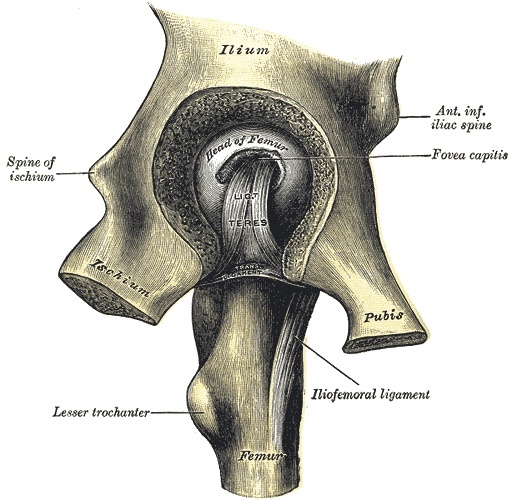
Figure 1. The hip joint opened, showing the femoral head, fovea, ligamentum teres, and capsule. Henry Vandyke Carter, Gray’s Anatomy (1918), public domain, via Wikimedia Commons.
The acetabulum is understood through the two-column (inverted-Y) concept of Judet and Letournel. The articular socket is cradled between the limbs of an inverted Y: the anterior column (iliopubic), running from the iliac crest down through the anterior wall to the superior pubic ramus, and the posterior column (ilioischial), running from the dense bone of the greater sciatic notch through the posterior wall to the ischial tuberosity. The two columns are joined to the axial skeleton by the sciatic buttress, meet medially as the quadrilateral plate, and support the weight-bearing dome (roof or tectum) superiorly.[6] The structures at risk are those of the pelvic ring: the sciatic nerve and superior gluteal bundle posteriorly, the femoral vessels and the corona mortis (the obturator-external iliac anastomosis) anteriorly, and the femoral-head-supplying MFCA, which is protected by keeping the short external rotators released at least 1.5 cm from their insertion.[7]
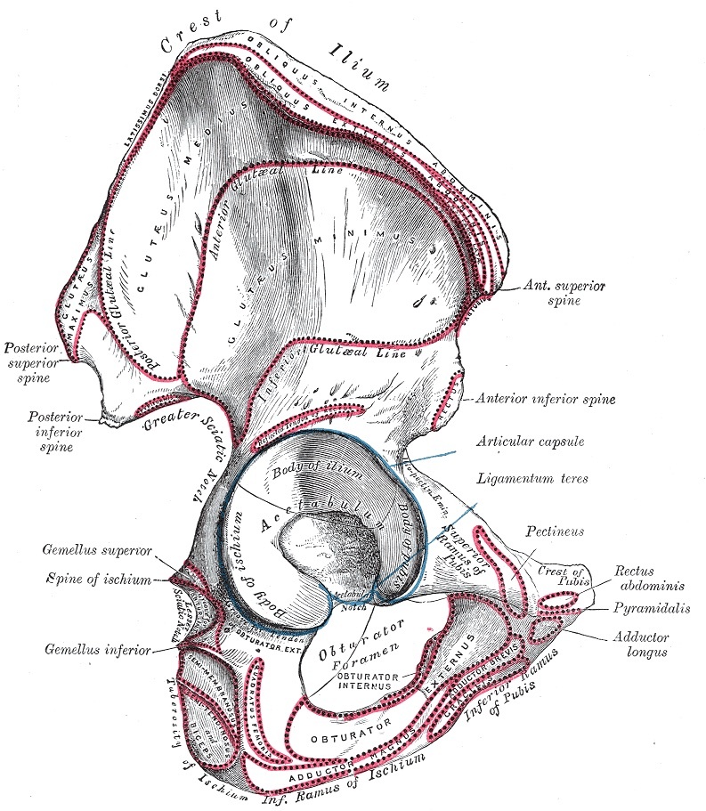
Figure 2. The innominate (hip) bone, showing the acetabulum formed by the ilium, ischium, and pubis, the basis of the two-column concept. Henry Vandyke Carter, Gray’s Anatomy (1918), public domain, via Wikimedia Commons.
Part II - Traumatic Hip Dislocation
2.1 Mechanism and direction
Hip dislocation is a high-energy injury, classically the “dashboard injury” of a flexed hip and knee struck from in front.[8] Posterior dislocations outnumber anterior by about nine to one (roughly 90% versus 10%); the hip position at impact determines the direction (flexion, adduction, and internal rotation drive a posterior dislocation; abduction and external rotation an anterior one), and less adduction at impact favours a fracture-dislocation (a posterior-wall or femoral-head fracture) over a pure dislocation.[9] Given the energy involved, associated injuries are common (head, chest, abdomen, and the ipsilateral knee from the same dashboard), and they may overshadow the dislocation itself.[10]
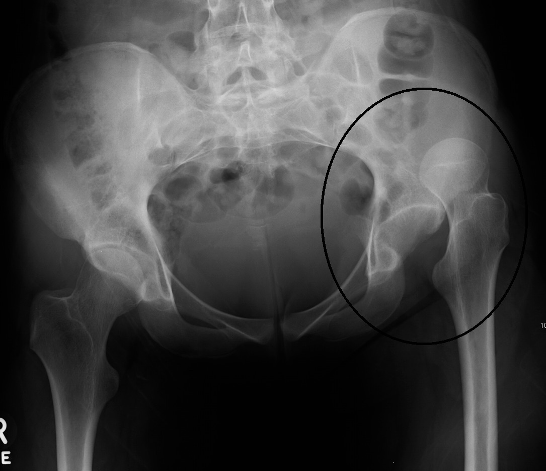
Figure 3. A posterior dislocation of the right hip on AP pelvis radiograph: the femoral head lies superolateral to the acetabulum. James Heilman, MD, CC BY-SA 3.0, via Wikimedia Commons.
2.2 Classification
Posterior dislocations are classified by Thompson-Epstein (type I with or without a minor fracture, II with a single large posterior-rim fragment, III with a comminuted rim, IV with an acetabular floor fracture, V with a femoral head fracture) and by Stewart-Milford (graded by the resulting stability, type III being a grossly unstable posterior wall).[11] The modern AO/OTA scheme codes the dislocation and any femoral-head fracture separately.[12] Whatever the scheme, the two questions that drive treatment are the same: is there an associated fracture, and is the hip stable after reduction?[13]
2.3 Assessment
The posture is diagnostic: the posterior dislocation holds the limb flexed, adducted, internally rotated, and shortened, the anterior dislocation abducted and externally rotated.[14] A prereduction sciatic nerve examination is mandatory (the peroneal division especially), as is a check of the distal pulses. Imaging starts with the AP pelvis, on which the diagnosis should be apparent and any associated femoral neck fracture (the great pitfall, which must be excluded before manipulation), femoral head fracture, or acetabular fracture sought.[15] After reduction, a fine-cut CT is obtained to confirm a concentric reduction and to detect incarcerated fragments, marginal impaction, and associated fractures; on CT the head should sit centred in the acetabular “bullseye.”[16]
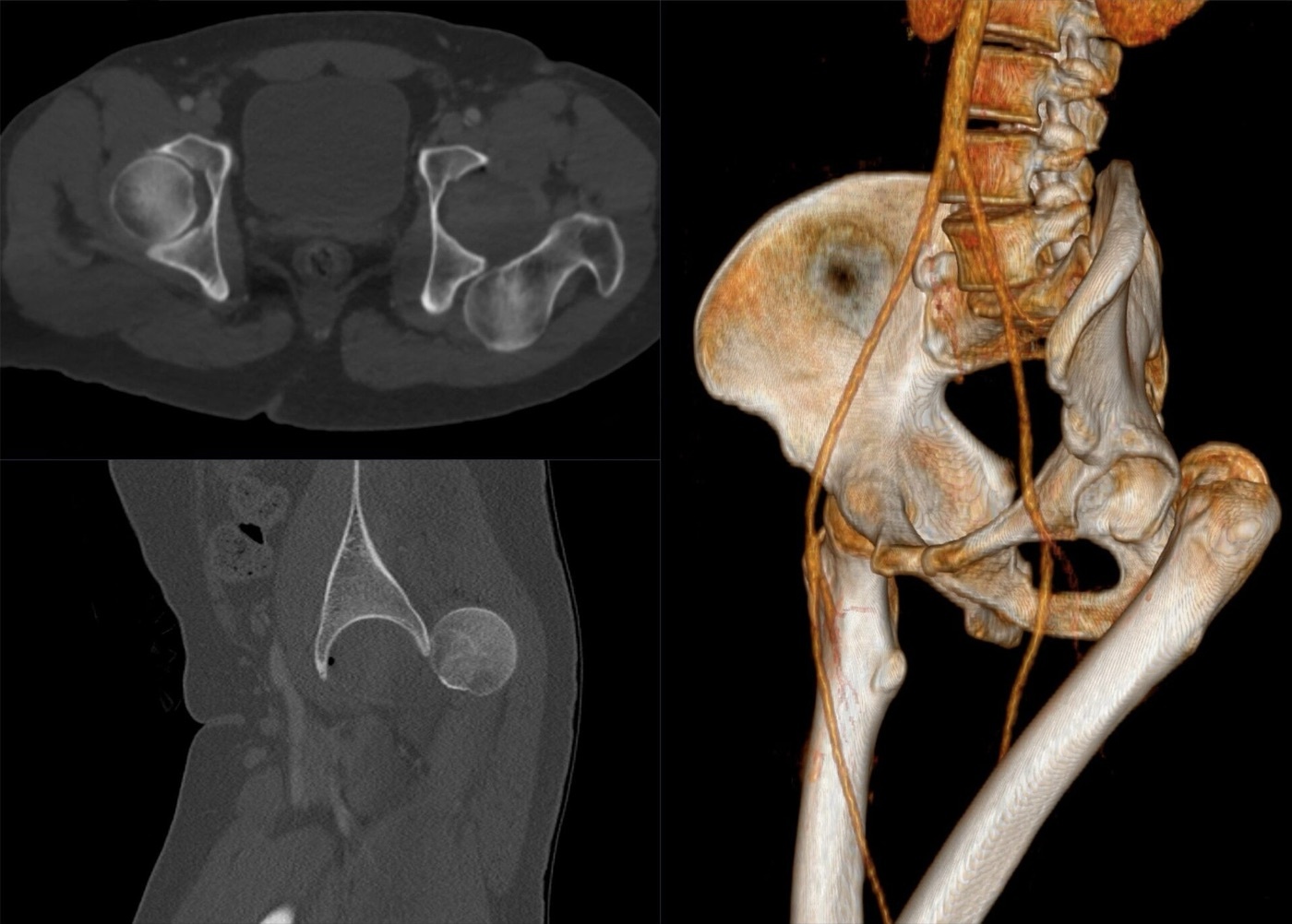
Figure 4. A traumatic posterior hip dislocation on CT (axial, sagittal, and 3D reconstruction). Hellerhoff, CC BY-SA 4.0, via Wikimedia Commons.
2.4 Treatment: an emergency
The dislocated hip is reduced emergently, because the risk of avascular necrosis rises steeply with the delay: AVN is about 0% to 10% when the hip is reduced within 6 hours, and the risk roughly quintuples (×5.6) with a delay beyond 12 hours (Kellam and Ostrum).[17] Reduction is by gentle in-line traction under sedation or general anaesthesia, using the Allis manoeuvre for a posterior dislocation (and the Walker modification for an anterior one), with no more than two attempts to avoid further damage or an iatrogenic femoral neck fracture; an irreducible dislocation goes urgently to open reduction.[18] After reduction, stability is assessed and a CT obtained: a concentric, stable reduction without a fracture needing fixation is treated nonoperatively (a small fragment in the cotyloid fossa not contacting the articular surface may be left), whereas an irreducible dislocation, a non-concentric reduction (incarcerated fragment or interposed soft tissue), or an associated fracture requiring fixation mandates surgery, approached from the side of the dislocation (posterior dislocations through a Kocher-Langenbeck approach).[19]
2.5 Complications
The complications are those foreshadowed by the anatomy.[20] Avascular necrosis is reported in about 1.7% to 40% of series and is time-to-reduction dependent (hence the 6-hour imperative); its changes appear within two years. Post-traumatic arthritis is the commonest long-term complication, more frequent after posterior dislocations and after fracture-dislocations (up to 50% when a femoral head fracture is present), driven both by the cartilage injury at impact and by any residual incongruity. Sciatic nerve injury is present in up to about 19% (the peroneal division), recovering in around 70%; a deficit that appears or worsens after reduction prompts exploration. Heterotopic ossification and recurrent instability (which essentially only follows an unfixed posterior-wall fracture) complete the list.[21]
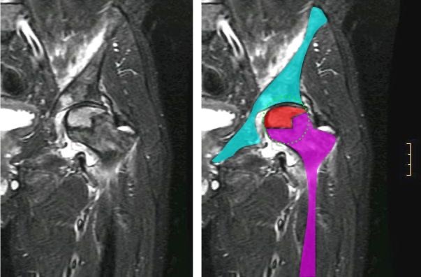
Figure 5. Osteonecrosis of the femoral head on MRI (plain image and colour-segmented overlay), the feared late complication of hip dislocation. Jmarchn, CC BY-SA 3.0, via Wikimedia Commons.
Part III - Femoral Head Fractures
Femoral head fractures occur by shear during a posterior dislocation, the head failing against the posterior acetabular rim and shearing off an anteromedial fragment.[22] They are classified by Pipkin, by the fragment’s relation to the fovea and by associated injuries: type I, a fracture below (caudad to) the fovea (infrafoveal); type II, a fracture above (cephalad to) the fovea (suprafoveal, involving the weight-bearing surface); type III, a femoral head fracture with an associated femoral neck fracture; and type IV, any of these with an associated acetabular fracture.[23]
Treatment follows the type.[24] A small Pipkin I (infrafoveal) fragment lies below the weight-bearing surface and is excised, or left in place if the reduction is congruent and the hip stable. A large Pipkin II (suprafoveal) fragment is part of the weight-bearing surface and requires anatomical reduction; if it does not reduce with the hip, it is fixed (through an anterior approach, with countersunk or headless compression screws sunk beneath the cartilage). The Pipkin III (head plus neck) carries a poor prognosis from the threat to the head’s blood supply and is fixed urgently in the young (or replaced in the elderly), and the Pipkin IV (with an acetabular fracture) is the most complex; types III and IV are contraindications to nonoperative care.[25] For the complex types and for combined head-and-acetabular injuries, a surgical (Ganz) hip dislocation through a trochanteric flip osteotomy gives the best access while protecting the medial femoral circumflex blood supply, and improves outcomes over the older approaches.[26]
Part IV - Acetabular Fractures
4.1 The two-column concept and radiographic assessment
The acetabular fracture is read against the two-column (inverted-Y) anatomy of Part I.[27] Imaging uses three plain views plus CT. On the AP pelvis, six cardinal lines of Letournel are traced, each a tangent of the X-ray beam to a region of bone: the iliopectineal line (the anterior column), the ilioischial line (the posterior column), the teardrop, the acetabular roof, the anterior wall, and the posterior wall.[28] The two 45° Judet oblique views complete the picture: the obturator oblique profiles the anterior column and the posterior wall (and shows the spur sign of a both-column fracture), while the iliac oblique profiles the posterior column and the anterior wall.[29] CT defines marginal impaction, intra-articular fragments, and the size of posterior-wall fragments.[30]
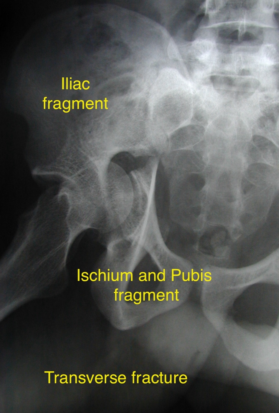
Figure 6. A transverse acetabular fracture on an oblique (Judet) radiograph, separating the iliac from the ischiopubic fragment. Dr. C. J. Thakkar, CC BY-SA 4.0, via Wikimedia Commons.
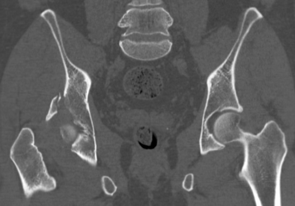
Figure 7. An acetabular fracture on axial CT. James Heilman, MD, CC BY-SA 4.0, via Wikimedia Commons.
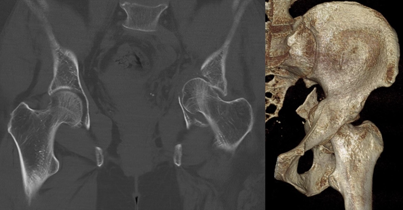
Figure 8. A central acetabular fracture-dislocation on CT (coronal reformat and 3D reconstruction): the femoral head is driven medially through the acetabulum. Hellerhoff, CC BY-SA 3.0, via Wikimedia Commons.
The weight-bearing dome is the region whose involvement most determines treatment, assessed by the roof-arc angles (Matta): the medial roof arc on the AP, the anterior roof arc on the obturator oblique, and the posterior roof arc on the iliac oblique, with the classic criterion that all three exceeding 45° indicates an intact dome suitable for nonoperative care (revised biomechanical thresholds are higher; roof arcs do not apply to posterior-wall or both-column fractures).[31]
4.2 The Judet-Letournel classification
The Judet-Letournel classification has ten patterns in two groups.[32] The five elementary patterns each break all or part of a single column: posterior wall (the commonest acetabular fracture overall, about 25%), posterior column, anterior wall, anterior column, and transverse (the transverse is grouped as elementary although it crosses both columns).[33] The five associated patterns combine these: posterior column with posterior wall; transverse with posterior wall; T-shaped; anterior column with a posterior hemitransverse; and the both-column fracture (the commonest associated pattern, about 23%, in which no part of the articular surface remains attached to the axial skeleton).[34] Two signs are worth knowing: the “spur sign” on the obturator oblique is pathognomonic of a both-column fracture, and secondary congruence describes how, in a both-column fracture, the freed articular fragments may rotate to remain congruent with the medially-migrated head, which can permit nonoperative treatment in the low-demand patient.[35]
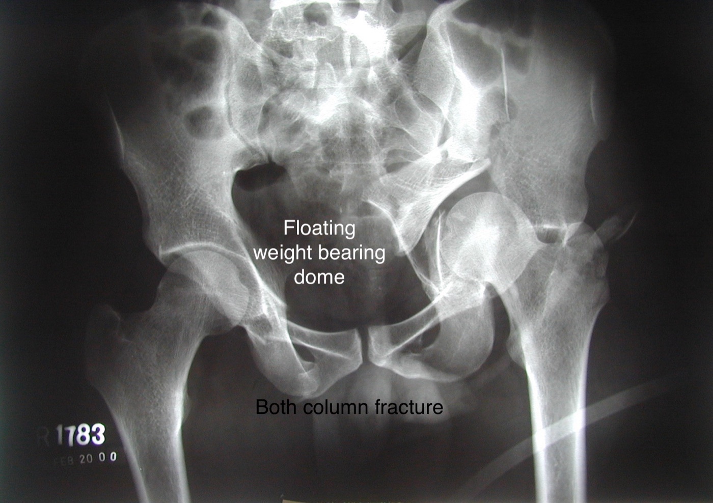
Figure 9. A both-column acetabular fracture with dissociation of the weight-bearing dome on AP radiograph. Dr. C. J. Thakkar, CC BY-SA 4.0, via Wikimedia Commons.
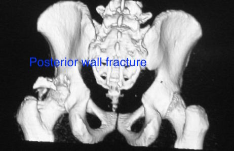
Figure 10. A posterior wall acetabular fracture on 3D CT reconstruction. Dr. C. J. Thakkar, CC BY-SA 4.0, via Wikimedia Commons.
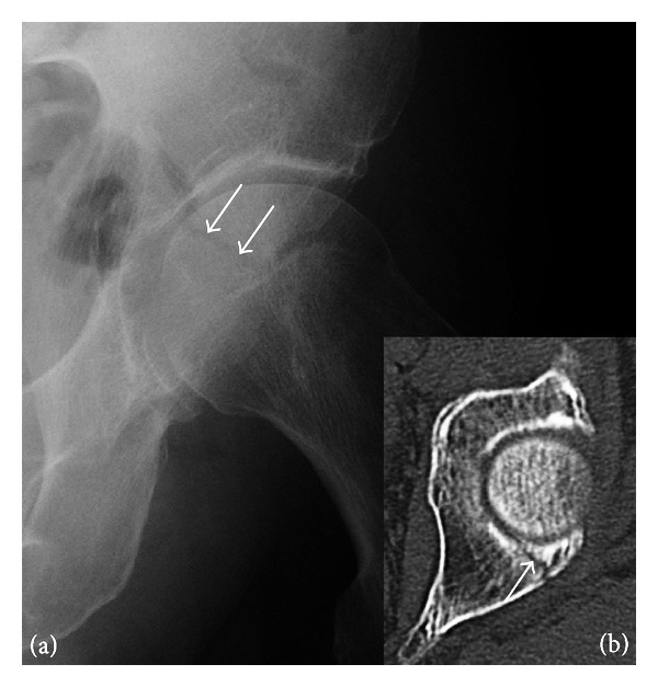
Figure 11. A subtle posterior acetabular wall fracture (radiograph with arrows; CT inset). Jarraya et al., CC BY 3.0, via Wikimedia Commons.
4.3 Treatment
The goal is a congruent, stable hip with the head reduced to an intact weight-bearing dome, and the single strongest predictor of a good long-term result (and of avoiding arthritis) is the quality of the reduction, displacement within 1 mm faring far better than more.[36] Nonoperative treatment suits the stable, congruent fracture with an intact dome (roof arcs over 45°), the low anterior-column or low transverse fracture, the both-column fracture with secondary congruence, and the infirm or severely osteoporotic patient, with protected weight-bearing.[37] Operative treatment is indicated for instability or incongruity, a displaced dome, or an intra-articular fragment, ideally within about two weeks (surgery is urgent only for an irreducible or recurrently dislocating hip, an incarcerated fragment, a progressive sciatic deficit, or an open fracture).[38]
Fixation is by interfragmentary lag screws and 3.5 mm reconstruction plates contoured along the columns, with spring (buttress) plates for posterior-wall comminution, taking great care to keep every screw out of the joint (confirmed by fluoroscopy in the obturator and iliac oblique planes before leaving the theatre).[39] The surgical approach is chosen by the fracture pattern: the Kocher-Langenbeck (posterior) for posterior-wall and posterior-column fractures (the sciatic nerve the structure at risk, protected by knee flexion); the ilioinguinal (anterior, with its three windows) and the now-preferred modified Stoppa / anterior intrapelvic approach for anterior-column, anterior-column-with-posterior-hemitransverse, and both-column fractures (the lateral femoral cutaneous nerve, femoral vessels, and corona mortis at risk); and the extended iliofemoral for selected complex or late fractures (at the cost of more heterotopic ossification).[40] In the elderly patient with an osteoporotic, comminuted fracture, dome impaction (the “gull sign”), or femoral-head damage, acute total hip arthroplasty (sometimes combined with limited fixation) is an increasingly chosen alternative to ORIF, though primary arthroplasty after a fracture has historically done less well than routine arthroplasty for arthritis.[41]
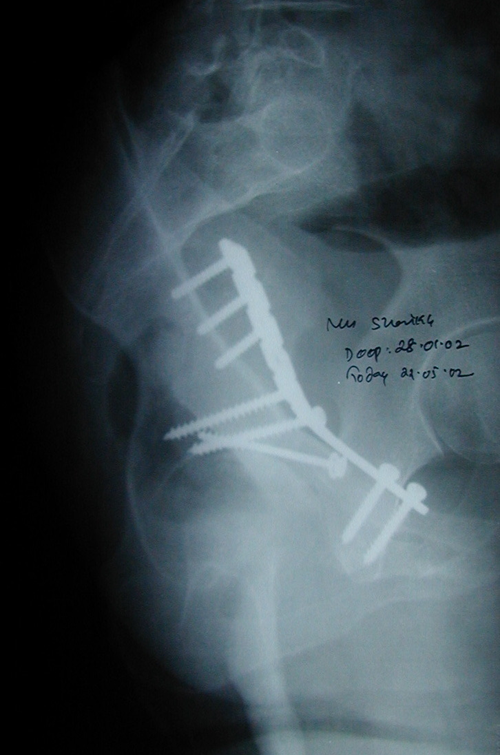
Figure 12. An acetabular fracture after open reduction and internal fixation with a reconstruction plate and screws (postoperative radiograph). Dr. C. J. Thakkar, CC BY-SA 4.0, via Wikimedia Commons.
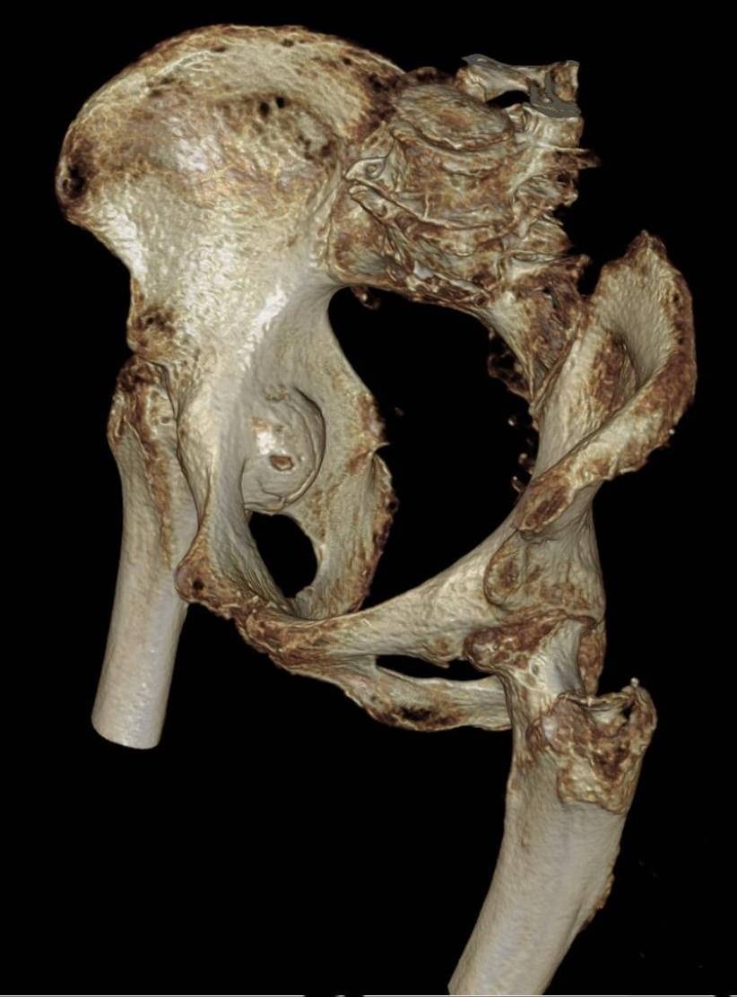
Figure 13. A 3D CT reconstruction of an acetabular fracture with protrusio of the femoral head. Hellerhoff, CC BY-SA 4.0, via Wikimedia Commons.
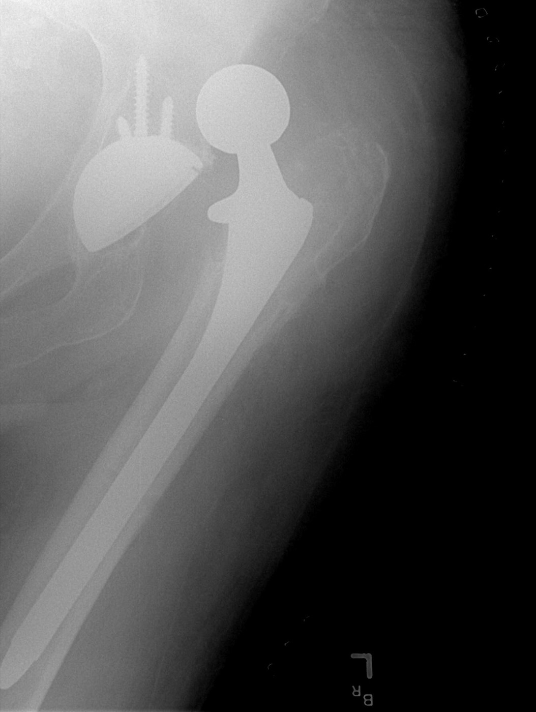
Figure 14. A dislocated total hip arthroplasty, an alternative to fixation in the elderly acetabular fracture. Bill Rhodes, CC BY 2.0, via Wikimedia Commons.
4.4 Complications
The complications parallel those of the dislocation.[42] Post-traumatic osteoarthritis is the commonest long-term complication (about 20%) and is governed by reduction quality; osteonecrosis of the femoral head (about 3% to 9% after operative treatment) follows the injury to its blood supply. Sciatic nerve injury (the L5/peroneal distribution) is mostly associated with the posterior and extended approaches; heterotopic ossification is very common radiographically (reported 18% to 90%), clinically important most after the extended iliofemoral and Kocher-Langenbeck approaches and prevented by indomethacin or single-dose radiation (about 700-800 cGy). Infection (about 5%), venous thromboembolism, and the destructive complication of an intra-articular screw complete the list.[43]
Part V - A Synthesis: How to Reason About the Injured Hip
Start with the clock and the blood supply. A dislocated hip is an emergency: examine the sciatic nerve, exclude a femoral neck fracture, and reduce it gently within hours (the Allis manoeuvre, no more than two attempts), because the avascular necrosis rate climbs with every hour the head stays out. Next, work out what came with the dislocation. Get a post-reduction CT, because a non-concentric reduction, an incarcerated fragment, a Pipkin femoral-head fracture, or a posterior-wall fracture changes everything, and an unstable posterior wall must be fixed. For the acetabular fracture, reason in columns and lines. Trace the iliopectineal and ilioischial lines on the AP, read the obturator and iliac obliques, and ask whether the weight-bearing dome is involved (the roof arcs) and whether the hip is congruent and stable. Classify it as one of the ten Judet-Letournel patterns, choose the approach from the pattern (Kocher-Langenbeck for the back, ilioinguinal or Stoppa for the front), and keep in mind that what you are after is an anatomical reduction, because reduction quality is the one thing that most determines whether this hip arthroses. In the elderly, weigh acute arthroplasty against fixation. Throughout, protect what the injury and your approach both threaten: the medial femoral circumflex artery, the sciatic nerve, and the articular cartilage, the structures whose loss turns a reduced hip into a painful one.
References
-
Rockwood & Green’s Fractures in Adults, pp.3519, 3539, 3588 (the femoral head’s largely retrograde supply via the medial femoral circumflex artery threatened by dislocation; AVN and post-traumatic arthritis the dominant complications; emergent reduction the goal); Rockwood & Green’s Fractures in Adults, pp.3496-3497 (reduction quality the main determinant of late arthritis after acetabular fracture); AO Principles of Fracture Management, p.771 (long-term outcome correlates with the quality of reduction).
-
Rockwood & Green’s Fractures in Adults, pp.3535-3540 (ball-and-socket, >400 N to distract; the femoral head supplied mainly by cervical branches of an extracapsular ring, MFCA posteriorly dominant and lateral femoral circumflex anteriorly; the ligamentum teres / foveal artery a minor contributor present in ~75% of hips; the retrograde supply kinked by dislocation).
-
Rockwood & Green’s Fractures in Adults, pp.3535-3540 (ball-and-socket, >400 N to distract; the femoral head supplied mainly by cervical branches of an extracapsular ring, MFCA posteriorly dominant and lateral femoral circumflex anteriorly; the ligamentum teres / foveal artery a minor contributor present in ~75% of hips; the retrograde supply kinked by dislocation).
-
Rockwood & Green’s Fractures in Adults, pp.3535-3540 (ball-and-socket, >400 N to distract; the femoral head supplied mainly by cervical branches of an extracapsular ring, MFCA posteriorly dominant and lateral femoral circumflex anteriorly; the ligamentum teres / foveal artery a minor contributor present in ~75% of hips; the retrograde supply kinked by dislocation).
-
Rockwood & Green’s Fractures in Adults, pp.3538, 3587 (the sciatic nerve behind the joint, separated by the short external rotators; the peroneal division most vulnerable, examined before and after reduction).
-
Rockwood & Green’s Fractures in Adults, pp.3372, 3400-3402 (the inverted-Y two-column concept of Judet and Letournel: anterior/iliopubic and posterior/ilioischial columns joined by the sciatic buttress, the quadrilateral plate medially, the weight-bearing dome superiorly; the femoral head supplied by the deep branch of the MFCA protected by the obturator externus tendon); AO Principles of Fracture Management, pp.765-766, 773 (the two columns and walls, sciatic buttress, quadrilateral plate, dome; corona mortis, superior gluteal artery, sciatic nerve at risk; release the short external rotators 1.5 cm from insertion to protect the MFCA).
-
Rockwood & Green’s Fractures in Adults, pp.3372, 3400-3402 (the inverted-Y two-column concept of Judet and Letournel: anterior/iliopubic and posterior/ilioischial columns joined by the sciatic buttress, the quadrilateral plate medially, the weight-bearing dome superiorly; the femoral head supplied by the deep branch of the MFCA protected by the obturator externus tendon); AO Principles of Fracture Management, pp.765-766, 773 (the two columns and walls, sciatic buttress, quadrilateral plate, dome; corona mortis, superior gluteal artery, sciatic nerve at risk; release the short external rotators 1.5 cm from insertion to protect the MFCA).
-
Rockwood & Green’s Fractures in Adults, pp.3519-3521, 3529 (high-energy dashboard mechanism; posterior:anterior ~9:1; hip position determines direction, flexion/adduction/internal rotation → posterior, abduction/external rotation → anterior; less adduction favours a fracture-dislocation; frequent associated head/chest/abdominal and ipsilateral knee injuries).
-
Rockwood & Green’s Fractures in Adults, pp.3519-3521, 3529 (high-energy dashboard mechanism; posterior:anterior ~9:1; hip position determines direction, flexion/adduction/internal rotation → posterior, abduction/external rotation → anterior; less adduction favours a fracture-dislocation; frequent associated head/chest/abdominal and ipsilateral knee injuries).
-
Rockwood & Green’s Fractures in Adults, pp.3519-3521, 3529 (high-energy dashboard mechanism; posterior:anterior ~9:1; hip position determines direction, flexion/adduction/internal rotation → posterior, abduction/external rotation → anterior; less adduction favours a fracture-dislocation; frequent associated head/chest/abdominal and ipsilateral knee injuries).
-
Rockwood & Green’s Fractures in Adults, pp.3529-3533 (Thompson-Epstein I-V and Stewart-Milford I-IV posterior-dislocation classifications; AO/OTA codes dislocation and femoral-head fracture separately; the operative questions are the associated fracture and post-reduction stability).
-
Rockwood & Green’s Fractures in Adults, pp.3529-3533 (Thompson-Epstein I-V and Stewart-Milford I-IV posterior-dislocation classifications; AO/OTA codes dislocation and femoral-head fracture separately; the operative questions are the associated fracture and post-reduction stability).
-
Rockwood & Green’s Fractures in Adults, pp.3529-3533 (Thompson-Epstein I-V and Stewart-Milford I-IV posterior-dislocation classifications; AO/OTA codes dislocation and femoral-head fracture separately; the operative questions are the associated fracture and post-reduction stability).
-
Rockwood & Green’s Fractures in Adults, pp.3522-3529 (posterior dislocation limb flexed/adducted/internally rotated/shortened, anterior abducted/externally rotated; mandatory prereduction sciatic examination and pulse check; AP pelvis the first study, exclude an associated femoral neck fracture before manipulation; post-reduction fine-cut CT for concentric reduction, incarcerated fragments, marginal impaction, and associated fractures).
-
Rockwood & Green’s Fractures in Adults, pp.3522-3529 (posterior dislocation limb flexed/adducted/internally rotated/shortened, anterior abducted/externally rotated; mandatory prereduction sciatic examination and pulse check; AP pelvis the first study, exclude an associated femoral neck fracture before manipulation; post-reduction fine-cut CT for concentric reduction, incarcerated fragments, marginal impaction, and associated fractures).
-
Rockwood & Green’s Fractures in Adults, pp.3522-3529 (posterior dislocation limb flexed/adducted/internally rotated/shortened, anterior abducted/externally rotated; mandatory prereduction sciatic examination and pulse check; AP pelvis the first study, exclude an associated femoral neck fracture before manipulation; post-reduction fine-cut CT for concentric reduction, incarcerated fragments, marginal impaction, and associated fractures).
-
Rockwood & Green’s Fractures in Adults, pp.3540-3550, 3588 (emergent reduction; AVN 0-10% if reduced within 6 hours, delay >12 hours raises AVN risk ×5.6 per Kellam & Ostrum; closed reduction by gentle in-line traction under sedation/GA, the Allis manoeuvre for posterior and the Walker modification for anterior dislocations, no more than two attempts to avoid damage/iatrogenic neck fracture; irreducible → urgent open reduction).
-
Rockwood & Green’s Fractures in Adults, pp.3540-3550, 3588 (emergent reduction; AVN 0-10% if reduced within 6 hours, delay >12 hours raises AVN risk ×5.6 per Kellam & Ostrum; closed reduction by gentle in-line traction under sedation/GA, the Allis manoeuvre for posterior and the Walker modification for anterior dislocations, no more than two attempts to avoid damage/iatrogenic neck fracture; irreducible → urgent open reduction).
-
Rockwood & Green’s Fractures in Adults, pp.3541, 3550-3554 (concentric stable reduction without a fracture needing fixation treated nonoperatively, a non-contacting cotyloid-fossa fragment may be left; surgery for irreducible dislocation, non-concentric reduction from an incarcerated fragment or interposed soft tissue, or an associated fracture; approached from the direction of dislocation, posterior via Kocher-Langenbeck).
-
Rockwood & Green’s Fractures in Adults, pp.3587-3589 (AVN 1.7-40%, time-to-reduction dependent, changes within 2 years; post-traumatic arthritis the commonest long-term complication, up to 50% with a femoral head fracture; sciatic nerve injury up to ~19%, peroneal division, recovery ~70%, exploration if a deficit appears/worsens after reduction; heterotopic ossification; recurrent instability essentially only with an unfixed posterior wall fracture).
-
Rockwood & Green’s Fractures in Adults, pp.3587-3589 (AVN 1.7-40%, time-to-reduction dependent, changes within 2 years; post-traumatic arthritis the commonest long-term complication, up to 50% with a femoral head fracture; sciatic nerve injury up to ~19%, peroneal division, recovery ~70%, exploration if a deficit appears/worsens after reduction; heterotopic ossification; recurrent instability essentially only with an unfixed posterior wall fracture).
-
Rockwood & Green’s Fractures in Adults, pp.3530, 3534, 3556 (femoral head fracture by shear during posterior dislocation, an anteromedial fragment; Pipkin I infrafoveal/caudad to the fovea, II suprafoveal/cephalad to the fovea, III with a femoral neck fracture, IV with an acetabular fracture).
-
Rockwood & Green’s Fractures in Adults, pp.3530, 3534, 3556 (femoral head fracture by shear during posterior dislocation, an anteromedial fragment; Pipkin I infrafoveal/caudad to the fovea, II suprafoveal/cephalad to the fovea, III with a femoral neck fracture, IV with an acetabular fracture).
-
Rockwood & Green’s Fractures in Adults, pp.3548, 3555-3558, 3572-3578 (Pipkin I excised or left if congruent and stable; Pipkin II requires anatomical reduction, fixed via an anterior approach with countersunk/headless screws beneath the cartilage if not reduced with the hip; Pipkin III poor prognosis, fixed urgently in the young or replaced in the elderly; III and IV contraindications to nonoperative care; the surgical (Ganz) dislocation via trochanteric flip osteotomy protects the MFCA and improves outcomes for III/IV and combined injuries); AO Principles of Fracture Management, pp.773, 780 (the Ganz surgical dislocation protecting the femoral-head blood supply).
-
Rockwood & Green’s Fractures in Adults, pp.3548, 3555-3558, 3572-3578 (Pipkin I excised or left if congruent and stable; Pipkin II requires anatomical reduction, fixed via an anterior approach with countersunk/headless screws beneath the cartilage if not reduced with the hip; Pipkin III poor prognosis, fixed urgently in the young or replaced in the elderly; III and IV contraindications to nonoperative care; the surgical (Ganz) dislocation via trochanteric flip osteotomy protects the MFCA and improves outcomes for III/IV and combined injuries); AO Principles of Fracture Management, pp.773, 780 (the Ganz surgical dislocation protecting the femoral-head blood supply).
-
Rockwood & Green’s Fractures in Adults, pp.3548, 3555-3558, 3572-3578 (Pipkin I excised or left if congruent and stable; Pipkin II requires anatomical reduction, fixed via an anterior approach with countersunk/headless screws beneath the cartilage if not reduced with the hip; Pipkin III poor prognosis, fixed urgently in the young or replaced in the elderly; III and IV contraindications to nonoperative care; the surgical (Ganz) dislocation via trochanteric flip osteotomy protects the MFCA and improves outcomes for III/IV and combined injuries); AO Principles of Fracture Management, pp.773, 780 (the Ganz surgical dislocation protecting the femoral-head blood supply).
-
Rockwood & Green’s Fractures in Adults, pp.3373-3377 (the six AP lines of Letournel: iliopectineal = anterior column, ilioischial = posterior column, teardrop, roof, anterior wall, posterior wall; the obturator oblique profiles the anterior column and posterior wall, the iliac oblique the posterior column and anterior wall; CT for marginal impaction and posterior-wall fragments); AO Principles of Fracture Management, pp.768-770 (the same six lines and two 45° Judet obliques, CT for comminution/impaction/intra-articular fragments).
-
Rockwood & Green’s Fractures in Adults, pp.3373-3377 (the six AP lines of Letournel: iliopectineal = anterior column, ilioischial = posterior column, teardrop, roof, anterior wall, posterior wall; the obturator oblique profiles the anterior column and posterior wall, the iliac oblique the posterior column and anterior wall; CT for marginal impaction and posterior-wall fragments); AO Principles of Fracture Management, pp.768-770 (the same six lines and two 45° Judet obliques, CT for comminution/impaction/intra-articular fragments).
-
Rockwood & Green’s Fractures in Adults, pp.3373-3377 (the six AP lines of Letournel: iliopectineal = anterior column, ilioischial = posterior column, teardrop, roof, anterior wall, posterior wall; the obturator oblique profiles the anterior column and posterior wall, the iliac oblique the posterior column and anterior wall; CT for marginal impaction and posterior-wall fragments); AO Principles of Fracture Management, pp.768-770 (the same six lines and two 45° Judet obliques, CT for comminution/impaction/intra-articular fragments).
-
Rockwood & Green’s Fractures in Adults, pp.3373-3377 (the six AP lines of Letournel: iliopectineal = anterior column, ilioischial = posterior column, teardrop, roof, anterior wall, posterior wall; the obturator oblique profiles the anterior column and posterior wall, the iliac oblique the posterior column and anterior wall; CT for marginal impaction and posterior-wall fragments); AO Principles of Fracture Management, pp.768-770 (the same six lines and two 45° Judet obliques, CT for comminution/impaction/intra-articular fragments).
-
Rockwood & Green’s Fractures in Adults, pp.3403-3404 (the dome is the superior weight-bearing third; Matta roof-arc angles, medial on the AP / anterior on the obturator oblique / posterior on the iliac oblique, all three >45° indicating an intact dome; Vrahas revised thresholds medial >45°/anterior >25°/posterior >70°; not applicable to posterior-wall or both-column fractures); AO Principles of Fracture Management, pp.770-771 (roof-arc >45° on all three views as a nonoperative criterion).
-
Rockwood & Green’s Fractures in Adults, pp.3379-3397 (the ten-pattern Judet-Letournel classification: five elementary [posterior wall ~25% the commonest, posterior column, anterior wall, anterior column, transverse] and five associated [posterior column + posterior wall, transverse + posterior wall, T-shaped, anterior column + posterior hemitransverse, both-column ~23% the commonest associated, defined by no articular surface remaining attached to the axial skeleton]); AO Principles of Fracture Management, pp.766-769 (the same 5 + 5 patterns, mapped to AO/OTA 62A/B/C).
-
Rockwood & Green’s Fractures in Adults, pp.3379-3397 (the ten-pattern Judet-Letournel classification: five elementary [posterior wall ~25% the commonest, posterior column, anterior wall, anterior column, transverse] and five associated [posterior column + posterior wall, transverse + posterior wall, T-shaped, anterior column + posterior hemitransverse, both-column ~23% the commonest associated, defined by no articular surface remaining attached to the axial skeleton]); AO Principles of Fracture Management, pp.766-769 (the same 5 + 5 patterns, mapped to AO/OTA 62A/B/C).
-
Rockwood & Green’s Fractures in Adults, pp.3379-3397 (the ten-pattern Judet-Letournel classification: five elementary [posterior wall ~25% the commonest, posterior column, anterior wall, anterior column, transverse] and five associated [posterior column + posterior wall, transverse + posterior wall, T-shaped, anterior column + posterior hemitransverse, both-column ~23% the commonest associated, defined by no articular surface remaining attached to the axial skeleton]); AO Principles of Fracture Management, pp.766-769 (the same 5 + 5 patterns, mapped to AO/OTA 62A/B/C).
-
Rockwood & Green’s Fractures in Adults, pp.3396-3397 (the spur sign on the obturator oblique pathognomonic of a both-column fracture; secondary congruence permitting nonoperative treatment in the low-demand patient). The spur sign appears in the Rockwood chapter; it is not mentioned in the mined AO chapter.
-
Rockwood & Green’s Fractures in Adults, pp.3403-3415, 3496-3497 (goal a congruent stable hip reduced to an intact dome; reduction quality the strongest outcome predictor, <1 mm best; nonoperative for stable congruent fractures with an intact dome, low anterior-column/transverse, both-column with secondary congruence, and the infirm/osteoporotic; operative for instability/incongruity/displaced dome/intra-articular fragment within ~2 weeks; emergency surgery for irreducible/recurrent dislocation, incarcerated fragment, progressive sciatic deficit, open fracture); AO Principles of Fracture Management, pp.770-771 (displacement/incongruity >1-2 mm unsatisfactory; nonoperative criteria).
-
Rockwood & Green’s Fractures in Adults, pp.3403-3415, 3496-3497 (goal a congruent stable hip reduced to an intact dome; reduction quality the strongest outcome predictor, <1 mm best; nonoperative for stable congruent fractures with an intact dome, low anterior-column/transverse, both-column with secondary congruence, and the infirm/osteoporotic; operative for instability/incongruity/displaced dome/intra-articular fragment within ~2 weeks; emergency surgery for irreducible/recurrent dislocation, incarcerated fragment, progressive sciatic deficit, open fracture); AO Principles of Fracture Management, pp.770-771 (displacement/incongruity >1-2 mm unsatisfactory; nonoperative criteria).
-
Rockwood & Green’s Fractures in Adults, pp.3403-3415, 3496-3497 (goal a congruent stable hip reduced to an intact dome; reduction quality the strongest outcome predictor, <1 mm best; nonoperative for stable congruent fractures with an intact dome, low anterior-column/transverse, both-column with secondary congruence, and the infirm/osteoporotic; operative for instability/incongruity/displaced dome/intra-articular fragment within ~2 weeks; emergency surgery for irreducible/recurrent dislocation, incarcerated fragment, progressive sciatic deficit, open fracture); AO Principles of Fracture Management, pp.770-771 (displacement/incongruity >1-2 mm unsatisfactory; nonoperative criteria).
-
Rockwood & Green’s Fractures in Adults, pp.3417-3455 (lag screws and 3.5 mm reconstruction plates along the columns, spring plates for posterior-wall comminution, keep screws extra-articular confirmed by fluoroscopy; Kocher-Langenbeck for posterior wall/column with the sciatic nerve protected by knee flexion, ilioinguinal [three windows] and modified Stoppa/AIP for anterior and both-column fractures with the LFCN/femoral vessels/corona mortis at risk, extended iliofemoral for complex/late fractures); AO Principles of Fracture Management, pp.773-789 (the same four approaches and pattern-specific fixation, the AIP/Stoppa now a mainstream replacement for the ilioinguinal).
-
Rockwood & Green’s Fractures in Adults, pp.3417-3455 (lag screws and 3.5 mm reconstruction plates along the columns, spring plates for posterior-wall comminution, keep screws extra-articular confirmed by fluoroscopy; Kocher-Langenbeck for posterior wall/column with the sciatic nerve protected by knee flexion, ilioinguinal [three windows] and modified Stoppa/AIP for anterior and both-column fractures with the LFCN/femoral vessels/corona mortis at risk, extended iliofemoral for complex/late fractures); AO Principles of Fracture Management, pp.773-789 (the same four approaches and pattern-specific fixation, the AIP/Stoppa now a mainstream replacement for the ilioinguinal).
-
Rockwood & Green’s Fractures in Adults, pp.3407-3408, 3501-3502 (the elderly osteoporotic/comminuted fracture, dome impaction the “gull sign”, femoral-head damage; acute THA, sometimes with limited fixation, an increasing alternative to ORIF; primary arthroplasty after fracture historically poorer than routine THA for arthritis); AO Principles of Fracture Management, p.789 (acute THA in the elderly for >40% articular impaction, head damage, or factors precluding reduction; combined limited ORIF + acute THA).
-
Rockwood & Green’s Fractures in Adults, pp.3496-3501 (post-traumatic arthritis the commonest long-term complication, governed by reduction quality; femoral-head osteonecrosis; sciatic/L5 nerve injury most with posterior and extended approaches; heterotopic ossification 18-90%, worst with the extended iliofemoral and Kocher-Langenbeck, prevented by indomethacin or single-dose radiation; infection ~5%; VTE; intra-articular hardware); AO Principles of Fracture Management, pp.789-790 (post-traumatic osteoarthritis ~20% the commonest, AVN 3-9%, HO 18-90%, infection 4-5%, DVT ~30%, PE 1.7%/fatal 0.3% on p.790; HO prophylaxis indomethacin 75 mg for 6 weeks or single-dose 800 cGy on p.789). The Rockwood hip chapter states the HO radiation dose as “700 Gy,” which is a units error for the standard single-fraction prophylactic dose of about 7 Gy (700 cGy); the AO figure of 800 cGy is used here.
-
Rockwood & Green’s Fractures in Adults, pp.3496-3501 (post-traumatic arthritis the commonest long-term complication, governed by reduction quality; femoral-head osteonecrosis; sciatic/L5 nerve injury most with posterior and extended approaches; heterotopic ossification 18-90%, worst with the extended iliofemoral and Kocher-Langenbeck, prevented by indomethacin or single-dose radiation; infection ~5%; VTE; intra-articular hardware); AO Principles of Fracture Management, pp.789-790 (post-traumatic osteoarthritis ~20% the commonest, AVN 3-9%, HO 18-90%, infection 4-5%, DVT ~30%, PE 1.7%/fatal 0.3% on p.790; HO prophylaxis indomethacin 75 mg for 6 weeks or single-dose 800 cGy on p.789). The Rockwood hip chapter states the HO radiation dose as “700 Gy,” which is a units error for the standard single-fraction prophylactic dose of about 7 Gy (700 cGy); the AO figure of 800 cGy is used here.
-
Rockwood & Green’s Fractures in Adults, pp.3538-3540, 3550, 3588.
-
Rockwood & Green’s Fractures in Adults, pp.3538-3540.
-
Rockwood & Green’s Fractures in Adults, pp.3522-3524, 3529.
-
Rockwood & Green’s Fractures in Adults, pp.3522, 3540-3546.
-
Rockwood & Green’s Fractures in Adults, pp.3550-3554.
-
Rockwood & Green’s Fractures in Adults, pp.3530, 3555-3558.
-
Rockwood & Green’s Fractures in Adults, pp.3587-3589.
-
Rockwood & Green’s Fractures in Adults, pp.3372, 3400; AO Principles of Fracture Management, pp.765-766.
-
Rockwood & Green’s Fractures in Adults, pp.3373-3377; AO Principles of Fracture Management, pp.768-769.
-
Rockwood & Green’s Fractures in Adults, pp.3379-3397; AO Principles of Fracture Management, pp.766-769.
-
Rockwood & Green’s Fractures in Adults, pp.3403-3411; AO Principles of Fracture Management, pp.770-771.
-
Rockwood & Green’s Fractures in Adults, pp.3496-3497; AO Principles of Fracture Management, pp.771, 790.
-
Rockwood & Green’s Fractures in Adults, pp.3419-3438; AO Principles of Fracture Management, pp.773-782.
-
Rockwood & Green’s Fractures in Adults, pp.3407-3408, 3501; AO Principles of Fracture Management, p.789.