Contents
- Part I - The Two Modes of Healing
- Part II - The Stages of Secondary Healing
- Part III - Perren’s Interfragmentary Strain Theory
- Part IV - The Biology of Repair
- Part V - Factors Influencing Healing
- Part VI - Bone Grafts, Substitutes, and Enhancement
- Part VII - Failure of Healing
- References
Part I - The Two Modes of Healing
Bone is one of the few tissues that can heal without a scar, restoring its original structure and strength. How it does so depends almost entirely on the mechanical environment the surgeon (or the splint) creates. There are two modes, and they are fundamentally different.[1]
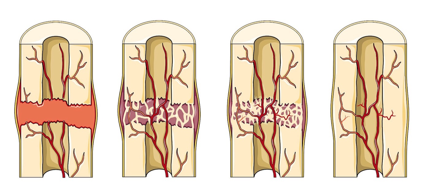
Figure 1. Secondary fracture healing: haematoma → soft (cartilaginous) callus → hard (bony) callus → remodelled bone, with the periosteal and endosteal blood supply. Source: Laboratoires Servier (Servier Medical Art), via Wikimedia Commons, CC BY-SA 3.0.
Figure 1. Secondary fracture healing: haematoma → soft (cartilaginous) callus → hard (bony) callus → remodelled bone, with the periosteal and endosteal blood supply. Source: Laboratoires Servier (Servier Medical Art), via Wikimedia Commons, CC BY-SA 3.0.
Primary (direct) healing occurs only under absolute stability (anatomical reduction with interfragmentary compression and almost no gap or motion) and proceeds without callus, by the same osteonal (Haversian) remodelling that renews normal bone.[2] It has two subtypes. In contact healing, osteoclastic cutting cones cross the fracture directly wherever the surfaces touch or are separated by only a minute gap (under about 0.01 mm, strain below 2%), and the trailing osteoblasts lay down new osteons. In gap healing, a small stable gap (up to roughly 0.5-1 mm) first fills with lamellar bone laid down transverse to the long axis, then remodels.[3]
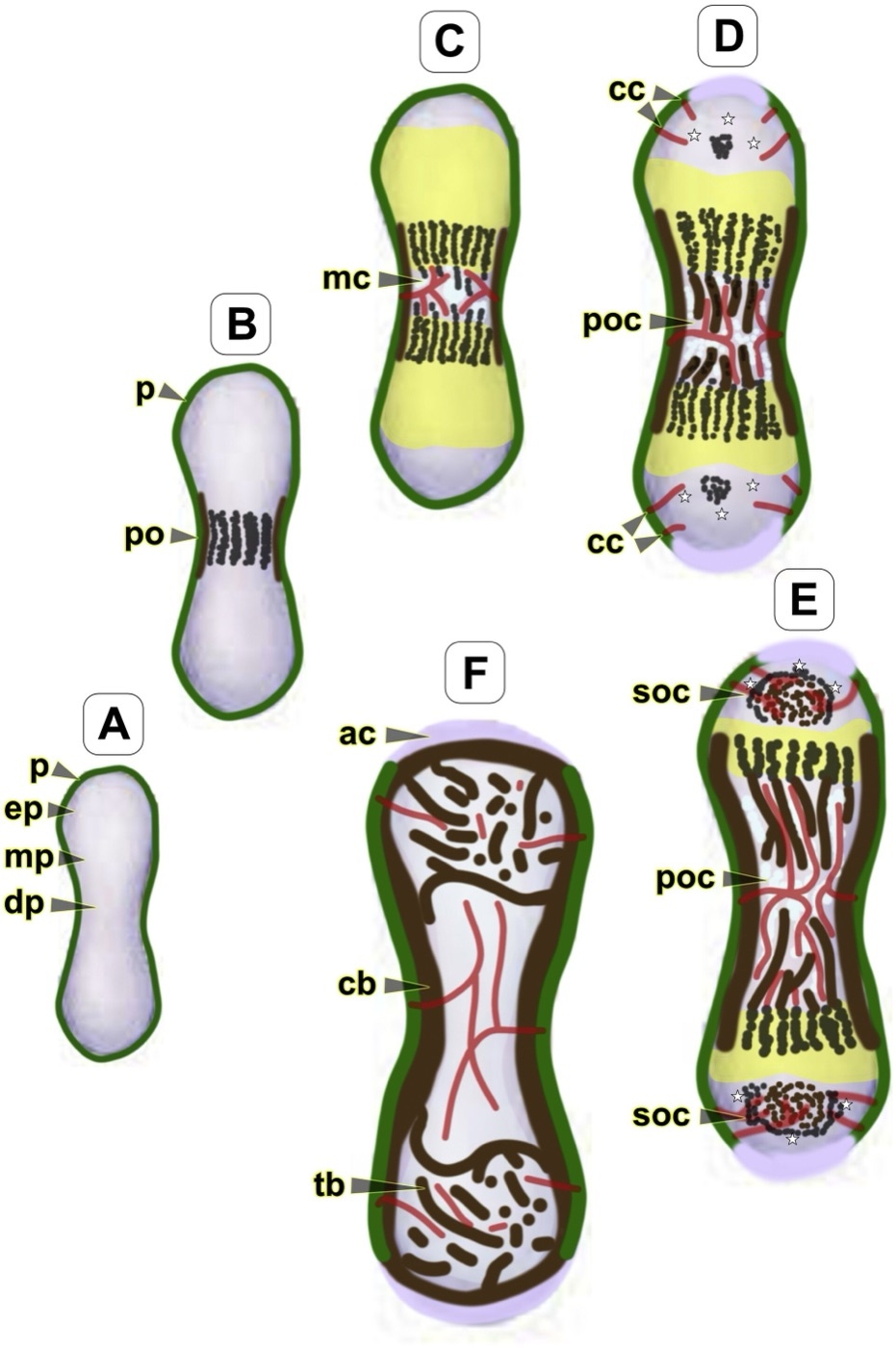
Figure 2. Schematic of endochondral bone development showing the osteoclastic cutting cone (cc) - the same mechanism that crosses the fracture in primary (Haversian) healing. From Khan et al. (2020), Arthritis Res Ther 22:168, PMC7488094, CC BY 4.0.
Figure 2. Schematic of endochondral bone development showing the osteoclastic cutting cone (cc) - the same mechanism that crosses the fracture in primary (Haversian) healing. From Khan et al. (2020), Arthritis Res Ther 22:168, PMC7488094, CC BY 4.0.
Secondary (indirect) healing is the natural, evolved process that occurs under relative stability (controlled micromotion from a cast, external fixator, intramedullary nail or bridging plate) and proceeds through callus, combining intramembranous ossification (forming hard callus peripherally) with endochondral ossification (a cartilage callus bridging the gap centrally).[4] This carries a clinical corollary worth memorising: a simple fracture must not be bridge-plated with relative stability, because the high strain across a single gap produces delayed union or nonunion. Simple patterns require absolute stability, whereas comminuted patterns heal well with relative stability.[5] Cancellous (metaphyseal) bone heals faster and more reliably than cortical bone, by intramembranous ossification with little external callus.[6]
Part II - The Stages of Secondary Healing
Secondary healing passes seamlessly through four overlapping stages.[7]
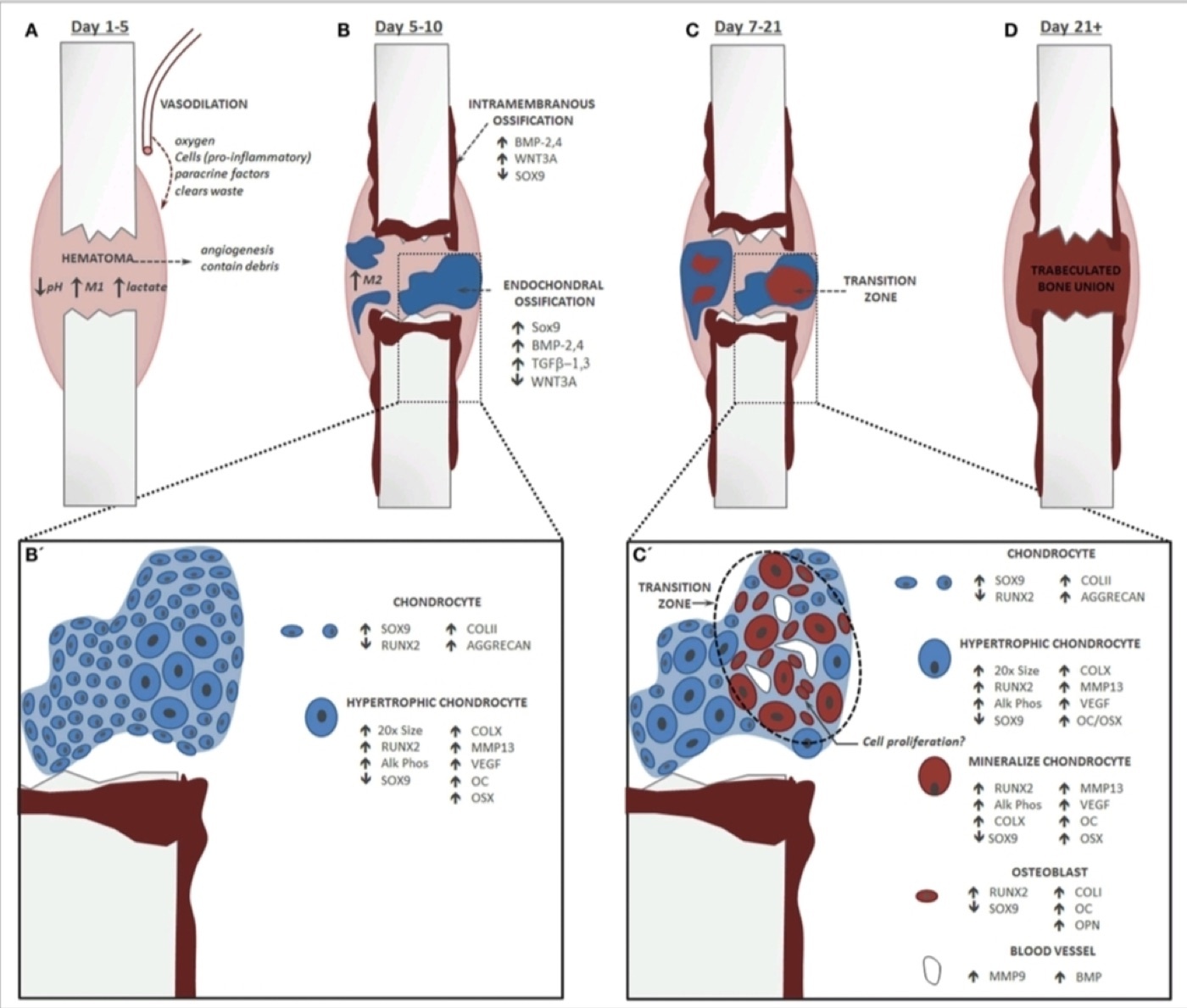
Figure 3. Temporal stages of fracture healing with the key cell types and signalling markers (SOX9, RUNX2, BMP-2/4, TGF-β, VEGF). From Bahney et al., PMC4318416, via Wikimedia Commons, CC BY 4.0.
Figure 3. Temporal stages of fracture healing with the key cell types and signalling markers (SOX9, RUNX2, BMP-2/4, TGF-β, VEGF). From Bahney et al., PMC4318416, via Wikimedia Commons, CC BY 4.0.
- Haematoma and inflammation (≈1-7 days). Ruptured vessels form a hypoxic haematoma; platelet degranulation and the inflammatory cells (neutrophils then macrophages) release cytokines (IL-1, IL-6, TNF-α) that recruit progenitor cells, while osteoclasts remove necrotic bone at the fragment ends.[8]
- Soft (fibrocartilaginous) callus (≈2-3 weeks). Periosteal and endosteal progenitors become osteoblasts (intramembranous bone away from the gap) while progenitors nearer the gap become chondrocytes, producing a cartilage callus (type II collagen, aggrecan) that steadies the fragments against shortening.[9]
- Hard (bony) callus (≈3-4 months). The cartilage callus now undergoes endochondral ossification: hypertrophic chondrocytes mineralise their matrix, VEGF drives vascular invasion, and osteoblasts replace the matrix with woven bone, converting the callus to rigid bone. Bridging begins at the periphery, where strain is lowest, and works inward.[10]
- Remodelling (months to years). The woven bone is slowly replaced by lamellar bone by coupled osteoclast-osteoblast activity, the medullary canal is restored, and the bone re-aligns to its loads (Wolff’s law).[11]
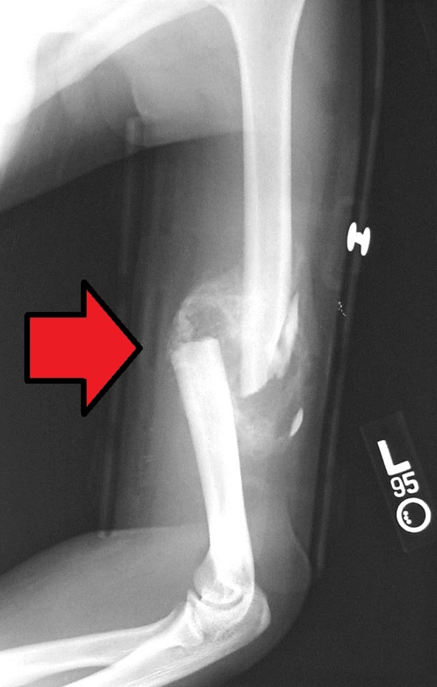
Figure 4. Bridging callus around a healing comminuted humeral shaft fracture (arrow). Source: Bill Rhodes, via Wikimedia Commons, CC BY 2.0.
Figure 4. Bridging callus around a healing comminuted humeral shaft fracture (arrow). Source: Bill Rhodes, via Wikimedia Commons, CC BY 2.0.
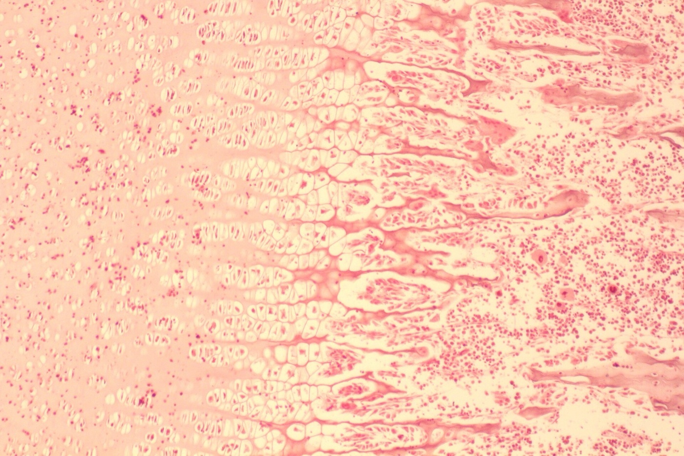
Figure 5. Endochondral ossification: resting/proliferating cartilage (left), hypertrophic cartilage (middle), and new cancellous bone (right). Source: Patbio, via Wikimedia Commons, CC BY-SA 4.0.
Figure 5. Endochondral ossification: resting/proliferating cartilage (left), hypertrophic cartilage (middle), and new cancellous bone (right). Source: Patbio, via Wikimedia Commons, CC BY-SA 4.0.
Part III - Perren’s Interfragmentary Strain Theory
The unifying mechanical principle, due to Perren, is that a tissue can only form and persist where the local strain is below its tolerance. Strain is the interfragmentary motion divided by the gap size, and the tolerances rise as the tissue is less mineralised: granulation tissue ~100%, cartilage ~15%, woven/cortical bone ~2%.[12] From this one idea most of fracture biology follows:
- Too much strain (an unstable fixation, or a single wide gap) prevents bony bridging despite an exuberant biological response, giving a hypertrophic nonunion. The remedy is mechanical stability, not biology.[13]
- Too little strain (a fixation so rigid, or a gap so wide, that there is no stimulus) also fails to form callus, and here the remedy is to permit motion (dynamisation).[14]
- Comminution lowers the strain at each gap, because the total movement is shared across several fracture planes. This is why multifragmentary fractures tolerate relative stability.[15]
- As the gap narrows during healing, strain rises; nature lowers it by enlarging the callus (reducing strain at its growing periphery) and by laying osteons in a spiral, spring-like pattern across the final gap.[16]
Part IV - The Biology of Repair
Cells. The osteochondral progenitors of the callus arise chiefly locally, from the cambium layer of the periosteum and from the endosteum (bone-marrow mesenchymal stem cells contribute mainly as paracrine “signalling cells”).[17] The effectors are osteoblasts (lay down type I collagen and osteoid), chondrocytes (the endochondral intermediary), and osteoclasts (haematopoietic-lineage cells that resorb bone in Howship’s lacunae).[18]
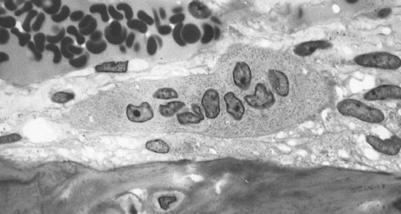
Figure 6. A multinucleated osteoclast on a bone surface (the bone-resorbing cell). Source: Robert M. Hunt, via Wikimedia Commons, public domain.
Figure 6. A multinucleated osteoclast on a bone surface (the bone-resorbing cell). Source: Robert M. Hunt, via Wikimedia Commons, public domain.
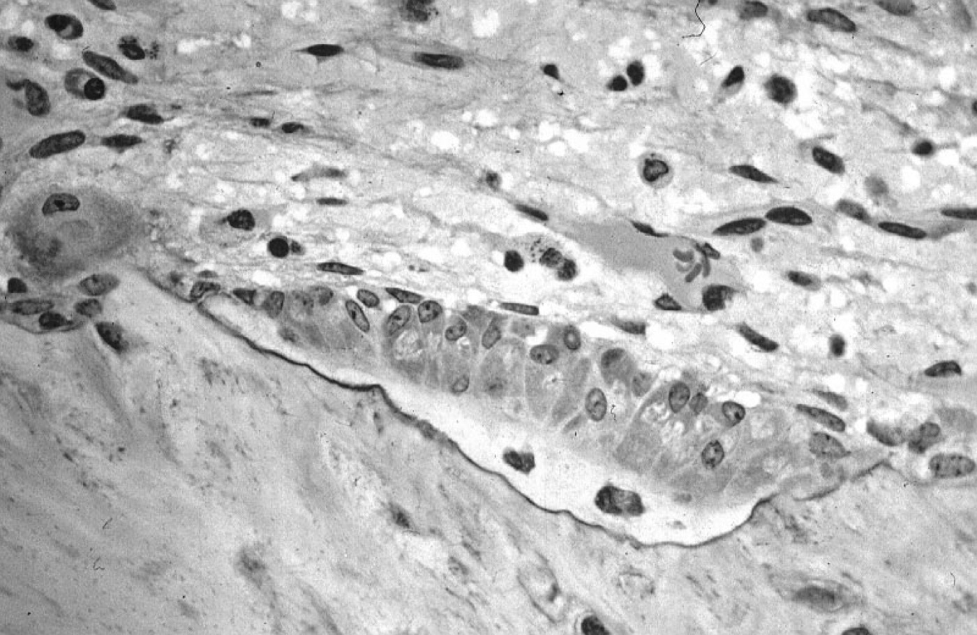
Figure 7. Active osteoblasts lining bone and forming osteoid, with embedded osteocytes. Source: Robert M. Hunt, via Wikimedia Commons, CC BY-SA 3.0.
Figure 7. Active osteoblasts lining bone and forming osteoid, with embedded osteocytes. Source: Robert M. Hunt, via Wikimedia Commons, CC BY-SA 3.0.
Signalling. Healing is orchestrated by the TGF-β superfamily, especially the bone morphogenetic proteins (BMPs), first described by Urist in the 1960s as the inductive principle of demineralised bone. Acting alongside them are TGF-β, PDGF, FGF-2, and VEGF (the angiogenic factor released by hypertrophic chondrocytes), and the Wnt, Notch and Hedgehog (Ihh) pathways.[19] Osteoclast formation and activity are governed by the RANK / RANKL / osteoprotegerin (OPG) axis with M-CSF: RANKL from osteoblastic cells drives osteoclast differentiation, while OPG is the decoy receptor that restrains it.[20]
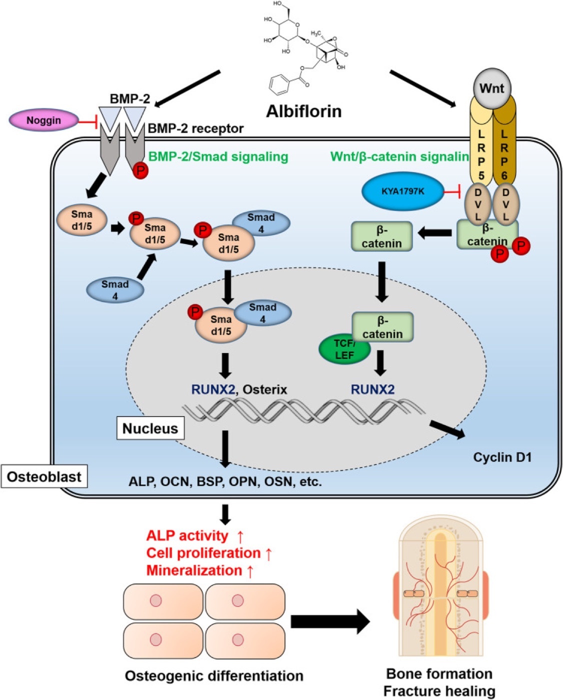
Figure 8. BMP-2/Smad and Wnt/β-catenin signalling converging on RUNX2/Osterix to drive osteoblast differentiation and fracture healing. From Front Pharmacol 2021;12:690113, PMC8327266, Fig. 8, CC BY 4.0.
Figure 8. BMP-2/Smad and Wnt/β-catenin signalling converging on RUNX2/Osterix to drive osteoblast differentiation and fracture healing. From Front Pharmacol 2021;12:690113, PMC8327266, Fig. 8, CC BY 4.0.
Blood supply. Bone is fed by three systems: the nutrient (medullary) artery, the periosteal vessels, and the metaphyseal/epiphyseal vessels. The medullary supply normally flows centrifugally (inside-out) to perfuse the inner cortex.[21] A fracture ruptures these vessels and cuts cortical perfusion by about half; the periosteal/extraosseous supply then becomes dominant. That is the central reason to preserve the soft-tissue attachments and to minimise periosteal stripping and bone-implant contact.[22]
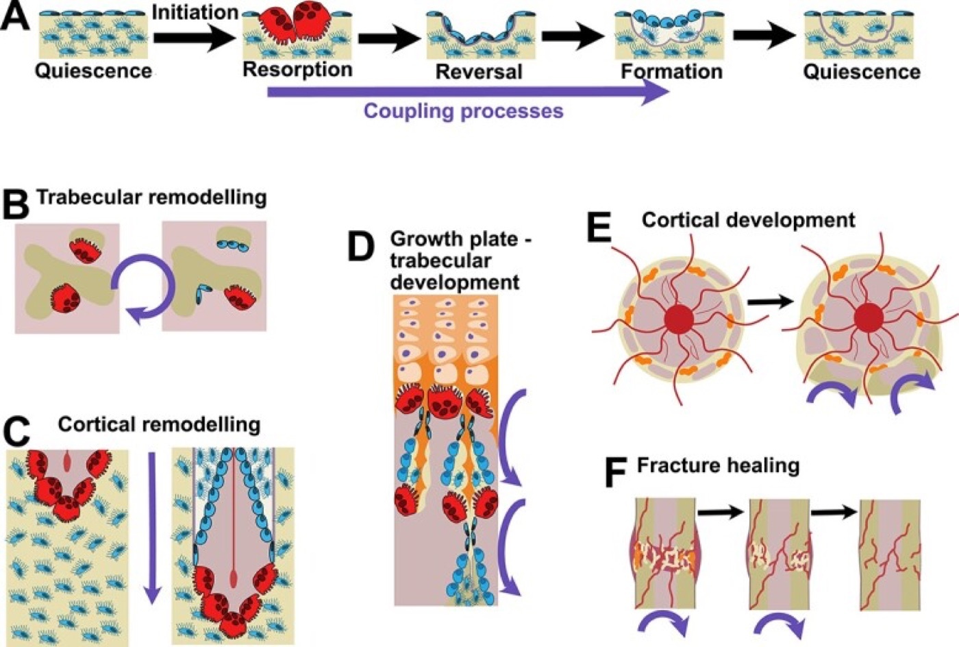
Figure 9. The bone-remodelling sequence and the cortical cutting cone of a basic multicellular unit (BMU) - advancing osteoclasts with trailing osteoblasts. From Sims (2024), PMC11425696, Fig. 1, CC BY 4.0.
Figure 9. The bone-remodelling sequence and the cortical cutting cone of a basic multicellular unit (BMU) - advancing osteoclasts with trailing osteoblasts. From Sims (2024), PMC11425696, Fig. 1, CC BY 4.0.
The diamond concept. Successful union requires several elements to come together, captured by Giannoudis’s “diamond concept”: mechanical stability, osteogenic cells, an osteoconductive scaffold, osteoinductive growth factors, and adequate vascularity (with host optimisation).[23]
Part V - Factors Influencing Healing
Local factors are dominated by blood supply and mechanical stability: a stripped periosteum, a high-energy soft-tissue injury, a wide bone gap, infection, and either excessive motion or excessive rigidity all impair healing; an intra-articular location demands anatomical reduction; and cancellous bone heals faster than cortical.[24]
Systemic/patient factors include age (slower, less robust, with a prolonged inflammatory phase), smoking/nicotine, diabetes (advanced glycation products and oxidative stress raising osteoclast and lowering osteoblast activity), NSAIDs and corticosteroids, malnutrition and vitamin-D deficiency, osteoporosis, and vascular disease; an occult endocrine abnormality is found in a high proportion of otherwise-unexplained nonunions.[25] The NSAID question is unsettled: COX-2 is mechanistically required for endochondral healing and NSAIDs inhibit it in animals, yet clinical evidence does not convincingly contraindicate short-term post-operative use.[26]
Part VI - Bone Grafts, Substitutes, and Enhancement
The biology of grafting rests on three properties: osteogenesis (the graft’s own living cells form bone), osteoconduction (the graft is a scaffold for ingrowth), and osteoinduction (graft growth factors recruit and differentiate host stem cells into bone).[27]
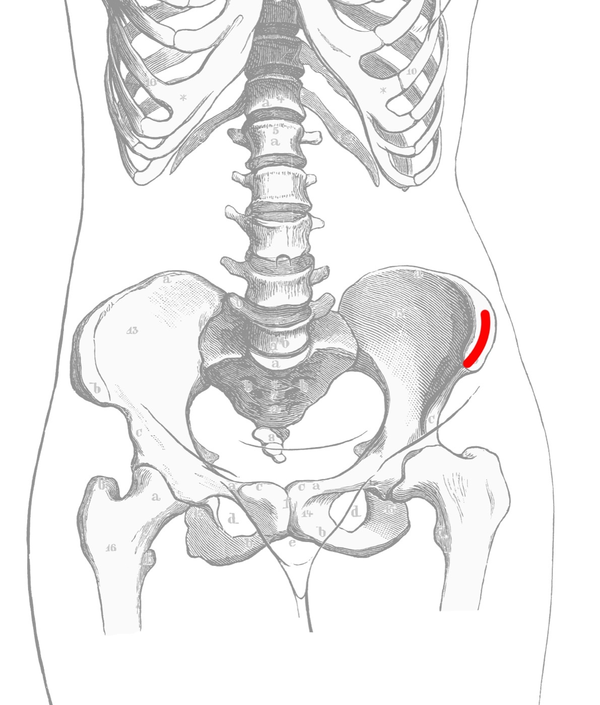
Figure 10. The anterior iliac crest, the standard cancellous/corticocancellous autograft harvest site. Source: Wikimedia Commons, public domain.
Figure 10. The anterior iliac crest, the standard cancellous/corticocancellous autograft harvest site. Source: Wikimedia Commons, public domain.
- Autograft is the gold standard, the only material with all three properties; cancellous autograft is osteogenic/conductive/inductive but non-structural and rapidly revascularised, while cortical (and vascularised) autograft is structural but slower to incorporate. The cost is donor-site morbidity (≈20% minor, ≈9% major), which the reamer-irrigator-aspirator (RIA) reduces.[28]
- Allograft is osteoconductive (± osteoinductive as DBM) but not osteogenic, with unlimited supply but a small disease-transmission risk and slow “creeping substitution”.[29]
- Bone-graft substitutes are osteoconductive scaffolds only: calcium sulfate (rapidly resorbed, now mainly an antibiotic carrier), calcium phosphate (slower, the cement form giving the highest compressive strength for early weight-bearing), tricalcium phosphate and hydroxyapatite ceramics, and demineralised bone matrix (DBM), which is osteoconductive and variably osteoinductive with high lot-to-lot variability. Optimal ingrowth needs interconnected pores of 300-500 µm.[30]
- Osteoinductive growth factors: recombinant BMP-2 (FDA-approved for open tibial fractures and lumbar fusion) and BMP-7/OP-1 (a humanitarian device for recalcitrant nonunion). Their efficacy is debated (positive in the BESTT open-tibia trial, negative in Aro’s), and they carry concerns of supraphysiologic dosing, heterotopic ossification and cost.[31]
- Biophysical stimulation: low-intensity pulsed ultrasound (LIPUS), electrical stimulation / pulsed electromagnetic fields (PEMF) and extracorporeal shock-wave therapy (ESWT) are safe and possibly effective (a meta-analysis put electrical stimulation at a 35% relative risk reduction of nonunion), but the evidence is generally low quality.[32] The only systemic anabolic agent is teriparatide (PTH 1-34).[33]
Part VII - Failure of Healing
When healing fails, the terms are: delayed union (healing achieved but over a prolonged course); malunion (healed with deformity); and nonunion (failure to heal).[34]
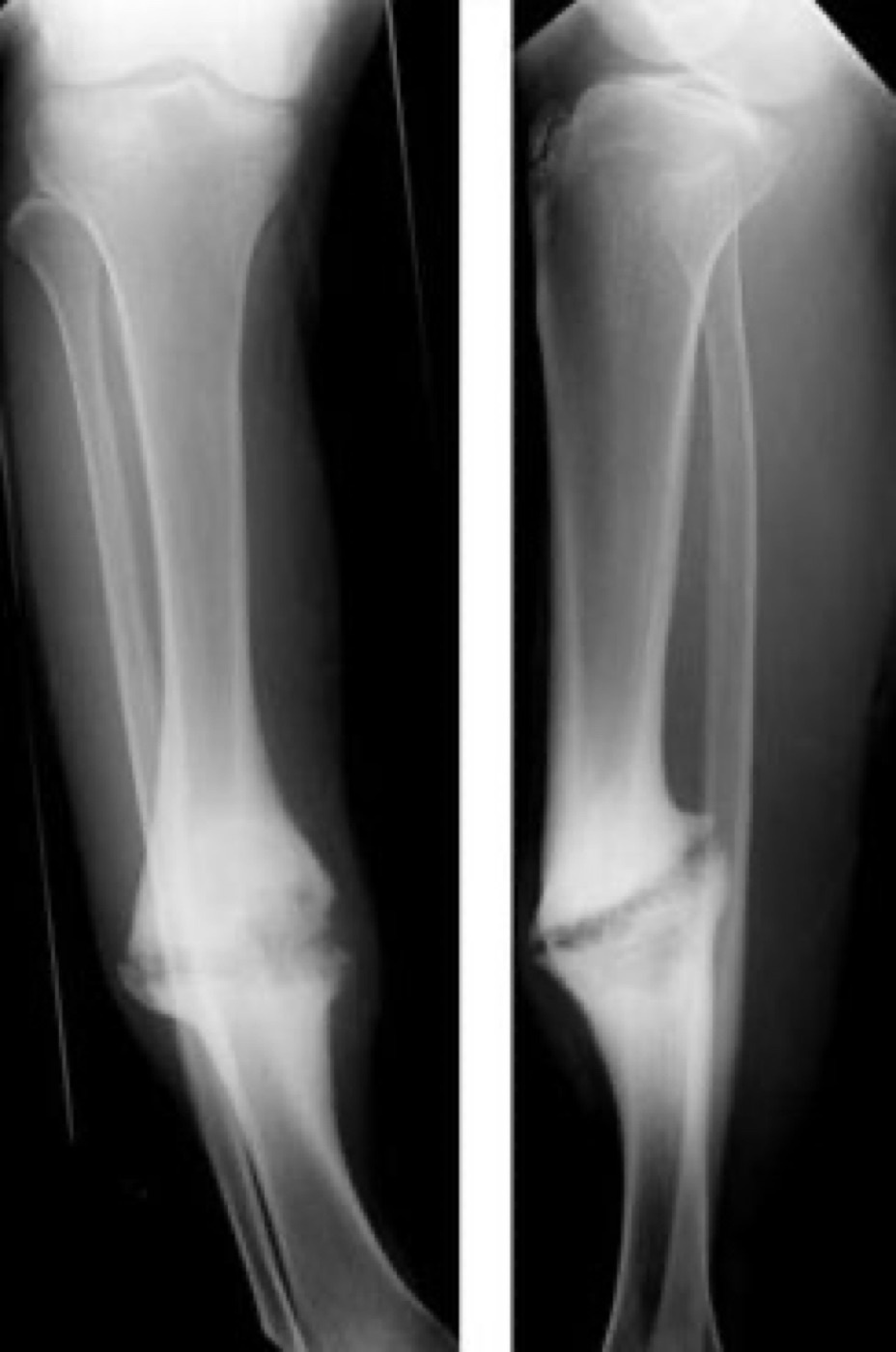
Figure 11. Hypertrophic (“elephant-foot”) nonunion of the tibia - abundant callus with a persistent fracture line, reflecting inadequate mechanical stability. Source: Lindsaydavidson, via Wikimedia Commons, CC BY 3.0.
Figure 11. Hypertrophic (“elephant-foot”) nonunion of the tibia - abundant callus with a persistent fracture line, reflecting inadequate mechanical stability. Source: Lindsaydavidson, via Wikimedia Commons, CC BY 3.0.
Nonunions divide by their biology:
- Hypertrophic nonunion: an abundant (“elephant-foot”) callus that fails to bridge because of inadequate mechanical stability. The treatment is stability (rigid fixation), not biology.[35]
- Atrophic nonunion: little or no callus, reflecting a failed biological response (poor vascularity, infection, prior radiation). The treatment addresses the biology (debridement, bone grafting).[36]
- Oligotrophic nonunion: an intermediate form with viable but insufficient callus, often after inadequate reduction.[37]
References
-
Rockwood & Green, ch. 2, p. 98; AO Principles ch. 1.2, p. 29.
-
AO p. 32, 39. Historically, primary healing was not the goal but an observed by-product of Danis’s rigid fixation aimed at early movement.
-
AO pp. 42-43; the precise µm thresholds are standard teaching, the textbook describing contact healing qualitatively as “contact or only a minute gap.” Direct healing is slower than callus healing, so the implant must stay stable for a long time.
-
AO p. 32, 37; Rockwood p. 133.
-
AO p. 37.
-
AO pp. 34, 42-43.
-
AO p. 32-33; Rockwood p. 103; Skeletal Trauma p. 150. The timelines below are AO’s.
-
AO p. 33; Rockwood pp. 98-101. A controlled pro-inflammatory phase is essential, but it must resolve for healing to progress.
-
AO p. 33; Rockwood p. 105 (Sox9-driven chondrogenesis).
-
AO p. 33; Rockwood pp. 105-106.
-
AO p. 33; Rockwood p. 107.
-
AO p. 38 (granulation 100%, bone 2%); the cartilage ~15% value is given at AO ch. 1.3 p. 47. Strain = ΔL/L, dimensionless.
-
AO p. 39; Rockwood p. 110.
-
AO p. 39.
-
AO p. 39.
-
AO p. 38.
-
Rockwood pp. 99, 132-134.
-
Rockwood pp. 99-100, 105.
-
Rockwood pp. 101-106.
-
Rockwood p. 100 (RANK/RANKL); OPG and the M-CSF role are standard teaching, not named in these extracts.
-
AO p. 32; Rockwood p. 99; the classic “centrifugal” description is from Rhinelander, though AO’s text uses “centripetal” for the same medullary system.
-
AO pp. 31-32. Devitalising the periosteum predisposes to nonunion.
-
Rockwood ch. 3 p. 125 enumerates the requirements (viable cells, vascularity, growth factors, stability); the “diamond concept” eponym (Giannoudis) is standard teaching.
-
AO pp. 30-32; Rockwood pp. 99, 109-110. Concomitant bone-plus-vascular injury carries a nonunion rate near 50%; the scaphoid’s tenuous blood supply explains its high nonunion rate.
-
Rockwood pp. 107-110.
-
Rockwood p. 140; Skeletal Trauma p. 100, a genuine source disagreement.
-
Skeletal Trauma p. 99; Rockwood p. 126.
-
Rockwood pp. 126-131; Skeletal Trauma pp. 100-101.
-
Rockwood pp. 128-130.
-
Skeletal Trauma pp. 102-109; Rockwood pp. 130-131.
-
Skeletal Trauma pp. 119-123; AO p. 33. Platelet-rich plasma is osteopromotive at best, with insufficient evidence to recommend routinely.
-
Rockwood pp. 143-144.
-
Rockwood pp. 108, 141.
-
Rockwood p. 109. About 2.5-10% of fractures fail to heal normally; every nonunion warrants a search for cause, including an endocrine work-up.
-
Rockwood p. 110; AO p. 39.
-
Rockwood pp. 109-110.
-
Standard teaching; these adult sources divide nonunions only into atrophic and hypertrophic.
-
AO pp. 32, 39.
-
AO p. 43; thresholds standard teaching.
-
AO pp. 32-33.
-
AO pp. 38-39, 47.
-
Rockwood pp. 99-106.
-
AO pp. 31-32.
-
Rockwood ch. 3 p. 125.
-
Skeletal Trauma p. 99; Rockwood p. 126.
-
Skeletal Trauma pp. 102-109.
-
Rockwood pp. 109-110; oligotrophic from standard teaching.