Contents
- Orientation: The Forearm Is a Joint, Not Two Long Bones
- Part I - Epidemiology and the Spectrum of Injury
- Part II - The Forearm as a Functional Unit: Applied Anatomy
- Part III - Classification
- Part IV - Clinical Assessment and Imaging
- Part V - The Galeazzi Fracture-Dislocation
- Part VI - The Monteggia Fracture-Dislocation (Cross-Reference)
- Part VII - The Essex-Lopresti Lesion and the Isolated (Nightstick) Ulnar Fracture
- Part VIII - Principles of Treatment
- Part IX - Surgical Approaches
- Part X - Both-Bone Forearm Fracture: Open Reduction and Internal Fixation
- Part XI - Intramedullary Nailing: A Selected Role
- Part XII - Open Fractures and External Fixation
- Part XIII - Complications
- Part XIV - A Synthesis: How to Reason About the Forearm
- References
Orientation: The Forearm Is a Joint, Not Two Long Bones
The single most useful idea in this whole topic is that the forearm is not really two parallel long bones at all. It is a joint. The radius and the ulna, locked together at the proximal radioulnar joint (PRUJ) above and the distal radioulnar joint (DRUJ) below, and tethered along their length by the interosseous membrane, exist to let the hand turn over. The radius carries the hand and rotates about the relatively fixed ulna, so that the palm can face up or down through an arc of roughly 150 degrees.[1] This is why a forearm-shaft fracture is treated quite differently from, say, a femoral or humeral shaft fracture. In the femur a few degrees of malrotation or a centimetre of bow is invisible to the patient. In the forearm the same error jams the rotation of the hand. Restoring length, axial alignment, rotation, and above all the natural lateral curvature of the radius (the radial bow) is therefore not cosmetic, it is functional, and it is the reason displaced adult forearm fractures demand anatomical reduction and rigid (absolute-stability) fixation, exactly as an articular fracture would.[2]
Two named fracture-dislocations dominate the topic because they capture this principle perfectly. The Galeazzi fracture (a distal-third radius fracture that disrupts the DRUJ) and the Monteggia fracture (a proximal ulna fracture that dislocates the radial head at the PRUJ) are both reminders that breaking one bone of a closed ring almost always injures the linked joint at the other end. The corollary, equally important, is therapeutic: if the broken bone is reduced anatomically, the dislocated radioulnar joint usually falls back into place on its own.[3]
Part I - Epidemiology and the Spectrum of Injury
Diaphyseal forearm fractures are roughly one-tenth as common as distal radius fractures, and while the incidence of distal radius fractures has climbed over the decades, the frequency of forearm-shaft fractures has stayed essentially flat.[4] The average yearly incidence in adults is about 1.35 per 10,000 (range 0-4 depending on age and sex), well below that of the humeral, femoral, or tibial shaft.[5] The age and sex distribution is bimodal: four-fifths of all forearm-shaft fractures occur in children, and among adults there is a striking and universal male predominance (the reported proportion of men ranges from 63% to 91%), concentrated in young men aged roughly 15 to 40 years injured by high-energy mechanisms.[6] Women have a lower incidence throughout life with a smaller second peak in the seventh decade, the low-energy osteoporotic tail of the distribution.[7]
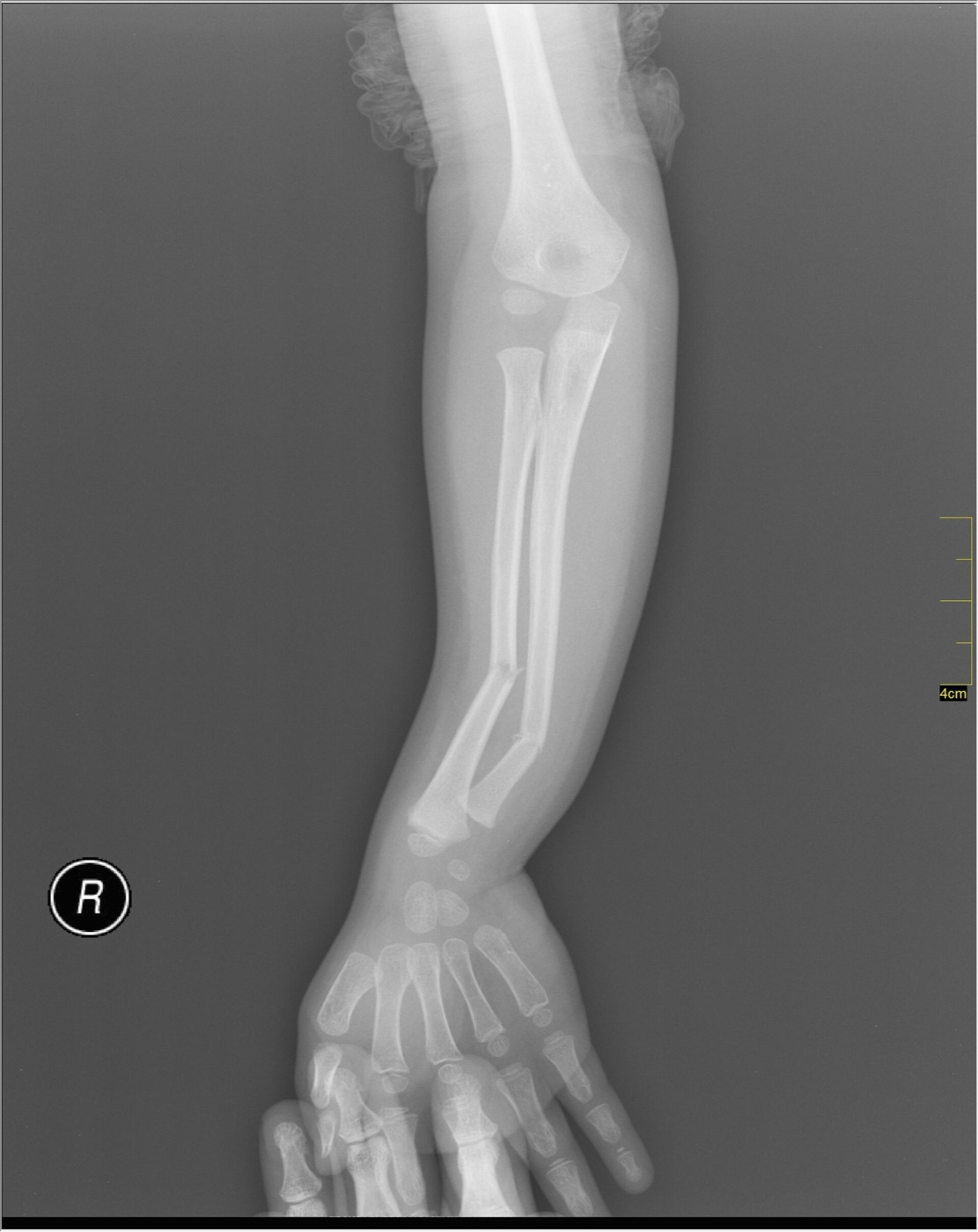
Figure 1. A displaced both-bone diaphyseal fracture of the radius and ulna (a paediatric example: four-fifths of forearm-shaft fractures occur in children). Nevit Dilmen, CC BY-SA 3.0, via Wikimedia Commons.
The mechanism shapes the injury. Most adult forearm-shaft fractures occur in young men with good bone, so they tend to be high-energy (road-traffic collisions, falls from height, sport, direct blows).[8] The energy explains the high proportion that are open and that are part of a polytrauma: open fractures range from under 10% in isolated radial-shaft injuries up to about 43% in both-bone forearm fractures, and roughly half of all forearm-shaft fractures occur in a multiply-injured patient.[9] Within the spectrum, Monteggia fractures make up about 13% and Galeazzi fractures about 23% of forearm fractures; a Galeazzi pattern (an associated DRUJ injury) is present in something like 7% to 25% of apparently isolated radial-shaft fractures.[10] The practical message is that the forearm-shaft fracture is rarely a benign, solitary event: it is frequently open, frequently high-energy, and frequently accompanied by an injured radioulnar joint or a second system.
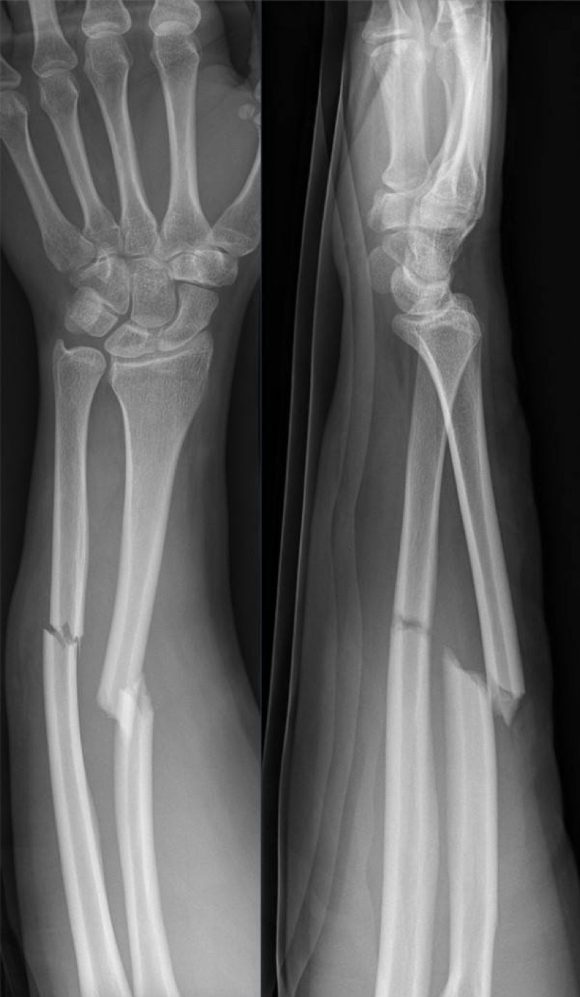
Figure 2. A displaced both-bone forearm-shaft fracture in a young adult from direct, high-energy trauma, AP and lateral radiographs. Hellerhoff, CC BY-SA 4.0, via Wikimedia Commons.
Part II - The Forearm as a Functional Unit: Applied Anatomy
2.1 The two bones and the radial bow
The radius averages about 25 cm long (range 21-29 cm) and is, in the words of the AO text, “a laterally bowed bone.”[11] That bow is the anatomical key to the whole topic. The radius actually has two curvatures, a larger one in the coronal plane (the major or lateral radial bow, running from the bicipital tuberosity to the ulnar corner of the distal articular surface) and a lesser anterior curve.[12] Schemitsch and Richards measured the normal bow on uninjured forearms and gave the numbers that now guide fixation: the maximum bow lies at about 60% of the length from the bicipital tuberosity to the distal articular surface, and its average magnitude is 15.3 mm.[13] Crucially, they showed that deviation from these values after fixation correlates directly with loss of forearm rotation, and that restoring the location of the bow to within about 4% and its magnitude to within about 1.5 mm of normal recovers at least 80% of rotation.[14] The bow is, in effect, the spacer that holds the radius away from the ulna and creates the room for it to rotate; flatten it and the hand stops turning.
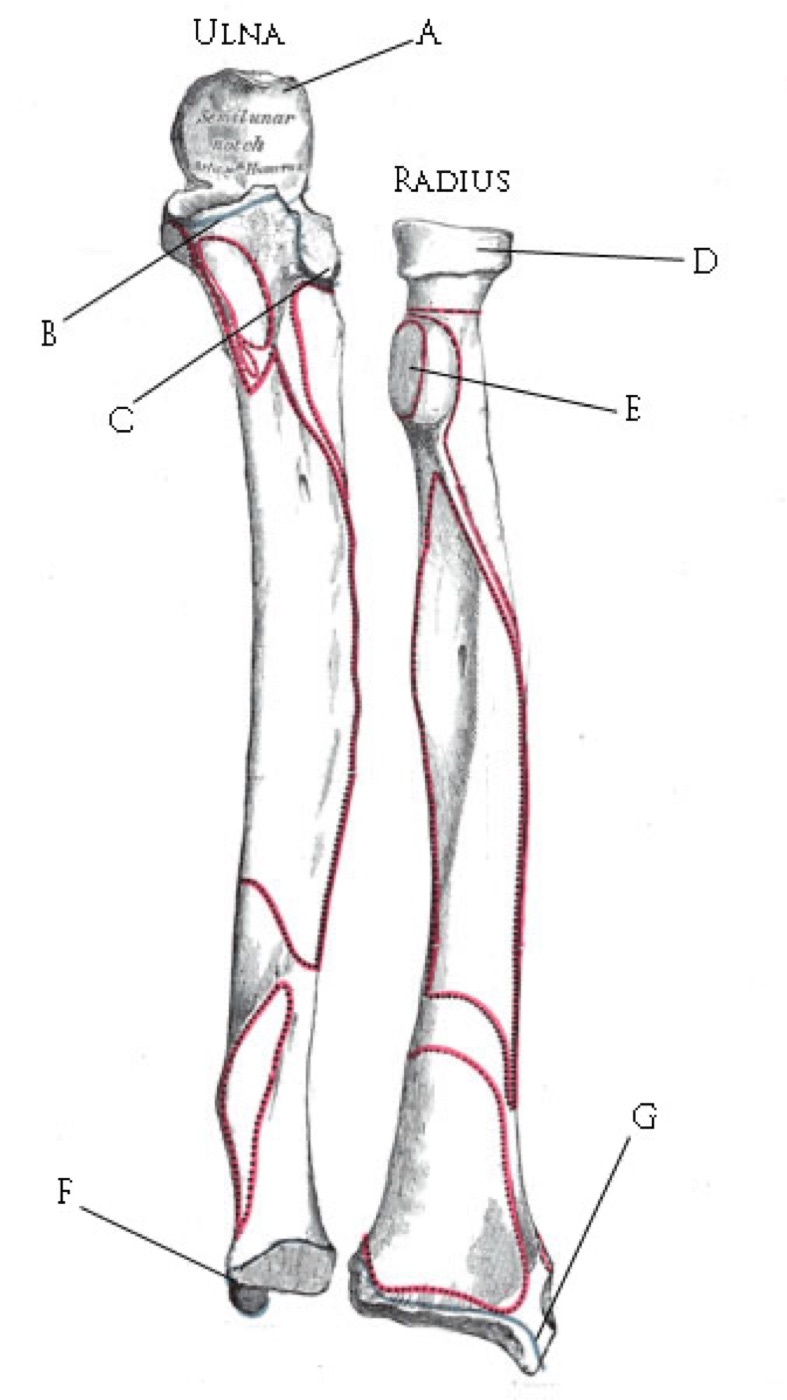
Figure 3. The radius and the ulna: the laterally bowed radius rotates about the straight ulna, the anatomical basis of pronation and supination. Illustration by Rafael Di Marco Barros after Gray’s Anatomy, public domain, via Wikimedia Commons.
The ulna is, by contrast, “a straight, posterior-medially positioned bone” that acts as the axis about which the radius rotates.[15] Its proximal end carries the olecranon and coronoid (forming the trochlear notch that grips the humeral trochlea) and, just distal and lateral, the radial notch, where the radial head articulates and the annular ligament inserts. Its distal end is the ulnar head and styloid, the base of the styloid being the anchor of the triangular fibrocartilage complex.[16] Because the ulna is straight and subcutaneous along its dorsal border, it is the easier bone to reduce and is often fixed first; its subcutaneous border is also why an isolated ulnar fracture from a direct blow is called a “nightstick” fracture.[17]
2.2 The linking structures: the two radioulnar joints and the interosseous membrane
Three sets of structures bind the radius and ulna into a single articulation. Proximally, the radial head sits in the radial (lesser sigmoid) notch of the ulna, held by the annular ligament, which blends with the lateral collateral ligament of the elbow (the PRUJ).[18] Distally, the ulnar head sits in the sigmoid notch of the radius, stabilised by the dorsal and palmar radioulnar ligaments and the triangular fibrocartilage complex (TFCC), the primary static stabiliser of the DRUJ (the DRUJ).[19] Along the shaft, the interosseous membrane (IOM) bridges the gap, and within it a marked thickening, the central band (about 3.5 cm wide, its fibres running proximal-radial to distal-ulnar at roughly 20° to the bone axis), is the key longitudinal stabiliser.[20] Hotchkiss showed that the central band provides about 71% of the longitudinal stiffness of the membrane once the radial head has been removed, which is exactly why a radial-head fracture combined with an IOM tear produces the longitudinal instability of the Essex-Lopresti lesion.[21] The interosseous space is widest in supination, a fact that bears on how the limb is positioned for surgery and immobilisation.
2.3 Muscle compartments and the deforming forces
The forearm holds four compartments (superficial volar, deep volar, dorsal, and the mobile wad of brachioradialis and the radial wrist extensors), which matters most for compartment syndrome.[22] For the surgeon reducing a fracture, the more immediate concern is the set of rotational deforming forces: the supinator and biceps supinate the proximal radius, while the pronator teres and pronator quadratus pronate.[23] The worst rotational deformity occurs in a radius fracture that lies distal to the supinator’s insertion but proximal to the pronator teres: the proximal fragment is dragged into full supination while the distal fragment pronates, so the surgeon must counter-rotate to reduce it. Fractures distal to the pronator teres are less deformed because the pronator teres balances the supinating pull.[24] Knowing where a fracture sits relative to these insertions tells you how the proximal fragment is rotated before you ever pick up the bone.
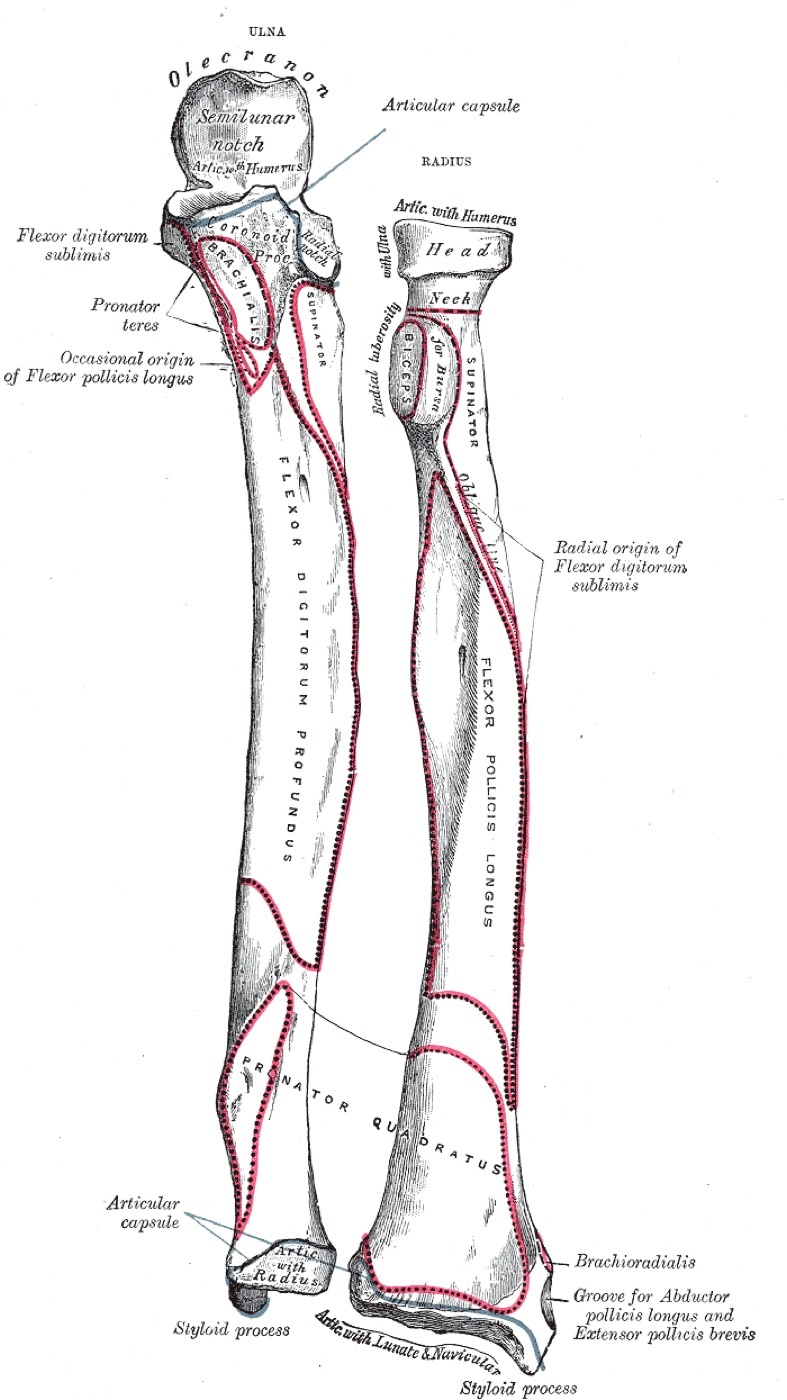
Figure 4. Bones of the forearm with their muscle attachments and the interosseous border, anterior aspect; the interosseous membrane spans the gap between the two bones along this border. Henry Vandyke Carter, Gray’s Anatomy (1918), public domain, via Wikimedia Commons.
2.4 The nerves and vessels at risk
The nerve that dominates forearm surgery is the posterior interosseous nerve (PIN), the deep motor branch of the radial nerve, which dives through the supinator (entering at the arcade of Frohse) to reach the dorsal compartment and run on the back of the interosseous membrane.[25] It is at risk in any exposure of the proximal radius. The governing rule, repeated in both source texts, is that pronation brings the proximal radius into view but supination protects the PIN by carrying it away from the dissection, so the supinator is released from the radius with the forearm supinated.[26] In the Henry approach the PIN must be protected when the supinator is reflected; in the Thompson approach the nerve can be palpated as a bulge about three finger-breadths distal to the radial head, where it may need to be directly identified.[27] The superficial radial nerve runs under the brachioradialis (vulnerable in the Henry approach), the anterior interosseous nerve runs on the front of the membrane between flexor pollicis longus and flexor digitorum profundus, and the dorsal cutaneous branch of the ulnar nerve emerges about 5-8 cm proximal to the wrist and is at risk in the distal ulnar exposure.[28] Vascular landmarks include the radial recurrent artery (the leash of Henry), which must be ligated for proximal radial exposure.[29]
Part III - Classification
3.1 The descriptive questions
Before any eponym or alphanumeric code, the useful classification is a list of questions: which bone or bones are broken; at what level (proximal, middle, or distal third); what is the pattern (transverse, oblique, comminuted, segmental); is the PRUJ or DRUJ disrupted; is it open or closed; and is the bone normal or previously deformed or implanted.[30] No single system captures all of these, so forearm fractures are most often described by location and degree of comminution.[31]
3.2 The AO/OTA classification
In the 2018 AO/OTA revision the radius and ulna are coded as separate bones to give greater flexibility, the diaphyseal segment being segment 2.[32] The codes are 2R2 for the radial shaft and 2U2 for the ulnar shaft, each then graded by morphology: type A simple, type B wedge, and type C multifragmentary (comminuted or segmental).[33] (The older Rockwood text still uses the legacy combined “22” forearm-shaft code with the same A/B/C grades, the radius and ulna distinguished within the subgroups; the modern separate-bone notation comes from the AO source.)[34] The classification is reliable enough for research but its complexity limits day-to-day clinical use, so most surgeons fall back on the descriptive scheme.[35]
3.3 The named patterns
Four eponymous or descriptive patterns are worth committing to memory, because each names a specific combination of fracture and joint injury:
- The nightstick fracture: an isolated ulnar-shaft fracture from a direct blow to the raised forearm. Because the ulna is subcutaneous it can break with relatively little energy and less soft-tissue injury, though a severely displaced one is prone to being open.[36]
- The Monteggia fracture-dislocation: a proximal or middle-third ulna fracture with dislocation of the radial head at the PRUJ, the radial head dislocating toward the apex of the ulnar deformity. It is classified by Bado into four types by the direction of radial-head dislocation (type 1 anterior, the commonest in children; type 2 posterior, up to 80% of adult Monteggias and often with a radial-head or coronoid fracture; type 3 lateral, almost exclusively in children; type 4 anterior with a fracture of both forearm bones at the same level, seen only in adults), with Jupiter subclassifying the adult type-2 by the level of the ulnar fracture (2A through the coronoid, 2B at the metaphyseal-diaphyseal junction, 2C diaphyseal, 2D complex).[37]
- The Galeazzi fracture-dislocation: a distal-third radius fracture with disruption of the DRUJ (the full account is in Part V).[38]
- The Essex-Lopresti lesion: a radial-head (or proximal radial) fracture with tearing of the interosseous membrane and DRUJ disruption, producing longitudinal (axial) instability of the forearm (the full account is in Part VII).[39]
Part IV - Clinical Assessment and Imaging
4.1 History and examination
The history separates the low-energy fall or direct blow from the high-energy collision or fall from height, and pain distant from the obvious injury should prompt a search for a second fracture or a radioulnar-joint injury.[40] Examination notes the swelling and deformity, inspects the skin for an open wound (most often on the ulnar side), and demands a thorough neurovascular examination of the radial nerve and PIN, the median nerve and its anterior interosseous branch, and the ulnar nerve, together with the radial and ulnar pulses.[41] Because the forearm fracture so often hides an injury at one of its ends, the elbow and wrist are examined deliberately for the tenderness and instability that betray a Monteggia or Galeazzi pattern.[42]
4.2 Compartment syndrome: a constant vigilance
The forearm is the second most frequent site of acute compartment syndrome, which complicates about 3% of forearm fractures, most often in men under 35.[43] The earliest and most sensitive clinical sign is pain out of proportion, worsened by passive extension of the digits; the diagnosis is confirmed when the compartment pressure comes within 30 mm Hg of the patient’s diastolic blood pressure.[44] Treatment is urgent fasciotomy. The AO text reduces it to a practical instruction: decompress the two anterior (volar) compartments and the one posterior (dorsal) fascial compartment, usually with a carpal-tunnel release added.[45] Even with prompt release, complications follow in around 41% of cases, neurological deficit being the most frequent, so the emphasis is on suspecting it early rather than treating it late.[46]
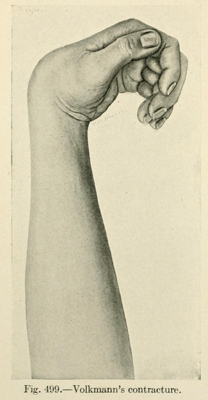
Figure 5. Volkmann ischaemic contracture of the hand, the end-stage deformity of an untreated forearm compartment syndrome. Mumford, The Practice of Surgery (1910), public domain, via Wikimedia Commons / Internet Archive.
4.3 Imaging
The minimum study is anteroposterior and lateral radiographs of the entire forearm, from the elbow to the wrist, because a film that excludes either joint will miss a Monteggia or a Galeazzi.[47] Dedicated elbow and wrist views are added when an isolated radial or ulnar fracture raises that suspicion: on the elbow film the axis of the radial neck must point at the capitellum (confirming the PRUJ), and on the wrist film the ulnar head must sit within the sigmoid notch with normal ulnar variance (confirming the DRUJ).[48] CT and MRI are rarely needed acutely, reserved for suspected rotational malunion, nonunion, or occult DRUJ/TFCC and interosseous-membrane injury; if a deformity cannot be quantified, a contralateral comparison film establishes the patient’s normal radial bow and ulnar variance.[49]
Part V - The Galeazzi Fracture-Dislocation
The Galeazzi is a fracture of the radial shaft (typically the distal third) with disruption of the DRUJ, and it carries the telling nickname “fracture of necessity” because in the adult, nonoperative treatment gives such uniformly poor results that operation is obligatory.[50] Its other names are the Piedmont fracture and the “reverse Monteggia.”[51] The mechanism is a torsional force with axial loading (a fall onto a hyperpronated, extended wrist), the fracture energy travelling distally to rupture the interosseous membrane, the radioulnar ligaments, and finally the TFCC, leaving the DRUJ unstable.[52]
The clinical problem is recognising the DRUJ injury, which is easy to miss. The strongest predictor is the level of the radial fracture: Rettig and Raskin showed that fractures within 7.5 cm of the distal articular (lunate-facet) surface carry a markedly higher rate of DRUJ instability, and the more distal the fracture, the more likely the joint is disrupted.[53] This gives a simple subclassification, type 1 within 7.5 cm (high instability, often needing open DRUJ repair) and type 2 more proximal.[54] Moore added that 5 mm of radial shortening on a standard wrist film correlates with DRUJ injury. It is fair to add the dissent: Tsismenakis and Tornetta found both the 7.5 cm rule and the 5 mm shortening unreliable as predictors and recommended an intraoperative DRUJ stress examination in every case instead.[55] The radiographic signs of the DRUJ injury itself are a fracture at the base of the ulnar styloid, widening of the DRUJ on the AP film, dorsal dislocation of the ulnar head on the lateral, and a change in ulnar variance of more than 5 mm.[56]
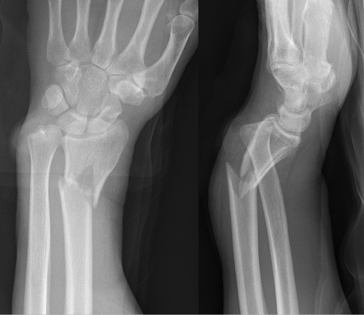
Figure 6. Galeazzi fracture-dislocation: a distal-third radius fracture with disruption of the distal radioulnar joint, AP and lateral radiographs. Hellerhoff, CC BY-SA 3.0, via Wikimedia Commons.
The treatment follows directly from the principle. The radius is reduced and plated (a 3.5 mm compression plate; the volar/Henry approach is most common for a distal-third fracture, though some prefer the dorsal Thompson approach to spare pronation), and in most cases anatomical reduction of the radius stabilises the DRUJ on its own.[57] The DRUJ is then tested and managed in three tiers:[58]
- Reduced and stable (stays put through pronation, supination, and neutral): immobilise in a long-arm cast or splint for 3 to 6 weeks, in the position of greatest stability.
- Reducible but unstable: hold the reduction with two K-wires (about 2.0 mm in adults) placed across the DRUJ, with the forearm in the position of stability (supination for a dorsal dislocation, pronation for a volar one), and protect it with a splint that crosses the elbow and wrist to prevent rotation and wire breakage; an associated displaced ulnar-styloid base fracture is fixed.
- Irreducible: first re-check the radial reduction (inadequate radial reduction is the commonest cause); if the radius is anatomical, the DRUJ is blocked by interposed soft tissue, classically the extensor carpi ulnaris tendon, and is opened and repaired together with the TFCC.
With anatomical reduction of the radius and a congruent DRUJ, satisfactory outcomes are reported in 80% to 92%, and primary DRUJ repair gives excellent results in about 95%; the price of inadequate radial reduction or a missed DRUJ injury is chronic instability and DRUJ arthritis.[59]
Part VI - The Monteggia Fracture-Dislocation (Cross-Reference)
The Monteggia is the mirror image of the Galeazzi and the same logic applies in reverse: fix the ulna anatomically and the radial head usually reduces itself.[60] It is uncommon, the treatment section putting it at only about 1% to 2% of forearm fractures (the chapter’s epidemiology section quotes a higher 13%, the difference reflecting how broadly the Monteggia pattern is counted). The ulna is plated, conventionally on its dorsal (tension) surface, often with a precontoured proximal plate, and the proximal ulnar dorsal angulation (PUDA) must be restored (a mean of about 6° of apex-dorsal angulation roughly 5 cm distal to the olecranon tip), because residual radial subluxation persists if the ulnar shape is wrong.[61] If the radial head will not reduce after the ulna is anatomically fixed, the surgeon must suspect either a malreduced ulna or buttonholing of the radial head through the annular ligament, and explore through a separate lateral incision (never a single combined approach, which invites synostosis).[62] Outcomes are best for Bado types 1 and 3 and worse for the adult types 2 and 4 and for injuries with an associated radial-head or coronoid fracture.[63]
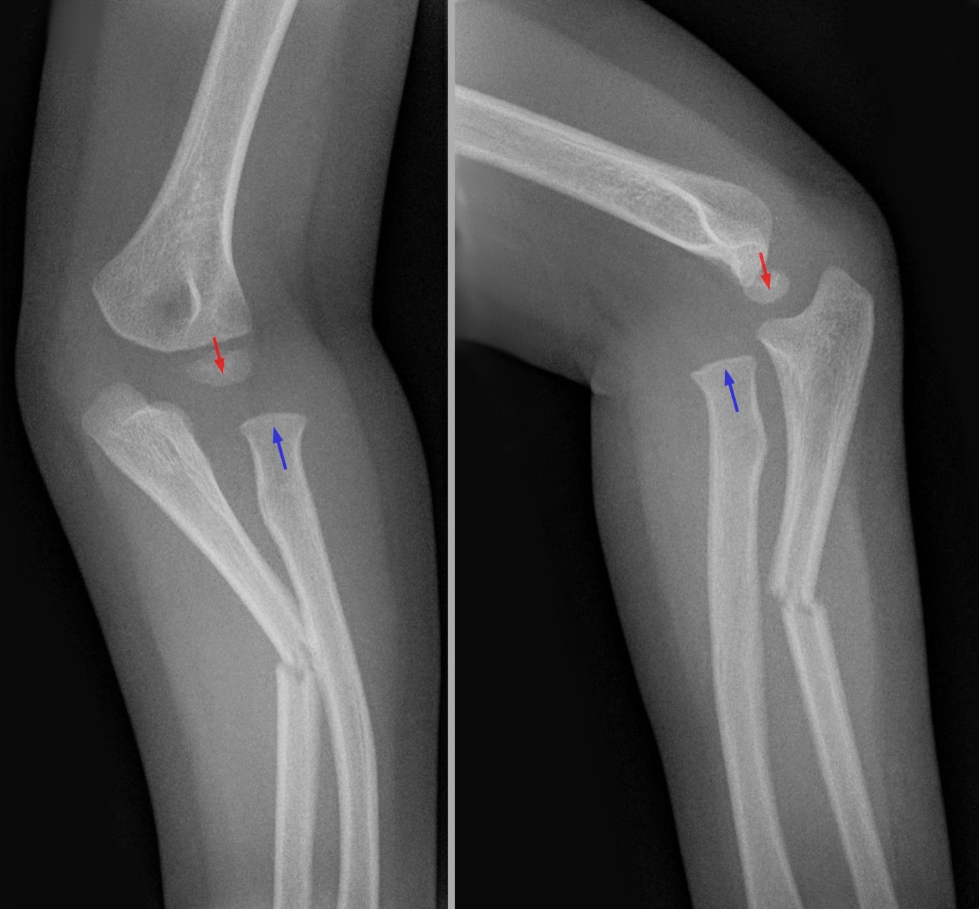
Figure 7. Monteggia fracture-dislocation: a proximal ulna fracture (blue arrow) with dislocation of the radial head (red arrow), AP and lateral radiographs. Hellerhoff, CC BY-SA 3.0, via Wikimedia Commons.
Part VII - The Essex-Lopresti Lesion and the Isolated (Nightstick) Ulnar Fracture
7.1 Essex-Lopresti
The Essex-Lopresti lesion is the longitudinal counterpart to the transverse Monteggia and Galeazzi: a fracture of the radial head or neck (or proximal radial shaft) combined with a tear of the interosseous membrane and disruption of the DRUJ, so that the radius can migrate proximally.[64] It is the clinical expression of the anatomy in Part II: with the radial head gone and the central band torn, the principal longitudinal restraints of the forearm are lost. Anatomical reconstruction of the radial head or neck is crucial, because even mild proximal radial displacement creates incongruity at the DRUJ, and failure to recognise and address the longitudinal instability at the outset leads to progressive proximal migration that is extremely difficult to reconstruct late.[65] The lesson is to examine the wrist in every radial-head fracture and never to excise a radial head in a forearm with a torn membrane.
7.2 The isolated ulnar (nightstick) fracture
The isolated ulnar-shaft fracture is the one forearm-shaft injury that is routinely treated without surgery, provided it is stable. The accepted threshold for nonoperative management is a fracture of the distal two-thirds of the ulna with less than 50% displacement and less than 10° of angulation; cadaver work shows that displacement beyond 50% implies an interosseous-membrane tear and instability.[66] Such fractures are managed in a below-elbow cast or, classically, a functional brace (Sarmiento), with good results: Sarmiento’s series reported about 96.5% good or excellent outcomes and 99% union (though with 35% lost to follow-up), and De Boeck reported 93% union in a short-arm cast.[67] The caveats are two. First, proximal-third ulnar fractures are treated operatively, because they tend to displace, threaten the PRUJ, and have lower union rates.[68] Second, even the “benign” nightstick fracture carries a notable rate of delayed or nonunion despite usually being a closed, simple injury, so it is followed to union rather than dismissed.[69] Displaced or angulated ulnar fractures beyond the threshold are plated like any other forearm-shaft fracture.
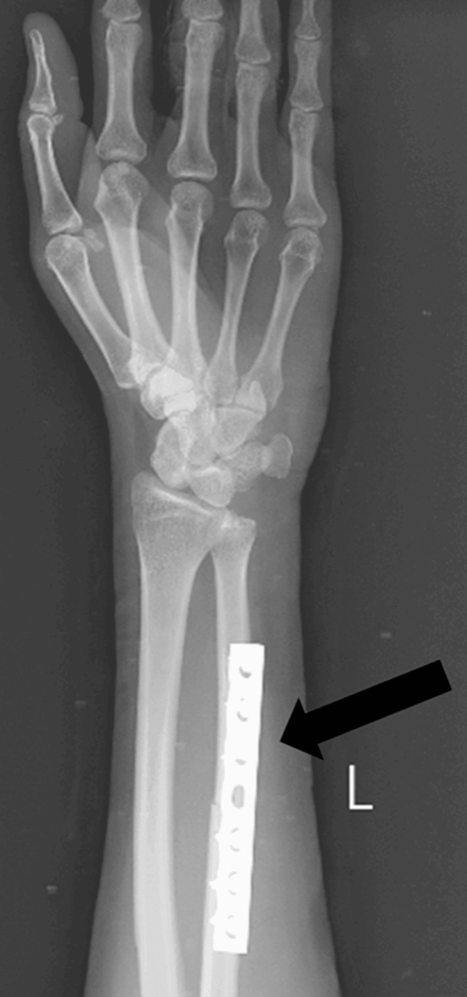
Figure 8. Isolated ulnar shaft (“nightstick”) fracture, here shown after plate fixation of a displaced fracture; the radius is intact. Ambekar et al., Cureus 2024;16(1):e53353, CC BY 4.0.
Part VIII - Principles of Treatment
The governing principle has already been stated and bears repeating because every technical decision flows from it: the forearm is a joint, so a displaced adult forearm-shaft fracture is reduced anatomically and fixed with absolute stability. The Rockwood text puts it plainly: the most important part of treatment is “restoring function by restoring normal anatomical relationships and form of the forearm by means of reestablishing length, alignment, rotation, and radial bow,” and “restoration of the radial bow is related to functional outcome, especially in regaining pronation and supination.”[70] The cost of getting it wrong is measurable: Tarr found that 15° of malunion costs about 27% of rotation, and Matthews that 20° produces a functionally significant 30% deficit in pronation-supination.[71]
The indications are therefore broad. Operation is the rule for displaced fractures of both bones, any displaced/angulated/rotated isolated fracture (the AO threshold being more than 10° of angulation or rotation), all the fracture-dislocations (Galeazzi, Monteggia, Essex-Lopresti), open fractures, and the floating or polytraumatised limb.[72] The narrow remaining role for nonoperative care is the simple, undisplaced or minimally displaced isolated fracture, in practice the stable nightstick fracture in a brace or cast.[73] Timing follows the diaphyseal rule (closed fractures are best fixed within the first 24 hours), and delay is specifically discouraged because it raises the risk of synostosis.[74]
Part IX - Surgical Approaches
9.1 The radius: Henry (anterior) versus Thompson (dorsal)
The Henry (volar/anterior) approach exposes the whole length of the radius and is the workhorse, particularly for the distal four-fifths.[75] The skin incision runs from the lateral edge of the biceps tendon to the radial styloid along the ulnar border of brachioradialis; the true internervous plane is between brachioradialis (radial nerve) and the pronator teres and flexor carpi radialis (median nerve).[76] The superficial radial nerve is protected under brachioradialis and the radial artery is retracted ulnarly with flexor carpi radialis. Proximally the recurrent radial vessels (the leash of Henry) are ligated, and the supinator is released with the forearm supinated to carry the PIN out of harm’s way; the middle third is exposed with the forearm pronated; the distal third by reflecting pronator quadratus.[77]
The Thompson (dorsal/posterolateral) approach runs from the lateral epicondyle to Lister’s tubercle through the interval between extensor carpi radialis brevis and extensor digitorum, and gives the best access to the proximal and middle radius.[78] It is avoided for distal-third fractures because of the higher risk of tendon irritation over the hardware and of PIN injury. In the proximal exposure the PIN runs through the supinator at right angles to its fibres and can be palpated as a bulge about three finger-breadths distal to the radial head; it is identified and protected before the supinator is reflected off the radius from ulnar to radial.[79]
9.2 The ulna
The ulna is approached along its subcutaneous border through the internervous plane between extensor carpi ulnaris (PIN) and flexor carpi ulnaris (ulnar nerve), but the plate is never placed on the subcutaneous crest itself; it is laid on the dorsal (extensor) or volar (flexor) surface, the side chosen before exposure (usually the side of greatest soft-tissue stripping) so that only one surface is dissected.[80] In the distal incision the dorsal cutaneous branch of the ulnar nerve is protected.[81]
9.3 One incision per bone
A cardinal rule unites all of this: use a separate incision for each bone and keep a broad skin bridge between them. Fixing both bones through a single combined approach increases the risk of nerve injury and, above all, of radioulnar synostosis, and it is not recommended.[82]
Part X - Both-Bone Forearm Fracture: Open Reduction and Internal Fixation
10.1 Why plates, and which plate
Compression plating is the gold standard for the displaced both-bone forearm fracture, and the historical series that established it are worth knowing: Anderson reported 98% radial and 96% ulnar union in 330 fractures (the study that “established the gold standard”), and Chapman established the 3.5 mm plate as the standard size (equal union to the 4.5 mm plate but with less refracture after removal).[83] Union rates with modern compression plating are consistently above 90% and usually 96% to 100%.[84] Locking plates were introduced in the hope of better biology but have shown no clinical advantage over conventional compression plates for routine forearm fractures, so a 3.5 mm limited-contact dynamic compression plate (LC-DCP) remains the AO implant of choice; locking screws are reserved for osteoporotic bone.[85]
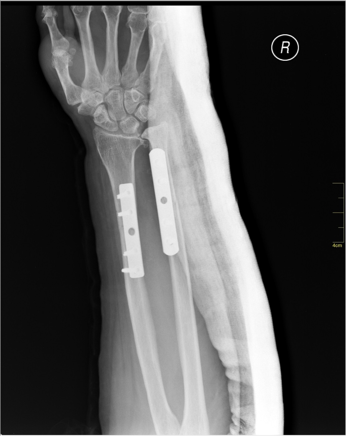
Figure 9. A both-bone forearm fracture after open reduction and internal fixation with plates and screws on both the radius and the ulna. Nevit Dilmen, CC BY-SA 3.0, via Wikimedia Commons.
10.2 The fixation rules
The fixation aims at absolute stability: a simple (type A) or wedge (type B) fracture is fixed with an interfragmentary lag screw plus a compression or protection (neutralisation) plate, while a multifragmentary (type C) fracture is bridge-plated to restore length, alignment, and rotation without disturbing the fragments.[86] The AO rule for plate length is six cortices, or three bicortical screws, in each main fragment, which for a simple fracture usually means a 7- or 8-hole plate; bridge plates for comminution are longer still.[87] There is a documented controversy here: biomechanical and clinical data (Crow; Lindvall and Sagi) suggest that four cortices (two screws) per side can suffice with a standard-length plate, but the prudent advice, especially in osteoporotic bone, remains three screws per fragment, because the loss of a single screw in a two-screw construct invites rotational failure.[88]
A few practical points complete the picture. The less comminuted bone is approached and reduced first (when both are simple, the ulna is usually fixed first because its straight shape guides the length and rotation of the radius; when both are comminuted, the ulna is fixed first and the tourniquet time reserved for the radius).[89] Restoring the radial bow is done by contouring the plate and confirming the reduction against a contralateral comparison film, then checking that full, symmetric pronation and supination are present on the table before closure.[90] Periosteal stripping is kept to about 1 mm at the fracture edges, and routine bone grafting of comminuted or open fractures is no longer considered necessary (Wright and many series show reproducible union without it); if grafting is needed for a defect, the graft is kept away from the interosseous membrane to avoid a cross-union.[91]
10.3 After surgery
When fixation is stable the fascia is left open and not repaired, the limb is dressed lightly, and early active motion of the fingers, wrist, elbow, and forearm rotation is begun, which gives significantly better function than prolonged immobilisation.[92] A circular cast is specifically avoided; a short volar or sugar-tong splint may be used for the first week for comfort. Radiographs are checked at 6 and 12 weeks, weight-bearing is usually allowed at 6 to 8 weeks, and lifting is limited (about 2.3 kg) until radiographic union, which averages 8 to 24 weeks and is longer in open fractures.[93] Routine implant removal is not recommended, because of the risks of nerve injury and refracture (discussed in Part XIII).[94]
Part XI - Intramedullary Nailing: A Selected Role
For the routine both-bone forearm fracture, plating is superior and intramedullary nailing has a limited, selected role.[95] The traditional objection is that a smooth nail cannot control rotation or restore the radial bow, so older nails required supplementary casting, which defeats the purpose of internal fixation. Nailing is reasonable for unstable patterns where plating is unattractive: segmental fractures, open fractures with a poor soft-tissue envelope, the polytrauma patient, and osteopenic bone, and it is the standard for paediatric forearm fractures (elastic nails), where AO notes excellent results in children but inadequate adult stability.[96] Contraindications include an open physis, a canal under 3 mm, active infection, and metaphyseal extension that prevents locking.[97]
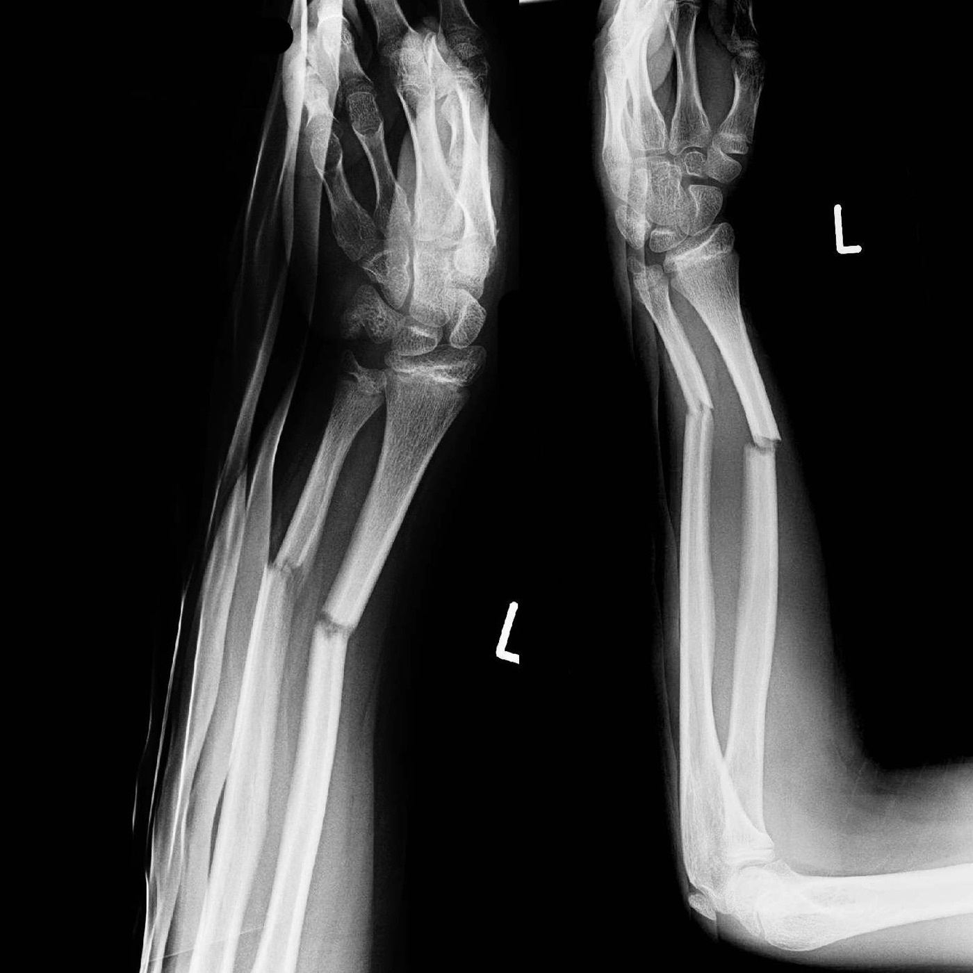
Figure 10. A displaced both-bone forearm fracture before fixation, AP and lateral radiographs. Thomas Zimmermann (THWZ), CC BY-SA 3.0 DE, via Wikimedia Commons.
Modern interlocked, pre-contoured nails that bend to match the radial bow have narrowed the gap. Comparative series (Ozkaya, Köse) find that the newest interlocking nails match plating for union, ROM, and functional scores while reducing blood loss and operative time, although the Rockwood summary still concludes that “no advantage has been shown over conventional screws and plates.”[98] Two technical hazards deserve mention: the PIN is at risk during proximal radial interlocking (the danger zone is within about 3 cm of the radial head, so the locking screw is placed in neutral rotation, with a small open incision if there is any doubt), and the nail is bent intraoperatively to mimic the radial bow even when pre-contoured.[99]
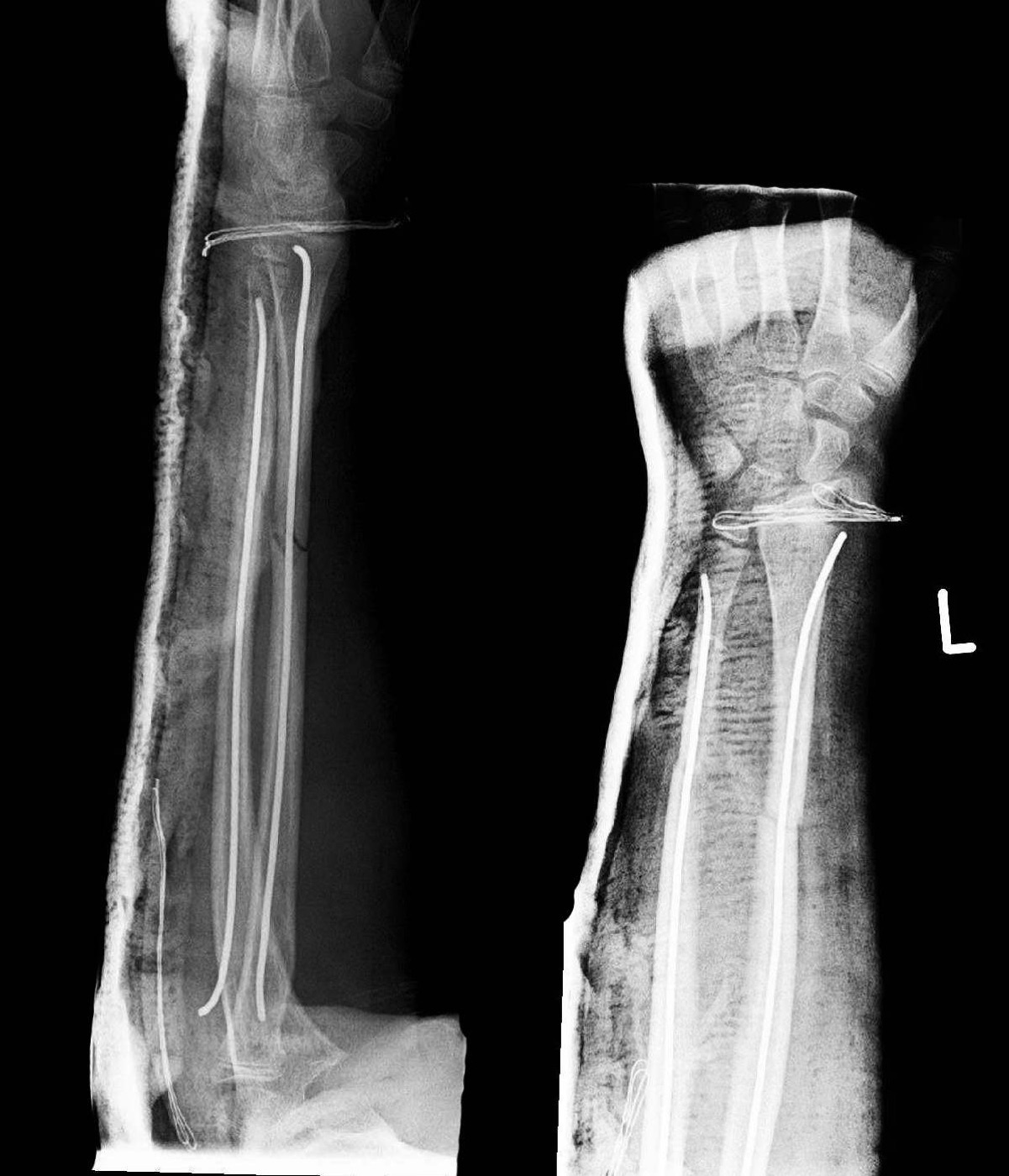
Figure 11. The same forearm stabilised with elastic intramedullary nails, the elastic-nail technique that is standard in children. Thomas Zimmermann (THWZ), CC BY-SA 3.0 DE, via Wikimedia Commons.
Part XII - Open Fractures and External Fixation
Open forearm fractures are common (up to 43% of both-bone injuries) and the great majority are Gustilo type I or II.[100] The principle, established by Chapman and Moed, is that immediate internal fixation is safe for types I, II, and IIIA after debridement, with satisfactory results in about 90%; results fall off for IIIB and IIIC, where the AO advice is to fix but to plan repeat debridement until soft-tissue coverage (a flap) is achieved.[101] Antibiotic cover is a first-generation cephalosporin for all, with gram-negative cover added for type III and anaerobic cover for farm or combat contamination.[102]
External fixation is reserved for the rare situation in which massive soft-tissue loss or contamination precludes plating.[103] Pins are placed on the subcutaneous ulnar surface percutaneously and into the radius through an open incision to avoid nerve injury. Because external fixation alone has a high rate of nonunion and malunion (especially malrotation), the recommended strategy is early conversion to plate fixation once the soft tissues allow, sometimes with bone grafting.[104] For a segmental bone defect, a bridge plate with a temporary cement spacer and Masquelet-technique autografting at 6 to 8 weeks (or a free vascularised fibula, or conversion to a single-bone forearm in catastrophic injury) is the reconstructive ladder.[105]
Part XIII - Complications
The standard list of complications is infection, malunion, nonunion, radioulnar synostosis, refracture, compartment syndrome, and nerve palsy.[106]
Infection is uncommon (0% to 3%; Anderson reported 3% of 330, with about a third of nonunions being septic), treated for the superficial case with oral antibiotics and for the deep case by repeat debridement, retention of stable hardware until union, and prolonged intravenous antibiotics.[107] Malunion is the complication that defines the forearm: it costs rotation in proportion to its size (Tarr 15° → 27% loss; Matthews 20° → 30% deficit) and is corrected, when symptomatic, by osteotomy of one or both bones, ideally within a year of injury, with care not to over-release the interosseous membrane and destabilise the DRUJ.[108] Nonunion occurs in 0% to 10% and reflects either inadequate mechanics (gapping, a weak or too-short plate, screws too close to the fracture) or inadequate biology (high energy, stripping, comminution); infection must always be excluded, and the treatment is revision fixation per AO principles with grafting for any defect, which heals up to 100% of cases.[109]
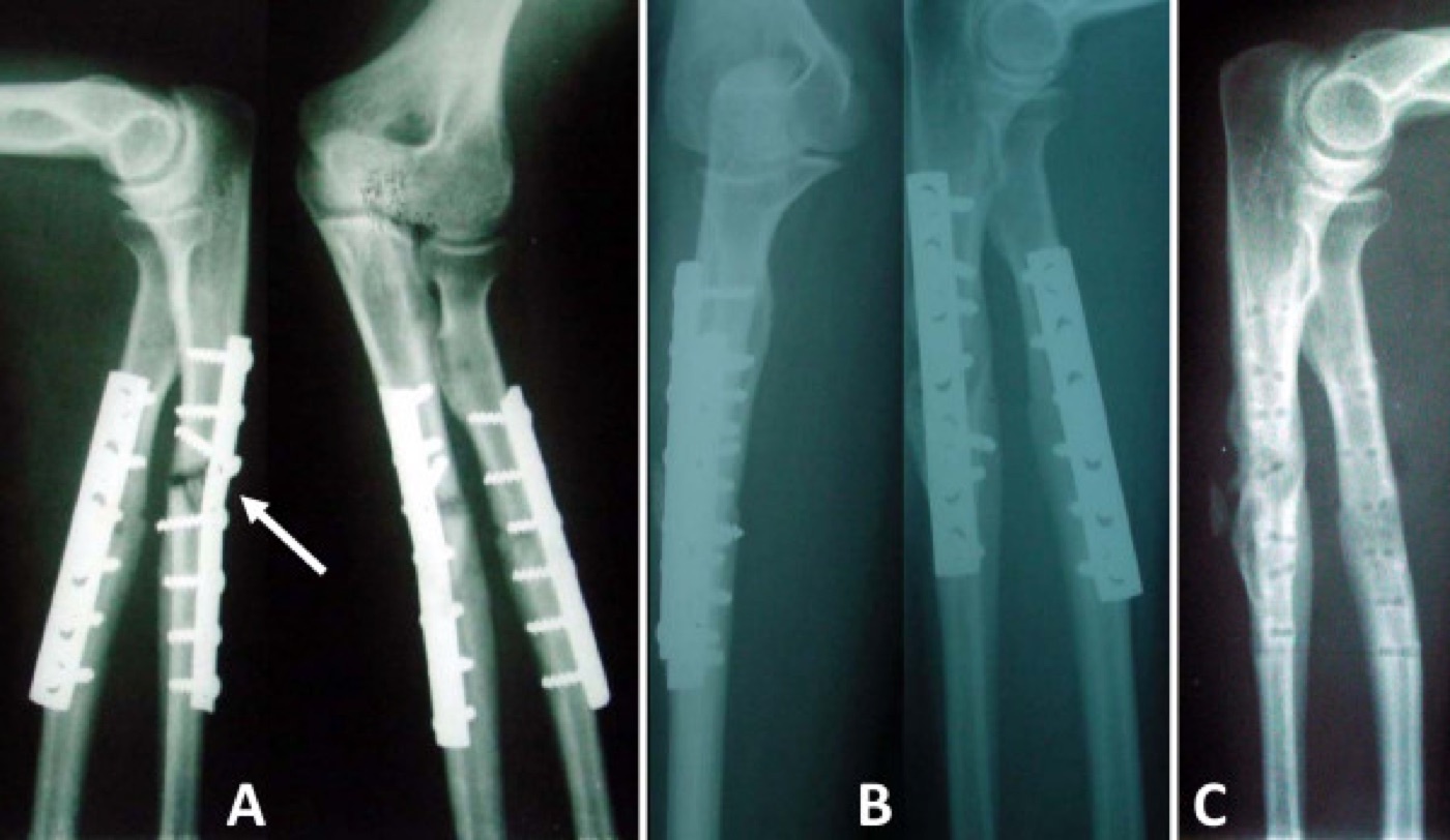
Figure 12. Diaphyseal forearm nonunion (ulnar shaft, arrow) and its revision by compression plating and bone grafting. Dos Reis et al., Ann Surg Innov Res 2009;3:5, CC BY 2.0.
Radioulnar synostosis (a bony cross-union bridging the two bones) is uncommon (about 1% to 6%, AO citing 2.6% to 6.6%) but disabling, since it abolishes rotation.[110] Its risk factors are the ones the surgeon can partly control: fractures of both bones at the same level, an interosseous-membrane injury, severe soft-tissue damage and comminution, delayed fixation, a single combined incision for both bones, bone graft placed in the interosseous space, postoperative casting, and a concomitant head injury (which raises the propensity to heterotopic bone).[111] Prevention is built into the technique already described (separate incisions, minimal stripping, graft away from the membrane, early motion). Treatment is excision of the mature cross-union once it has fully formed (usually 6 to 9 months, when the heterotopic bone shows trabeculation), often with interposition of fat and adjuvant indomethacin or low-dose irradiation to prevent recurrence.[112]
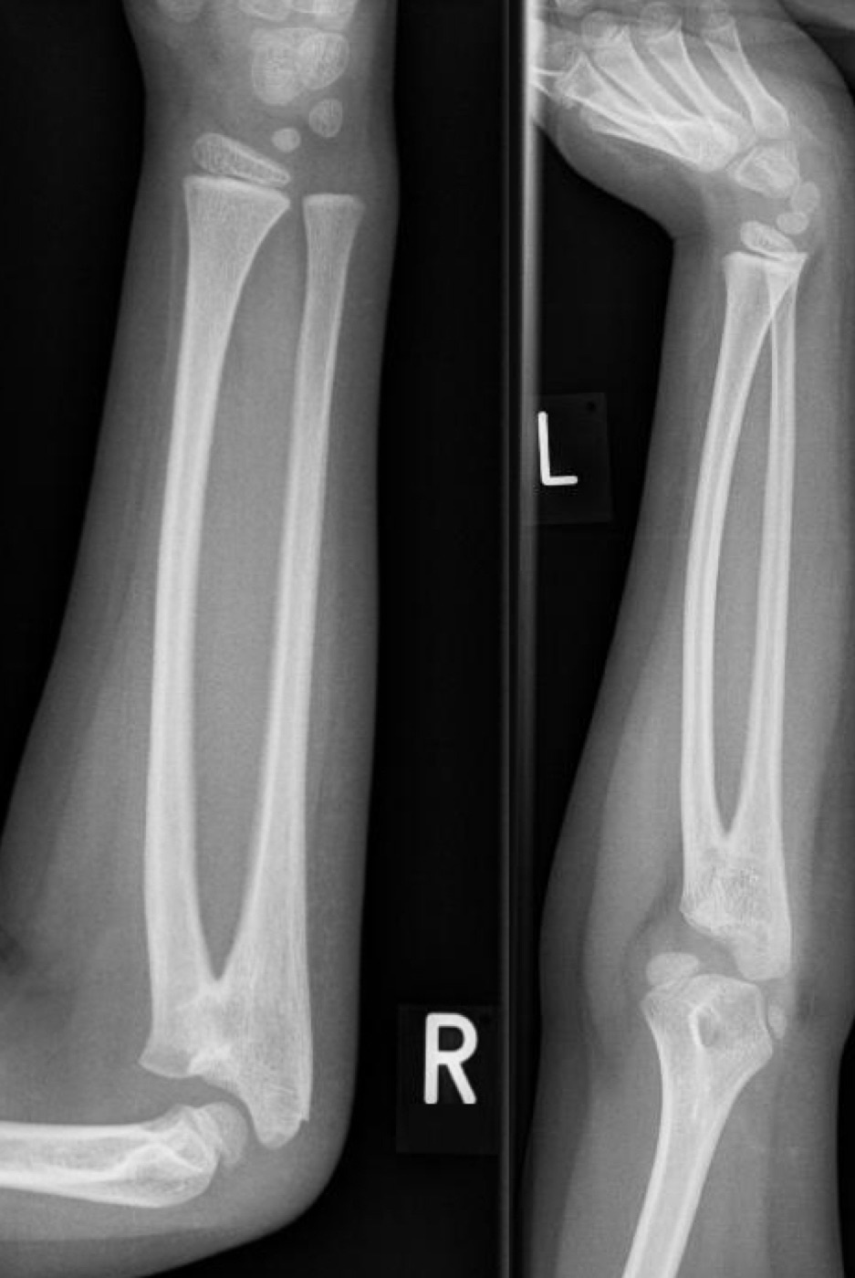
Figure 13. Radioulnar synostosis: a bony bridge (cross-union) between the radius and the ulna that abolishes forearm rotation. This example is a congenital cross-union; the bony-bridge morphology is the same as that of the post-traumatic complication. Kinderradiologie Olgahospital, Klinikum Stuttgart, CC BY-SA 3.0, via Wikimedia Commons.
Refracture after plate removal is the reason removal is discouraged: it occurs in up to 18% (AO 3.5% to 25%), through the original site or an empty screw hole.[113] When removal is unavoidable, the risk is reduced by delaying it 12 to 18 months (AO: not within 12 months) and protecting the limb in a splint or brace for several weeks afterwards; the 3.5 mm plate has markedly reduced the refracture rate compared with the old 4.5 mm plate.[114] Nerve palsy is led by the PIN, classically after proximal radial fixation through the Thompson approach (lower with the volar approach), and most palsies are neurapraxias that recover; a temporary loss of flexor pollicis longus from anterior interosseous nerve traction can follow Henry exposure and usually resolves.[115] Compartment syndrome and its sequela, Volkmann ischaemic contracture, complete the list and are managed as in Part IV, by early recognition and fasciotomy.[116]
Part XIV - A Synthesis: How to Reason About the Forearm
The forearm rewards one habit of thought above all: treat it as a joint. When you see a forearm fracture, ask not only what is broken but what is dislocated, because a fracture of one bone in this closed ring nearly always injures the linked joint at the other end. A distal-third radius fracture is a Galeazzi until the DRUJ is proven intact; a proximal ulna fracture is a Monteggia until the radial head is proven located; a radial-head fracture with a sore wrist is an Essex-Lopresti until the interosseous membrane is proven whole. That is why the films must run from elbow to wrist every time. Once the diagnosis is made, the treatment principle is the same throughout: restore length, alignment, rotation, and the radial bow, and fix with absolute stability, because a few degrees of malrotation here jam the hand in a way they never would in the femur. Reduce the broken bone anatomically and the dislocated radioulnar joint usually reduces itself; plate the simple fracture with compression and bridge the comminuted one, always through separate incisions, and move the limb early. Then watch for the complications the anatomy predicts: the synostosis born of a violated interosseous space, the lost rotation of a flattened radial bow, the PIN palsy of an over-zealous proximal exposure, and the compartment syndrome of a high-energy, swollen forearm. A forearm fixed anatomically and mobilised early turns the hand over; one left bowed, short, or malrotated does not, and that single functional fact governs the whole of this topic.
References
-
Rockwood & Green’s Fractures in Adults, pp.2470, 2494 (the geometric relationship of radius and ulna renders the forearm a functional joint; the radius rotates about the ulna, which acts as the axis, allowing ~150° of rotation); AO Principles of Fracture Management, p.677 (“The bowed morphology of the radius allows rotation of the radius around the ulna, which acts as an axis”).
-
Rockwood & Green’s Fractures in Adults, pp.2501, 2504; AO Principles of Fracture Management, pp.687, 691 (“Functional outcome largely depends on reconstruction of the radial bow and perfect anatomical restoration of the proximal and distal radioulnar joints”).
-
Rockwood & Green’s Fractures in Adults, p.2504; AO Principles of Fracture Management, pp.678-679.
-
Rockwood & Green’s Fractures in Adults, p.2470 (diaphyseal forearm fractures ~1/10 as frequent as distal radius fractures; adult incidence 1.35 per 10,000, range 0-4; distal radius incidence rising while shaft frequency stable). AO Principles of Fracture Management, p.677 records forearm fractures as 10-14% of all fractures in the 1980-1996 AO documentation and a >200% rise in surgically-treated forearm fractures from 1996 to 2006.
-
Rockwood & Green’s Fractures in Adults, p.2470 (diaphyseal forearm fractures ~1/10 as frequent as distal radius fractures; adult incidence 1.35 per 10,000, range 0-4; distal radius incidence rising while shaft frequency stable). AO Principles of Fracture Management, p.677 records forearm fractures as 10-14% of all fractures in the 1980-1996 AO documentation and a >200% rise in surgically-treated forearm fractures from 1996 to 2006.
-
Rockwood & Green’s Fractures in Adults, pp.2470-2471 (four-fifths occur in children; male predominance 63-91%; peak in males 15-40 years; female peak in the seventh decade).
-
Rockwood & Green’s Fractures in Adults, pp.2470-2471 (four-fifths occur in children; male predominance 63-91%; peak in males 15-40 years; female peak in the seventh decade).
-
Rockwood & Green’s Fractures in Adults, pp.2471, 2482 (high-energy mechanism in young men; open-fracture frequency <10% isolated radius to 43% both-bone; ~half occur in polytrauma).
-
Rockwood & Green’s Fractures in Adults, pp.2471, 2482 (high-energy mechanism in young men; open-fracture frequency <10% isolated radius to 43% both-bone; ~half occur in polytrauma).
-
Rockwood & Green’s Fractures in Adults, pp.2471, 2480 (Monteggia 13%, Galeazzi 23% of forearm fractures; DRUJ injury in 7-25% of isolated radial-shaft fractures).
-
Rockwood & Green’s Fractures in Adults, p.2493 (radius mean length 25 cm, range 21-29 cm; two curvatures, the major radial bow from bicipital tuberosity to the ulnar aspect of the distal articular surface); AO Principles of Fracture Management, p.677 (the radius is “a laterally bowed bone”).
-
Rockwood & Green’s Fractures in Adults, p.2493 (radius mean length 25 cm, range 21-29 cm; two curvatures, the major radial bow from bicipital tuberosity to the ulnar aspect of the distal articular surface); AO Principles of Fracture Management, p.677 (the radius is “a laterally bowed bone”).
-
Rockwood & Green’s Fractures in Adults, pp.2493-2494, 2520 (Schemitsch & Richards: maximum radial bow at 60 ± 0.7% of length, magnitude 15.3 ± 0.3 mm; restoring location within 4.3 ± 0.7% and magnitude within 1.5 ± 0.2 mm yields ≥80% of normal rotation; deviation correlates with reduced rotation).
-
Rockwood & Green’s Fractures in Adults, pp.2493-2494, 2520 (Schemitsch & Richards: maximum radial bow at 60 ± 0.7% of length, magnitude 15.3 ± 0.3 mm; restoring location within 4.3 ± 0.7% and magnitude within 1.5 ± 0.2 mm yields ≥80% of normal rotation; deviation correlates with reduced rotation).
-
Rockwood & Green’s Fractures in Adults, pp.2494-2495 (the ulna as the axis of rotation; proximal trochlear and radial notches; distal head and styloid as the TFCC anchor; subcutaneous dorsal border); AO Principles of Fracture Management, p.677.
-
Rockwood & Green’s Fractures in Adults, pp.2494-2495 (the ulna as the axis of rotation; proximal trochlear and radial notches; distal head and styloid as the TFCC anchor; subcutaneous dorsal border); AO Principles of Fracture Management, p.677.
-
Rockwood & Green’s Fractures in Adults, pp.2494-2495 (the ulna as the axis of rotation; proximal trochlear and radial notches; distal head and styloid as the TFCC anchor; subcutaneous dorsal border); AO Principles of Fracture Management, p.677.
-
Rockwood & Green’s Fractures in Adults, pp.2492, 2496 (PRUJ stabilised by the annular ligament blending with the lateral collateral ligament; DRUJ stabilised by the dorsal and palmar radioulnar ligaments and the TFCC, the static DRUJ stabiliser); AO Principles of Fracture Management, p.678 (the TFCC joins the bones distally; the annular and lateral collateral ligaments proximally; the IOM along the shaft).
-
Rockwood & Green’s Fractures in Adults, pp.2492, 2496 (PRUJ stabilised by the annular ligament blending with the lateral collateral ligament; DRUJ stabilised by the dorsal and palmar radioulnar ligaments and the TFCC, the static DRUJ stabiliser); AO Principles of Fracture Management, p.678 (the TFCC joins the bones distally; the annular and lateral collateral ligaments proximally; the IOM along the shaft).
-
Rockwood & Green’s Fractures in Adults, p.2495 (central band ~3.5 cm wide, fibres ~20° to the axis, provides 71% of longitudinal IOM stiffness after radial-head resection - Hotchkiss; interosseous space greatest in full supination); AO Principles of Fracture Management, p.678 (the IOM comprises the proximal oblique cord, dorsal oblique accessory cord, central band, accessory band, and distal oblique bundle).
-
Rockwood & Green’s Fractures in Adults, p.2495 (central band ~3.5 cm wide, fibres ~20° to the axis, provides 71% of longitudinal IOM stiffness after radial-head resection - Hotchkiss; interosseous space greatest in full supination); AO Principles of Fracture Management, p.678 (the IOM comprises the proximal oblique cord, dorsal oblique accessory cord, central band, accessory band, and distal oblique bundle).
-
Rockwood & Green’s Fractures in Adults, p.2499 (four compartments: superficial volar, deep volar, dorsal, and mobile wad).
-
Rockwood & Green’s Fractures in Adults, pp.2499-2500 (main supinators the supinator and biceps, main pronators the pronator teres and quadratus; greatest rotational deformity in fractures distal to the supinator and proximal to the pronator teres insertion).
-
Rockwood & Green’s Fractures in Adults, pp.2499-2500 (main supinators the supinator and biceps, main pronators the pronator teres and quadratus; greatest rotational deformity in fractures distal to the supinator and proximal to the pronator teres insertion).
-
Rockwood & Green’s Fractures in Adults, pp.2497-2498, 2501, 2516-2517 (PIN as the deep radial branch through the supinator at the arcade of Frohse; supinate to protect, pronate to expose); AO Principles of Fracture Management, pp.684, 686 (“Pronation will give the best exposure of the proximal radius but it must be remembered that supination gives the best protection to the posterior interosseous nerve”; PIN palpable ~3 finger-breadths distal to the radial head).
-
Rockwood & Green’s Fractures in Adults, pp.2497-2498, 2501, 2516-2517 (PIN as the deep radial branch through the supinator at the arcade of Frohse; supinate to protect, pronate to expose); AO Principles of Fracture Management, pp.684, 686 (“Pronation will give the best exposure of the proximal radius but it must be remembered that supination gives the best protection to the posterior interosseous nerve”; PIN palpable ~3 finger-breadths distal to the radial head).
-
Rockwood & Green’s Fractures in Adults, pp.2497-2498, 2501, 2516-2517 (PIN as the deep radial branch through the supinator at the arcade of Frohse; supinate to protect, pronate to expose); AO Principles of Fracture Management, pp.684, 686 (“Pronation will give the best exposure of the proximal radius but it must be remembered that supination gives the best protection to the posterior interosseous nerve”; PIN palpable ~3 finger-breadths distal to the radial head).
-
Rockwood & Green’s Fractures in Adults, pp.2500-2501, 2510-2511 (superficial radial nerve under brachioradialis; AIN between FPL and FDP; recurrent radial artery/leash of Henry); AO Principles of Fracture Management, p.683 (dorsal cutaneous branch of the ulnar nerve branches 5-8 cm from the wrist crease).
-
Rockwood & Green’s Fractures in Adults, pp.2500-2501, 2510-2511 (superficial radial nerve under brachioradialis; AIN between FPL and FDP; recurrent radial artery/leash of Henry); AO Principles of Fracture Management, p.683 (dorsal cutaneous branch of the ulnar nerve branches 5-8 cm from the wrist crease).
-
Rockwood & Green’s Fractures in Adults, p.2484 (descriptive questions; most often classified by location in thirds and by comminution; no single system captures all variables).
-
Rockwood & Green’s Fractures in Adults, p.2484 (descriptive questions; most often classified by location in thirds and by comminution; no single system captures all variables).
-
AO Principles of Fracture Management, p.678 (2018 revision codes the radius and ulna separately: 2R2A/B/C simple/wedge/multifragmentary radial diaphysis, 2U2A/B/C for the ulna). Rockwood & Green’s Fractures in Adults, p.2485 uses the older combined “22” segment code with A/B/C grades and radius/ulna subgroups; the modern 2R2/2U2 alphanumeric form is taken from the AO text.
-
AO Principles of Fracture Management, p.678 (2018 revision codes the radius and ulna separately: 2R2A/B/C simple/wedge/multifragmentary radial diaphysis, 2U2A/B/C for the ulna). Rockwood & Green’s Fractures in Adults, p.2485 uses the older combined “22” segment code with A/B/C grades and radius/ulna subgroups; the modern 2R2/2U2 alphanumeric form is taken from the AO text.
-
AO Principles of Fracture Management, p.678 (2018 revision codes the radius and ulna separately: 2R2A/B/C simple/wedge/multifragmentary radial diaphysis, 2U2A/B/C for the ulna). Rockwood & Green’s Fractures in Adults, p.2485 uses the older combined “22” segment code with A/B/C grades and radius/ulna subgroups; the modern 2R2/2U2 alphanumeric form is taken from the AO text.
-
Rockwood & Green’s Fractures in Adults, p.2485 (AO/OTA utility in forearm management is mainly restricted to research because of the complex nomenclature and low reliability).
-
Rockwood & Green’s Fractures in Adults, pp.2472, 2480 (nightstick fracture = isolated ulnar-shaft fracture from a direct blow as the arm is raised to protect the body; subcutaneous ulna may break with minor energy but is prone to being open when displaced); AO Principles of Fracture Management, p.677 (nightstick fractures classically from defence against blunt trauma, with a high rate of delayed or nonunion despite usually being closed and simple).
-
Rockwood & Green’s Fractures in Adults, pp.2473, 2489-2490 (Monteggia = proximal ulna fracture with radial-head dislocation; Bado I-IV with type 2 up to 80% of adult cases; Jupiter subtypes 2A-2D of the Bado type-2).
-
Rockwood & Green’s Fractures in Adults, pp.2480, 2490 (Galeazzi definition); AO Principles of Fracture Management, p.680 (Essex-Lopresti lesion: proximal radial fracture with DRUJ instability and a torn interosseous membrane causing axial instability).
-
Rockwood & Green’s Fractures in Adults, pp.2480, 2490 (Galeazzi definition); AO Principles of Fracture Management, p.680 (Essex-Lopresti lesion: proximal radial fracture with DRUJ instability and a torn interosseous membrane causing axial instability).
-
Rockwood & Green’s Fractures in Adults, pp.2479, 2483 (history, inspection for open wounds on the ulnar side, thorough neurologic examination of radial/PIN, median/AIN, and ulnar nerves); AO Principles of Fracture Management, p.677 (assessment of radial and ulnar pulses and median, ulnar, and radial nerve function is critical; inspect for tense compartments and joint instability).
-
Rockwood & Green’s Fractures in Adults, pp.2479, 2483 (history, inspection for open wounds on the ulnar side, thorough neurologic examination of radial/PIN, median/AIN, and ulnar nerves); AO Principles of Fracture Management, p.677 (assessment of radial and ulnar pulses and median, ulnar, and radial nerve function is critical; inspect for tense compartments and joint instability).
-
Rockwood & Green’s Fractures in Adults, pp.2479, 2483 (history, inspection for open wounds on the ulnar side, thorough neurologic examination of radial/PIN, median/AIN, and ulnar nerves); AO Principles of Fracture Management, p.677 (assessment of radial and ulnar pulses and median, ulnar, and radial nerve function is critical; inspect for tense compartments and joint instability).
-
Rockwood & Green’s Fractures in Adults, p.2482 (forearm the 2nd most frequent site of acute compartment syndrome; ~3% of forearm fractures; pain on passive digital extension the most sensitive test; diagnostic threshold within 30 mm Hg of diastolic BP; complications in 41% even with prompt treatment). The dedicated fasciotomy/Volkmann-contracture narrative sits on chapter pages preceding the mined treatment extract; the assessment data here are from the captured anatomy/assessment text.
-
Rockwood & Green’s Fractures in Adults, p.2482 (forearm the 2nd most frequent site of acute compartment syndrome; ~3% of forearm fractures; pain on passive digital extension the most sensitive test; diagnostic threshold within 30 mm Hg of diastolic BP; complications in 41% even with prompt treatment). The dedicated fasciotomy/Volkmann-contracture narrative sits on chapter pages preceding the mined treatment extract; the assessment data here are from the captured anatomy/assessment text.
-
AO Principles of Fracture Management, p.690 (immediate decompression of the two anterior compartments and one posterior fascial compartment).
-
Rockwood & Green’s Fractures in Adults, p.2482 (forearm the 2nd most frequent site of acute compartment syndrome; ~3% of forearm fractures; pain on passive digital extension the most sensitive test; diagnostic threshold within 30 mm Hg of diastolic BP; complications in 41% even with prompt treatment). The dedicated fasciotomy/Volkmann-contracture narrative sits on chapter pages preceding the mined treatment extract; the assessment data here are from the captured anatomy/assessment text.
-
Rockwood & Green’s Fractures in Adults, pp.2483-2484 (AP and lateral radiographs of the whole forearm from elbow to wrist; dedicated elbow and wrist views to exclude Monteggia and Galeazzi; radial-neck axis must align with the capitellum; ulnar head within the sigmoid notch); AO Principles of Fracture Management, p.677 (two-plane x-rays including elbow and wrist).
-
Rockwood & Green’s Fractures in Adults, pp.2483-2484 (AP and lateral radiographs of the whole forearm from elbow to wrist; dedicated elbow and wrist views to exclude Monteggia and Galeazzi; radial-neck axis must align with the capitellum; ulnar head within the sigmoid notch); AO Principles of Fracture Management, p.677 (two-plane x-rays including elbow and wrist).
-
Rockwood & Green’s Fractures in Adults, p.2483 (CT/MRI rarely needed acutely; contralateral films for baseline variance); AO Principles of Fracture Management, p.677 (contralateral comparison film if deformity cannot be quantified).
-
Rockwood & Green’s Fractures in Adults, pp.2480, 2490 (Galeazzi = radial-shaft fracture with DRUJ dislocation; nicknames “fracture of necessity,” Piedmont fracture, reverse Monteggia); AO Principles of Fracture Management, p.679 (Galeazzi termed the “fracture of necessity,” describing the requirement for ORIF). The Rockwood extract gives the eponym “fracture of necessity” but does not attribute the term to a named author, so no attribution is asserted here.
-
Rockwood & Green’s Fractures in Adults, pp.2480, 2490 (Galeazzi = radial-shaft fracture with DRUJ dislocation; nicknames “fracture of necessity,” Piedmont fracture, reverse Monteggia); AO Principles of Fracture Management, p.679 (Galeazzi termed the “fracture of necessity,” describing the requirement for ORIF). The Rockwood extract gives the eponym “fracture of necessity” but does not attribute the term to a named author, so no attribution is asserted here.
-
Rockwood & Green’s Fractures in Adults, p.2473 (torsional force plus axial loading, with sequential rupture of the IOM, distal radioulnar ligaments, and TFCC).
-
Rockwood & Green’s Fractures in Adults, pp.2480, 2490-2491 (Rettig & Raskin: radial fractures within 7.5 cm of the lunate facet at higher risk of DRUJ disruption, defining type 1; Moore: 5 mm of radial shortening correlates with DRUJ injury; Tsismenakis & Tornetta: poor reliability of both, recommend an intraoperative DRUJ-instability examination).
-
Rockwood & Green’s Fractures in Adults, pp.2480, 2490-2491 (Rettig & Raskin: radial fractures within 7.5 cm of the lunate facet at higher risk of DRUJ disruption, defining type 1; Moore: 5 mm of radial shortening correlates with DRUJ injury; Tsismenakis & Tornetta: poor reliability of both, recommend an intraoperative DRUJ-instability examination).
-
Rockwood & Green’s Fractures in Adults, pp.2480, 2490-2491 (Rettig & Raskin: radial fractures within 7.5 cm of the lunate facet at higher risk of DRUJ disruption, defining type 1; Moore: 5 mm of radial shortening correlates with DRUJ injury; Tsismenakis & Tornetta: poor reliability of both, recommend an intraoperative DRUJ-instability examination).
-
Rockwood & Green’s Fractures in Adults, pp.2483-2484 (radiographic signs of DRUJ injury: ulnar styloid base fracture, DRUJ widening on AP, dorsal ulnar dislocation on lateral, ulnar variance change >5 mm).
-
Rockwood & Green’s Fractures in Adults, p.2539 (radius plated, volar approach most common, some prefer dorsal Thompson; anatomical radial reduction usually stabilises the DRUJ); AO Principles of Fracture Management, p.679 (radius stabilised with an LC-DCP 3.5 normally reduces the DRUJ without further treatment).
-
Rockwood & Green’s Fractures in Adults, pp.2539-2540 (the three-tier DRUJ algorithm: stable → immobilise 3-6 weeks; reducible-unstable → two 2 mm K-wires in the stable position; irreducible → open repair for interposed ECU/extensors with TFCC repair; immobilisation position dorsal→supination, volar→pronation); AO Principles of Fracture Management, p.679 (DRUJ cross-pinning with 2.0 mm K-wires in adults, splint crossing elbow and wrist; dorsal exploration for an interposed extensor carpi ulnaris tendon). Outcomes: anatomical radius and DRUJ reduction give satisfactory results in 80-92%, and primary DRUJ repair is excellent in about 95% (Rockwood pp.2539-2540).
-
Rockwood & Green’s Fractures in Adults, pp.2539-2540 (the three-tier DRUJ algorithm: stable → immobilise 3-6 weeks; reducible-unstable → two 2 mm K-wires in the stable position; irreducible → open repair for interposed ECU/extensors with TFCC repair; immobilisation position dorsal→supination, volar→pronation); AO Principles of Fracture Management, p.679 (DRUJ cross-pinning with 2.0 mm K-wires in adults, splint crossing elbow and wrist; dorsal exploration for an interposed extensor carpi ulnaris tendon). Outcomes: anatomical radius and DRUJ reduction give satisfactory results in 80-92%, and primary DRUJ repair is excellent in about 95% (Rockwood pp.2539-2540).
-
Rockwood & Green’s Fractures in Adults, pp.2537-2539 (Monteggia ~1-2% of forearm fractures; ulna plated dorsally; restore the PUDA, mean ~6° apex-dorsal ~5 cm distal to the olecranon tip; an intact TFCC and distal IOM mean reducing the ulna usually reduces the radial head); AO Principles of Fracture Management, p.678 (“If the ulna is correctly reduced and stabilized, the radial head reduces spontaneously in most cases”; early open reduction and fixation necessary).
-
Rockwood & Green’s Fractures in Adults, pp.2537-2539 (Monteggia ~1-2% of forearm fractures; ulna plated dorsally; restore the PUDA, mean ~6° apex-dorsal ~5 cm distal to the olecranon tip; an intact TFCC and distal IOM mean reducing the ulna usually reduces the radial head); AO Principles of Fracture Management, p.678 (“If the ulna is correctly reduced and stabilized, the radial head reduces spontaneously in most cases”; early open reduction and fixation necessary).
-
Rockwood & Green’s Fractures in Adults, pp.2538-2539 (persistent radial subluxation usually means a malreduced ulna or interposition/buttonholing; explore via a separate lateral incision; single combined approach risks synostosis; Bado 1 and 3 do best, 2 and 4 worse); AO Principles of Fracture Management, pp.678-679 (if the PRUJ remains unstable, explore via a separate lateral incision or extension with detachment of the anconeus and supinator; the annular ligament may need repair). The detailed Monteggia discussion belongs to the elbow chapter.
-
Rockwood & Green’s Fractures in Adults, pp.2538-2539 (persistent radial subluxation usually means a malreduced ulna or interposition/buttonholing; explore via a separate lateral incision; single combined approach risks synostosis; Bado 1 and 3 do best, 2 and 4 worse); AO Principles of Fracture Management, pp.678-679 (if the PRUJ remains unstable, explore via a separate lateral incision or extension with detachment of the anconeus and supinator; the annular ligament may need repair). The detailed Monteggia discussion belongs to the elbow chapter.
-
AO Principles of Fracture Management, p.680 (Essex-Lopresti: proximal radial fracture with DRUJ instability; proximal migration tears the IOM and causes axial instability; even mild proximal radial displacement creates DRUJ incongruity; late reconstruction extremely difficult). The named lesion does not appear in the mined Rockwood forearm-shaft extract, which describes the analogous radial-head/IOM/DRUJ complex without the eponym; the eponym and account here are from the AO text.
-
AO Principles of Fracture Management, p.680 (Essex-Lopresti: proximal radial fracture with DRUJ instability; proximal migration tears the IOM and causes axial instability; even mild proximal radial displacement creates DRUJ incongruity; late reconstruction extremely difficult). The named lesion does not appear in the mined Rockwood forearm-shaft extract, which describes the analogous radial-head/IOM/DRUJ complex without the eponym; the eponym and account here are from the AO text.
-
Rockwood & Green’s Fractures in Adults, pp.2489, 2501-2502, 2542 (stable isolated ulnar fracture = <50% displacement and <10° angulation in the distal two-thirds; displacement >50% implies IOM disruption and instability; below-elbow immobilisation). The chapter’s dedicated nightstick-treatment narrative precedes the mined treatment extract; the thresholds quoted are from the captured classification and Authors’-Preferred-Treatment text.
-
Rockwood & Green’s Fractures in Adults, p.2503 (Sarmiento functional bracing of isolated ulnar fractures: 96.5% good/excellent, 99% healing, 35% lost to follow-up; De Boeck 93% healing with a short-arm cast); AO Principles of Fracture Management, p.681 (a simple undisplaced shaft fracture may be treated by brace or cast; the classic nonoperative candidate is the isolated ulnar/nightstick fracture treated with functional braces).
-
Rockwood & Green’s Fractures in Adults, pp.2542, 2551 (proximal-third ulnar fractures typically treated operatively because they displace, cause PRUJ instability, and have decreased union rates); AO Principles of Fracture Management, p.677 (nightstick fractures carry a high rate of delayed or nonunion despite usually being closed and simple).
-
Rockwood & Green’s Fractures in Adults, pp.2542, 2551 (proximal-third ulnar fractures typically treated operatively because they displace, cause PRUJ instability, and have decreased union rates); AO Principles of Fracture Management, p.677 (nightstick fractures carry a high rate of delayed or nonunion despite usually being closed and simple).
-
Rockwood & Green’s Fractures in Adults, pp.2520, 2527 (“restoring length, alignment, rotation, and radial bow”; “restoration of the radial bow is related to functional outcome”; Tarr 15° malunion → 27% loss of rotation; Matthews 20° → 30% prono-supination deficit); AO Principles of Fracture Management, p.687.
-
Rockwood & Green’s Fractures in Adults, pp.2520, 2527 (“restoring length, alignment, rotation, and radial bow”; “restoration of the radial bow is related to functional outcome”; Tarr 15° malunion → 27% loss of rotation; Matthews 20° → 30% prono-supination deficit); AO Principles of Fracture Management, p.687.
-
AO Principles of Fracture Management, p.681 (surgery for displaced both-bone fractures; displaced, >10° rotated or >10° angulated isolated fractures; Monteggia, Galeazzi, Essex-Lopresti; open fractures; polytrauma/floating or bilateral injuries; nonoperative only for simple undisplaced isolated fractures); Rockwood & Green’s Fractures in Adults, pp.2501-2502, 2542 (the Authors’ algorithm: isolated distal two-thirds ulna <50%/<10° nonoperative, proximal-third ulna operative, all radius fractures except completely nondisplaced get ORIF).
-
AO Principles of Fracture Management, p.681 (surgery for displaced both-bone fractures; displaced, >10° rotated or >10° angulated isolated fractures; Monteggia, Galeazzi, Essex-Lopresti; open fractures; polytrauma/floating or bilateral injuries; nonoperative only for simple undisplaced isolated fractures); Rockwood & Green’s Fractures in Adults, pp.2501-2502, 2542 (the Authors’ algorithm: isolated distal two-thirds ulna <50%/<10° nonoperative, proximal-third ulna operative, all radius fractures except completely nondisplaced get ORIF).
-
AO Principles of Fracture Management, p.682 (closed forearm fractures best operated within the first 24 hours; prolonged delay increases the risk of radioulnar synostosis).
-
Rockwood & Green’s Fractures in Adults, pp.2510-2511, 2516-2517 (Henry approach: incision from the biceps tendon to the radial styloid; internervous plane brachioradialis vs pronator teres/FCR; superficial radial nerve under brachioradialis; ligate the recurrent radial artery; supinate to protect the PIN when releasing the supinator; pronate for the middle third; reflect pronator quadratus distally); AO Principles of Fracture Management, pp.684-685 (the five muscles reflected, distal to proximal: pronator quadratus, flexor pollicis longus, pronator teres, flexor digitorum superficialis, supinator; supinate for proximal exposure to protect the PIN).
-
Rockwood & Green’s Fractures in Adults, pp.2510-2511, 2516-2517 (Henry approach: incision from the biceps tendon to the radial styloid; internervous plane brachioradialis vs pronator teres/FCR; superficial radial nerve under brachioradialis; ligate the recurrent radial artery; supinate to protect the PIN when releasing the supinator; pronate for the middle third; reflect pronator quadratus distally); AO Principles of Fracture Management, pp.684-685 (the five muscles reflected, distal to proximal: pronator quadratus, flexor pollicis longus, pronator teres, flexor digitorum superficialis, supinator; supinate for proximal exposure to protect the PIN).
-
Rockwood & Green’s Fractures in Adults, pp.2510-2511, 2516-2517 (Henry approach: incision from the biceps tendon to the radial styloid; internervous plane brachioradialis vs pronator teres/FCR; superficial radial nerve under brachioradialis; ligate the recurrent radial artery; supinate to protect the PIN when releasing the supinator; pronate for the middle third; reflect pronator quadratus distally); AO Principles of Fracture Management, pp.684-685 (the five muscles reflected, distal to proximal: pronator quadratus, flexor pollicis longus, pronator teres, flexor digitorum superficialis, supinator; supinate for proximal exposure to protect the PIN).
-
Rockwood & Green’s Fractures in Adults, pp.2517-2518 (Thompson approach: lateral epicondyle to Lister’s tubercle; interval ECRB-EDC; not a true internervous plane; avoided in distal-third fractures for higher tendon-irritation and PIN risk); AO Principles of Fracture Management, p.686 (PIN runs through the supinator at right angles to its fibres, palpable ~3 finger-breadths distal to the radial head; reflect the supinator ulnar to radial).
-
Rockwood & Green’s Fractures in Adults, pp.2517-2518 (Thompson approach: lateral epicondyle to Lister’s tubercle; interval ECRB-EDC; not a true internervous plane; avoided in distal-third fractures for higher tendon-irritation and PIN risk); AO Principles of Fracture Management, p.686 (PIN runs through the supinator at right angles to its fibres, palpable ~3 finger-breadths distal to the radial head; reflect the supinator ulnar to radial).
-
Rockwood & Green’s Fractures in Adults, pp.2509, 2523 (ulna approached along the subcutaneous border via the ECU-FCU plane; plate placed dorsally or volarly, not on the subcutaneous border; decide the side before exposure); AO Principles of Fracture Management, p.683 (plate on the posterolateral or anterior surface, not the subcutaneous border; protect the dorsal cutaneous branch of the ulnar nerve).
-
Rockwood & Green’s Fractures in Adults, pp.2509, 2523 (ulna approached along the subcutaneous border via the ECU-FCU plane; plate placed dorsally or volarly, not on the subcutaneous border; decide the side before exposure); AO Principles of Fracture Management, p.683 (plate on the posterolateral or anterior surface, not the subcutaneous border; protect the dorsal cutaneous branch of the ulnar nerve).
-
AO Principles of Fracture Management, p.682 (“As a rule, a separate incision for each bone should be used, preserving a broad skin bridge”; a single approach for both bones increases nerve-injury and synostosis risk and is not recommended); Rockwood & Green’s Fractures in Adults, p.2538 (a single combined approach places the elbow at unnecessary risk of synostosis).
-
Rockwood & Green’s Fractures in Adults, pp.2528-2530 (compression plating the established standard; Anderson 98% radial / 96% ulnar union in 330 fractures, “established gold standard”; Chapman established the 3.5 mm plate as standard with less refracture than 4.5 mm; modern union rates >90%, usually 96-100%).
-
Rockwood & Green’s Fractures in Adults, pp.2528-2530 (compression plating the established standard; Anderson 98% radial / 96% ulnar union in 330 fractures, “established gold standard”; Chapman established the 3.5 mm plate as standard with less refracture than 4.5 mm; modern union rates >90%, usually 96-100%).
-
Rockwood & Green’s Fractures in Adults, p.2529 (locking constructs show no discrete clinical benefit over standard compression plates); AO Principles of Fracture Management, p.682 (“a plate 3.5 is the ideal size”; recommend the LC-DCP; “nonlocking screws give good results and locking head screws are not commonly required”).
-
AO Principles of Fracture Management, pp.687-688 (simple and wedge fractures fixed with absolute stability and a lag screw; multifragmentary fractures bridge-plated with relative stability but still requiring exact length, alignment, and rotation; “It is mandatory to fix a simple fracture with interfragmentary compression to produce absolute stability”); Rockwood & Green’s Fractures in Adults, pp.2523-2526 (technique by pattern: transverse compression plating with prebend, oblique lag-screw plus neutralisation, comminuted bridge plating). Plate length: AO p.682 (“six cortices or three bicortical screws in each main fragment,” usually a 7- or 8-hole plate).
-
AO Principles of Fracture Management, pp.687-688 (simple and wedge fractures fixed with absolute stability and a lag screw; multifragmentary fractures bridge-plated with relative stability but still requiring exact length, alignment, and rotation; “It is mandatory to fix a simple fracture with interfragmentary compression to produce absolute stability”); Rockwood & Green’s Fractures in Adults, pp.2523-2526 (technique by pattern: transverse compression plating with prebend, oblique lag-screw plus neutralisation, comminuted bridge plating). Plate length: AO p.682 (“six cortices or three bicortical screws in each main fragment,” usually a 7- or 8-hole plate).
-
Rockwood & Green’s Fractures in Adults, p.2523 (Crow and Lindvall & Sagi: four cortices/two screws per side can be stable with a standard-length plate; but loosening of one screw in a two-screw construct risks rotational instability, so the author advises three screws per fragment in intact shaft, especially in osteoporotic bone).
-
AO Principles of Fracture Management, p.688 (reduce the simpler-fractured bone first as a guide to the other’s length and rotation; check that forearm rotation is full and symmetric after fixing both); Rockwood & Green’s Fractures in Adults, pp.2522-2523, 2525, 2542-2543 (approach the less comminuted bone first; ulna usually first; reserve tourniquet time for the radius; contralateral films indispensable for length and radial bow).
-
AO Principles of Fracture Management, p.688 (reduce the simpler-fractured bone first as a guide to the other’s length and rotation; check that forearm rotation is full and symmetric after fixing both); Rockwood & Green’s Fractures in Adults, pp.2522-2523, 2525, 2542-2543 (approach the less comminuted bone first; ulna usually first; reserve tourniquet time for the radius; contralateral films indispensable for length and radial bow).
-
AO Principles of Fracture Management, pp.687-688 (periosteal stripping limited to ~1 mm; bone grafting historically overestimated and much less important with limited stripping; keep any graft away from the interosseous membrane); Rockwood & Green’s Fractures in Adults, p.2525 (routine acute grafting not necessary
- Wright; many series report union without grafting).
-
AO Principles of Fracture Management, p.690 (functional treatment with early active motion; circular cast avoided; radiographs at 6 and 12 weeks; weight-bearing at 6-8 weeks; implant removal not indicated in asymptomatic patients); Rockwood & Green’s Fractures in Adults, pp.2525-2526 (fascia left open; soft dressing and early ROM superior to immobilisation; lifting limited to ~2.27 kg until union at 8-24 weeks; hardware removal not routine).
-
AO Principles of Fracture Management, p.690 (functional treatment with early active motion; circular cast avoided; radiographs at 6 and 12 weeks; weight-bearing at 6-8 weeks; implant removal not indicated in asymptomatic patients); Rockwood & Green’s Fractures in Adults, pp.2525-2526 (fascia left open; soft dressing and early ROM superior to immobilisation; lifting limited to ~2.27 kg until union at 8-24 weeks; hardware removal not routine).
-
AO Principles of Fracture Management, p.690 (functional treatment with early active motion; circular cast avoided; radiographs at 6 and 12 weeks; weight-bearing at 6-8 weeks; implant removal not indicated in asymptomatic patients); Rockwood & Green’s Fractures in Adults, pp.2525-2526 (fascia left open; soft dressing and early ROM superior to immobilisation; lifting limited to ~2.27 kg until union at 8-24 weeks; hardware removal not routine).
-
Rockwood & Green’s Fractures in Adults, pp.2531-2532 (IM nailing a limited/selected role: segmental, open, poor soft tissues, polytrauma, osteopenic bone; plate fixation remains the gold standard; older nails give no rotational control and need a cast); AO Principles of Fracture Management, p.682 (the role of locked IM nails “is still to be defined as questions persist about their ability to control rotation”; elastic nails excellent in children but inadequate for early adult motion).
-
Rockwood & Green’s Fractures in Adults, pp.2531-2532 (IM nailing a limited/selected role: segmental, open, poor soft tissues, polytrauma, osteopenic bone; plate fixation remains the gold standard; older nails give no rotational control and need a cast); AO Principles of Fracture Management, p.682 (the role of locked IM nails “is still to be defined as questions persist about their ability to control rotation”; elastic nails excellent in children but inadequate for early adult motion).
-
Rockwood & Green’s Fractures in Adults, pp.2531-2532 (contraindications: open physis, canal <3 mm, active infection, concomitant radial head/neck fracture, metaphyseal fractures preventing locking).
-
Rockwood & Green’s Fractures in Adults, pp.2532-2537 (newest interlocking nails match ORIF for union/ROM/DASH with less blood loss and operating time - Ozkaya, Köse; summary statement “no advantage has been shown over conventional screws and plates”; PIN at risk during proximal radial interlocking, screw <3 cm from the radial head, open incision if in doubt; bend the radial nail to mimic the lateral bow).
-
Rockwood & Green’s Fractures in Adults, pp.2532-2537 (newest interlocking nails match ORIF for union/ROM/DASH with less blood loss and operating time - Ozkaya, Köse; summary statement “no advantage has been shown over conventional screws and plates”; PIN at risk during proximal radial interlocking, screw <3 cm from the radial head, open incision if in doubt; bend the radial nail to mimic the lateral bow).
-
Rockwood & Green’s Fractures in Adults, pp.2540-2542 (open in ~10% of isolated to ~43% of both-bone fractures; types I+II ~80%; immediate fixation satisfactory in 90% for I, II, IIIA - Chapman, Moed; worse for IIIB/C); AO Principles of Fracture Management, p.690 (open fractures can be treated by immediate internal fixation with results comparable to closed fractures).
-
Rockwood & Green’s Fractures in Adults, pp.2540-2542 (open in ~10% of isolated to ~43% of both-bone fractures; types I+II ~80%; immediate fixation satisfactory in 90% for I, II, IIIA - Chapman, Moed; worse for IIIB/C); AO Principles of Fracture Management, p.690 (open fractures can be treated by immediate internal fixation with results comparable to closed fractures).
-
Rockwood & Green’s Fractures in Adults, p.2542 (immediate gram-positive cover for all; add gram-negative for type 3; add anaerobic cover for farm/combat contamination; types 1/2/3A single washout and immediate ORIF, 3B repeat I&D until coverage, 3C fix then vascular repair).
-
AO Principles of Fracture Management, p.690 (external fixation when the plate cannot be covered; radius pins inserted open to avoid nerve/vessel damage; external fixation alone gives a high rate of nonunion and malunion, especially malrotation, so convert early to plate fixation; bone defects managed by bridge plate plus cement spacer and Masquelet autografting at 6-8 weeks, free vascularised fibula, or single-bone forearm); Rockwood & Green’s Fractures in Adults, pp.2541-2542 (external fixation for massive soft-tissue disruption; Schuind 91.5% union; Smith & Cooney 16% nonunion with frequent conversion to plating).
-
AO Principles of Fracture Management, p.690 (external fixation when the plate cannot be covered; radius pins inserted open to avoid nerve/vessel damage; external fixation alone gives a high rate of nonunion and malunion, especially malrotation, so convert early to plate fixation; bone defects managed by bridge plate plus cement spacer and Masquelet autografting at 6-8 weeks, free vascularised fibula, or single-bone forearm); Rockwood & Green’s Fractures in Adults, pp.2541-2542 (external fixation for massive soft-tissue disruption; Schuind 91.5% union; Smith & Cooney 16% nonunion with frequent conversion to plating).
-
AO Principles of Fracture Management, p.690 (external fixation when the plate cannot be covered; radius pins inserted open to avoid nerve/vessel damage; external fixation alone gives a high rate of nonunion and malunion, especially malrotation, so convert early to plate fixation; bone defects managed by bridge plate plus cement spacer and Masquelet autografting at 6-8 weeks, free vascularised fibula, or single-bone forearm); Rockwood & Green’s Fractures in Adults, pp.2541-2542 (external fixation for massive soft-tissue disruption; Schuind 91.5% union; Smith & Cooney 16% nonunion with frequent conversion to plating).
-
Rockwood & Green’s Fractures in Adults, p.2543 (complications: infection, malunion, nonunion, radioulnar synostosis, refracture, compartment syndrome); AO Principles of Fracture Management, pp.690-691 (nerve palsy, compartment syndrome, CRPS, synostosis, nonunion, refracture).
-
Rockwood & Green’s Fractures in Adults, pp.2543-2546 (infection 0-3%, Anderson 3% of 330, ~1/3 of nonunions septic; superficial treated with oral antibiotics, deep with repeat I&D, retain stable hardware, prolonged IV antibiotics; malunion costs rotation in proportion - Tarr 15°/27%, Matthews 20°/30%; correction by osteotomy within a year, avoiding over-aggressive IOM release).
-
Rockwood & Green’s Fractures in Adults, pp.2543-2546 (infection 0-3%, Anderson 3% of 330, ~1/3 of nonunions septic; superficial treated with oral antibiotics, deep with repeat I&D, retain stable hardware, prolonged IV antibiotics; malunion costs rotation in proportion - Tarr 15°/27%, Matthews 20°/30%; correction by osteotomy within a year, avoiding over-aggressive IOM release).
-
Rockwood & Green’s Fractures in Adults, pp.2546-2548 (nonunion 0-10%, inadequate mechanics or biology, ~1/3 septic; revision ORIF with selective grafting heals up to 100%); AO Principles of Fracture Management, p.691 (nonunion 3.7-10.3%, most often technical; “bridge plating should not be used for simple fracture patterns”).
-
AO Principles of Fracture Management, pp.690-691 (synostosis incidence 2.6-6.6%; risk factors fractures at the same level, IOM injury, severe soft-tissue damage and comminution, delayed fixation, a combined single approach, cancellous grafting, postoperative cast, head injury; treatment excision once mature at 6-9 months, with adjuvant indomethacin or irradiation); Rockwood & Green’s Fractures in Adults, pp.2548-2549 (synostosis 1-6%; risk factors closed head injury, high energy, same-level fractures with comminution; minimise by avoiding graft/fragments/metal in the interosseous space; excision with fat interposition).
-
AO Principles of Fracture Management, pp.690-691 (synostosis incidence 2.6-6.6%; risk factors fractures at the same level, IOM injury, severe soft-tissue damage and comminution, delayed fixation, a combined single approach, cancellous grafting, postoperative cast, head injury; treatment excision once mature at 6-9 months, with adjuvant indomethacin or irradiation); Rockwood & Green’s Fractures in Adults, pp.2548-2549 (synostosis 1-6%; risk factors closed head injury, high energy, same-level fractures with comminution; minimise by avoiding graft/fragments/metal in the interosseous space; excision with fat interposition).
-
AO Principles of Fracture Management, pp.690-691 (synostosis incidence 2.6-6.6%; risk factors fractures at the same level, IOM injury, severe soft-tissue damage and comminution, delayed fixation, a combined single approach, cancellous grafting, postoperative cast, head injury; treatment excision once mature at 6-9 months, with adjuvant indomethacin or irradiation); Rockwood & Green’s Fractures in Adults, pp.2548-2549 (synostosis 1-6%; risk factors closed head injury, high energy, same-level fractures with comminution; minimise by avoiding graft/fragments/metal in the interosseous space; excision with fat interposition).
-
Rockwood & Green’s Fractures in Adults, pp.2549-2550 (refracture up to 18% through the original site or an empty screw hole; delay removal 12-18 months plus external protection; 3.5 mm plates have lower refracture than the old 4.5 mm); AO Principles of Fracture Management, p.691 (“plate removal from the forearm bears a significant risk of refracture and is no longer recommended”; 3.5-25%; early removal within 12 months increases risk; titanium plates may be at even more risk).
-
Rockwood & Green’s Fractures in Adults, pp.2549-2550 (refracture up to 18% through the original site or an empty screw hole; delay removal 12-18 months plus external protection; 3.5 mm plates have lower refracture than the old 4.5 mm); AO Principles of Fracture Management, p.691 (“plate removal from the forearm bears a significant risk of refracture and is no longer recommended”; 3.5-25%; early removal within 12 months increases risk; titanium plates may be at even more risk).
-
Rockwood & Green’s Fractures in Adults, p.2549 (PIN the most commonly injured nerve, classically after proximal radial fixation via the posterior Thompson approach, lower with the volar approach; transient FPL loss from AIN traction after Henry plating); AO Principles of Fracture Management, p.690 (PIN, AIN, superficial radial, and dorsal ulnar cutaneous nerves at risk; compartment syndrome decompressed by releasing the two anterior and one posterior compartments).
-
Rockwood & Green’s Fractures in Adults, p.2549 (PIN the most commonly injured nerve, classically after proximal radial fixation via the posterior Thompson approach, lower with the volar approach; transient FPL loss from AIN traction after Henry plating); AO Principles of Fracture Management, p.690 (PIN, AIN, superficial radial, and dorsal ulnar cutaneous nerves at risk; compartment syndrome decompressed by releasing the two anterior and one posterior compartments).
-
Rockwood & Green’s Fractures in Adults, pp.2470, 2494, 2501; AO Principles of Fracture Management, pp.677, 687.
-
Rockwood & Green’s Fractures in Adults, pp.2493-2494, 2520.
-
Rockwood & Green’s Fractures in Adults, pp.2480, 2490, 2539; AO Principles of Fracture Management, p.679.
-
Rockwood & Green’s Fractures in Adults, pp.2480, 2490-2491, 2539-2540; AO Principles of Fracture Management, p.679.
-
Rockwood & Green’s Fractures in Adults, pp.2473, 2489-2490, 2537-2539; AO Principles of Fracture Management, p.678.
-
AO Principles of Fracture Management, p.680.
-
Rockwood & Green’s Fractures in Adults, pp.2489, 2501-2503, 2542; AO Principles of Fracture Management, p.681.
-
AO Principles of Fracture Management, pp.682, 687-688; Rockwood & Green’s Fractures in Adults, pp.2522-2530.
-
Rockwood & Green’s Fractures in Adults, pp.2510-2511, 2516-2518; AO Principles of Fracture Management, pp.684, 686.
-
AO Principles of Fracture Management, p.682; Rockwood & Green’s Fractures in Adults, p.2538.
-
Rockwood & Green’s Fractures in Adults, pp.2531-2537; AO Principles of Fracture Management, p.682.
-
AO Principles of Fracture Management, pp.690-691; Rockwood & Green’s Fractures in Adults, pp.2548-2549.
-
Rockwood & Green’s Fractures in Adults, pp.2549-2550; AO Principles of Fracture Management, p.691.
-
Rockwood & Green’s Fractures in Adults, p.2482; AO Principles of Fracture Management, p.690.