Contents
- Orientation
- Part I - The Foot in Trauma: Anatomy, Columns, and the Energy Principle
- Part II - Talus Fractures: Vascular Anatomy and the Problem of Avascular Necrosis
- Part III - Talar Neck, Body, and Process Fractures, and Subtalar Dislocation
- Part IV - Calcaneus Fractures: Anatomy, Böhler’s Angle, and Classification
- Part V - Calcaneus Treatment and the Operative Debate
- Part VI - Midfoot Injuries: Navicular, Cuboid, and Cuneiform
- Part VII - The Lisfranc (Tarsometatarsal) Injury
- Part VIII - Forefoot: Metatarsal, Sesamoid, and Phalangeal Fractures
- Part IX - Compartment Syndrome of the Foot and the Mangled Foot
- Part X - Paediatric Foot Fractures
- References
Orientation
The foot is not one bone but a chain of twenty-six, arranged into a hindfoot, a midfoot and a forefoot that together act as a flexible shock absorber at heel strike and a rigid lever at toe-off. A fracture anywhere along that chain threatens the architecture. Because the joints tolerate no incongruity and the soft-tissue envelope is thin, the injuries gathered under this konspekt topic have earned a reputation for being undertreated, misdiagnosed, and disabling out of proportion to how they look on a radiograph.[1]
Two organising ideas run through the whole topic. The first is the division of the foot into a medial and a lateral column, with the talus and navicular as the keystones of the medial column and the calcaneus and cuboid as the keystones of the lateral; restoring the length and alignment of each column is the recurring surgical goal.[2] The second is the energy principle. High-energy axial loads crush the talus and calcaneus (a fall from height, a dashboard injury), so these fractures cluster with spine, pilon and contralateral heel injuries and carry a severe soft-tissue penalty. Many midfoot and forefoot injuries, by contrast, are low-energy and subtle, and their danger lies in being missed.[3]
A Bulgarian glossary and a viva-voce appendix close the document. One fact recurs more than any other: anatomical reduction of the articular surface and restoration of column length are the determinants the surgeon can control, while the energy absorbed at impact, the chondral injury, and the patient’s social circumstances are the ones he cannot.
Part I - The Foot in Trauma: Anatomy, Columns, and the Energy Principle
The hindfoot carries roughly half the body’s weight through the calcaneus, transmits it through the subtalar joint, and depends on a talus that is more than half covered in cartilage and has no muscular or tendinous attachments of its own.[4] That articular design comes at a price. The blood supply is precarious and retrograde, which makes osteonecrosis the signature complication of the talus, and the joint surfaces tolerate not even a millimetre of incongruity, which makes post-traumatic arthritis the signature complication of the subtalar joint after a calcaneal fracture.
The midfoot is built around two keystones and a transverse arch. The navicular is the keystone of the medial column and one of the foot’s essential joints, with about 37 degrees of sagittal motion at the talonavicular articulation; the cuboid is the keystone of the lateral column.[5] At the distal end of the midfoot the metatarsal bases meet the cuneiforms and cuboid at the Lisfranc (tarsometatarsal) joint, which derives most of its stability from the recessed second metatarsal base wedged between the cuneiforms and from the Lisfranc ligament. One anatomical quirk matters more than any other here: there is no ligament joining the first and second metatarsals, so the Lisfranc ligament, running from the medial cuneiform to the second metatarsal base, is the only link across that gap.[6]
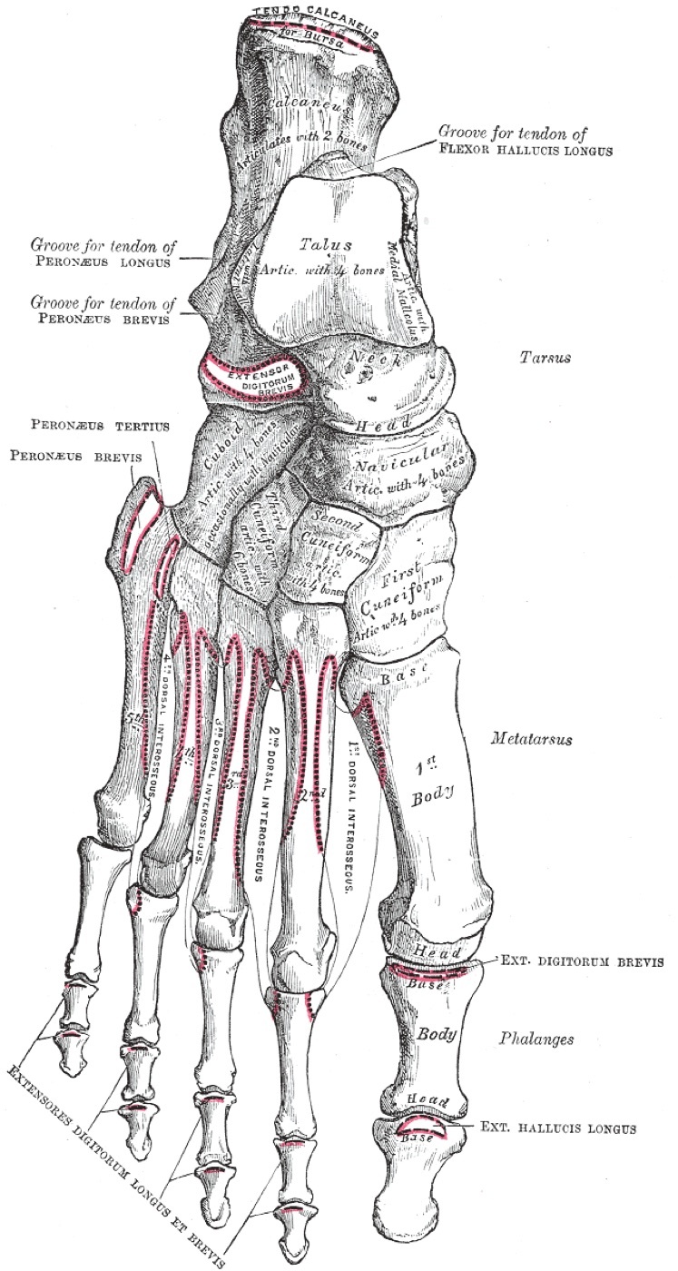
Figure 1. Bones of the foot (dorsal view): the hindfoot (talus, calcaneus), the midfoot bones (navicular, cuboid and the three cuneiforms) and the forefoot metatarsals and phalanges. Gray’s Anatomy (1918), public domain, via Wikimedia Commons.
The forefoot spreads load across the metatarsal heads while staying mobile in the sagittal plane to accommodate uneven ground, the mobility rising in a cascade from the rigid second and third rays out to the highly mobile fourth and fifth.[7] This accounts for two clinical observations: stress fractures favour the second and third metatarsals, and in a metatarsal fracture the only deformity that truly matters is sagittal-plane malalignment, which redistributes pressure under the heads. The first ray bears about a third of body weight and is the preferred ray for push-off; the fifth metatarsal base is a watershed that gives the field its most famous eponym, the Jones fracture.
Part II - Talus Fractures: Vascular Anatomy and the Problem of Avascular Necrosis
Talus fractures are uncommon, making up about 2 per cent of lower-limb fractures and 5 to 7 per cent of foot injuries, yet they rank second among tarsal fractures after the calcaneus.[8] They occur in younger men, frequently in polytrauma, and one anatomical fact dominates them: the blood supply is retrograde and precarious, so a displaced fracture of the talar neck can starve the body and produce osteonecrosis.
Three arteries feed the talus and form an anastomotic network: the posterior tibial artery (through the artery of the tarsal canal and a deltoid branch), the dorsalis pedis (anterior tibial), and the peroneal artery.[9] The artery of the tarsal canal is the main supply to the talar body, and the deltoid branch must be preserved at all costs, because in a high Hawkins-grade injury it may be the only remaining source. For that reason the deep deltoid ligament is left intact during a medial malleolar osteotomy, and the medial side is handled with care in every talar approach.[10] Necrosis most often affects the posterolateral talar dome, the watershed farthest from the medial neck.
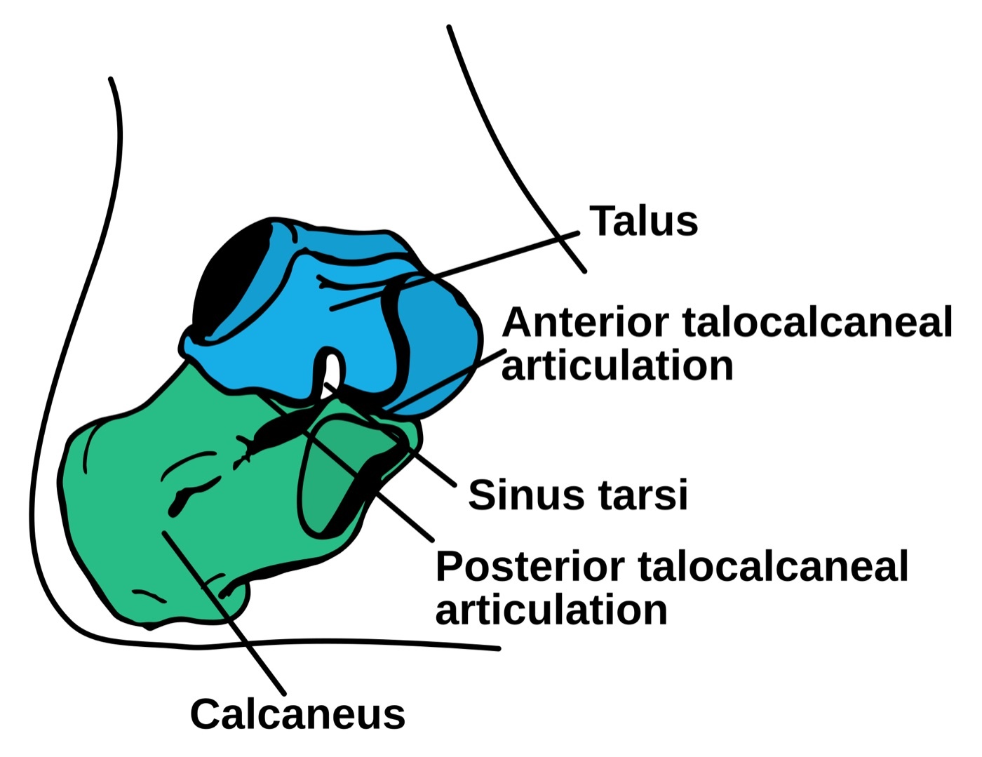
Figure 2. Subtalar (talocalcaneal) joint anatomy: the talus sitting over the calcaneus, with the anterior and posterior talocalcaneal facets and the intervening sinus tarsi. The talus has no muscular attachments, which is why its blood supply is retrograde and precarious. Vertenflow, CC BY-SA 3.0, via Wikimedia Commons.
The anatomy also fixes a distinction worth knowing. The lateral process of the talus is the dividing line between a neck fracture, which exits in front of the posterior facet, and a body fracture, which exits into it. That line predicts outcome, since body fractures damage both the ankle and the subtalar joint and fare worse than neck fractures.[11]
Part III - Talar Neck, Body, and Process Fractures, and Subtalar Dislocation
Talar neck fractures and the Hawkins classification
The talar neck is the commonest site, fractured by forced dorsiflexion driving the neck against the anterior tibia (or, in the cadaveric refinement, by an axial plantar force on a dorsiflexed foot, the aviator’s or accelerator-pedal mechanism).[12] The Hawkins classification grades the neck fracture by the joints it disrupts and remains the most important prognostic tool: type I is nondisplaced; type II adds subtalar subluxation or dislocation; type III adds dislocation of the body from both the ankle and subtalar joints; and type IV, added by Canale and Kelly, adds talonavicular dislocation.[13] The Vallier modification splits type II into IIA (subluxated subtalar joint) and IIB (dislocated), a distinction that carries real prognostic weight.
The classification predicts osteonecrosis almost linearly. Type I carries essentially no risk; type II overall runs from 0 to 50 per cent (with IIA near zero and IIB around 25 per cent); type III runs from roughly 41 to 75 per cent, lower in modern series; and open injuries reach 69 to 86 per cent.[14] Vallier’s observation is the prognostic anchor: necrosis with collapse never occurred in a fracture without subtalar dislocation. Its radiographic counterpart is the Hawkins sign, a subchondral lucency seen on the mortise view at six to eight weeks. Because resorbing that subchondral bone requires intact blood flow, the sign is a favourable one; its absence, or frank sclerosis, is worrying but does not guarantee necrosis.[15]
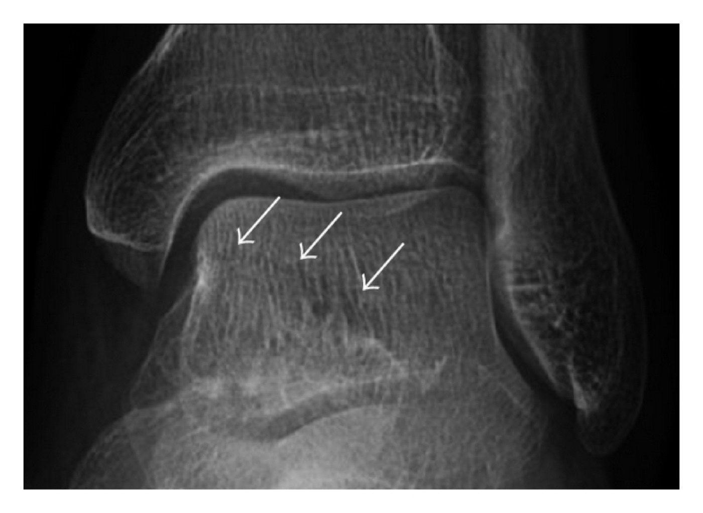
Figure 3. A talar fracture that is subtle on the plain radiograph (arrows); talar fractures are easily missed and frequently need CT for confirmation. Jarraya et al., CC BY 3.0, via Wikimedia Commons.
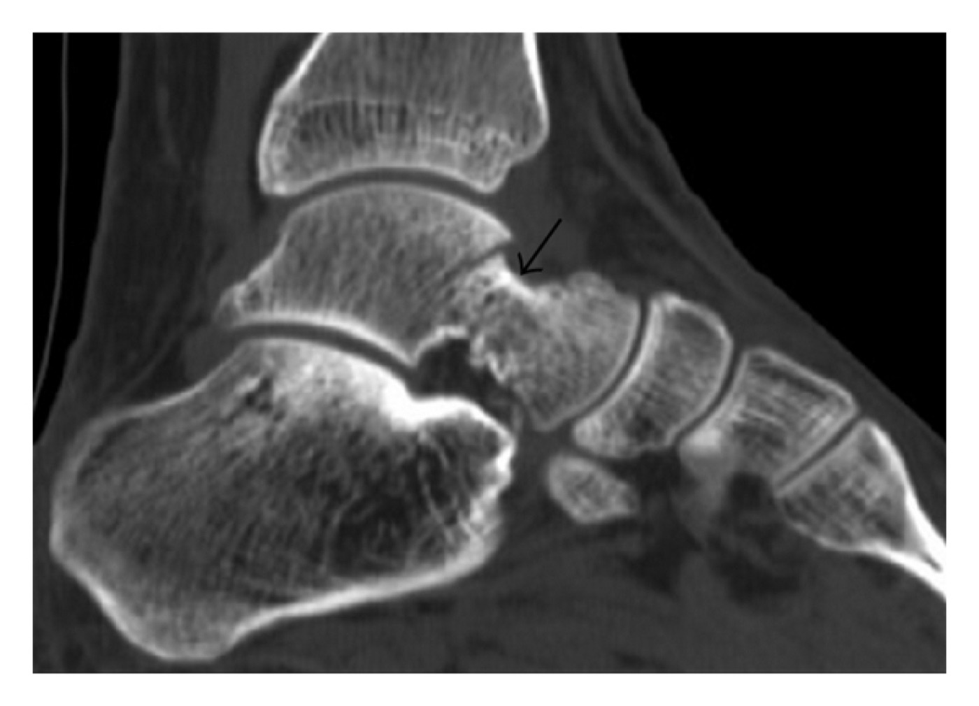
Figure 4. The same talar fracture confirmed on sagittal CT (arrow), which is routine for characterising talar injuries and planning fixation. Jarraya et al., CC BY 3.0, via Wikimedia Commons.
Management follows from the vascular anatomy. A truly nondisplaced fracture can be treated nonoperatively once CT confirms it, but every displaced neck fracture needs anatomical reduction, and a dislocated talar body is a surgical emergency that threatens the skin and the posterior tibial neurovascular bundle.[16] The gold-standard exposure is the dual anteromedial and anterolateral approach, which lets the surgeon judge length and rotation on both sides; a medial malleolar osteotomy is not needed for neck fractures but is often required to reach the posterior body in body fractures.[17] The cardinal technical pitfall is varus malunion from over-compressing comminuted medial bone. To avoid it, the surgeon fixes the lateral side to length first, often with a plate, then places the medial screw as a position screw rather than a lag screw, confirming alignment on the Canale view. One point bears emphasis: although a dislocated body must be reduced urgently, the timing of definitive fixation does not affect the necrosis rate, so it can wait for an experienced surgeon and healthy soft tissues.[18]
Body and process fractures
Body fractures involve the ankle and subtalar surfaces, account for 13 to 20 per cent of talar injuries, and carry high rates of arthritis (tibiotalar around 65 per cent, subtalar 35 per cent); they usually need a malleolar osteotomy for exposure and mini-fragment screws countersunk beneath the cartilage.[19] Two named process fractures recur in vivas. The lateral process fracture (the “snowboarder’s fracture”) follows dorsiflexion with eversion and external rotation, shows best on the mortise view, and classically produces persistent pain after what looks like an ankle sprain; small fragments are excised, larger ones or those with subtalar instability fixed.[20] The posterior process fracture follows extreme plantarflexion or ligamentous avulsion. Its medial-tubercle variant is the Cedell fracture, the posterolateral-tubercle eponym the Shepherd fracture (standard teaching), and an os trigonum must be told apart from an acute injury.[21]
Subtalar (peritalar) dislocation
In a subtalar dislocation the talus stays in the mortise while the foot dislocates around it at the subtalar and talonavicular joints. Inversion produces the medial dislocation (“basketball foot”), the commonest (about 75 per cent in RG, 85 per cent in Miller); eversion produces the higher-energy lateral dislocation.[22] The blocks to reduction are predictable and worth knowing. A medial dislocation is blocked by the extensor digitorum brevis, the retinaculum and the peroneal tendons, whereas a lateral dislocation is blocked by the posterior tibial tendon and the other medial flexors. Reduction is urgent and is performed with the knee flexed to relax the gastrocnemius; about 10 per cent of medial and 15 to 20 per cent of lateral dislocations cannot be reduced closed and need open reduction. Every case warrants a post-reduction CT, since associated tarsal fractures accompany roughly 90 per cent of these injuries.[23]
Part IV - Calcaneus Fractures: Anatomy, Böhler’s Angle, and Classification
The calcaneus is the most commonly fractured tarsal bone, accounting for about 65 per cent of tarsal injuries, typically in young working men after a fall from height or a dashboard injury, with about 75 per cent of fractures involving the posterior facet of the subtalar joint.[24] The mechanism is axial. The lateral process of the talus is driven like a wedge into the angle of Gissane, creating a primary fracture line that splits the bone into an anteromedial (sustentacular) fragment and a posterolateral (tuberosity) fragment. The sustentaculum tali is the “constant fragment,” held to the talus by strong ligaments, and the rest of the reconstruction is built against it as a reference.[25]
Two radiographic angles are examination staples. Böhler’s angle (normally 25 to 40 degrees) flattens when the posterior facet is depressed; a value that stays above about 15 degrees supports nonoperative care, while a value below zero predicts a worse result and a higher rate of fusion.[26] The critical angle of Gissane (normally 120 to 145 degrees) increases with intra-articular depression. The Harris axial view shows heel width, varus and shortening, and Broden’s views profile the posterior facet, but CT is the modality that defines the injury, and the coronal CT underpins the modern classification.
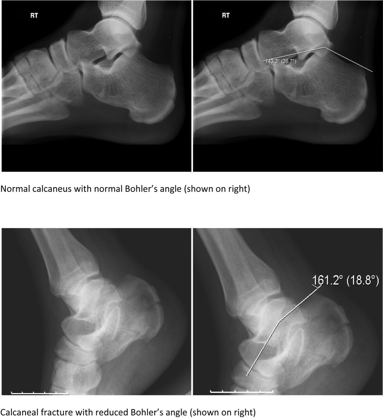
Figure 5. Böhler’s tuber-joint angle on the lateral view: a normal calcaneus (top) compared with a calcaneal fracture in which the angle is flattened by depression of the posterior facet (bottom). Gilo1969, CC BY-SA 3.0, via Wikimedia Commons.
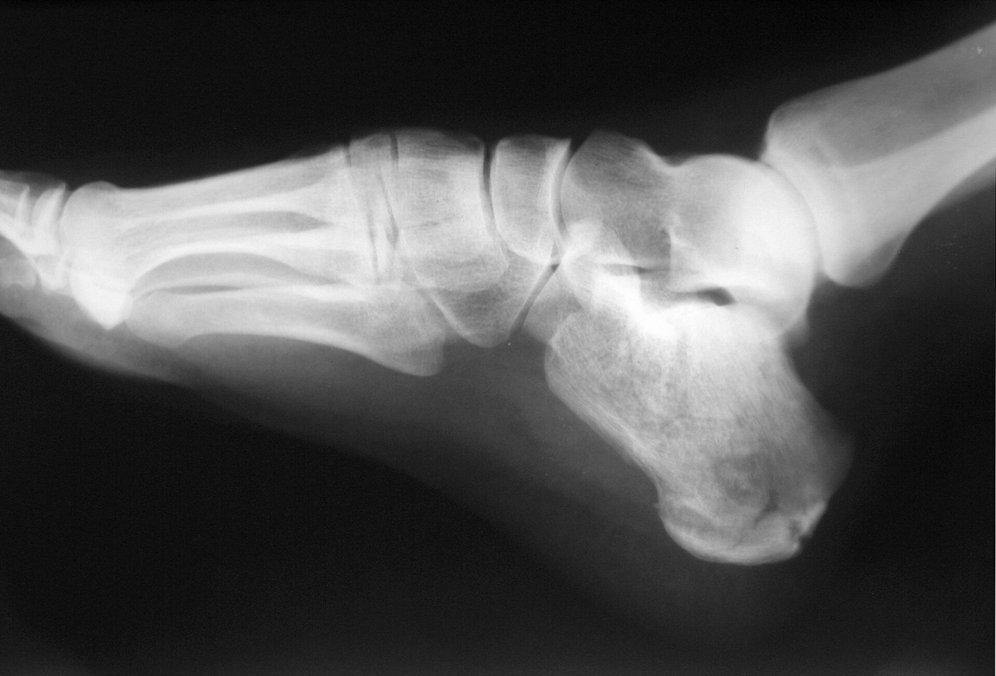
Figure 6. Lateral radiograph of a calcaneal fracture with a depressed posterior facet after a fall from height. Jojo, CC BY-SA 3.0, via Wikimedia Commons.
The Essex-Lopresti scheme divides intra-articular fractures by the secondary fracture line into a tongue type, in which the articular fragment stays attached to the tuberosity, and a joint-depression type, in which the articular fragment is separate.[27] The system in universal use is the Sanders CT classification, which counts the posterior-facet fragments on the coronal image: type I is nondisplaced, type II has two fragments (one fracture line), type III has three, and type IV has four or more or is comminuted. Sanders tracks both treatment and prognosis; type III fractures are about four times more likely than type II to need a later subtalar fusion (47 versus 19 per cent), and type IV is usually managed by primary subtalar arthrodesis.[28]
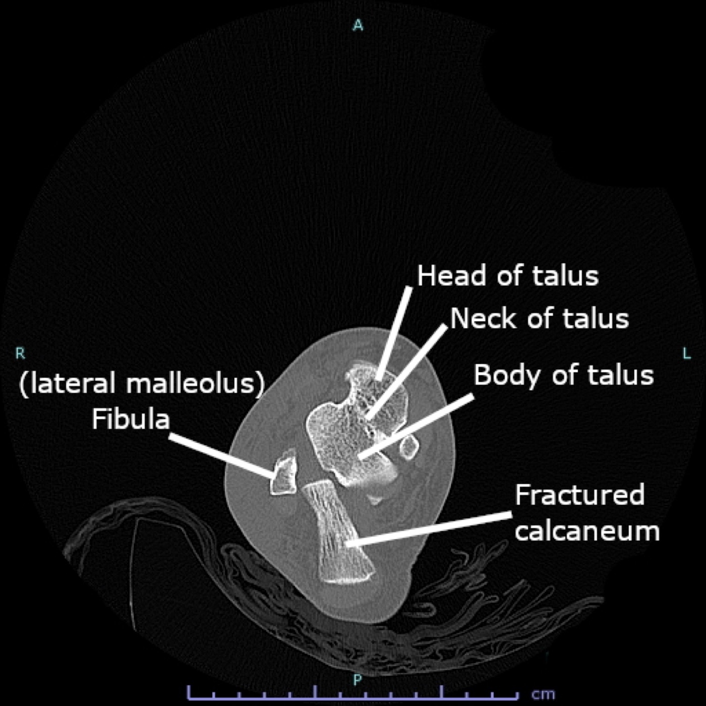
Figure 7. Axial CT of a comminuted calcaneal fracture (the Sanders classification is built on the coronal reformat through the posterior facet). Cerevisae, CC BY-SA 4.0, via Wikimedia Commons.
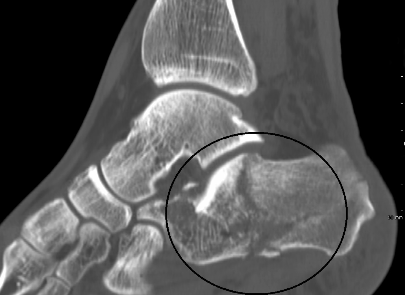
Figure 8. Sagittal CT of a fractured calcaneus (circled), profiling the posterior-facet depression and the tuberosity. James Heilman, MD, CC BY-SA 3.0, via Wikimedia Commons.
One extra-articular pattern demands particular vigilance: the tongue-type tuberosity fracture, a skin-necrosis emergency. The Achilles pulls the superior tuberosity fragment proximally so that it tents the thin posterior heel skin, producing necrosis in up to 21 per cent of presentations and amputation in 29 per cent of those, so it demands emergent reduction even in a smoker.[29]
Part V - Calcaneus Treatment and the Operative Debate
Treatment aims to restore the articular surface, the height, the width, the length and the alignment of the heel, leaving a plantigrade foot that can be braced and shod.[30] Nondisplaced fractures, minimally displaced extra-articular fractures, and patients with prohibitive comorbidities (peripheral vascular disease, poorly controlled diabetes, heavy smoking) are treated nonoperatively with early motion and protected weight bearing for six to twelve weeks.
When surgery is chosen, the soft tissues dictate timing: the surgeon waits for the wrinkle sign to return to the lateral hindfoot, usually one to two weeks after injury.[31] The historical workhorse is the extensile lateral approach, which gives the best view of the posterior facet at a high wound-complication cost, with edge necrosis at the apex of the L-shaped incision and delayed healing in 25 to 30 per cent of cases (deep infection much lower, around 1 to 4 per cent). The sinus tarsi (limited) approach and percutaneous techniques have gained ground because they achieve similar reductions with far fewer wound problems and can be done earlier.[32]
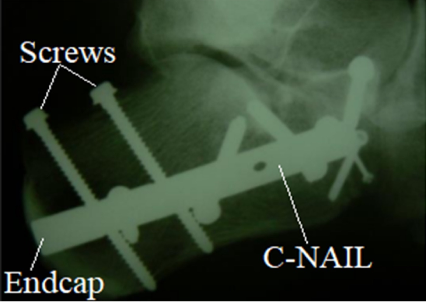
Figure 9. Calcaneal fracture after osteosynthesis with a minimally invasive intraosseous nail (C-Nail) and screws, an alternative to extensile-approach lateral plating that reduces the wound-complication risk. Fry72, CC BY-SA 4.0, via Wikimedia Commons.
The reconstruction proceeds from the constant sustentacular fragment outward: a Schanz pin disimpacts the tuberosity to correct varus and restore length, the articular fragments are reduced and fixed to the sustentaculum with a lag screw, the lateral wall is replaced, and a low-profile plate buttresses the construct.[33] When a Sanders IV fracture is past realistic reconstruction, primary subtalar arthrodesis combined with restoration of heel shape is the treatment of choice.
The operative-versus-nonoperative debate is among the most studied in foot trauma, and the honest answer is nuanced. The Canadian (Buckley) trial found surgery better for specific subgroups (women, younger patients, lighter workloads, non-compensation cases, higher initial Böhler angle, anatomical CT reductions), and nonoperative patients were several times more likely to need a later subtalar fusion. The UK Heel trial and the Ågren trial, by contrast, found few overall functional advantages to surgery and a real complication cost.[34] The two camps reconcile around a single message: the best results come from well-selected operative candidates treated by high-volume surgeons, and the worst from complications in patients who should not have had surgery. Even with anatomical reduction, radiographic subtalar arthritis approaches 100 per cent (symptomatic in about 30 per cent), and foot compartment syndrome complicates up to 10 to 17 per cent of calcaneal fractures.[35]
Part VI - Midfoot Injuries: Navicular, Cuboid, and Cuneiform
Midfoot injuries are rare (about 5 per cent of foot injuries) and notoriously missed, up to 30 per cent of the time, almost always combining osseous and ligamentous components.[36] The tarsal navicular, the keystone of the medial column, fractures as a cortical avulsion (commonest), a tuberosity avulsion (the posterior tibial tendon pulling off the tuberosity in eversion), or a body fracture, graded by the Sangeorzan classification into a transverse (horizontal) fracture splitting the navicular into dorsal and plantar fragments (type I), a vertical fracture cleaving it into medial and lateral fragments (type II, the group into which most stress fractures fall), and a comminuted body fracture (type III). The navicular also hosts the classic athletic stress fracture, favoured by a long second ray or a cavus foot, accounting for 14 per cent of all stress fractures, often invisible on plain films, and best seen on CT.[37]
Treatment turns on column length and articular congruity, not the fracture pattern. Displacement under 2 mm with an intact medial column is treated in a cast, while displacement of 2 mm or more, articular incongruity over 1 mm, or column shortening calls for ORIF that restores length and the joint surface; bone loss over 40 per cent prompts a talonavicular (or naviculocuneiform) arthrodesis. The most common sequel is talonavicular arthritis.[38]
The cuboid, keystone of the lateral column, is most often injured as an avulsion with a sprain, but the high-yield pattern is the “nutcracker” fracture, in which forced abduction crushes the cuboid between the calcaneus and the metatarsal bases, shortening the lateral column and producing a planovalgus foot. A nutcracker fracture is never isolated, and lateral-column length must be restored, often with a bridge plate and bone graft.[39] Cuneiform injuries are rarely isolated and usually accompany a Lisfranc injury; the “gap sign” of intercuneiform widening points to Lisfranc ligament rupture, and a medial-cuneiform fracture must be told apart from a bipartite cuneiform by its coronal (rather than horizontal) cleavage.[40]
Part VII - The Lisfranc (Tarsometatarsal) Injury
The Lisfranc injury is the high-yield midfoot topic. It is rare (0.1 to 0.4 per cent of fractures) but misdiagnosed in up to 20 per cent of cases, often because a low-energy athletic injury reduces spontaneously and is dismissed as a sprain.[41] The pathoanatomy centres on the recessed second metatarsal base, the keystone locked between the cuneiforms, and the Lisfranc ligament that runs from the medial cuneiform to that base, the only link across the unligamented first-to-second-metatarsal gap. The mechanism is either a direct crush, which brings major soft-tissue injury and a high rate of compartment syndrome, or, more often, an indirect axial load on a plantarflexed foot, which ruptures the weaker dorsal ligaments first.[42]
The diagnosis rests on weight-bearing and stress radiographs, since non-weight-bearing films miss subtle instability. The key signs are loss of the normal alignment of the second metatarsal base with the middle cuneiform (lateral translation of the second metatarsal is diagnostic and mandates surgery), widening of the first-to-second intermetatarsal space beyond the normal 2.5 mm, and the “fleck sign,” a small avulsion from the second metatarsal base or medial cuneiform that is pathognomonic of a Lisfranc ligament injury.[43] Any malalignment of 1 mm or more is pathological, and MRI categorises stability with about 94 per cent sensitivity. The athletic sprain spectrum is staged by Nunley and Vertullo (stage I, no diastasis but a positive bone scan; stage II, 1 to 5 mm diastasis; stage III, over 5 mm with arch collapse), while the older Quénu-Küss / Hardcastle / Myerson schemes describe the displacement patterns as homolateral, isolated or divergent.[44]
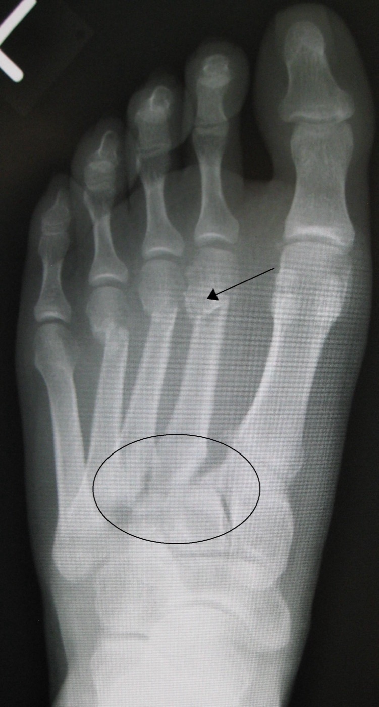
Figure 10. AP radiograph of a Lisfranc (tarsometatarsal) injury with malalignment at the base of the second metatarsal. James Heilman, MD, CC BY 3.0, via Wikimedia Commons.
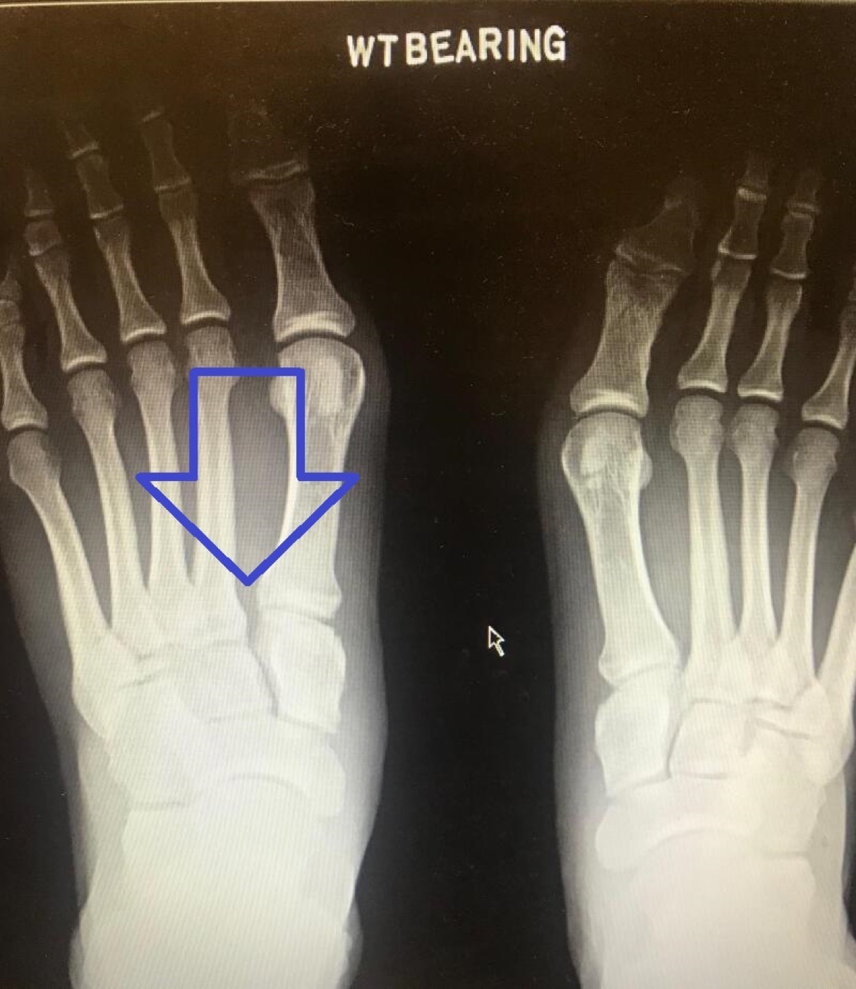
Figure 11. Bilateral weight-bearing radiographs showing subtle diastasis of the left first-second intermetatarsal space from a Lisfranc ligament rupture; comparison with the normal side is the diagnostic key. Mark A. Dreyer, DPM, CC BY 4.0, via Wikimedia Commons.
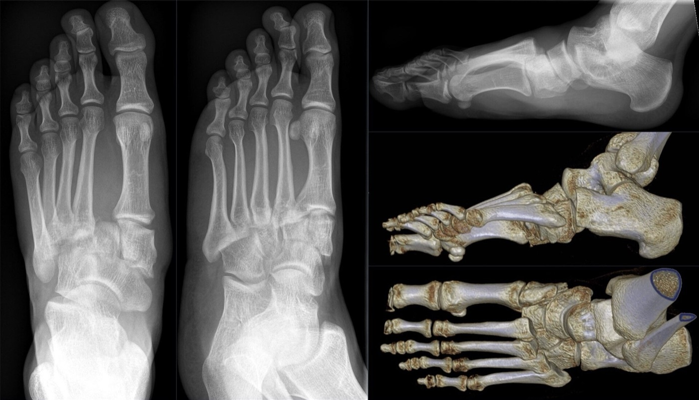
Figure 12. Lisfranc fracture-dislocation with lateral displacement of the lesser metatarsals, shown on radiographs and three-dimensional CT reconstruction. Hellerhoff, CC BY-SA 4.0, via Wikimedia Commons.
Only a truly stable, nondisplaced injury (malalignment under 1 mm, no displacement on stress or weight-bearing views, no bony injury on CT) is treated nonoperatively. Everything else requires anatomical reduction and stable fixation, since anatomical reduction of the medial column dominates outcome.[45] The two operative strategies are ORIF (transarticular screws, including the “Lisfranc screw” from the medial cuneiform into the second metatarsal base, or increasingly dorsal bridge plates that spare the joint cartilage) and primary arthrodesis. Primary arthrodesis is particularly favoured for purely ligamentous high-energy injuries, where one randomised trial found no difference in SMFA or SF-36 against ORIF but fewer reoperations and lower costs.[46] Throughout, the medial three columns may be fixed or fused, but the mobile fourth and fifth tarsometatarsal joints are stabilised only temporarily with K-wires removed at six weeks, never fused. The dominant late complication is post-traumatic arthritis, salvaged by midfoot arthrodesis.
Part VIII - Forefoot: Metatarsal, Sesamoid, and Phalangeal Fractures
Metatarsal fractures
Metatarsal fractures make up about 35 per cent of all foot fractures, most often of the fifth.[47] The first metatarsal bears about a third of body weight and tolerates little malalignment, so instability on stress radiographs or a plantarly displaced head calls for fixation. The central metatarsals (second to fourth) tolerate injury better, but the sagittal plane is unforgiving: more than 10 degrees of sagittal deviation or 3 to 4 mm of translation should be corrected to prevent transfer metatarsalgia, and when all three are fractured the construct loses the intermetatarsal ligaments that would otherwise splint it and becomes inherently unstable.[48] Any fracture of a metatarsal base, especially the second, should prompt a search for a Lisfranc injury.
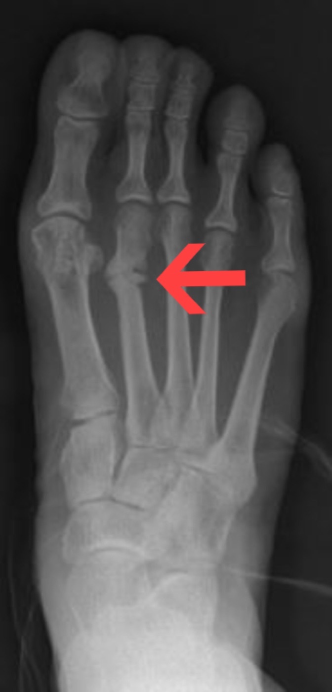
Figure 13. Healing stress (march) fracture of the second metatarsal shaft with periosteal callus (arrow); stress fractures favour the relatively immobile second and third rays. Personalo, CC BY-SA 3.0, via Wikimedia Commons.
The fifth metatarsal is the examination favourite, divided into three proximal zones. Zone 1 is the tuberosity avulsion (“pseudo-Jones”), caused by inversion pulling on the lateral plantar aponeurosis, and it heals reliably in a stiff-soled shoe. Zone 2 is the true Jones fracture at the metaphyseal-diaphyseal junction, a watershed region with a 15 to 25 per cent nonunion rate. Zone 3 is the proximal diaphyseal stress fracture, the most nonunion-prone of all and the one most clearly tied to a cavovarus foot.[49] The treatment gradient follows the blood supply: zone 1 heals nonoperatively, zone 2 goes into a boot for the non-athlete but earns an intramedullary screw in the elite athlete (where screw fixation gives 95 per cent union versus 66 per cent), and zone 3 is fixed by intramedullary screw as the procedure of choice, with a hindfoot osteotomy considered when the foot is in varus.[50] The other common fifth-metatarsal injury, the spiral dancer’s fracture of the distal shaft, heals well nonoperatively.
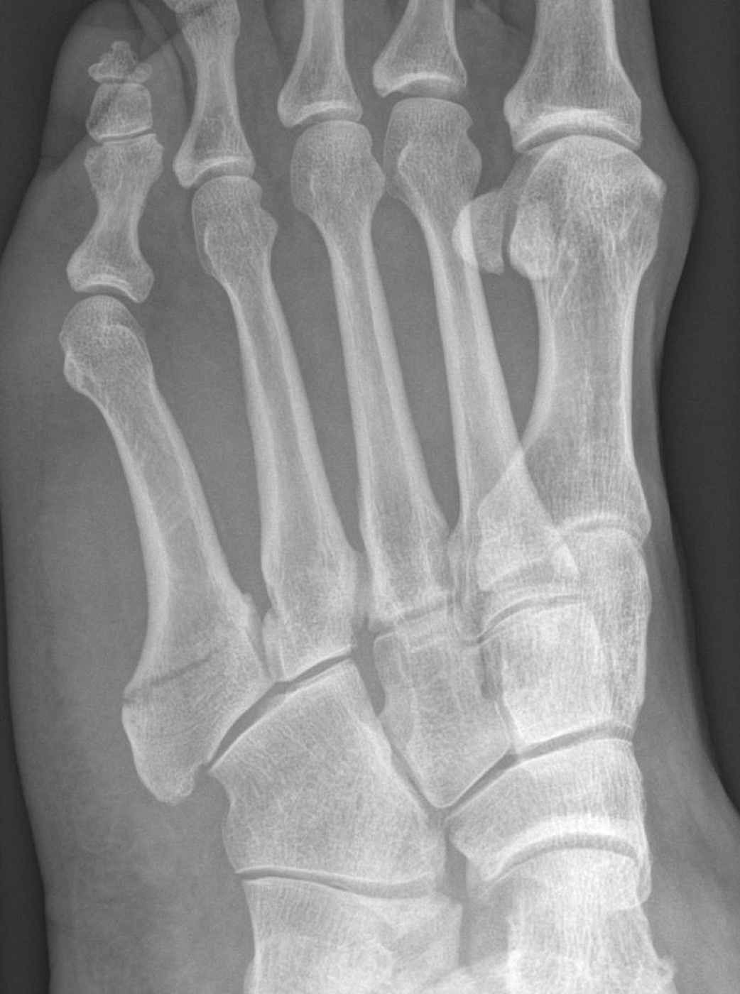
Figure 14. Oblique radiograph of a true Jones fracture at the metaphyseal-diaphyseal junction of the fifth metatarsal, a watershed region prone to nonunion. Hellerhoff, CC BY-SA 4.0, via Wikimedia Commons.
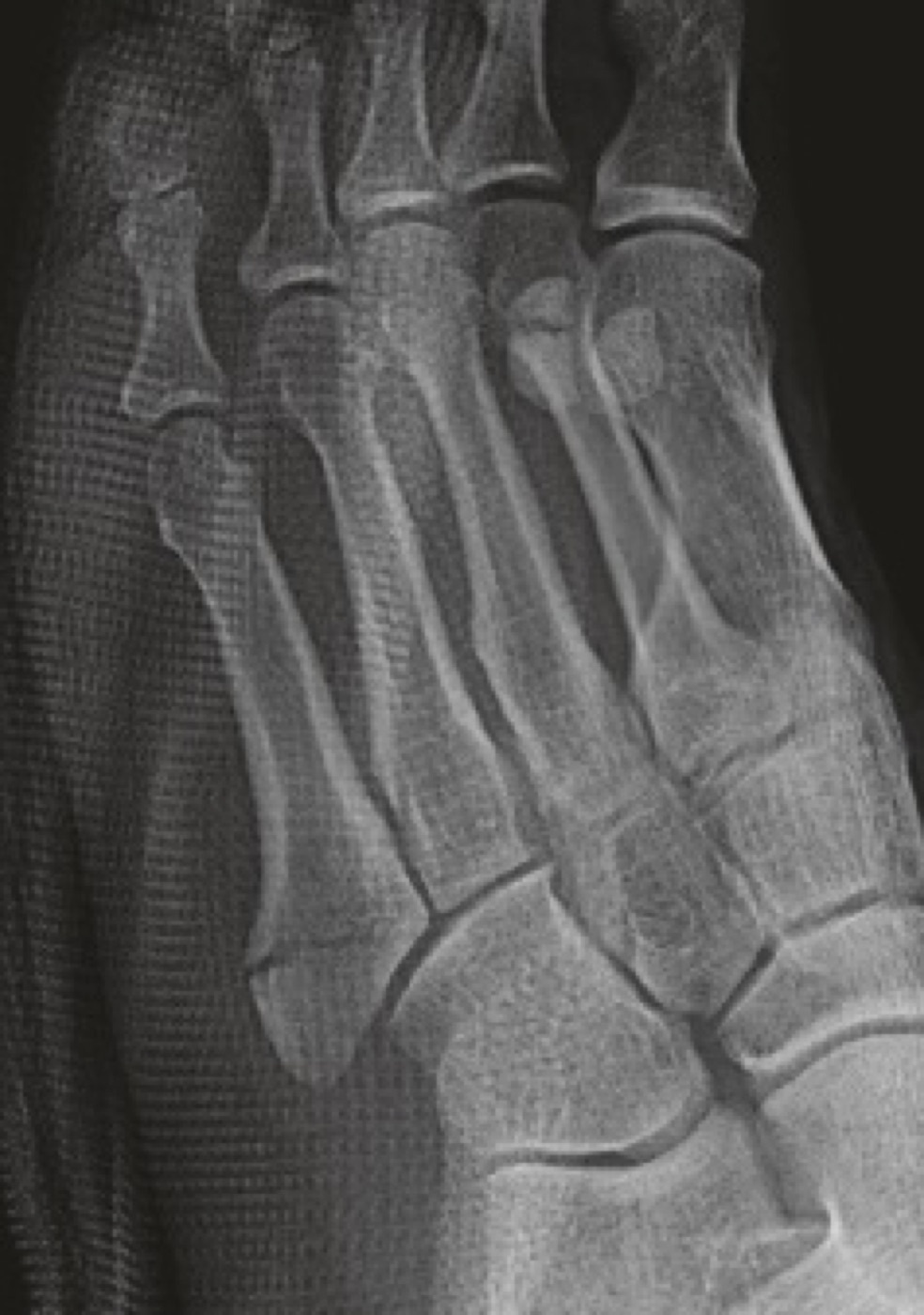
Figure 15. Fracture of the proximal fifth metatarsal at the border between the metaphysis and the tuberosity. Park et al., CC BY 4.0, via Wikimedia Commons.
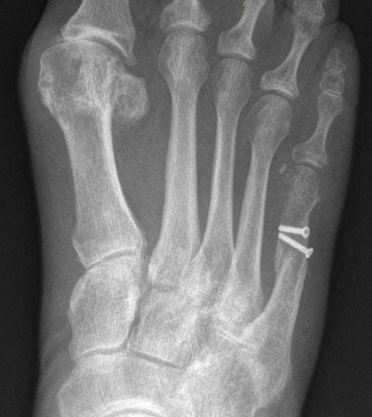
Figure 16. Postoperative radiograph after internal fixation of a fifth-metatarsal fracture with two screws. Jmarchn, CC BY-SA 3.0, via Wikimedia Commons.
Sesamoid and phalangeal fractures
The hallucal sesamoids transmit up to half of body weight, rising to about 300 per cent at push-off, and the larger medial (tibial) sesamoid is the one more often injured.[51] Its sharp margins distinguish a fracture from a bipartite sesamoid, which has smooth, rounded edges. Most are treated nonoperatively; a persistent painful nonunion may need partial excision through a medial or dorsal approach, but both sesamoids are never excised, since that produces a cock-up deformity. Phalangeal fractures are the commonest forefoot injuries, usually treated by buddy taping and a stiff-soled shoe; the great toe matters more functionally than the lesser toes, and a distal phalanx fracture with a nail-bed laceration is an open fracture.[52] Turf toe is a hyperextension sprain of the first metatarsophalangeal joint, and a dorsal dislocation of that joint with an intact intersesamoid ligament (type I) is irreducible and needs open reduction.
Part IX - Compartment Syndrome of the Foot and the Mangled Foot
Compartment syndrome of the foot is the most important soft-tissue emergency in this topic. The foot is now described as having nine compartments (medial, lateral, four interosseous, and three central: superficial, deep/adductor, and the intermediate or calcaneal compartment). The calcaneal compartment is the critical one: it communicates with the deep posterior compartment of the leg through the tarsal tunnel and houses the quadratus plantae, whose ischaemic contracture produces the claw-toe and cavus deformity of an untreated syndrome.[53] Crush injuries and calcaneal fractures are the usual causes (a crush roughly doubles the risk), and normal pulses and capillary refill do not exclude it.
Diagnosis rests on clinical suspicion (intractable pain, pain on passive toe stretch) supported by compartment pressure measurement, with fasciotomy indicated when the pressure is within about 30 mm Hg of diastolic (Miller: above 30 mm Hg or within 20 of diastolic).[54] Decompression combines two dorsal incisions and a medial incision, the medial incision being the one that reliably releases the calcaneal compartment; dorsal incisions alone are inadequate, and that is where the controversy lies. Miller presses one teaching point in particular: an untreated foot compartment syndrome leaves not merely claw toes but chronic neuropathic pain that is difficult or impossible to treat, so benign neglect is not acceptable.[55]
The mangled (multiply injured) foot sits at the other end of the spectrum, a high-energy crush or blast injury managed in stages with serial debridement, external fixation and K-wire transfixation, early “fix and flap” soft-tissue cover, and a salvage-versus-amputation decision governed by the “life before limb” principle. The most quoted predictor of amputation is a fracture of all five metatarsals in the setting of mid- or hindfoot trauma after high-energy injury.[56]
Part X - Paediatric Foot Fractures
The child’s foot is largely cartilaginous and flexible until late adolescence. For that reason tarsal fractures are rare in children (under 1 per cent of fractures) while metatarsal and toe fractures are common (7 to 9 per cent), and interpretation is hard amid multiple ossification centres (the medial cuneiform does not ossify until age four).[57] Comparison radiographs of the opposite foot are the most useful diagnostic aid.
Paediatric talus fractures mirror the adult anatomy: forced dorsiflexion fractures the neck (with a 25 to 30 per cent rate of associated medial malleolar fracture), the blood supply enters through the neck and the deltoid branch, and outcome is graded by the Letts and Gibeault classification (types I and II minimally displaced with low necrosis risk, type III displaced, type IV with body dislocation and expected necrosis).[58] Osteonecrosis in children does not usually prevent healing, and the Hawkins sign at six to eight weeks again signals a vascular body. Calcaneal fractures are often occult, diagnosis delayed in 30 to 50 per cent; in young children they are mostly extra-articular (75 per cent) and treated nonoperatively, with ORIF reserved for displaced intra-articular fractures in adolescents.[59]
In the midfoot, a fracture of the second metatarsal base again marks a tarsometatarsal injury, often picked up only on the comparison view; these are pinned and casted.[60] Metatarsal fractures are the commonest paediatric foot fractures (the first in younger children, the fifth in older ones), and at the fifth-metatarsal base the task is to tell a Jones fracture, an avulsion and the normal apophysis apart, the key being that the apophysis lies parallel to the shaft and does not enter the joint (the Iselin apophysitis eponym is standard teaching). One pinning pitfall is worth knowing: if the metatarsophalangeal joint is not held reduced during K-wiring, the result is an extension contracture and a painful prominent metatarsal head.[61] Finally, an open fracture of the great-toe proximal phalanx under a nail-bed laceration is the paediatric Seymour fracture, treated as an open physeal injury.[62]
References
-
The topic spans four anatomical regions, each a chapter in its own right: the talus (Rockwood & Green ch.65), the calcaneus (ch.66), and the midfoot and forefoot (ch.67), supplemented by the AO hindfoot and midfoot/forefoot sections (6.10.1 and 6.10.2), Miller’s Review for exam framing, and Lovell & Winter’s for the paediatric foot. Every claim below carries its page citation; facts that are genuine established teaching but absent from these sources are flagged as standard teaching rather than given a false citation.
-
The medial column is the talus, navicular, the three cuneiforms and the first three metatarsals; the lateral column is the calcaneus, cuboid and the fourth and fifth metatarsals (RG p.4774). The lateral column is the more mobile and is meant to stay that way, which is why fusing the fourth and fifth tarsometatarsal joints is avoided (RG p.4833-4834).
-
The “fall-from-height” cluster (lumbar spine in 6-20 per cent, contralateral heel, pilon) is the reason every high-energy hindfoot injury demands examination of the spine and the opposite foot (RG p.4717). Up to 30 per cent of midfoot injuries are missed or treated late, and missed Lisfranc injuries do worse than those treated acutely (RG p.4774, p.4816).
-
The talus is over 50-60 per cent cartilage with no muscle or tendon attachments (RG p.4652-4653; AO p.994; Miller p.616); the calcaneus carries about 50 per cent of weight-bearing load, with the talus placed eccentrically and medially (RG p.4714).
-
RG p.4774, p.4781. The only tendon inserting entirely within the midfoot is the tibialis posterior, whose plantar slips connect all the midfoot bones and lock them into a rigid lever during toe-off (RG p.4775).
-
RG p.4776, p.4817; AO p.1010; Miller p.608. The Lisfranc ligament is the largest and strongest of the tarsometatarsal ligaments, about 8-10 mm wide and 5-6 mm thick, and its interosseous bundle is the stiffest component (Miller p.608).
-
RG p.4848. The second and third tarsometatarsal joints permit only 0.6 and 1.6 mm of sagittal motion respectively, while the fourth and fifth permit 9.6 and 10.2 mm (RG p.4775-4776).
-
RG p.4645; Miller p.616. In one level-1 series, 44 per cent of talus-fracture patients were polytrauma victims, of whom 59 per cent had ipsilateral injuries (RG p.4645).
-
RG p.4655-4656; AO p.994; Miller p.616. The artery of the tarsal canal and the artery of the tarsal sinus form an anastomotic sling beneath the neck.
-
RG p.4667; AO p.999; Miller p.616. Miller frames it by Hawkins type: type I disrupts one of three sources, type II disrupts two (leaving the deltoid branch), type III theoretically disrupts all three, and type IV may additionally lose the head and neck supply (Miller p.616).
-
RG p.4652; AO p.994; Miller p.617. AO ranks outcome by injury: lateral process fractures do best, then neck, then body (AO p.1000).
-
RG p.4639; Miller p.616. Peterson could not reproduce neck fractures by pure hyperdorsiflexion but did so with a dorsiflexed ankle and a plantar axial force (RG p.4639).
-
RG p.4651-4652; Miller p.616-617. True type I injuries are rare with modern imaging; any displacement is treated as type II (RG p.4651).
-
RG p.4682-4683; AO p.999; Miller p.617-618. MRI-based modern data give lower rates than the historical figures.
-
RG p.4686; AO p.999; Miller p.618.
-
RG p.4658, p.4701; AO p.994-995. Absolute operative indications include any displacement on CT, debris in any joint, a noncongruent reduction, skin compromise, an open fracture, or neurovascular compromise (AO p.995).
-
RG p.4661, p.4667; Miller p.617; AO p.999.
-
RG p.4688, p.4701; AO p.996, p.1000; Miller p.617. Early fixation may even raise wound-healing complications (Miller p.617).
-
RG p.4652, p.4666-4669; Miller p.617, p.619.
-
RG p.4642, p.4676-4678; Miller p.619. The lateral talocalcaneal ligament attaches to the lateral process (Miller p.619).
-
RG p.4643-4644. The Shepherd eponym for the posterolateral tubercle fracture is not used in these sources; RG names only the Cedell fracture for the medial tubercle.
-
RG p.4694; Miller p.623. The discrepancy in the medial percentage between the two sources is noted; both are widely quoted.
-
RG p.4698-4699; Miller p.623-624. Avascular necrosis after subtalar dislocation runs 0-10 per cent in closed injuries but up to 50 per cent in open ones (RG p.4701).
-
RG p.4712; AO p.981; Miller p.620. The fall-from-height cluster (lumbar spine 6-20 per cent, bilateral 3-7.9 per cent, contralateral heel) must always be sought (RG p.4717).
-
RG p.4713-4715, p.4728; AO p.986-987. The sustentaculum sits 2.5 cm below the medial malleolus and carries the FHL tendon in a groove on its undersurface, which is at risk from medially directed screws (RG p.4713).
-
RG p.4721; AO p.982; Miller p.622.
-
RG p.4714-4715; AO p.984.
-
RG p.4726-4727, p.4759; AO p.984; Miller p.622-623.
-
RG p.4719; AO p.981; Miller p.619. The Beavis type II “beak” fracture corresponds to this pattern.
-
RG p.4712-4713; AO p.988.
-
AO p.985-986; RG p.4744; Miller p.622. Calcaneal fracture blisters are more often blood-filled and predict more scarring (RG p.4717-4718).
-
RG p.4743-4744, p.4756, p.4758; AO p.987, p.992; Miller p.622. The flexor hallucis longus is at risk at the sustentaculum during lateral-to-medial screws (Miller p.622).
-
RG p.4746-4749; AO p.988-989; Miller p.623. Locking plates allow fixation of all patterns but are not biomechanically stronger than conventional fixation (AO p.989).
-
RG p.4756-4757, p.4759-4761; AO p.992, p.994.
-
RG p.4718, p.4759; Miller p.625. The chief virtue of operative treatment, even when function is similar, is that restored height and width make any later subtalar fusion far more successful (Miller p.623).
-
RG p.4774; plain-film sensitivity for navicular fractures is only 33 per cent against CT (RG p.4778).
-
RG p.4778-4783; Miller p.613, p.615. The current recommendation for a navicular stress fracture is non-weight-bearing cast immobilisation for six to eight weeks, with surgery (a transverse screw placed dorsomedial to plantar-lateral) reserved for displaced fractures or nonunion rather than as primary treatment (Miller p.615-616).
-
RG p.4782-4791; Miller p.616. The two most common complications of navicular fractures are degenerative arthritis and avascular necrosis (Miller p.616).
-
RG p.4795, p.4805-4807; AO p.1008; Miller p.611-612. A useful rule throughout the midfoot is to avoid transfixing the cuboid to the navicular or cuneiforms, which would abolish the necessary interplay between the columns (RG p.4800).
-
RG p.4807-4810; Miller p.613.
-
RG p.4816; AO p.1011. Plantar ecchymosis is a classic clinical pointer, though it may be absent in subtle athletic injuries (RG p.4818).
-
RG p.4817-4818; Miller p.608.
-
RG p.4820; Miller p.608-609.
-
RG p.4822-4823; Miller p.609. The classification schemes are acknowledged to be of little value for guiding treatment or predicting outcome (Miller p.609).
-
RG p.4823-4824, p.4839; Miller p.609.
-
RG p.4833, p.4839; AO p.1014; Miller p.610-611.
-
RG p.4840; the fifth metatarsal accounts for about 68 per cent of metatarsal fractures, the first only about 1.5 per cent, and the central metatarsals about 10 per cent (RG p.4841, p.4847, p.4856).
-
RG p.4848-4849; Miller p.605.
-
RG p.4856-4859; AO p.1018; Miller p.605-606. The relative frequencies are roughly 93 per cent zone 1, 4 per cent zone 2, 3 per cent zone 3 (RG p.4856).
-
RG p.4858-4864; Miller p.605-606. “Watch for the Jones fracture in the varus foot” (Miller p.606).
-
RG p.4865-4866.
-
RG p.4870-4878; Miller p.604, p.606.
-
RG p.4886-4889; Miller p.625. The incidence after a calcaneal fracture is classically quoted as up to 10 per cent (Myerson), about 17 per cent in Miller, but only about 1 per cent in one large registry of isolated hindfoot injuries (RG p.4889; Miller p.625).
-
RG p.4892; Miller p.625.
-
RG p.4892-4896; Miller p.625.
-
RG p.4879-4880; AO p.1018; Miller p.604. Notably, a talar fracture, a lower Sanders calcaneal grade, an ankle dislocation and preserved sensation are not predictive of amputation (Miller p.604).
-
Lovell p.5775-5776. Most paediatric tarsal fractures are nondisplaced and may be an underreported cause of the limping toddler (Lovell p.5776).
-
Lovell p.5776-5777. Closed treatment is appropriate for fractures angulated under 5 degrees and displaced under 2 mm; displaced fractures need ORIF through an anteromedial approach with lag screws (Lovell p.5777).
-
Lovell p.5785-5786. The Schmidt and Weiner classification adds a third category for calcaneal fractures with Achilles-insertion loss and major soft-tissue injury, the lawnmower injury (Lovell p.5785).
-
Lovell p.5786.
-
Lovell p.5790-5791.
-
Lovell p.5796. Köhler disease of the navicular and the Iselin apophysis are the two paediatric mimics to keep in mind; both are standard teaching not detailed in this source.
-
RG p.4655-4656, p.4667; AO p.994; Miller p.616.
-
RG p.4651-4652, p.4682-4683; AO p.999; Miller p.616-617.
-
RG p.4686; AO p.999; Miller p.618.
-
RG p.4642, p.4676-4678; Miller p.619.
-
RG p.4721; Miller p.622.
-
RG p.4726-4727, p.4759; Miller p.622-623.
-
RG p.4719; AO p.981; Miller p.619.
-
RG p.4820-4822; Miller p.608-609.
-
RG p.4824-4834, p.4839; AO p.1014; Miller p.609-611.
-
RG p.4856-4864; Miller p.605-606.
-
RG p.4886-4896; Miller p.625.
-
Lovell p.5775-5777.