Contents
- Orientation: A High-Energy Diaphyseal Fracture Treated by the Intramedullary Nail
- Part I - Definition, Epidemiology, and Mechanism of Injury
- Part II - Applied Anatomy and the Deforming Forces
- Part III - Assessment, Associated Injuries, and Imaging
- Part IV - Classification
- Part V - Systemic Injury, Blood Loss, and the Timing of Fixation
- Part VI - Treatment
- Part VII - Special Fracture Patterns and Associated Injuries
- Part VIII - Complications
- Part IX - Paediatric Femoral Shaft Fractures
- Part X - A Synthesis: How to Reason About the Femoral Shaft Fracture
- References
Orientation: A High-Energy Diaphyseal Fracture Treated by the Intramedullary Nail
The femoral shaft fracture is, in the skeletally mature patient with healthy bone, a marker of high-energy transfer to the body, and it dominates two themes that recur throughout its management. The first is the systemic patient: the femur is wrapped in a thick, vascular muscular envelope, the fracture is frequently part of a polytrauma, and the surgeon must reason about blood loss, fat embolism, the timing of fixation, and the search for an occult ipsilateral femoral neck fracture before reaching for an implant. The second is the operation itself: the modern standard of care is a reamed, statically locked, antegrade intramedullary nail, a load-sharing implant that allows immediate weight bearing and unites reliably, and the technical battle is fought over the starting point, the amount of reaming, and the restoration of length, alignment, and rotation without leaving the limb malrotated.[1] The bone heals well, largely because of its rich circumferential muscular blood supply, so the questions that decide outcome are systemic (when and how to fix the multiply-injured patient) and technical (entry point and rotation), rather than whether the fracture will unite.[2]
Part I - Definition, Epidemiology, and Mechanism of Injury
The femoral shaft (diaphysis) is conventionally taken to run from about 2 cm below the lesser trochanter to about 8 cm proximal to the knee joint, the tubular segment between the proximal and distal metaphyses; the narrowest point of the medullary canal, the isthmus, lies in the middle third, so that distal-third fractures are also called infraisthmal.[3] The injury has a striking age-and-sex distribution: Rockwood describes a bimodal pattern with peaks in young men (around 15 and 25 years) from high-energy trauma and in women over 75 from low-energy falls, while the AO text frames the same data as a trimodal distribution (a low childhood peak, a high young-adult peak, and a rising elderly peak driven by osteoporosis, bisphosphonate-associated fractures, periprosthetic fractures, and the greater activity of older people).[4] The reported incidence is 10 to 37 fractures per 100,000 population per year.[5]
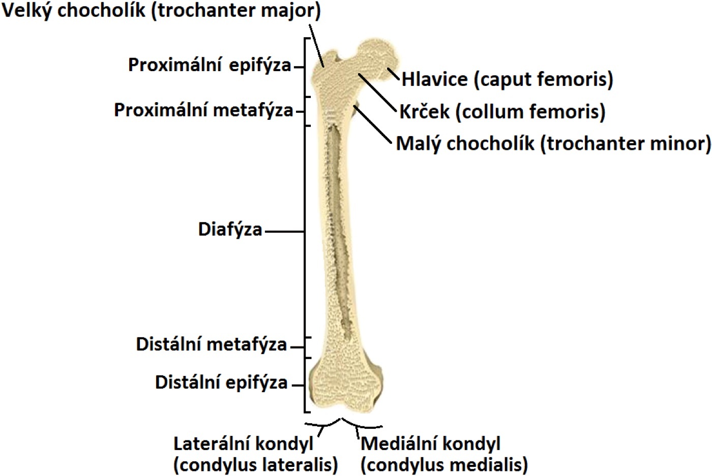
Figure 1. Regional anatomy of the femur: the proximal and distal epiphyses and metaphyses flank the diaphysis (shaft), the segment that fractures here. Illustration by Karel Frydrýšek (Fry72), CC BY-SA 4.0, via Wikimedia Commons.
The mechanism follows the energy. In young men the leading cause is the motor-vehicle collision, with motorcycle crashes, falls from height, pedestrian injuries, and gunshot wounds also common; in the elderly the fracture usually follows a low-energy fall from standing on poor-quality bone.[6] An estimated 250 Nm is needed to fracture a healthy adult femur, rising to as much as 8,000 Nm if the load is purely compressive, which is why a femoral shaft fracture in a young person flags the likelihood of other serious injuries.[7] The fracture pattern itself encodes the mechanism: bending loads produce a transverse fracture, often with a compression-side butterfly fragment; torsion produces a spiral or oblique fracture (the typical low-energy elderly pattern); axial loading drives the associated hip and knee injuries; and increasing energy produces increasing comminution and displacement.[8] A distinct group is the atypical (bisphosphonate-associated) fracture, seen with a mild increase in the 50-to-70-year range, which is transverse, begins at the lateral cortex with localised cortical thickening, may be heralded by prodromal thigh pain, and is frequently bilateral; it is treated with a locked reamed nail, the drug is stopped, and the opposite femur is imaged.[9]
Part II - Applied Anatomy and the Deforming Forces
The femur is the largest long bone in the body. Its shaft is not straight but carries an anterolateral bow; Rockwood gives an average radius of curvature of 120 ± 36 cm (Egol’s study of 948 femurs), while the AO text quotes approximately 1.5 m, and the practical point is that most nails are designed straighter than the canal (radii of roughly 186 to 300 cm), so a mismatch between the bow of the bone and the bow of the nail can cause difficulty seating the nail, anterior cortical penetration distally, sagittal malalignment, or even bursting of the proximal femur.[10] The posterior cortex thickens into the linea aspera, the ridge that anchors the thigh muscles and the intermuscular septa, and the thigh is divided into three compartments: the anterior (quadriceps, sartorius, iliopsoas, pectineus; the femoral artery, vein, and nerve), the medial (the adductors, gracilis, obturator externus; the profunda femoris and obturator vessels and the obturator nerve), and the posterior (the hamstrings and part of adductor magnus; the sciatic nerve and branches of the profunda femoris).[11]
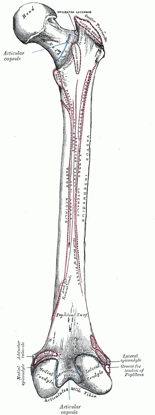
Figure 2. Posterior view of the femur showing the linea aspera and the muscle attachments along the shaft. Gray’s Anatomy (Henry Vandyke Carter, 1918), public domain, via Wikimedia Commons.
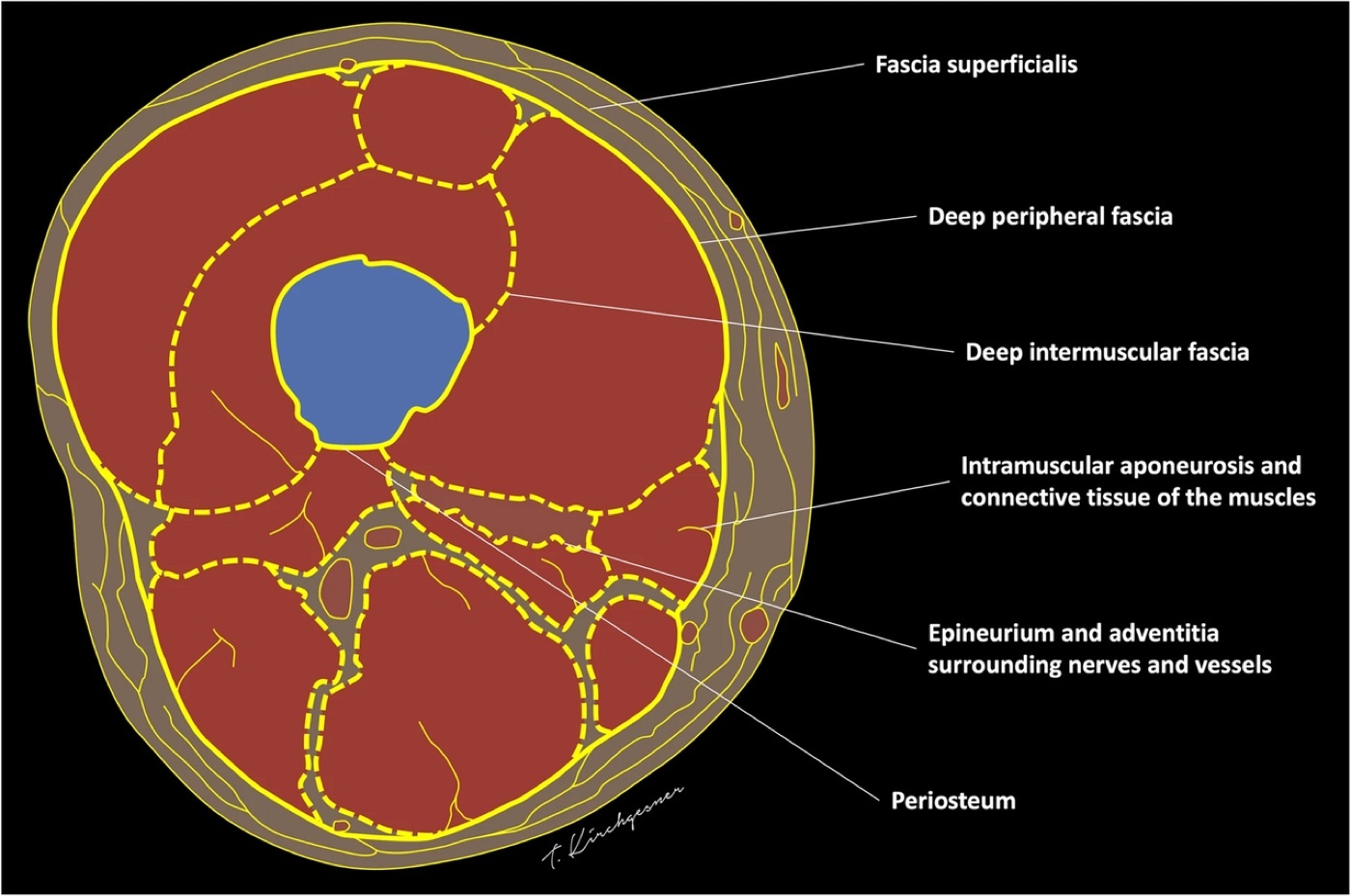
Figure 3. Transverse section of the thigh: the femur lies within the three muscle compartments, bounded by the fascial and intermuscular planes. Illustration by Thomas Kirchgesner, CC BY 4.0, via Wikimedia Commons.
The blood supply explains why reaming and displacement are tolerated. The principal nutrient artery arises from the profunda femoris (from its second perforating branch) and enters the bone along the linea aspera, supplying (depending on the source) the inner one-third (Rockwood) or inner two-thirds (AO) of the cortex and the medulla, while the periosteal vessels, fed by the surrounding muscles, supply the outer cortex; in the intact bone, flow runs from the medullary canal outward.[12] When the medullary supply is disrupted by a displaced fracture, by reaming, or by a nail, the periosteal arteries take over and the direction of flow reverses, which is why a well-vascularised muscular envelope, gently handled, allows the femur to revascularise its cortex and heal even after reaming; the perforating vessels run roughly perpendicular to the bone at intervals of about 3 cm and are therefore seldom extensively stripped, so the surgical lesson is to limit soft-tissue stripping off the linea aspera and to preserve the periosteal vessels.[13]
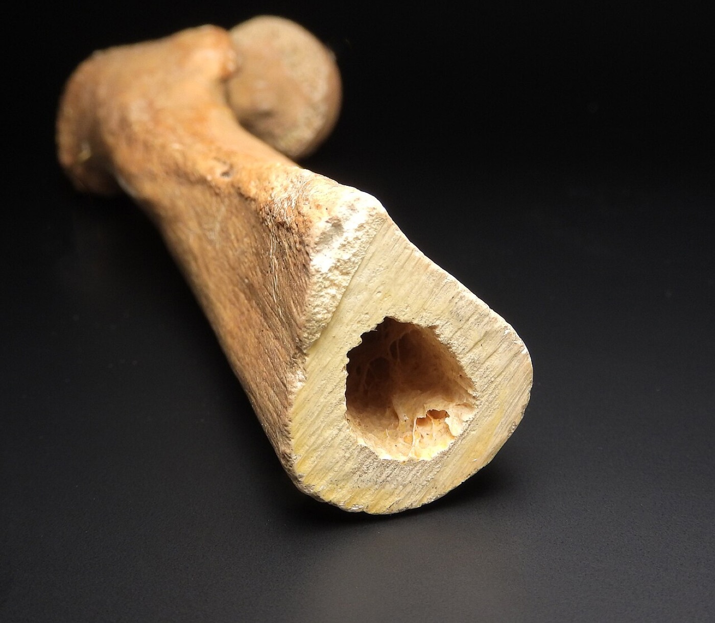
Figure 4. Cross-section through the femoral diaphysis showing the thick cortical wall and the central medullary canal that an intramedullary nail occupies. Image by MAKY.OREL, CC0, via Wikimedia Commons.
The deforming muscle forces are essential and depend on the level of the fracture.[14] In proximal-third fractures the proximal fragment flexes (pull of the iliopsoas on the lesser trochanter), abducts (pull of the gluteus medius/minimus on the greater trochanter), and externally rotates (the short external rotators), a triad that makes proximal fractures the hardest to reduce and most prone to a varus, flexed malposition. In distal-third / metaphyseal fractures the shaft is medialised by the adductors, and the gastrocnemius pulls the short distal fragment into extension (apex-posterior / recurvatum). At almost any level the limb shortens because the hamstrings and quadriceps contract across the fracture.[15] Understanding these vectors, rather than relying on simple in-line traction, is what tells the surgeon how to reduce each pattern (for example, flexing and adducting the proximal fragment to meet the shaft, or supporting the distal fragment to counter the gastrocnemius).[16]
Part III - Assessment, Associated Injuries, and Imaging
Assessment begins with ATLS, because the thigh is very vascular and because the femoral shaft fracture is so often one injury among many.[17] The alert patient reports pain, deformity, and inability to bear weight; the limb looks shortened and angulated and often arrives in a Hare traction splint. The limb must be inspected circumferentially for open wounds (a small posterior or medial wound is easily missed and any wound should be treated as open until proven otherwise), the joints above and below examined for dislocation and ligamentous injury, and a careful neurovascular examination performed. Imaging is radiographs of the entire femur including the hip and knee in two planes, supplemented by views of the contralateral femur when length or the femoral bow is uncertain (useful in elderly stress fractures), and by review of any abdomino-pelvic CT for occult proximal-femoral and acetabular injury.[18]
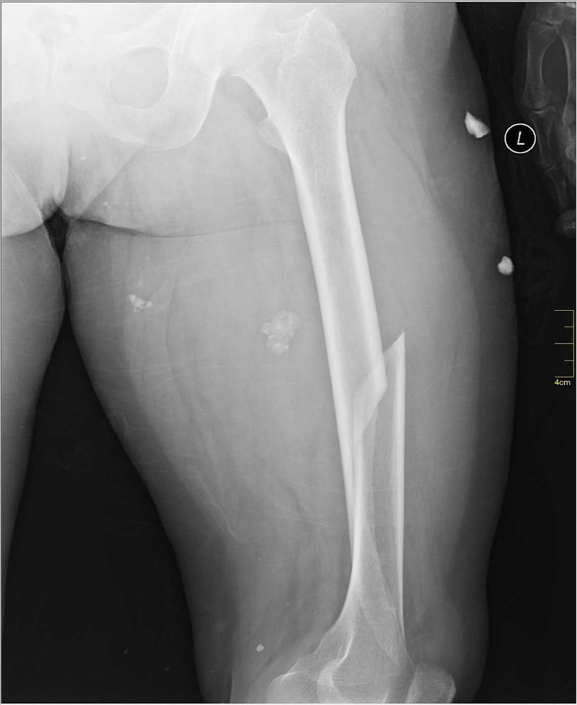
Figure 5. Displaced oblique/spiral fracture of the femoral shaft, AP radiograph. Image by Nevit Dilmen, CC BY-SA 3.0, via Wikimedia Commons.
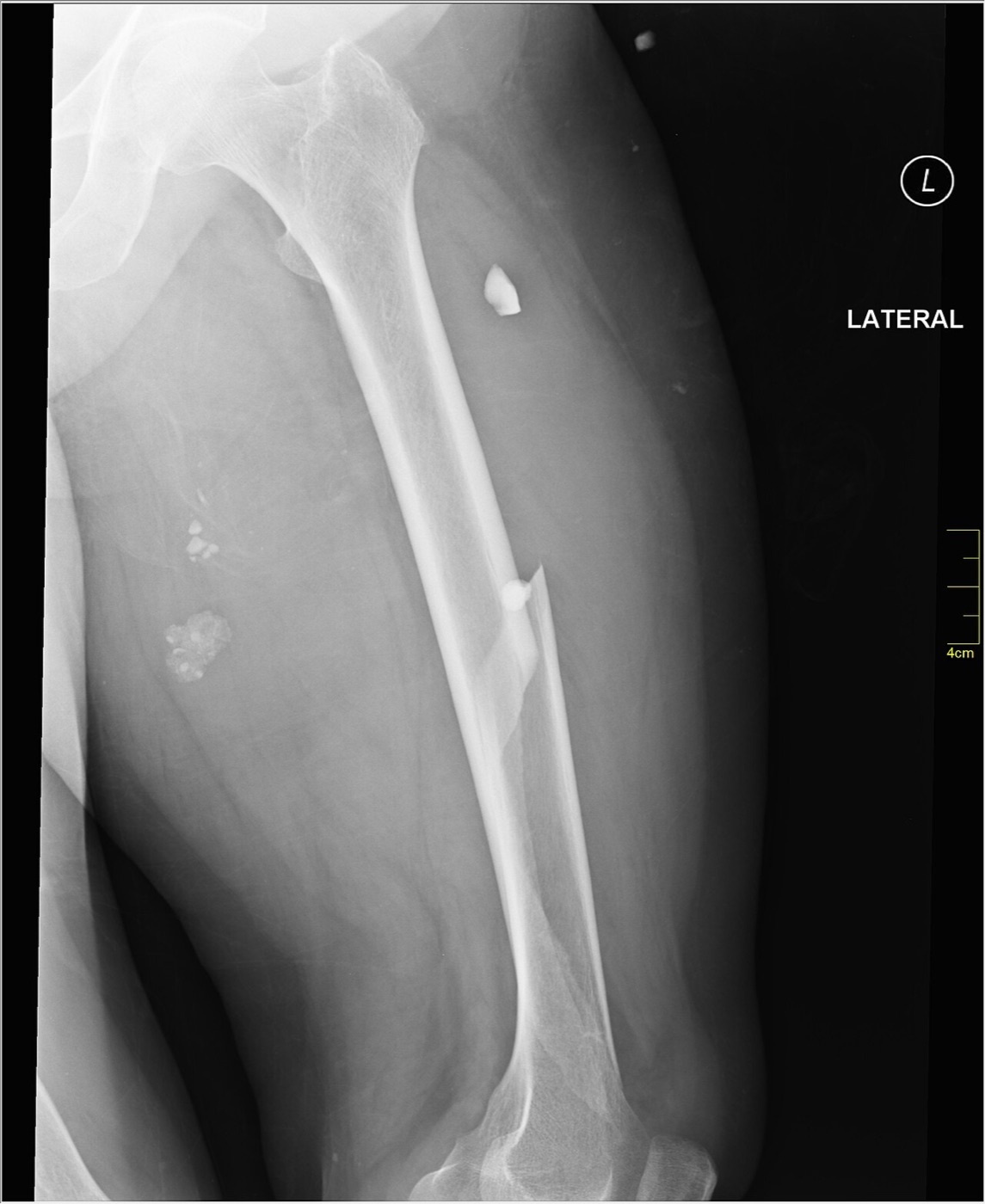
Figure 6. The same femoral shaft fracture on the lateral radiograph, confirming displacement in two planes. Image by Nevit Dilmen, CC BY-SA 3.0, via Wikimedia Commons.
The single most important associated injury, and a classic examination trap, is the ipsilateral femoral neck fracture, present in roughly 1 to 9 % (Rockwood), 2.5 to 6 % (AO), or under 10 % (Miller) of high-energy shaft fractures, and missed on initial presentation in 20 to 50 % of cases.[19] These neck fractures are typically nondisplaced, vertically oriented, and basicervical, overshadowed by more obvious injuries and degraded on a poorly rotated trauma film. Because a missed neck fracture risks osteonecrosis, nonunion, and post-traumatic arthritis, a dedicated screening protocol is mandatory: a fine-cut (2 mm) CT through the femoral neck, a dedicated internal-rotation AP view of the hip, intraoperative fluoroscopic lateral imaging of the neck before shaft fixation, and orthogonal hip radiographs after nailing; instituting this protocol reduced missed neck fractures by 91 %.[20] No single test is perfect. O’Toole found CT and plain films each had high false-negative rates (sensitivity 64 % and 56 %) but high specificity (96 % and 94 %), so that combining modalities reduced the miss rate to about 3 %, and any patient with hip or groin pain after shaft fixation should have dedicated hip radiographs, since a neck fracture can be revealed only later.[21]
The other associated injuries deserve a structured search.[22] A review of more than 26,000 femur fractures found the commonest internal-organ injuries to be lung (18.9 %), intracranial (13.5 %), and liver (6.2 %), and the commonest bony injuries to be the tibia/fibula (20.5 %) and ribs/sternum (19.1 %). Knee ligament and meniscal injuries are common but unreliable to detect until the femur is stabilised: arthroscopic series report ligamentous injury in roughly half of patients, so the knee should be examined under anaesthesia after fixation. Vascular injury is uncommon (Kluger found it in 1.6 % of 765 closed shaft fractures) but is screened with pulses and an ankle-brachial index, a value below 0.90 carrying high sensitivity and specificity for arterial injury. Nerve injury (obturator, femoral, sciatic) is rare in closed fractures. Open fractures require thorough circumferential examination, early antibiotics, and a sterile dressing.[23]
Part IV - Classification
Two systems matter for the examination.[24] The Winquist-Hansen classification grades comminution and was historically used to predict axial and rotational stability before locked nails were routine: grade 0 has no comminution; grade I, a small butterfly fragment (under 25 % of the width) with at least 75 % cortical contact; grade II, a fragment of about 25-50 % with at least 50 % contact; grade III, a large fragment of about 50-75 % with minimal contact; and grade IV, complete circumferential comminution with no cortical contact, including segmental fractures. Miller’s version adds a grade V (segmental bone loss).[25] More comminuted patterns (grades III-IV) were the ones thought to require a statically locked nail, but with universal static locking the classification “no longer predicts either treatment or postoperative management” and is now mainly descriptive.[26]
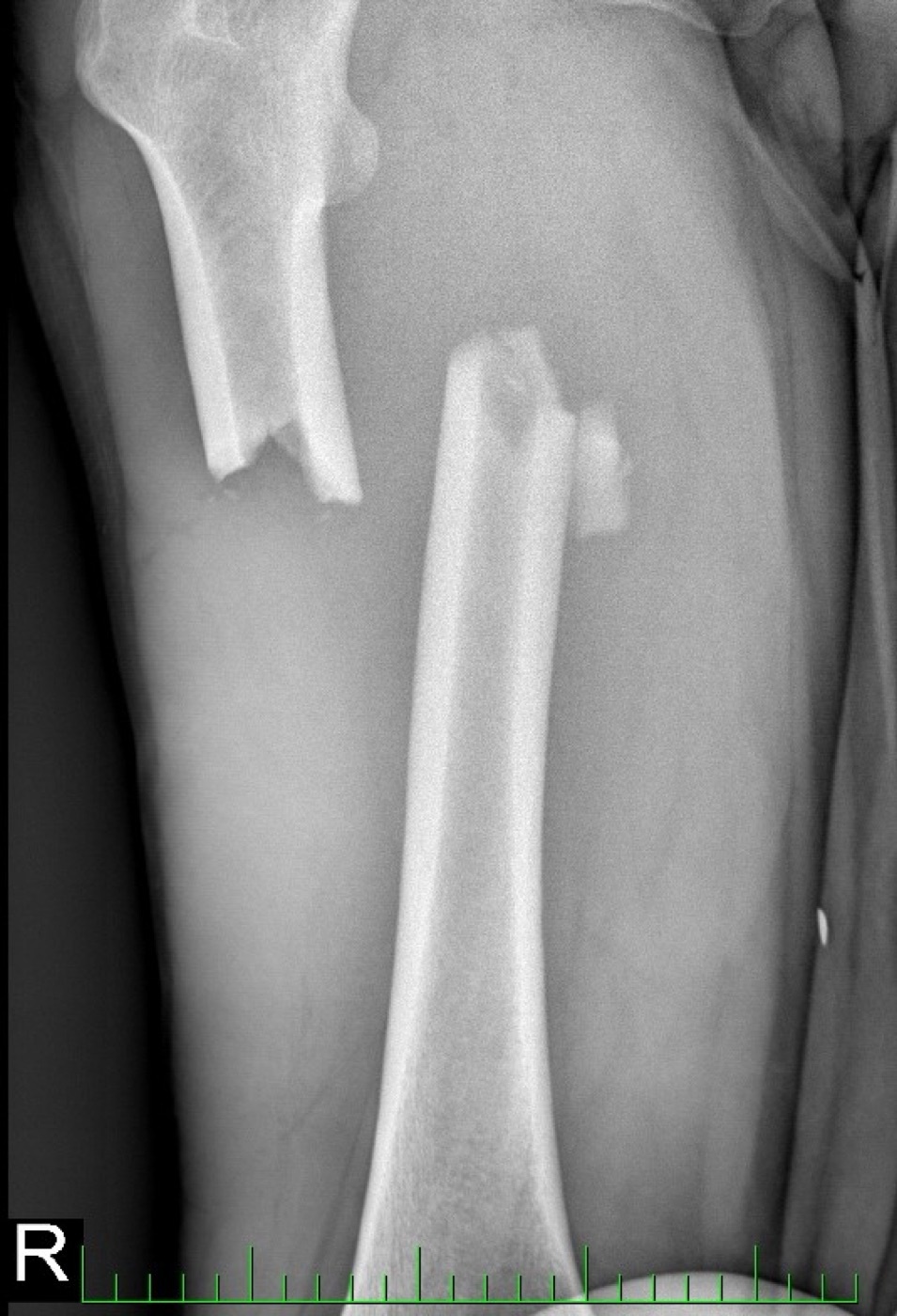
Figure 7. Displaced transverse/short-oblique fracture of the proximal femoral shaft. Image by Marek2kkk, CC BY-SA 4.0, via Wikimedia Commons.
The AO/OTA classification codes the femur as bone 3 and the diaphyseal segment as 2, so a femoral shaft fracture is region 32, subdivided into 32A (simple: spiral A1, oblique A2, transverse A3**)**, 32B (wedge), and 32C (complex / multifragmentary).[27] Within the diaphysis the pattern does not really change the method of fixation (a reamed, locked IM nail), but the classification aids preoperative planning, predicts the difficulty of achieving correct rotation, and flags associated soft-tissue injury, blood loss, and delayed-union risk. Open fractures are described with the Gustilo-Anderson system; the mined extracts give its grades only in the tibial context, so it is applied to the femur by the standard teaching that grade I is a clean wound under 1 cm, grade II a wound over 1 cm without extensive soft-tissue damage, and grade III a high-energy or contaminated injury (IIIA with adequate cover, IIIB requiring flap coverage, IIIC with a vascular injury requiring repair).[28]
Part V - Systemic Injury, Blood Loss, and the Timing of Fixation
The femoral shaft fracture is a systemic event.[29] An isolated closed fracture can lose up to 1.5 L of blood in the first 48 hours, and about 40 % of patients with an isolated fracture are transfused, averaging 2.5 units; nonetheless, in an otherwise healthy patient with a short prehospital time, an isolated femoral fracture should not by itself cause haemorrhagic shock, so a hypotensive patient demands a search for other bleeding (chest, abdomen, pelvis, other fractures). Bilateral and open fractures are different and can exsanguinate. The fracture also triggers a systemic inflammatory response, and resuscitation is judged by clearing the metabolic acidosis: the patient is considered adequately resuscitated when the base excess is under 4 mmol/L and falling and the lactate is under 2 mmol/L.[30]
Fat embolism syndrome and ARDS are the feared pulmonary complications, and early skeletal stabilisation reduces both.[31] Fat embolism syndrome classically appears 24-72 hours after injury with the triad of hypoxaemia, neurologic change, and a petechial rash, and is more frequent after bilateral or multiple long-bone fractures (the Gurd-and-Wilson major/minor criteria and the roughly 1-5 % incidence are standard teaching, the extracts simply linking early fixation to reduced fat embolism and unreamed nailing to reduced fat embolisation of uncertain significance).[32] Reaming and even nail insertion raise intramedullary pressure and embolise marrow contents; peak embolisation on echocardiography occurs at canal opening and nail insertion, even with unreamed nails, more than with reaming itself, and reamed nailing elevates pulmonary inflammatory markers such as IL-6, the basis of the “second hit” concept. Yet the bulk of clinical evidence shows that, with adequate resuscitation, reamed nailing does not increase ARDS, pneumonia, organ failure, or death, and the dominant determinants of pulmonary dysfunction are the severity of the chest injury and the quality of resuscitation, not the implant.[33]
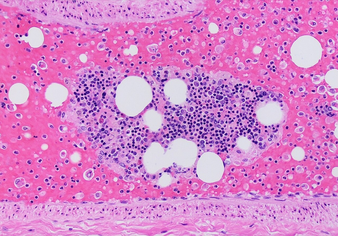
Figure 8. Pulmonary fat embolism: a pulmonary artery containing fat vacuoles together with a bone-marrow fragment (H\&E), a systemic complication of long-bone fractures. Image by Mikael Häggström, CC0, via Wikimedia Commons.
The timing of fixation has moved through three paradigms.[34] Early Total Care (ETC), definitive fixation within 24 hours, grew from Bone’s 1989 randomised trial showing fewer pulmonary complications with early stabilisation, and Nahm and others confirmed lower complication rates and shorter stays with early fixation in the multiply injured. Damage Control Orthopaedics (DCO), temporary external fixation with conversion to a nail once the patient is stable, was adopted for the under-resuscitated or physiologically unstable patient, reducing blood loss, hypothermia, and inflammatory-mediator load. The current synthesis is Early Appropriate Care: early definitive fixation is best provided the patient is adequately resuscitated (pH, lactate, and base deficit corrected), and DCO is reserved for those who are not. Pape’s data warn specifically that immediate reamed nailing in a patient with a significant chest injury increased ARDS and mortality, whereas Starr found that delaying fixation in a head-injured patient made pulmonary complications 45 times more likely without protecting the brain; the decision therefore depends on physiology, not the anatomical injury, and bodies such as EAST nonetheless recommend stabilisation within 24 hours for most patients.[35]
Part VI - Treatment
6.1 Nonoperative treatment
Nonoperative treatment of the adult femoral shaft fracture is now largely historical, reserved for the patient too sick for surgery, for settings where surgery is unavailable, and (as a separate topic) for young children.[36] Its tools are skeletal traction (a distal-femoral or proximal-tibial pin, about 15-20 lb or 15 % of body weight, often via a Thomas splint with a Pearson attachment) and cast bracing; the distal-femoral pin is placed medial-to-lateral to avoid the femoral artery, and the proximal-tibial pin lateral-to-medial to avoid the peroneal nerve. Union rates with traction are high (97-100 %), but the price is prolonged recumbency, knee stiffness, and residual shortening or angular malunion, and the AO text states bluntly that traction or cast-brace treatment “has unacceptable outcomes and should only be used when surgery is not an option.”[37]
6.2 External fixation
External fixation is a tool for rapid stabilisation, applied most often as damage control in the unstable polytrauma patient, for severe open injuries needing repeated access, and to stabilise the bone at correct length before vascular repair.[38] It is quick, can be applied in the ICU, and keeps pins outside the zone of injury, but its drawbacks limit it to a temporary role: pin-tract infection rates exceed 50 %, angular deformity is common after removal, and pins tethering the quadriceps or iliotibial band cause knee stiffness. The key principle is timely conversion to an intramedullary nail: this is safe within about two weeks, but the infection risk rises if conversion is delayed beyond 14 days, with reported infection rates of only 1.7-3.6 % and union of about 97 % when conversion is prompt.[39]
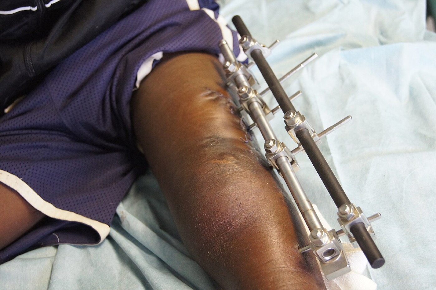
Figure 9. Monolateral external fixator applied to the femur, used for damage control and temporary stabilisation. Image by Ortopedikus, CC BY-SA 4.0, via Wikimedia Commons.
6.3 Plate fixation
Plating has declined with the refinement of nails but retains clear niches: a very narrow or deformed canal, a fracture adjacent to a deformity, malunion, or existing implant (periprosthetic/peri-implant), proximal or distal extension into the metaphysis, an associated vascular injury exposed during a medial approach, an ipsilateral neck fracture treated with a separate device, and nonunion reconstruction.[40] Open plating gives direct, anatomic reduction but at the cost of extensive dissection, blood loss, and periosteal insult; submuscular (MIPO) bridge plating preserves the fracture biology, its main downside being malreduction. The construct is typically a 4.5-mm narrow or broad plate with about four screws / eight cortices per side (or, in MIPO, a longer plate with a screw density of 0.4-0.5 and only three or four well-spread screws per side). Crucially, the plate is not a load-sharing implant: it sits eccentrically, so immediate full weight bearing risks failure, and weight bearing is generally protected until callus appears (around 6-12 weeks).[41]
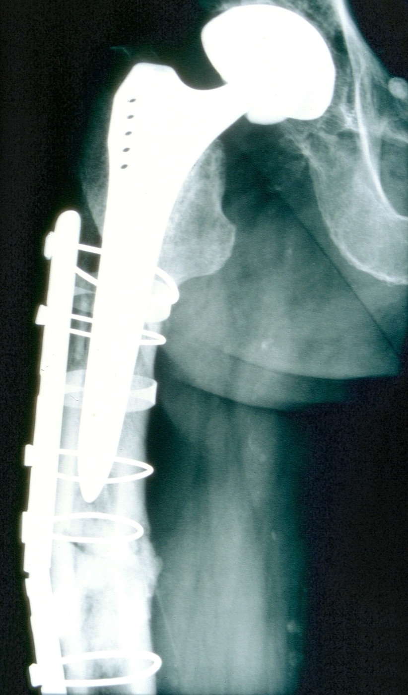
Figure 10. Periprosthetic femoral shaft fracture about a cemented stem, stabilised with a lateral plate, screws, and cerclage cables. Image by Mehlauge, CC BY-SA 3.0, via Wikimedia Commons.
6.4 Antegrade intramedullary nailing - the gold standard
The reamed, statically locked, antegrade intramedullary nail is the standard treatment for the great majority of femoral shaft fractures, an internal splint that load-shares with the bone, permits immediate weight bearing, and unites reliably; very few shaft fractures cannot be nailed (true contraindications are a canal too narrow to accept a nail, a canal-obstructing deformity or retained implant).[42] The biomechanical logic is that bending rigidity scales with the cube of the nail radius and torsional rigidity with the fourth power, so reaming to accept a larger nail makes the construct stiffer; because a locking-screw hole that exceeds half the nail diameter substantially weakens the nail, femoral interlocking screws are 5-6 mm, and a comminuted fracture without cortical contact can still bear weight on a 12-mm nail with two distal screws. All femoral nails are locked statically (two screws proximally and distally) because stability cannot be reliably judged in theatre; dynamic locking is reserved for delayed union or nonunion when length and rotation are assured. The nail should end at about the mid-patella distally.[43]
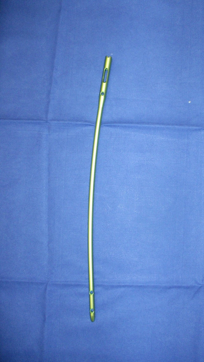
Figure 11. An antegrade interlocking intramedullary femoral nail, showing the proximal and distal locking holes and the anterior bow. Image by Hentsch, public domain, via Wikimedia Commons.
The central technical debate is the starting point.[44] The piriformis (more correctly trochanteric) fossa lies in line with the canal, so a straight piriformis-entry nail reproduces the anatomic axis, but the fossa is hard to reach (especially in the obese or supine patient), it endangers the branches of the medial femoral circumflex artery (raising a theoretical risk of femoral-head osteonecrosis, though in skeletally mature adults this is confined to case reports), and a start that is too anterior generates hoop stresses that can burst the proximal femur or fracture the neck. The greater-trochanteric entry nail, with a 4-6° proximal lateral bend, is easier to access (less time and radiation, useful in the obese), but if the start is too lateral (the natural tendency, since the bone is harder medially) the fracture falls into varus, so the start is taken just medial to the tip of the trochanter and matched to the nail’s bend. A trochanteric or more-lateral entry is mandatory in the adolescent with open physes to protect the femoral head. Soft-tissue studies show the piriformis start injures the obturator internus and medial femoral circumflex branches and risks the superior gluteal nerve (a Trendelenburg gait), while the trochanteric start injures the gluteus medius and piriformis tendons; randomised data show similar union and alignment but shorter operative and fluoroscopy times with the trochanteric start, and abductor weakness and gait abnormality after piriformis nailing.[45]
The operation proceeds by establishing length (a fracture table, manual traction, a distal-femoral traction pin, or a femoral distractor), identifying the start point, opening the canal, passing a ball-tipped guidewire centrally across a reduced fracture, reaming in 0.5-1.0 mm increments until cortical chatter and then 1.0-1.5 mm further (so the nail diameter is about 1-2 mm smaller than the last reamer), and inserting a statically locked nail of the correct length and rotation.[46] Distal locking is done freehand with a “perfect-circle” technique under a perpendicular image intensifier. The recurring pitfalls are loss of length (preventable with paralysis, traction, and contralateral films), coronal/sagittal malreduction (correct start point and nail-bow matching), and especially malrotation, which the surgeon guards against by matching the contralateral lesser-trochanter profile, reading the cortical step sign (most useful on the lateral view), matching anteversion, and obtaining a postoperative CT if uncertain; and before leaving theatre the surgeon re-examines the knee for ligament injury and the femoral neck for an occult fracture.[47]
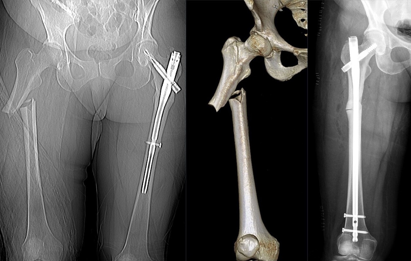
Figure 12. A femoral fracture before fixation, on 3D CT reconstruction, and after intramedullary nailing (post-operative radiograph). Image by Hellerhoff, CC BY-SA 4.0, via Wikimedia Commons.
6.5 Retrograde intramedullary nailing
Retrograde nailing, entered through the intercondylar notch just anterior to Blumensaat’s line and in line with the canal (the distal femur having 5-9° of valgus), has no absolute indications but several strong relative ones, all of which exploit easy supine access to a knee-level start: the morbidly obese (a difficult antegrade start), the pregnant (less fetal radiation), distal-third shaft fractures (easier alignment), the ipsilateral femoral neck fracture (the neck is fixed first with a separate device, then the shaft nailed retrograde), the ipsilateral acetabular fracture (avoiding an antegrade incision in the surgical field), the floating knee and bilateral fractures (one supine set-up), an existing knee arthrotomy, and proximal-femoral hardware.[48] The nail must be countersunk beneath the articular cartilage, since even 1 mm of prominence increases patellar contact, and it should end just above the lesser trochanter. Outcomes match antegrade nailing when nails are size-matched, the principal difference being that knee pain is commoner after retrograde nailing (about 36 %) and hip pain after antegrade nailing (10-40 %); for a proximal fracture, at least 5 cm of intact diaphysis should remain to avoid malalignment, and active knee sepsis is a contraindication.[49]
6.6 Reamed versus unreamed nailing
The evidence strongly favours reaming.[50] Reaming transiently disrupts the endosteal supply (reducing cortical blood flow by as much as 83 %, with recovery by about 12 weeks) but stimulates a sixfold increase in periosteal flow within 30 minutes, deposits osteoinductive reamings at the fracture (about 24 % of the reamings), and allows a larger, stiffer nail. The landmark Canadian Orthopaedic Trauma Society randomised trial found a nonunion rate of 1.7 % with reaming versus 7.5 % without (a relative risk of nonunion about 4.5 times greater without reaming, using small-diameter nails), and meta-analyses (Bhandari, Duan, Li) confirm lower nonunion, implant failure, and reoperation with reaming without any increase in ARDS or mortality.[51] The historical fear that reaming caused lethal pulmonary embolism has not been borne out: peak embolisation occurs at canal opening and nail insertion even with unreamed nails, and unreamed nailing’s only consistent advantages are slightly shorter operative time and a theoretical reduction in fat embolisation of uncertain clinical significance.[52]
6.7 The treatment algorithm
A workable decision sequence, drawn from the Rockwood authors’ preferred treatment, runs as follows.[53] First, is the patient stable? If not, temporise with external fixation or balanced skeletal traction until resuscitated. Then, is the fracture open? A clean open fracture is debrided and definitively nailed with a reamed locked nail; a grossly contaminated canal is externally fixed and re-debrided at 24-48 hours before nailing. Next, is there a femoral neck fracture? The neck takes priority and is fixed anatomically with its own device (a sliding hip screw plus a derotation screw, or cannulated screws), the shaft then nailed retrograde, and even a nondisplaced neck fracture warrants two implants. Then patient and bone factors: a canal-obstructing deformity or retained implant calls for plating; the older, osteoporotic patient is served by a reamed cephalomedullary nail (protecting the neck); the morbidly obese and the pregnant patient by a retrograde nail. Finally, location decides direction: a proximal fracture is best nailed antegrade, a distal fracture retrograde, and the governing principle is that all nails are reamed and statically locked, proximally and distally, regardless of pattern.[54]
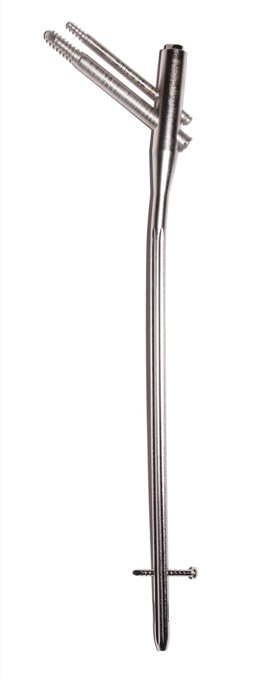
Figure 13. A cephalomedullary (reconstruction) nail, with cephalic screws into the head and neck and a distal locking bolt, the construct chosen when the femoral neck must also be protected. Image by Bullenwächter, CC BY 3.0, via Wikimedia Commons.
Part VII - Special Fracture Patterns and Associated Injuries
The ipsilateral femoral neck-shaft fracture, already discussed as a diagnostic trap, is also a reconstruction problem.[55] Because a delayed or malreduced neck fracture risks osteonecrosis, nonunion, and arthritis, most surgeons fix the neck first and anatomically, generally with two separate implants (a fixed-angle device or cannulated screws for the neck, and a retrograde nail or a plate for the shaft); a single cephalomedullary “reconstruction” nail is usually avoided for a displaced neck because the nail is not the optimal neck implant, its start point lies at the neck fracture, and correct anteversion for the head screws is hard to achieve, all raising the risk of neck malreduction and osteonecrosis. A reconstruction nail is acceptable for a nondisplaced neck or an associated intertrochanteric-plus-shaft pattern. If the neck fracture is found after nailing, a nondisplaced one is fixed with cannulated screws around the nail; a displaced one means removing the nail, fixing the neck, and re-nailing.[56]
The remaining patterns each carry their own rule.[57] Gunshot fractures: kinetic energy is ½mv², so low-velocity injuries (under about 2,000 ft/sec, the typical handgun) behave like closed fractures and are nailed (antegrade or retrograde) without formal soft-tissue débridement, whereas high-velocity and shotgun wounds need thorough, often staged débridement before nailing (retained intra-articular lead can cause systemic plumbism, standard teaching). Floating knee (ipsilateral femoral and tibial shaft fractures) is fixed with a retrograde femoral nail and an antegrade tibial nail, ideally through one knee incision, the femur first. Bilateral femoral fractures carry a markedly higher mortality (Copeland reported 25.9 % versus 11.7 %; later series 5.6-7 % versus 1.5-2 %) and more systemic injury. Vascular injury is generally stabilised first with an external fixator (or a temporary shunt) so the vascular surgeon can repair the vessel at correct length, aiming to restore flow within about 6 hours, with a low threshold for fasciotomy. With a head injury, fixation does not appear to worsen the brain, but intraoperative hypotension must be avoided, and delaying fixation makes pulmonary complications far more likely.[58]
Part VIII - Complications
Malrotation is the commonest complication after intramedullary nailing of comminuted shaft fractures.[59] A rotational malalignment greater than 10° is reported in up to 40 % of patients and greater than 15° in up to 19 %; deformities over 15° are symptomatic in a substantial minority (external malrotation being less well tolerated than internal). It is hard to detect; clinical examination missed a 20° deformity in nearly half of one CT series, and the surgeon must compare the limb with the contralateral side before leaving theatre using the lesser-trochanter profile, the cortical step sign, and anteversion matching, with CT the most sensitive confirmatory test. Patient position matters: supine nailing tends to internal malrotation, lateral nailing to external malrotation, and the fracture table produces more rotational malunion than manual traction; correction means unlocking, derotating, and re-locking.[60]
Other complications follow from the construct and the approach.[61] Angular malalignment (over 5°) is uncommon at the tight isthmus but rises for proximal and distal fractures (Ricci found 30 % of proximal versus 2 % of isthmal fractures malaligned) and is countered with correct start points, reaming the reduced fracture, multiple interlocking screws, and blocking (Poller) screws placed on the concavity of the deformity. Leg-length discrepancy accompanies comminution, and significant shortening (around 4 cm) shifts the mechanical axis medially. Nerve injuries are mostly positional: the pudendal nerve is the commonest iatrogenic palsy (about 10 % in Brumback’s series, from traction against the perineal post, causing numbness and erectile dysfunction), the peroneal nerve from the contralateral leg in a well-leg holder, and the sciatic nerve from overdistraction. Compartment syndrome of the thigh is rare (about 1-2 %) but can occur even after closed nailing and is decompressed through all three compartments. Heterotopic ossification about the entry site is commoner after reamed antegrade nailing (36 % versus 9 % unreamed) but rarely symptomatic, and the residual hip pain of antegrade nailing (10-40 %) and knee pain of retrograde nailing (~36 %) reflect the entry sites.[62]
Nonunion is uncommon after reamed, statically locked nailing, about 0.9-1.1 % in the two largest series, and its two great predictors are an open fracture and unreamed nailing.[63] A femoral nonunion is profoundly disabling, comparable to end-stage hip arthritis. Treatment is matched to the type: dynamization is simple but unreliable; reamed exchange nailing (a larger nail by 1-3 mm) succeeds in roughly 53-96 % and works best for isthmal, hypertrophic nonunions; augmentation plating around the retained nail (with bone graft) reliably heals atrophic and non-isthmal nonunions while preserving weight bearing. Infection is rare, under 1 % after nailing of a closed fracture and 2.4-4.8 % after an open fracture, and if it presents early with a stable implant, débridement with implant retention and antibiotics achieves union in about 71 % (Berkes); a chronically infected nail is removed, the canal reamed and debrided, and stability maintained, sometimes with an antibiotic-cement-coated nail. A broken implant almost always signifies an underlying nonunion, the screw (being thinner) breaking before the nail; and elective nail removal is not routine: it carries up to 10 % wound complications, helps about 78 % of patients with localised implant pain, but causes new symptoms in about 20 % of those who were asymptomatic.[64]
Part IX - Paediatric Femoral Shaft Fractures
Paediatric femoral shaft fractures account for 1-2 % of childhood fractures, with a bimodal distribution peaking at 2-3 years and again in adolescence; the cortex thickens after age 5, which parallels the falling incidence in mid-childhood.[65] The mechanism shifts with age, from minor falls in toddlers through bicycle accidents in the 4-to-7 group to motor-vehicle collisions in adolescents, and non-accidental injury must always be considered in the child who is not yet walking: abuse accounts for about 65 % of femur fractures in infants under one year once obvious causes are excluded, and should be suspected across the 0-to-3-year range, with osteogenesis imperfecta and metabolic disease in the differential for the infant. Children heal fast and remodel vigorously until about age 10 in girls and 12 in boys (up to 25° of midshaft angulation correcting in a child under 13), and they show overgrowth averaging about 1 cm in the 2-to-9-year group; Hougaard showed that fractures uniting with up to 3 cm of shortening recover to under 2 cm, which is the rationale for accepting modest shortening in the young child.[66]
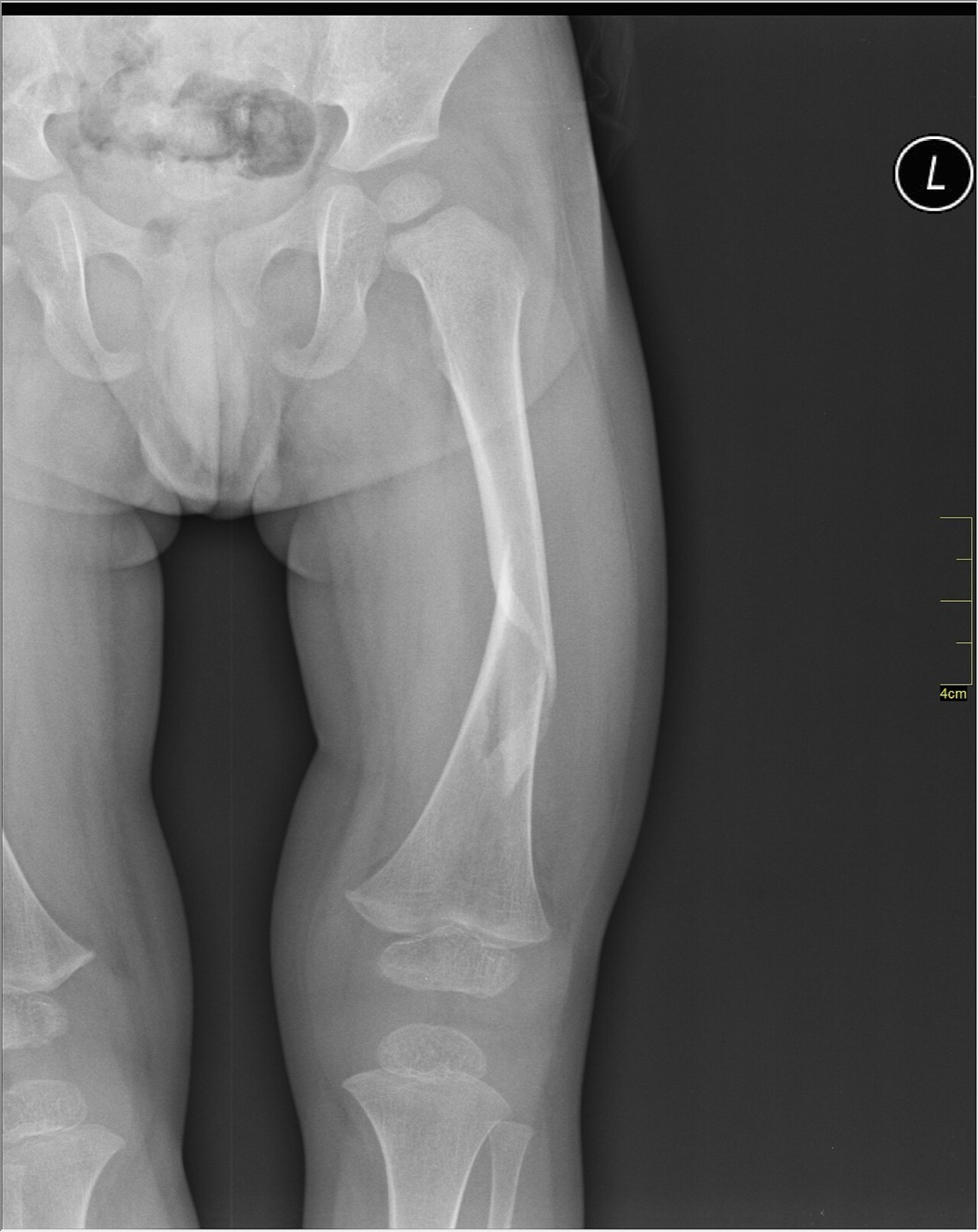
Figure 14. Paediatric transverse fracture of the femoral shaft with open growth plates, AP radiograph. Image by Nevit Dilmen, CC BY-SA 3.0, via Wikimedia Commons.
Treatment is therefore age- and weight-based.[67] The infant under 6 months is treated with a Pavlik harness, a splint, or a spica cast for a few weeks. From 6 months to 5 years, an isolated fracture is treated with early (often walking) spica casting with the hip and knee flexed about 30-40° (accepting under 3 cm of shortening and 20-30° of angulation, distal-third fractures within 20°), escalating to traction, external fixation, or elastic nails if it shortens more than 3 cm; care with cast application avoids the compartment syndrome and peroneal palsy caused by excessive traction or a short-leg-cast-with-90/90-traction technique. From 5 to 11 years, the workhorse is flexible (titanium elastic) intramedullary nailing for length-stable transverse diaphyseal fractures (two retrograde C-shaped nails each about 40 % of the canal diameter, together filling roughly 80 %, commonly 3.5 mm), with submuscular bridge plating or external fixation preferred for length-unstable, comminuted patterns; flexible nails become unreliable above 50 kg or 11 years or with comminution. In the adolescent (11 years to maturity), treatment is trochanteric (lateral greater-trochanteric) entry rigid nailing or submuscular plating, and the single highest-yield rule is that rigid nailing through the piriformis fossa must be avoided because of the risk of iatrogenic femoral-head osteonecrosis, a risk that persists as long as the proximal femoral physis remains open.[68]
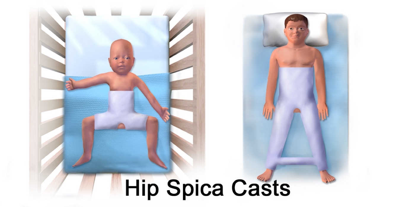
Figure 15. Hip spica casts, used for femoral shaft fractures in young children. Illustration by BruceBlaus, CC BY-SA 4.0, via Wikimedia Commons.
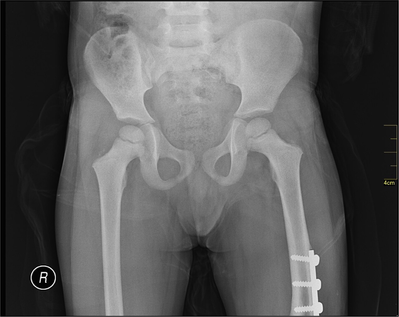
Figure 16. Paediatric femoral shaft fracture stabilised with a lateral (submuscular) bridge plate and screws. Image by Nevit Dilmen, CC BY-SA 3.0, via Wikimedia Commons.
Part X - A Synthesis: How to Reason About the Femoral Shaft Fracture
The femoral shaft fracture is best approached on two levels. The patient comes first: it is a high-energy injury until proven otherwise, so resuscitate, search hard for the occult ipsilateral femoral neck fracture with a dedicated protocol, and decide on the timing of fixation by physiology, not anatomy: early definitive nailing when the patient is resuscitated, damage-control external fixation when not, and particular caution with the chest-injured patient. The operation comes second, and its standard answer is the reamed, statically locked, antegrade intramedullary nail, a load-sharing implant that lets the patient bear weight immediately and unites reliably. The technical work is to choose the entry point sensibly (a trochanteric start that is easy and safe in most adults, mandatory in the child to protect the femoral head; a retrograde start for the obese, the pregnant, the distal fracture, the floating knee, and the ipsilateral neck or acetabular injury), to ream and lock every nail, and above all to restore length, alignment, and rotation without leaving the limb malrotated, malrotation being the commonest and most overlooked complication. The bone will heal; the surgeon’s job is to keep the patient alive through the systemic insult and to hand back a femur of the right length and rotation. In children, the same fracture is a different disease, governed by remodelling and overgrowth and treated by age, with the one inviolable rule that the immature femoral head’s blood supply forbids a piriformis-entry rigid nail.
References
-
Rockwood & Green’s Fractures in Adults, pp.3823, 3828, 3831 (high-energy mechanism, the reamed locked antegrade nail the gold standard, restoration of length/alignment/rotation, the deforming muscle forces); AO Principles of Fracture Management, pp.809, 813 (femoral shaft fractures as markers of high-energy transfer; the closed reamed interlocking IM nail the undisputed gold standard providing relative stability and callus healing; the femur “generally heals at a high rate, probably because of a good blood supply from circumferential muscles”); Miller’s Review of Orthopaedics, p.920 (IM nail for most femoral shaft fractures, union rates over 95%).
-
Rockwood & Green’s Fractures in Adults, pp.3823, 3828, 3831 (high-energy mechanism, the reamed locked antegrade nail the gold standard, restoration of length/alignment/rotation, the deforming muscle forces); AO Principles of Fracture Management, pp.809, 813 (femoral shaft fractures as markers of high-energy transfer; the closed reamed interlocking IM nail the undisputed gold standard providing relative stability and callus healing; the femur “generally heals at a high rate, probably because of a good blood supply from circumferential muscles”); Miller’s Review of Orthopaedics, p.920 (IM nail for most femoral shaft fractures, union rates over 95%).
-
Miller’s Review of Orthopaedics, p.920 (femoral shaft defined from 2.0 cm below the lesser trochanter to 8 cm from the knee joint); Rockwood & Green’s Fractures in Adults, pp.3823, 3826 (bimodal distribution with peaks at 15 and 25 in young males and over 75 in elderly females; the isthmus in the middle third and the infraisthmal designation of distal-third fractures); AO Principles of Fracture Management, p.809 (trimodal age distribution, the elderly peak from osteoporosis/bisphosphonates/periprosthetic fractures, and an incidence of 10-37 per 100,000 per year).
-
Miller’s Review of Orthopaedics, p.920 (femoral shaft defined from 2.0 cm below the lesser trochanter to 8 cm from the knee joint); Rockwood & Green’s Fractures in Adults, pp.3823, 3826 (bimodal distribution with peaks at 15 and 25 in young males and over 75 in elderly females; the isthmus in the middle third and the infraisthmal designation of distal-third fractures); AO Principles of Fracture Management, p.809 (trimodal age distribution, the elderly peak from osteoporosis/bisphosphonates/periprosthetic fractures, and an incidence of 10-37 per 100,000 per year).
-
Miller’s Review of Orthopaedics, p.920 (femoral shaft defined from 2.0 cm below the lesser trochanter to 8 cm from the knee joint); Rockwood & Green’s Fractures in Adults, pp.3823, 3826 (bimodal distribution with peaks at 15 and 25 in young males and over 75 in elderly females; the isthmus in the middle third and the infraisthmal designation of distal-third fractures); AO Principles of Fracture Management, p.809 (trimodal age distribution, the elderly peak from osteoporosis/bisphosphonates/periprosthetic fractures, and an incidence of 10-37 per 100,000 per year).
-
Rockwood & Green’s Fractures in Adults, pp.3823, 3828 (motor-vehicle collisions the leading cause in the young, low-energy falls in the elderly; 250 Nm to fracture a healthy femur, up to 8,000 Nm in pure compression; bending → transverse ± butterfly, torsion → spiral/oblique, axial → hip/knee injury, increasing energy → comminution); AO Principles of Fracture Management, p.831 and Miller’s Review of Orthopaedics, p.918 (the atypical bisphosphonate-associated femoral fracture: transverse, lateral-cortex origin with cortical beaking, prodromal thigh pain, frequently bilateral, treated with a locked reamed nail with the drug stopped and the contralateral femur imaged).
-
Rockwood & Green’s Fractures in Adults, pp.3823, 3828 (motor-vehicle collisions the leading cause in the young, low-energy falls in the elderly; 250 Nm to fracture a healthy femur, up to 8,000 Nm in pure compression; bending → transverse ± butterfly, torsion → spiral/oblique, axial → hip/knee injury, increasing energy → comminution); AO Principles of Fracture Management, p.831 and Miller’s Review of Orthopaedics, p.918 (the atypical bisphosphonate-associated femoral fracture: transverse, lateral-cortex origin with cortical beaking, prodromal thigh pain, frequently bilateral, treated with a locked reamed nail with the drug stopped and the contralateral femur imaged).
-
Rockwood & Green’s Fractures in Adults, pp.3823, 3828 (motor-vehicle collisions the leading cause in the young, low-energy falls in the elderly; 250 Nm to fracture a healthy femur, up to 8,000 Nm in pure compression; bending → transverse ± butterfly, torsion → spiral/oblique, axial → hip/knee injury, increasing energy → comminution); AO Principles of Fracture Management, p.831 and Miller’s Review of Orthopaedics, p.918 (the atypical bisphosphonate-associated femoral fracture: transverse, lateral-cortex origin with cortical beaking, prodromal thigh pain, frequently bilateral, treated with a locked reamed nail with the drug stopped and the contralateral femur imaged).
-
Rockwood & Green’s Fractures in Adults, pp.3823, 3828 (motor-vehicle collisions the leading cause in the young, low-energy falls in the elderly; 250 Nm to fracture a healthy femur, up to 8,000 Nm in pure compression; bending → transverse ± butterfly, torsion → spiral/oblique, axial → hip/knee injury, increasing energy → comminution); AO Principles of Fracture Management, p.831 and Miller’s Review of Orthopaedics, p.918 (the atypical bisphosphonate-associated femoral fracture: transverse, lateral-cortex origin with cortical beaking, prodromal thigh pain, frequently bilateral, treated with a locked reamed nail with the drug stopped and the contralateral femur imaged).
-
AO Principles of Fracture Management, p.810 (largest long bone; radius of curvature ≈1.5 m; lateral cortical flares requiring plate contouring); Rockwood & Green’s Fractures in Adults, pp.3829, 3831, 3833 (anterolateral bow, radius of curvature 120 ± 36 cm per Egol, nails straighter at 186-300 cm, and the consequences of nail-bow mismatch - difficult seating, anterior cortical penetration, sagittal malalignment, proximal-femur bursting; the linea aspera; the three thigh compartments and their muscular and neurovascular contents, reproduced from RG Fig 56-8).
-
AO Principles of Fracture Management, p.810 (largest long bone; radius of curvature ≈1.5 m; lateral cortical flares requiring plate contouring); Rockwood & Green’s Fractures in Adults, pp.3829, 3831, 3833 (anterolateral bow, radius of curvature 120 ± 36 cm per Egol, nails straighter at 186-300 cm, and the consequences of nail-bow mismatch - difficult seating, anterior cortical penetration, sagittal malalignment, proximal-femur bursting; the linea aspera; the three thigh compartments and their muscular and neurovascular contents, reproduced from RG Fig 56-8).
-
Rockwood & Green’s Fractures in Adults, pp.3834-3835 (nutrient vessel from the profunda femoris entering along the linea aspera, supplying the inner one-third of the cortex; periosteal supply from surrounding muscles; normal medullary-to-periosteal flow; reversal of flow and periosteal takeover after disruption by fracture/reaming/nail; perforators perpendicular at ~3 cm intervals; limit linea-aspera stripping); AO Principles of Fracture Management, pp.810-811 (nutrient artery from the second perforating artery supplying the inner two-thirds of cortex and medulla, periosteal arteries the outer one-third; preserve periosteal vessels and perforating arteries).
-
Rockwood & Green’s Fractures in Adults, pp.3834-3835 (nutrient vessel from the profunda femoris entering along the linea aspera, supplying the inner one-third of the cortex; periosteal supply from surrounding muscles; normal medullary-to-periosteal flow; reversal of flow and periosteal takeover after disruption by fracture/reaming/nail; perforators perpendicular at ~3 cm intervals; limit linea-aspera stripping); AO Principles of Fracture Management, pp.810-811 (nutrient artery from the second perforating artery supplying the inner two-thirds of cortex and medulla, periosteal arteries the outer one-third; preserve periosteal vessels and perforating arteries).
-
Rockwood & Green’s Fractures in Adults, p.3831 (RG Fig 56-7: proximal fragment flexed by iliopsoas, abducted by the gluteals, and externally rotated by the short rotators; distal/meta-diaphyseal medialisation by the adductors; the gastrocnemius creating an extension deformity of the distal fragment; generalised shortening from hamstrings and quadriceps; the need to strategise reduction around these forces). The discrete “midshaft adductors → varus” mapping and the recurvatum descriptor for the gastrocnemius pull are standard teaching consistent with the source, which attributes adductor medialisation specifically to the distal meta-diaphysis.
-
Rockwood & Green’s Fractures in Adults, p.3831 (RG Fig 56-7: proximal fragment flexed by iliopsoas, abducted by the gluteals, and externally rotated by the short rotators; distal/meta-diaphyseal medialisation by the adductors; the gastrocnemius creating an extension deformity of the distal fragment; generalised shortening from hamstrings and quadriceps; the need to strategise reduction around these forces). The discrete “midshaft adductors → varus” mapping and the recurvatum descriptor for the gastrocnemius pull are standard teaching consistent with the source, which attributes adductor medialisation specifically to the distal meta-diaphysis.
-
Rockwood & Green’s Fractures in Adults, p.3831 (RG Fig 56-7: proximal fragment flexed by iliopsoas, abducted by the gluteals, and externally rotated by the short rotators; distal/meta-diaphyseal medialisation by the adductors; the gastrocnemius creating an extension deformity of the distal fragment; generalised shortening from hamstrings and quadriceps; the need to strategise reduction around these forces). The discrete “midshaft adductors → varus” mapping and the recurvatum descriptor for the gastrocnemius pull are standard teaching consistent with the source, which attributes adductor medialisation specifically to the distal meta-diaphysis.
-
Rockwood & Green’s Fractures in Adults, pp.3824-3826 (ATLS-based assessment; the vascular thigh and significant closed blood loss; the Hare traction splint; circumferential inspection and the “open until proven otherwise” rule; examination of joints above and below and neurovascular status; whole-femur radiographs including hip and knee, contralateral films for length/bow, and review of abdomino-pelvic CT); AO Principles of Fracture Management, p.810 (radiographs in two orthogonal planes including the joint above and below; assessment of soft-tissue injury and neurovascular status).
-
Rockwood & Green’s Fractures in Adults, pp.3824-3826 (ATLS-based assessment; the vascular thigh and significant closed blood loss; the Hare traction splint; circumferential inspection and the “open until proven otherwise” rule; examination of joints above and below and neurovascular status; whole-femur radiographs including hip and knee, contralateral films for length/bow, and review of abdomino-pelvic CT); AO Principles of Fracture Management, p.810 (radiographs in two orthogonal planes including the joint above and below; assessment of soft-tissue injury and neurovascular status).
-
Rockwood & Green’s Fractures in Adults, pp.3826, 3892-3893 (ipsilateral neck fracture in 1-9 % of high-energy shaft fractures, missed in 20-50 %, typically nondisplaced/vertical/basicervical; the four-part screening protocol - internal-rotation AP, intraoperative fluoroscopy, fine-cut 2-mm CT, postoperative hip films - reducing misses by 91 %; O’Toole sensitivity 64 % CT / 56 % radiographs, specificity 96 % / 94 %, combined miss 3 %); AO Principles of Fracture Management, p.825 (2.5-6 % association, often undisplaced and more vertical, frequently missed); Miller’s Review of Orthopaedics, pp.920, 922 (uncommon <10 %, missed up to 50 %; nondisplaced/vertical/basicervical; dedicated hip radiographs for any postoperative hip pain).
-
Rockwood & Green’s Fractures in Adults, pp.3826, 3892-3893 (ipsilateral neck fracture in 1-9 % of high-energy shaft fractures, missed in 20-50 %, typically nondisplaced/vertical/basicervical; the four-part screening protocol - internal-rotation AP, intraoperative fluoroscopy, fine-cut 2-mm CT, postoperative hip films - reducing misses by 91 %; O’Toole sensitivity 64 % CT / 56 % radiographs, specificity 96 % / 94 %, combined miss 3 %); AO Principles of Fracture Management, p.825 (2.5-6 % association, often undisplaced and more vertical, frequently missed); Miller’s Review of Orthopaedics, pp.920, 922 (uncommon <10 %, missed up to 50 %; nondisplaced/vertical/basicervical; dedicated hip radiographs for any postoperative hip pain).
-
Rockwood & Green’s Fractures in Adults, pp.3826, 3892-3893 (ipsilateral neck fracture in 1-9 % of high-energy shaft fractures, missed in 20-50 %, typically nondisplaced/vertical/basicervical; the four-part screening protocol - internal-rotation AP, intraoperative fluoroscopy, fine-cut 2-mm CT, postoperative hip films - reducing misses by 91 %; O’Toole sensitivity 64 % CT / 56 % radiographs, specificity 96 % / 94 %, combined miss 3 %); AO Principles of Fracture Management, p.825 (2.5-6 % association, often undisplaced and more vertical, frequently missed); Miller’s Review of Orthopaedics, pp.920, 922 (uncommon <10 %, missed up to 50 %; nondisplaced/vertical/basicervical; dedicated hip radiographs for any postoperative hip pain).
-
Rockwood & Green’s Fractures in Adults, pp.3823-3825 (review of 26,357 femur fractures - lung 18.9 %, intracranial 13.5 %, liver 6.2 %, tibia/fibula 20.5 %, ribs/sternum 19.1 %; knee ligament/meniscal injury in ~50 % and the need to examine after stabilisation; Kluger 1.6 % vascular injury in 765 closed fractures; ABI < 0.90 threshold; uncommon nerve injury; open-fracture assessment and ER management).
-
Rockwood & Green’s Fractures in Adults, pp.3823-3825 (review of 26,357 femur fractures - lung 18.9 %, intracranial 13.5 %, liver 6.2 %, tibia/fibula 20.5 %, ribs/sternum 19.1 %; knee ligament/meniscal injury in ~50 % and the need to examine after stabilisation; Kluger 1.6 % vascular injury in 765 closed fractures; ABI < 0.90 threshold; uncommon nerve injury; open-fracture assessment and ER management).
-
Rockwood & Green’s Fractures in Adults, pp.3826-3827 (Winquist-Hansen grades 0-IV defined by butterfly size and cortical contact, the historical link to static locking for grades III-IV, and the statement that the classification no longer predicts treatment); Miller’s Review of Orthopaedics, pp.920-921 (Winquist-Hansen I-IV with grade V = segmental bone loss).
-
Rockwood & Green’s Fractures in Adults, pp.3826-3827 (Winquist-Hansen grades 0-IV defined by butterfly size and cortical contact, the historical link to static locking for grades III-IV, and the statement that the classification no longer predicts treatment); Miller’s Review of Orthopaedics, pp.920-921 (Winquist-Hansen I-IV with grade V = segmental bone loss).
-
Rockwood & Green’s Fractures in Adults, pp.3826-3827 (Winquist-Hansen grades 0-IV defined by butterfly size and cortical contact, the historical link to static locking for grades III-IV, and the statement that the classification no longer predicts treatment); Miller’s Review of Orthopaedics, pp.920-921 (Winquist-Hansen I-IV with grade V = segmental bone loss).
-
Rockwood & Green’s Fractures in Adults, pp.3827-3828 (AO/OTA femur = 3, diaphysis = 2 → region 32; 32A simple [spiral/oblique/transverse], 32B wedge, 32C complex; the A1/A2/A3 numerals are standard teaching, the patterns being named but not numbered in the extract; pattern does not change fixation but aids planning); AO Principles of Fracture Management, p.811 (32A simple, 32B wedge, 32C multifragmentary; classification predicts reduction difficulty, soft-tissue injury, blood loss, and delayed union). The Gustilo-Anderson grades are quoted in these sources only for the tibia (Miller’s Review of Orthopaedics, p.926) and are applied here to the open femur as standard teaching.
-
Rockwood & Green’s Fractures in Adults, pp.3827-3828 (AO/OTA femur = 3, diaphysis = 2 → region 32; 32A simple [spiral/oblique/transverse], 32B wedge, 32C complex; the A1/A2/A3 numerals are standard teaching, the patterns being named but not numbered in the extract; pattern does not change fixation but aids planning); AO Principles of Fracture Management, p.811 (32A simple, 32B wedge, 32C multifragmentary; classification predicts reduction difficulty, soft-tissue injury, blood loss, and delayed union). The Gustilo-Anderson grades are quoted in these sources only for the tibia (Miller’s Review of Orthopaedics, p.926) and are applied here to the open femur as standard teaching.
-
AO Principles of Fracture Management, pp.809, 813 (isolated closed fracture up to 1.5 L blood loss in 48 h, not generally the cause of shock; bilateral/open fractures can exsanguinate; SIRS; resuscitation thresholds base excess < 4 mmol/L trending down and lactate < 2 mmol/L); Rockwood & Green’s Fractures in Adults, p.3824 (≈40 % of isolated fractures transfused, averaging 2.5 units; hypotension warrants a search for other sources).
-
AO Principles of Fracture Management, pp.809, 813 (isolated closed fracture up to 1.5 L blood loss in 48 h, not generally the cause of shock; bilateral/open fractures can exsanguinate; SIRS; resuscitation thresholds base excess < 4 mmol/L trending down and lactate < 2 mmol/L); Rockwood & Green’s Fractures in Adults, p.3824 (≈40 % of isolated fractures transfused, averaging 2.5 units; hypotension warrants a search for other sources).
-
Rockwood & Green’s Fractures in Adults, pp.3887-3888, 3895 (early fixation reduces fat embolism and ARDS; bilateral fractures raise fat-embolism risk; peak canal pressure and embolisation at canal opening and nail insertion even with unreamed nails; reamed nailing raises IL-6 and the “second-hit” concept; Bosse and others showing no difference in ARDS/pneumonia/PE/organ failure/death between reamed nailing and plating); Miller’s Review of Orthopaedics, p.920 (unreamed nails associated with decreased fat embolisation of unclear clinical relevance). The Gurd-and-Wilson criteria, the 24-72-hour petechial triad, and the ~1-5 % incidence of clinical fat embolism syndrome are standard teaching not detailed in these extracts.
-
Rockwood & Green’s Fractures in Adults, pp.3887-3888, 3895 (early fixation reduces fat embolism and ARDS; bilateral fractures raise fat-embolism risk; peak canal pressure and embolisation at canal opening and nail insertion even with unreamed nails; reamed nailing raises IL-6 and the “second-hit” concept; Bosse and others showing no difference in ARDS/pneumonia/PE/organ failure/death between reamed nailing and plating); Miller’s Review of Orthopaedics, p.920 (unreamed nails associated with decreased fat embolisation of unclear clinical relevance). The Gurd-and-Wilson criteria, the 24-72-hour petechial triad, and the ~1-5 % incidence of clinical fat embolism syndrome are standard teaching not detailed in these extracts.
-
Rockwood & Green’s Fractures in Adults, pp.3887-3888, 3895 (early fixation reduces fat embolism and ARDS; bilateral fractures raise fat-embolism risk; peak canal pressure and embolisation at canal opening and nail insertion even with unreamed nails; reamed nailing raises IL-6 and the “second-hit” concept; Bosse and others showing no difference in ARDS/pneumonia/PE/organ failure/death between reamed nailing and plating); Miller’s Review of Orthopaedics, p.920 (unreamed nails associated with decreased fat embolisation of unclear clinical relevance). The Gurd-and-Wilson criteria, the 24-72-hour petechial triad, and the ~1-5 % incidence of clinical fat embolism syndrome are standard teaching not detailed in these extracts.
-
Rockwood & Green’s Fractures in Adults, pp.3887-3888, 3891-3892, 3918 (Bone 1989 RCT - early fixation, fewer pulmonary complications; Nahm/Vallier Early Appropriate Care; the ETC → DCO → Early Appropriate Care evolution; Pape - increased ARDS/mortality with immediate reamed nailing in chest trauma; Starr - delay in head injury made pulmonary complications 45× more likely without exacerbating the head injury; EAST recommendation for stabilisation within 24 h); AO Principles of Fracture Management, p.813 (early fixation reduces morbidity/mortality, usually within 24 h; “the decision depends on the physiology of the patient, not the anatomical injury”); Miller’s Review of Orthopaedics, pp.920-921 (damage-control principles - provisional external fixation, reductions in blood loss/hypothermia/inflammatory mediators, conversion to nail within 3 weeks, especially with chest injury).
-
Rockwood & Green’s Fractures in Adults, pp.3887-3888, 3891-3892, 3918 (Bone 1989 RCT - early fixation, fewer pulmonary complications; Nahm/Vallier Early Appropriate Care; the ETC → DCO → Early Appropriate Care evolution; Pape - increased ARDS/mortality with immediate reamed nailing in chest trauma; Starr - delay in head injury made pulmonary complications 45× more likely without exacerbating the head injury; EAST recommendation for stabilisation within 24 h); AO Principles of Fracture Management, p.813 (early fixation reduces morbidity/mortality, usually within 24 h; “the decision depends on the physiology of the patient, not the anatomical injury”); Miller’s Review of Orthopaedics, pp.920-921 (damage-control principles - provisional external fixation, reductions in blood loss/hypothermia/inflammatory mediators, conversion to nail within 3 weeks, especially with chest injury).
-
Rockwood & Green’s Fractures in Adults, pp.3835-3837 (indications for nonoperative treatment; skeletal traction technique, pin directions to protect the femoral artery and peroneal nerve, 15-20 lb or 15 % body weight, the Thomas/Pearson frame; cast bracing; union 97-100 % but knee stiffness and malunion, with shortening up to 2 cm); AO Principles of Fracture Management, p.811 (traction/cast-brace has unacceptable outcomes and should be used only when surgery is not an option; traction as temporary stabilisation).
-
Rockwood & Green’s Fractures in Adults, pp.3835-3837 (indications for nonoperative treatment; skeletal traction technique, pin directions to protect the femoral artery and peroneal nerve, 15-20 lb or 15 % body weight, the Thomas/Pearson frame; cast bracing; union 97-100 % but knee stiffness and malunion, with shortening up to 2 cm); AO Principles of Fracture Management, p.811 (traction/cast-brace has unacceptable outcomes and should be used only when surgery is not an option; traction as temporary stabilisation).
-
Rockwood & Green’s Fractures in Adults, pp.3837-3840, 3881-3882 (external fixation for damage control, open injuries, and vascular stabilisation; pin-tract infection over 50 %; angular deformity and knee stiffness; 5-6 mm pins, ≥4 pins/2 per side; conversion to nail within 2 weeks with infection 1.7-3.6 % and union ~97 %); AO Principles of Fracture Management, pp.813, 815 (damage control, conversion within 2 weeks, infection rising after 14 days, staged care lowering IL-6).
-
Rockwood & Green’s Fractures in Adults, pp.3837-3840, 3881-3882 (external fixation for damage control, open injuries, and vascular stabilisation; pin-tract infection over 50 %; angular deformity and knee stiffness; 5-6 mm pins, ≥4 pins/2 per side; conversion to nail within 2 weeks with infection 1.7-3.6 % and union ~97 %); AO Principles of Fracture Management, pp.813, 815 (damage control, conversion within 2 weeks, infection rising after 14 days, staged care lowering IL-6).
-
Rockwood & Green’s Fractures in Adults, pp.3840-3846 (declining use; relative indications - narrow canal, malunion-adjacent, proximal/distal extension, vascular injury, ipsilateral neck, periprosthetic, nonunion; open vs MIPO trade-offs; 4.5-mm narrow/broad plate, ≥8 cortices or 4 screws per side, plate length ≥10 holes for simple patterns; the plate as a non-load-bearing eccentric implant with higher failure on early weight bearing; protected weight bearing 6-12 weeks); AO Principles of Fracture Management, pp.812-814, 820-821 (relative indications for plating; long broad LCP 4.5 with ≥8 cortices per side; MIPO plate-screw density 0.4-0.5, 5-6 holes per side and usually 3-4 screws, preserving endosteal and periosteal supply).
-
Rockwood & Green’s Fractures in Adults, pp.3840-3846 (declining use; relative indications - narrow canal, malunion-adjacent, proximal/distal extension, vascular injury, ipsilateral neck, periprosthetic, nonunion; open vs MIPO trade-offs; 4.5-mm narrow/broad plate, ≥8 cortices or 4 screws per side, plate length ≥10 holes for simple patterns; the plate as a non-load-bearing eccentric implant with higher failure on early weight bearing; protected weight bearing 6-12 weeks); AO Principles of Fracture Management, pp.812-814, 820-821 (relative indications for plating; long broad LCP 4.5 with ≥8 cortices per side; MIPO plate-screw density 0.4-0.5, 5-6 holes per side and usually 3-4 screws, preserving endosteal and periosteal supply).
-
AO Principles of Fracture Management, pp.809, 813, 825 (reamed, locked, antegrade nailing the undisputed standard; load-sharing implant with early weight bearing; static locking with two screws top and bottom, dynamic locking only for delayed/nonunion; antegrade nail ends at mid-patella); Rockwood & Green’s Fractures in Adults, pp.3828-3831, 3846-3847 (bending ∝ radius³, torsion ∝ radius⁴; the
50 % hole-weakening threshold and 5-6 mm screws; the Brumback weight-bearing model; indications and the true contraindications of a narrow canal, canal-obstructing deformity, or retained implant; static locking recommended for all).
-
AO Principles of Fracture Management, pp.809, 813, 825 (reamed, locked, antegrade nailing the undisputed standard; load-sharing implant with early weight bearing; static locking with two screws top and bottom, dynamic locking only for delayed/nonunion; antegrade nail ends at mid-patella); Rockwood & Green’s Fractures in Adults, pp.3828-3831, 3846-3847 (bending ∝ radius³, torsion ∝ radius⁴; the
50 % hole-weakening threshold and 5-6 mm screws; the Brumback weight-bearing model; indications and the true contraindications of a narrow canal, canal-obstructing deformity, or retained implant; static locking recommended for all).
-
Rockwood & Green’s Fractures in Adults, pp.3857-3859, 3866-3868, 3885 (the piriformis/trochanteric fossa terminology; straight piriformis nails vs trochanteric nails with a 4-6° proximal bend and the far-lateral “bald spot”; too-lateral start → varus, too-anterior start → hoop-stress bursting or neck fracture; soft-tissue injuries at each start - piriformis tendon, obturator internus/externus, medial femoral circumflex branches, superior gluteal nerve/Trendelenburg vs gluteus medius; trochanteric/more-lateral start mandatory with open physes; Streubel - start just medial to the tip in up to 70 %; randomised data showing shorter operative/fluoroscopy times and better early function with trochanteric entry); AO Principles of Fracture Management, p.819 (entry at the piriformis fossa in line with the canal or at the greater-trochanter tip; harder medial bone driving a too-lateral entry and varus, so choose an entry medial to the trochanter tip); Miller’s Review of Orthopaedics, p.920 (anterior piriformis start → hoop stress and iatrogenic comminution; trochanteric start risks medial comminution and varus with a straight nail).
-
Rockwood & Green’s Fractures in Adults, pp.3857-3859, 3866-3868, 3885 (the piriformis/trochanteric fossa terminology; straight piriformis nails vs trochanteric nails with a 4-6° proximal bend and the far-lateral “bald spot”; too-lateral start → varus, too-anterior start → hoop-stress bursting or neck fracture; soft-tissue injuries at each start - piriformis tendon, obturator internus/externus, medial femoral circumflex branches, superior gluteal nerve/Trendelenburg vs gluteus medius; trochanteric/more-lateral start mandatory with open physes; Streubel - start just medial to the tip in up to 70 %; randomised data showing shorter operative/fluoroscopy times and better early function with trochanteric entry); AO Principles of Fracture Management, p.819 (entry at the piriformis fossa in line with the canal or at the greater-trochanter tip; harder medial bone driving a too-lateral entry and varus, so choose an entry medial to the trochanter tip); Miller’s Review of Orthopaedics, p.920 (anterior piriformis start → hoop stress and iatrogenic comminution; trochanteric start risks medial comminution and varus with a straight nail).
-
Rockwood & Green’s Fractures in Adults, pp.3852-3856, 3859-3866 (the key surgical steps - length, start point, guidewire, reaming, locked nail; reaming in 0.5-1.0 mm increments to cortical chatter then 1.0-1.5 mm further; freehand perfect-circle distal locking; reduction aids - Schanz pins, femoral distractor, finger/collinear tools; malrotation detection by lesser-trochanter profile, cortical step sign, anteversion matching, and postoperative CT; the antegrade pitfalls table; final knee and femoral-neck checks); AO Principles of Fracture Management, pp.822, 824 (closed/indirect reduction, the F-tool and intramedullary reduction tools, blocking/Poller screws for distal fractures, reaming to cortical chatter, nail diameter 1-2 mm below the largest reamer per Miller’s Review p.920).
-
Rockwood & Green’s Fractures in Adults, pp.3852-3856, 3859-3866 (the key surgical steps - length, start point, guidewire, reaming, locked nail; reaming in 0.5-1.0 mm increments to cortical chatter then 1.0-1.5 mm further; freehand perfect-circle distal locking; reduction aids - Schanz pins, femoral distractor, finger/collinear tools; malrotation detection by lesser-trochanter profile, cortical step sign, anteversion matching, and postoperative CT; the antegrade pitfalls table; final knee and femoral-neck checks); AO Principles of Fracture Management, pp.822, 824 (closed/indirect reduction, the F-tool and intramedullary reduction tools, blocking/Poller screws for distal fractures, reaming to cortical chatter, nail diameter 1-2 mm below the largest reamer per Miller’s Review p.920).
-
AO Principles of Fracture Management, pp.811-812, 820, 825, 833 (no absolute but multiple relative indications for retrograde nailing - obesity, ipsilateral neck/shaft, floating knee through one incision, multiple injuries, bilateral fractures, spine injury, pregnancy, uncontaminated knee arthrotomy, ipsilateral pelvic/acetabular fracture, proximal-femoral hardware; intercondylar-notch entry anterior to Blumensaat’s line; nail ends above the lesser trochanter; countersink beneath the articular surface; knee pain vs hip pain; union equal when size-matched); Rockwood & Green’s Fractures in Adults, pp.3869-3874, 3886 (distal femur 5-9° valgus, start at the apex of Blumensaat’s line; relative contraindications - subtrochanteric pattern, limited knee flexion, patella baja; Morgan 1-mm prominence and patellar contact; knee pain 36 % retrograde vs 9 % antegrade and hip pain 10 % antegrade vs 4 % retrograde; ≥5 cm intact diaphysis for proximal fractures).
-
AO Principles of Fracture Management, pp.811-812, 820, 825, 833 (no absolute but multiple relative indications for retrograde nailing - obesity, ipsilateral neck/shaft, floating knee through one incision, multiple injuries, bilateral fractures, spine injury, pregnancy, uncontaminated knee arthrotomy, ipsilateral pelvic/acetabular fracture, proximal-femoral hardware; intercondylar-notch entry anterior to Blumensaat’s line; nail ends above the lesser trochanter; countersink beneath the articular surface; knee pain vs hip pain; union equal when size-matched); Rockwood & Green’s Fractures in Adults, pp.3869-3874, 3886 (distal femur 5-9° valgus, start at the apex of Blumensaat’s line; relative contraindications - subtrochanteric pattern, limited knee flexion, patella baja; Morgan 1-mm prominence and patellar contact; knee pain 36 % retrograde vs 9 % antegrade and hip pain 10 % antegrade vs 4 % retrograde; ≥5 cm intact diaphysis for proximal fractures).
-
Rockwood & Green’s Fractures in Adults, pp.3883-3884, 3910 (reaming reduces cortical flow up to 83 % with recovery by 12 weeks, increases periosteal flow sixfold within 30 minutes, deposits ~24 % of reamings at the fracture; the Canadian Orthopaedic Trauma Society RCT - nonunion 1.7 % reamed vs 7.5 % unreamed, relative risk ~4.5×; Bhandari/Duan/Li meta-analyses favouring reaming with no ARDS/mortality penalty; peak embolisation at canal opening/insertion even unreamed); AO Principles of Fracture Management, pp.813, 833 (antegrade reamed union 95 %, unreamed nonunion 7.5 % vs 1.6 %; resuscitation and chest-injury severity more decisive than the device for pulmonary dysfunction); Miller’s Review of Orthopaedics, p.920 (reamed nailing for most fractures, higher union; unreamed associated with decreased fat embolisation of unclear relevance).
-
Rockwood & Green’s Fractures in Adults, pp.3883-3884, 3910 (reaming reduces cortical flow up to 83 % with recovery by 12 weeks, increases periosteal flow sixfold within 30 minutes, deposits ~24 % of reamings at the fracture; the Canadian Orthopaedic Trauma Society RCT - nonunion 1.7 % reamed vs 7.5 % unreamed, relative risk ~4.5×; Bhandari/Duan/Li meta-analyses favouring reaming with no ARDS/mortality penalty; peak embolisation at canal opening/insertion even unreamed); AO Principles of Fracture Management, pp.813, 833 (antegrade reamed union 95 %, unreamed nonunion 7.5 % vs 1.6 %; resuscitation and chest-injury severity more decisive than the device for pulmonary dysfunction); Miller’s Review of Orthopaedics, p.920 (reamed nailing for most fractures, higher union; unreamed associated with decreased fat embolisation of unclear relevance).
-
Rockwood & Green’s Fractures in Adults, pp.3883-3884, 3910 (reaming reduces cortical flow up to 83 % with recovery by 12 weeks, increases periosteal flow sixfold within 30 minutes, deposits ~24 % of reamings at the fracture; the Canadian Orthopaedic Trauma Society RCT - nonunion 1.7 % reamed vs 7.5 % unreamed, relative risk ~4.5×; Bhandari/Duan/Li meta-analyses favouring reaming with no ARDS/mortality penalty; peak embolisation at canal opening/insertion even unreamed); AO Principles of Fracture Management, pp.813, 833 (antegrade reamed union 95 %, unreamed nonunion 7.5 % vs 1.6 %; resuscitation and chest-injury severity more decisive than the device for pulmonary dysfunction); Miller’s Review of Orthopaedics, p.920 (reamed nailing for most fractures, higher union; unreamed associated with decreased fat embolisation of unclear relevance).
-
Rockwood & Green’s Fractures in Adults, pp.3878-3880 (Authors’ Preferred Treatment, Algorithm 56-1: stability first [external fixation or skeletal traction if unstable]; open fractures debrided and nailed, grossly contaminated canals externally fixed with repeat débridement at 24-48 h; femoral neck given priority with a sliding hip screw and derotation screw or cannulated screws and a retrograde shaft nail, two devices even for a nondisplaced neck; plating for canal-obstructing factors; reamed cephalomedullary nail for the older/osteoporotic patient; retrograde nail for the morbidly obese and the pregnant; proximal fractures antegrade and distal fractures retrograde; all nails reamed and locked proximally and distally).
-
Rockwood & Green’s Fractures in Adults, pp.3878-3880 (Authors’ Preferred Treatment, Algorithm 56-1: stability first [external fixation or skeletal traction if unstable]; open fractures debrided and nailed, grossly contaminated canals externally fixed with repeat débridement at 24-48 h; femoral neck given priority with a sliding hip screw and derotation screw or cannulated screws and a retrograde shaft nail, two devices even for a nondisplaced neck; plating for canal-obstructing factors; reamed cephalomedullary nail for the older/osteoporotic patient; retrograde nail for the morbidly obese and the pregnant; proximal fractures antegrade and distal fractures retrograde; all nails reamed and locked proximally and distally).
-
Rockwood & Green’s Fractures in Adults, pp.3892-3894 (fix the neck first and anatomically; two separate implants preferred over a single reconstruction nail for a displaced neck because of malreduction/osteonecrosis risk; reconstruction nail acceptable for a nondisplaced neck; management of a neck fracture discovered after nailing); AO Principles of Fracture Management, pp.825-827 (neck given priority, two-implant strategy, the “miss-a-nail” technique of placing lag screws anterior to an already-seated nail); Miller’s Review of Orthopaedics, p.922 (neck fixed first with a 135° fixed-angle device or parallel screws, then a retrograde nail or plate; a cephalomedullary nail for a displaced ipsilateral neck increases malreduction and osteonecrosis).
-
Rockwood & Green’s Fractures in Adults, pp.3892-3894 (fix the neck first and anatomically; two separate implants preferred over a single reconstruction nail for a displaced neck because of malreduction/osteonecrosis risk; reconstruction nail acceptable for a nondisplaced neck; management of a neck fracture discovered after nailing); AO Principles of Fracture Management, pp.825-827 (neck given priority, two-implant strategy, the “miss-a-nail” technique of placing lag screws anterior to an already-seated nail); Miller’s Review of Orthopaedics, p.922 (neck fixed first with a 135° fixed-angle device or parallel screws, then a retrograde nail or plate; a cephalomedullary nail for a displaced ipsilateral neck increases malreduction and osteonecrosis).
-
Rockwood & Green’s Fractures in Adults, pp.3889-3896 (gunshot - KE = ½mv², low-velocity < ~2,000 ft/sec treated as closed and nailed without formal débridement, high-velocity/shotgun needing staged débridement; floating knee handling; bilateral mortality 25.9 % vs 11.7 % [Copeland], 5.6 % vs 1.5 % [Nork], 7 % vs 2 % [O’Toole]; vascular injury - external fixator or shunt first, restore flow ideally within 6 h, early fasciotomy; head injury - early fixation does not exacerbate the brain but delay raises pulmonary complications, Starr 45×); Miller’s Review of Orthopaedics, p.921 (floating knee - retrograde femur and antegrade tibia, with fat embolism/ARDS risk). Lead toxicity from retained intra-articular bullets and the Fraser classification of the floating knee are standard teaching not detailed in these extracts.
-
Rockwood & Green’s Fractures in Adults, pp.3889-3896 (gunshot - KE = ½mv², low-velocity < ~2,000 ft/sec treated as closed and nailed without formal débridement, high-velocity/shotgun needing staged débridement; floating knee handling; bilateral mortality 25.9 % vs 11.7 % [Copeland], 5.6 % vs 1.5 % [Nork], 7 % vs 2 % [O’Toole]; vascular injury - external fixator or shunt first, restore flow ideally within 6 h, early fasciotomy; head injury - early fixation does not exacerbate the brain but delay raises pulmonary complications, Starr 45×); Miller’s Review of Orthopaedics, p.921 (floating knee - retrograde femur and antegrade tibia, with fat embolism/ARDS risk). Lead toxicity from retained intra-articular bullets and the Fraser classification of the floating knee are standard teaching not detailed in these extracts.
-
Rockwood & Green’s Fractures in Adults, pp.3904-3906 (malrotation
10° in up to 40 %, > 15° in up to 19 % [Braten], symptomatic over 15°, external worse than internal; clinical examination missing 20° in nearly half; detection by lesser-trochanter profile, cortical step sign, and anteversion, with CT most sensitive; correction by unlocking/derotating/re-locking); Miller’s Review of Orthopaedics, p.921 (malunion the most common complication of nailing highly comminuted shaft fractures; supine → internal, lateral → external, fracture table → more rotational malunion).
-
Rockwood & Green’s Fractures in Adults, pp.3904-3906 (malrotation
10° in up to 40 %, > 15° in up to 19 % [Braten], symptomatic over 15°, external worse than internal; clinical examination missing 20° in nearly half; detection by lesser-trochanter profile, cortical step sign, and anteversion, with CT most sensitive; correction by unlocking/derotating/re-locking); Miller’s Review of Orthopaedics, p.921 (malunion the most common complication of nailing highly comminuted shaft fractures; supine → internal, lateral → external, fracture table → more rotational malunion).
-
Rockwood & Green’s Fractures in Adults, pp.3900-3909 (angular malalignment > 5°, 30 % proximal vs 2 % isthmal [Ricci], blocking/Poller screws on the concavity; leg-length discrepancy with comminution; pudendal palsy ~10 % [Brumback] from the perineal post with sensory loss and erectile dysfunction; peroneal palsy from the well-leg holder; sciatic stretch from overdistraction; thigh compartment syndrome ~1-2 % decompressed through all three compartments; heterotopic ossification 36 % reamed vs 9 % unreamed; hip pain 10-40 % antegrade and knee pain ~36 % retrograde); Miller’s Review of Orthopaedics, p.921 (pudendal injury from excessive traction; 4 cm shortening → medial mechanical-axis deviation).
-
Rockwood & Green’s Fractures in Adults, pp.3900-3909 (angular malalignment > 5°, 30 % proximal vs 2 % isthmal [Ricci], blocking/Poller screws on the concavity; leg-length discrepancy with comminution; pudendal palsy ~10 % [Brumback] from the perineal post with sensory loss and erectile dysfunction; peroneal palsy from the well-leg holder; sciatic stretch from overdistraction; thigh compartment syndrome ~1-2 % decompressed through all three compartments; heterotopic ossification 36 % reamed vs 9 % unreamed; hip pain 10-40 % antegrade and knee pain ~36 % retrograde); Miller’s Review of Orthopaedics, p.921 (pudendal injury from excessive traction; 4 cm shortening → medial mechanical-axis deviation).
-
Rockwood & Green’s Fractures in Adults, pp.3910-3916 (nonunion 0.9-1.1 % after reamed statically locked nailing, the predictors being open fracture and unreamed nailing; dynamization unreliable; reamed exchange nailing 53-96 %, increase diameter 1-3 mm, best for isthmal/hypertrophic; augmentation plating around the nail with graft for atrophic/non-isthmal; infection < 1 % closed and 2.4-4.8 % open, Berkes 71 % union with early débridement and retention; broken implants signifying nonunion, screws breaking before nails; nail removal - up to 10 % wound complications, 78 % helped if localised pain, 20 % develop new symptoms); Miller’s Review of Orthopaedics, p.921 (infection, nonunion, and delayed union each < 5 % of closed fractures; hypertrophic nonunion → exchange nailing).
-
Rockwood & Green’s Fractures in Adults, pp.3910-3916 (nonunion 0.9-1.1 % after reamed statically locked nailing, the predictors being open fracture and unreamed nailing; dynamization unreliable; reamed exchange nailing 53-96 %, increase diameter 1-3 mm, best for isthmal/hypertrophic; augmentation plating around the nail with graft for atrophic/non-isthmal; infection < 1 % closed and 2.4-4.8 % open, Berkes 71 % union with early débridement and retention; broken implants signifying nonunion, screws breaking before nails; nail removal - up to 10 % wound complications, 78 % helped if localised pain, 20 % develop new symptoms); Miller’s Review of Orthopaedics, p.921 (infection, nonunion, and delayed union each < 5 % of closed fractures; hypertrophic nonunion → exchange nailing).
-
Lovell & Winter’s Pediatric Orthopaedics, pp.5617-5619 (1-2 % of childhood fractures; bimodal at 2-3 years and adolescence; cortex thickening after age 5; mechanism by age; non-accidental injury - abuse in ~65 % of infant femur fractures after excluding obvious causes, suspect across 0-3 years and consider osteogenesis imperfecta/metabolic disease; remodelling to ~10 years in girls and 12 in boys, 25° midshaft correcting under age 13; overgrowth averaging ~1 cm at 2-9 years; Hougaard - fractures with ≤ 3 cm shortening recover to < 2 cm).
-
Lovell & Winter’s Pediatric Orthopaedics, pp.5617-5619 (1-2 % of childhood fractures; bimodal at 2-3 years and adolescence; cortex thickening after age 5; mechanism by age; non-accidental injury - abuse in ~65 % of infant femur fractures after excluding obvious causes, suspect across 0-3 years and consider osteogenesis imperfecta/metabolic disease; remodelling to ~10 years in girls and 12 in boys, 25° midshaft correcting under age 13; overgrowth averaging ~1 cm at 2-9 years; Hougaard - fractures with ≤ 3 cm shortening recover to < 2 cm).
-
Lovell & Winter’s Pediatric Orthopaedics, pp.5619-5628, 5657-5665 (age-based algorithm - Pavlik/splint/spica under 6 months; early/walking spica 6 months-5 years with hip/knee flexion 30-40°, accepting < 3 cm shortening and 20-30° angulation, distal-third within 20°, escalating beyond 3 cm shortening; compartment-syndrome and peroneal-palsy warning with excessive traction and the short-leg-cast-90/90 technique; flexible titanium elastic nails 5-11 years for length-stable diaphyseal fractures, each ~40 % of the canal and two filling ~80 %, usually 3.5 mm, retrograde C-shaped; submuscular plating or external fixation for length-unstable/comminuted patterns; flexible nails unreliable over 50 kg or 11 years; adolescent trochanteric/lateral greater-trochanteric rigid nailing or submuscular plating; rigid piriformis-entry nailing to be avoided because of femoral-head osteonecrosis while the proximal physis is open). That the osteonecrosis arises from injury to the deep branch of the medial femoral circumflex artery at the piriformis entry is standard teaching consistent with the source, which attributes the risk to the piriformis entry without naming the vessel.
-
Lovell & Winter’s Pediatric Orthopaedics, pp.5619-5628, 5657-5665 (age-based algorithm - Pavlik/splint/spica under 6 months; early/walking spica 6 months-5 years with hip/knee flexion 30-40°, accepting < 3 cm shortening and 20-30° angulation, distal-third within 20°, escalating beyond 3 cm shortening; compartment-syndrome and peroneal-palsy warning with excessive traction and the short-leg-cast-90/90 technique; flexible titanium elastic nails 5-11 years for length-stable diaphyseal fractures, each ~40 % of the canal and two filling ~80 %, usually 3.5 mm, retrograde C-shaped; submuscular plating or external fixation for length-unstable/comminuted patterns; flexible nails unreliable over 50 kg or 11 years; adolescent trochanteric/lateral greater-trochanteric rigid nailing or submuscular plating; rigid piriformis-entry nailing to be avoided because of femoral-head osteonecrosis while the proximal physis is open). That the osteonecrosis arises from injury to the deep branch of the medial femoral circumflex artery at the piriformis entry is standard teaching consistent with the source, which attributes the risk to the piriformis entry without naming the vessel.
-
Miller’s Review of Orthopaedics, p.920; Rockwood & Green’s Fractures in Adults, pp.3826, 3831.
-
AO Principles of Fracture Management, pp.809, 813, 825; Rockwood & Green’s Fractures in Adults, pp.3823, 3880.
-
Rockwood & Green’s Fractures in Adults, pp.3857-3859, 3866-3868; AO Principles of Fracture Management, p.819.
-
AO Principles of Fracture Management, pp.811-812, 833; Rockwood & Green’s Fractures in Adults, pp.3869-3870, 3874.
-
Rockwood & Green’s Fractures in Adults, pp.3883-3884; AO Principles of Fracture Management, p.833; Miller’s Review of Orthopaedics, p.920.
-
Rockwood & Green’s Fractures in Adults, pp.3826, 3892-3893; Miller’s Review of Orthopaedics, pp.920, 922.
-
Rockwood & Green’s Fractures in Adults, pp.3892-3894; Miller’s Review of Orthopaedics, p.922.
-
AO Principles of Fracture Management, p.813; Rockwood & Green’s Fractures in Adults, pp.3887-3888, 3892, 3918.
-
Rockwood & Green’s Fractures in Adults, pp.3904-3906; Miller’s Review of Orthopaedics, p.921.
-
Rockwood & Green’s Fractures in Adults, pp.3889-3890.
-
Lovell & Winter’s Pediatric Orthopaedics, pp.5619-5628.
-
Rockwood & Green’s Fractures in Adults, pp.3824, 3887-3888, 3895; AO Principles of Fracture Management, p.809.