Contents
- Orientation: A Fracture Defined by Its Blood Supply
- Part I - Epidemiology and the Fragility Fracture
- Part II - Applied Anatomy and Blood Supply
- Part III - Assessment and Imaging
- Part IV - Classification
- Part V - Treatment
- Part VI - Complications
- Part VII - A Synthesis: How to Reason About the Femoral Neck Fracture
- References
Orientation: A Fracture Defined by Its Blood Supply
One anatomical fact explains the femoral neck fracture: the neck lies inside the hip capsule, has no periosteum, and is fed by a retrograde, capsule-bound blood supply. That single fact explains almost everything about the injury. With no periosteum, the fracture heals by endosteal union alone, which is slow. And because the blood supply runs up the neck in the retinacular vessels, a displaced fracture tears those vessels and threatens the head with avascular necrosis and nonunion. From this follows the central management decision, which is not really “how do I fix it?” but “should I keep the head or replace it?” The answer turns on three things: whether the fracture is displaced, the age and physiological status of the patient, and the bone quality. The working rule is easy to state and forms the spine of the whole topic: young patients and undisplaced fractures keep the head (internal fixation); displaced fractures in the older patient get an arthroplasty.[1]
Part I - Epidemiology and the Fragility Fracture
Hip fractures are a major public-health burden. They make up about 20% of the operative orthopaedic-trauma workload, and intracapsular femoral neck fractures account for over half (55-59%) of all hip fractures.[2] The lifetime risk of a hip fracture is 40-50% in women and 13-22% in men, and the worldwide number was projected to rise from 1.66 million in 1990 toward 6.26 million by 2050, though incidence has recently levelled or fallen in some Western populations.[3] The distribution is bimodal but heavily skewed to the elderly: the typical patient is an older woman who falls from standing height onto the trochanter, whereas only about 3% occur in patients under 60, usually men, from high-energy trauma.[4] About 15% of femoral neck fractures are undisplaced; the rest are displaced and predominate in elderly women. The underlying problem is osteoporosis, with each 1-SD fall in femoral-neck bone density raising fracture risk about 2.6-fold.[5]
Part II - Applied Anatomy and Blood Supply
Two anatomical facts govern the femoral neck fracture. The first is that the intracapsular neck has no cambial (periosteal) layer, so it heals by endosteal union alone, one reason union is slow.[6] The second is its blood supply, which comes from three sources: the retinacular (capsular) vessels, intramedullary vessels, and the foveal artery of the ligamentum teres. Of these the retinacular vessels are by far the most important.[7] They arise from the deep branch of the medial femoral circumflex artery (MFCA), which ascends the posterosuperior neck within the retinacula of Weitbrecht as the superior retinacular (ascending/lateral epiphyseal) arteries to supply the weight-bearing femoral head. A cadaveric study attributes about 82% of head perfusion to the MFCA and only 18% to the lateral femoral circumflex artery, while the foveal artery is a minor and usually insufficient contributor.[8] This is the crux of the topic: a displaced subcapital fracture tears these retinacular vessels and so jeopardises the head, producing the risk of avascular necrosis and nonunion.[9]
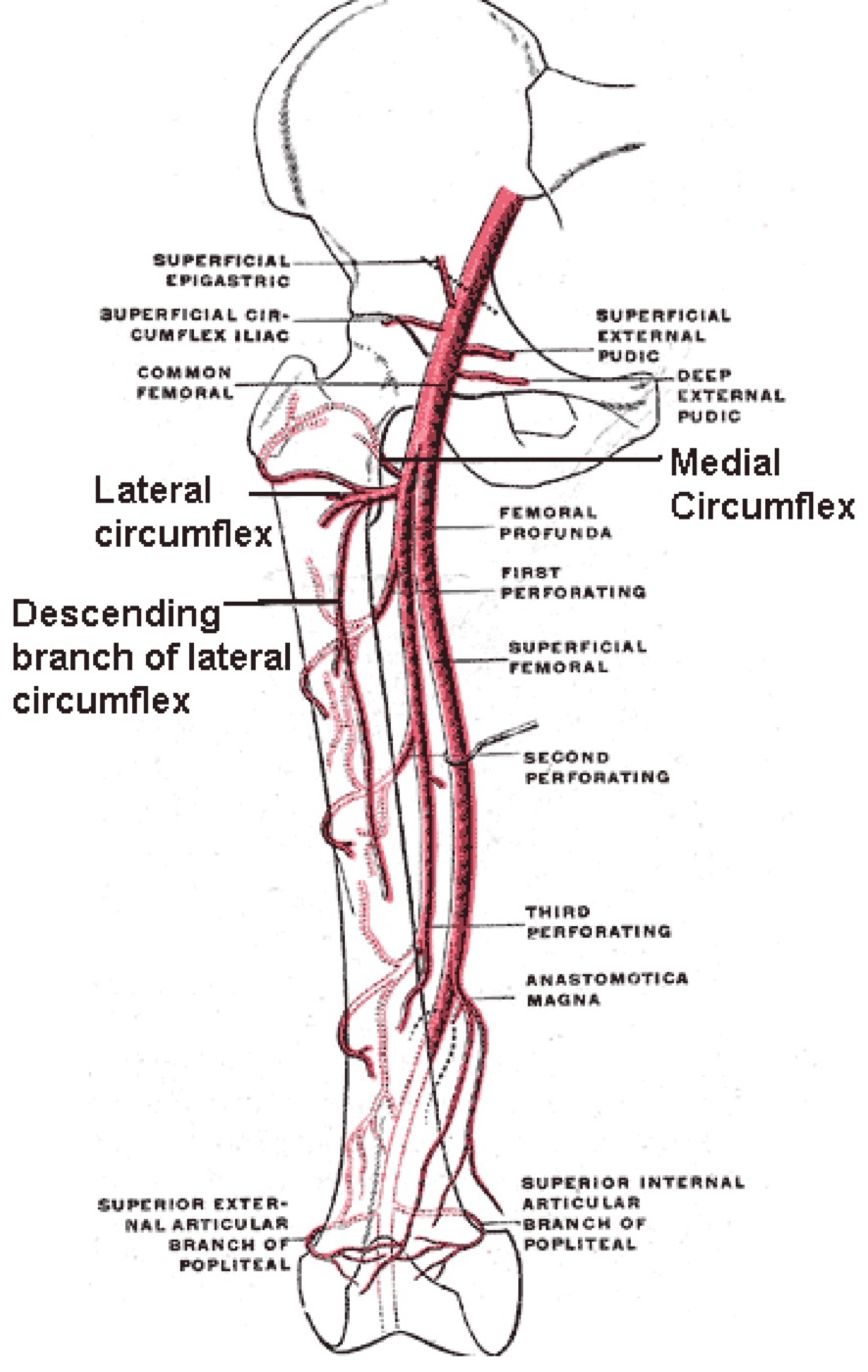
Figure 1. The femoral circumflex arteries: the medial femoral circumflex (dominant) gives the retinacular vessels that supply the femoral head. Diagram by Mikael Häggström after Gray’s Anatomy, public domain, via Wikimedia Commons.
The remaining anatomy fills in the clinical picture. The calcar femorale, a dense vertical plate of bone under the lesser trochanter, reinforces the posteroinferior neck and is the buttress an inferior screw should engage.[10] The neck-shaft angle averages about 128-130° (less is coxa vara, more is coxa valga) and anteversion is about 15-25°.[11] For defining “intracapsular” the key point is that the capsule reaches the intertrochanteric line anteriorly, but the lateral half of the neck is extracapsular posteriorly, which is the anatomical basis of the basicervical (extracapsular) distinction. The iliopsoas pull externally rotates the shaft of a displaced fracture, giving the classic deformity.[12]
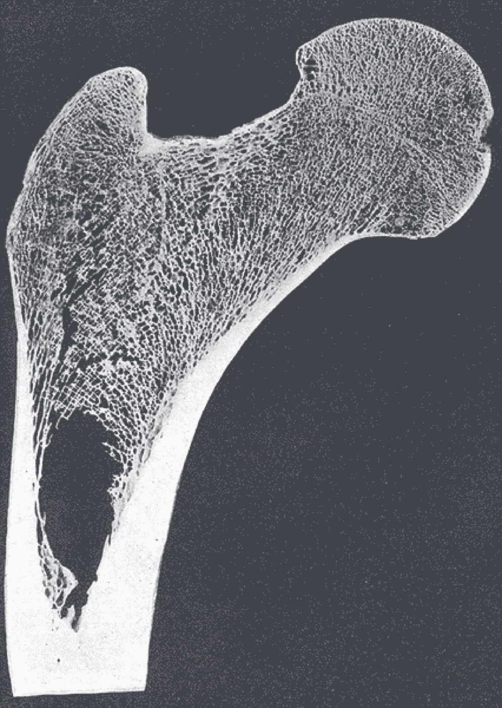
Figure 2. Frontal section of the proximal femur showing the head, neck, and the calcar femorale with its trabecular architecture. Henry Vandyke Carter, Gray’s Anatomy, public domain, via Wikimedia Commons.
Part III - Assessment and Imaging
The mechanism is a simple low-energy fall in about 90% of elderly patients, the force transmitted through the greater trochanter.[13] A displaced fracture gives the classic presentation of a shortened, externally rotated, painful limb, clinically indistinguishable from an extracapsular hip fracture, whereas an undisplaced fracture may show only painful movement.[14] Since 25-30% of older patients are cognitively impaired and surgery is urgent, the workup centres on medical optimisation rather than routine cardiac over-investigation, which only delays surgery, and preoperative skin traction is no longer recommended.[15]
Plain AP pelvis and lateral hip radiographs identify most fractures.[16] The pitfall to watch for is the occult (radiographically normal) fracture: when clinical suspicion persists with normal films, MRI is the recommended investigation, being the most sensitive, radiation-free, and able to show a haemarthrosis or a soft-tissue cause, with CT a reasonable alternative.[17]
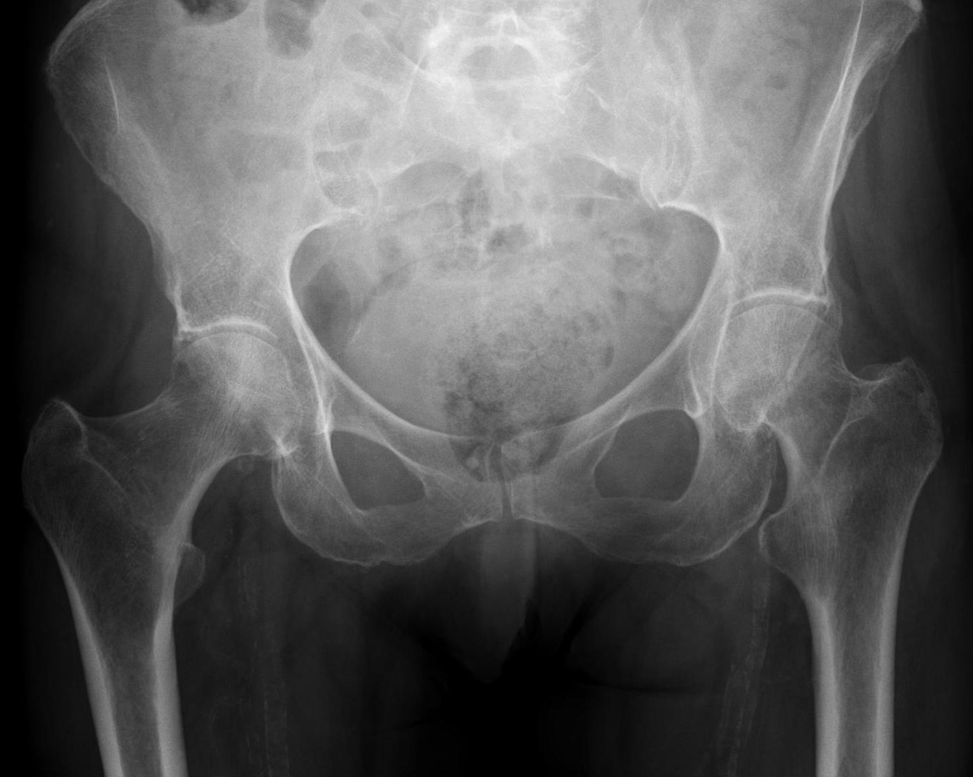
Figure 3. A displaced subcapital (medial) femoral neck fracture on AP radiograph. Hellerhoff, CC BY-SA 4.0, via Wikimedia Commons.
Part IV - Classification
The decisive distinction is simply displaced versus undisplaced (and intracapsular versus extracapsular), because that is what determines treatment and predicts the complications. The named subdivisions are less reliable than they look.[18] The Garden classification grades displacement on the AP film by the trabecular alignment: Garden I, an incomplete valgus-impacted fracture; Garden II, complete but undisplaced; Garden III, incompletely displaced (the head and neck still in contact, the retinacular attachments partly intact); and Garden IV, completely displaced.[19] In practice it is collapsed into nondisplaced/impacted (I-II) versus displaced (III-IV), the reliable and treatment-relevant division, since interobserver agreement across all four grades is poor at only about 22%.[20] The Pauwels classification grades the angle of the fracture line from the horizontal (type 1 under 30°, type 2 between 30° and 50°, type 3 over 50°). The more vertical the line, the greater the shear across it, so the Pauwels-3 (vertical) fracture is more prone to fixation failure and nonunion, a concept most relevant in younger, high-energy fractures.[21] The AO/OTA system codes the femoral neck as 31B (B1 subcapital/minimally displaced, B2 transcervical, B3 displaced subcapital).[22]
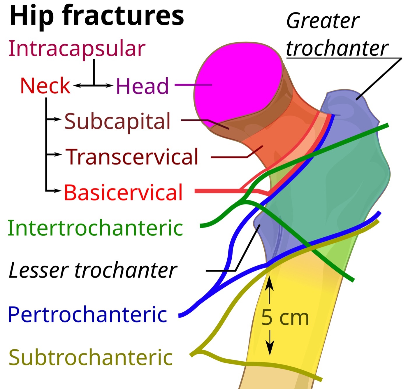
Figure 4. Classification of proximal femoral fractures by anatomic level: intracapsular (subcapital, transcervical, basicervical) versus extracapsular (intertrochanteric, subtrochanteric). Diagram by Mikael Häggström, CC0, via Wikimedia Commons.
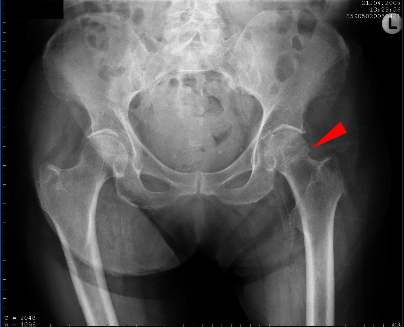
Figure 5. A nondisplaced medial femoral neck fracture on AP radiograph (arrow). Sjoehest, CC BY-SA 3.0, via Wikimedia Commons.
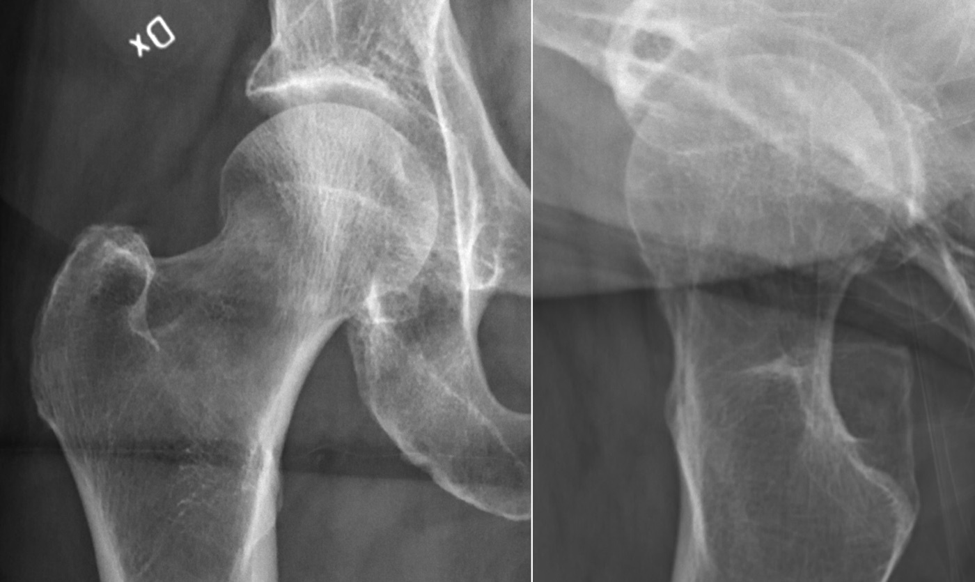
Figure 6. A subtle valgus-impacted femoral neck fracture (Garden I), with a sclerotic impaction line across the neck. Mikael Häggström, CC0, via Wikimedia Commons.
Part V - Treatment
5.1 The decision: keep the head or replace it
Every treatment choice follows from displacement, age, cognition, demand, and bone quality.[23] Undisplaced fractures are fixed at any age. Displaced fractures in younger patients are reduced and fixed to preserve the head. Displaced fractures in older patients are replaced. The genuinely contested zone is the “young old” (about 60-80 years), where practice varies widely. The modern trend, supported by the reoperation data, is to extend arthroplasty down to about 60 in the independent, cognitively intact patient.[24] Nonoperative treatment is reserved for the rare patient who cannot survive or tolerate surgery (a non-ambulator, terminal illness), since an undisplaced fracture managed nonoperatively displaces in roughly 19-46% of cases.[25]
5.2 Undisplaced fractures (Garden I-II): internal fixation
The undisplaced or valgus-impacted fracture is fixed in situ, most often with three cannulated screws (commonly an inverted triangle, the inferior screw buttressing the calcar) or a sliding hip screw (SHS).[26] Union exceeds 90%, with an overall nonunion of about 7% and late avascular necrosis of about 6%.[27] The randomised FAITH trial compared the sliding hip screw with cannulated screws and found no difference in the reoperation rate at 24 months (around 20-22% in both), with a slightly higher avascular necrosis after the sliding hip screw (about 9% versus 5%). Neither implant is clearly superior.[28] A newer locking plate (the Targon, and in current practice the Femoral Neck System) is an alternative, though without proven superiority.[29]
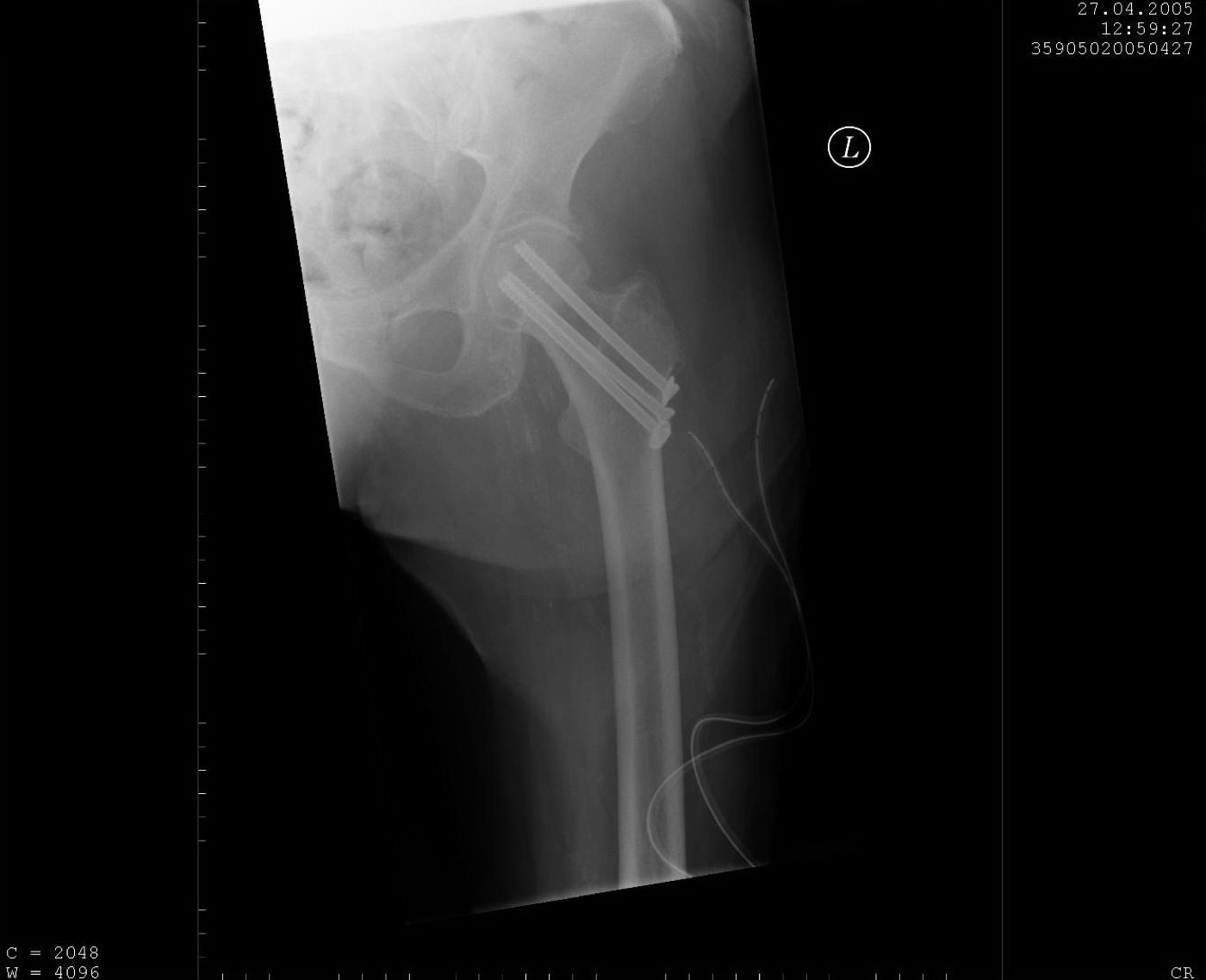
Figure 7. A femoral neck fracture fixed with cannulated screws. Sjoehest, CC BY-SA 3.0, via Wikimedia Commons.
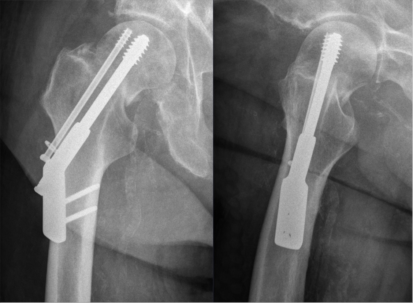
Figure 8. A dynamic (sliding) hip screw with an antirotation screw, two projections. Hellerhoff, CC BY-SA 4.0, via Wikimedia Commons.
5.3 Young patients with displaced fractures: reduce and fix urgently
In the patient under about 60, a displaced fracture is treated by prompt anatomical reduction (closed, or open through a Smith-Petersen approach) and internal fixation to preserve the head, accepting a high failure rate because arthroplasty in a young patient is undesirable.[30] The quality of reduction is paramount: avascular necrosis is lowest with an anatomical reduction, a slight valgus is more forgiving than varus (20° of varus carries about a 55% risk of failure), and the inferior screw should sit within a few millimetres of the calcar.[31] A vertical (Pauwels-3) fracture is better held by a fixed-angle device than by sliding screws, one series giving nonunion of about 8% versus 19%.[32] There is reasonable evidence that earlier reduction reduces avascular necrosis and nonunion in the young, so these fractures are treated as a priority. The honest caveat is that the evidence for routine emergency out-of-hours surgery, and for capsulotomy to decompress the haematoma, is actually weak.[33]
5.4 Displaced fractures in the older patient: arthroplasty
For the displaced fracture in the older patient, the weight of evidence strongly favours arthroplasty, because internal fixation of a displaced fracture has a reoperation rate of about 30-50% versus under 10% after replacement.[34] The choice is then between hemiarthroplasty and total hip arthroplasty (THA). The randomised and registry data show a consistent trade-off: THA gives better function and a lower reoperation/revision rate, while hemiarthroplasty gives a lower dislocation rate (the pooled figures are roughly: revision 9.6% hemi versus 5.4% THA; dislocation 2.5% hemi versus 7.9% THA).[35] So THA is chosen for the fit, independent, cognitively intact older patient (only about a quarter to a third of all hip-fracture patients), and modern hemiarthroplasty for the frailer, lower-demand majority. The landmark trial framing this question is the HEALTH trial of THA versus hemiarthroplasty.[36] A cemented stem is currently favoured because the registry data show a higher revision rate (largely from periprosthetic fracture) with uncemented stems, though caution with cement pressurisation is needed in the frail cardiac patient. A posterior approach raises the dislocation rate, so an anterior or lateral approach is preferred, and a bipolar head causes less acetabular erosion than a unipolar.[37]
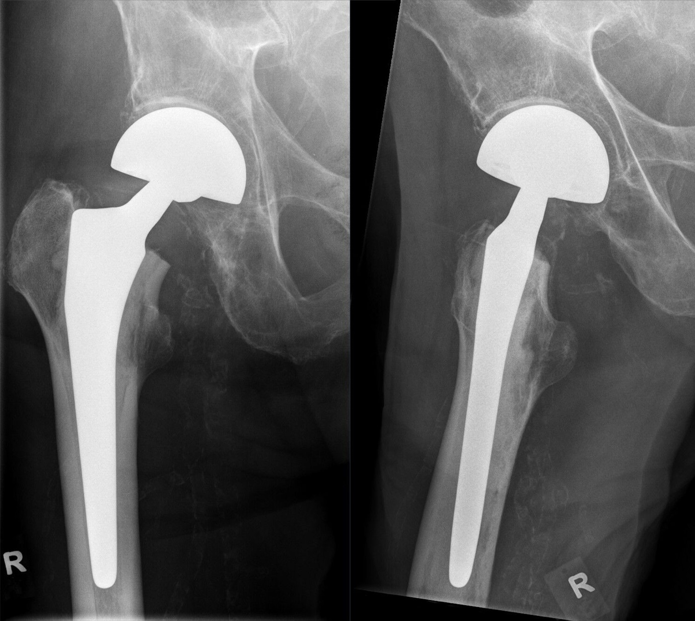
Figure 9. A bipolar hemiarthroplasty of the right hip, two projections. Hellerhoff, CC BY-SA 4.0, via Wikimedia Commons.
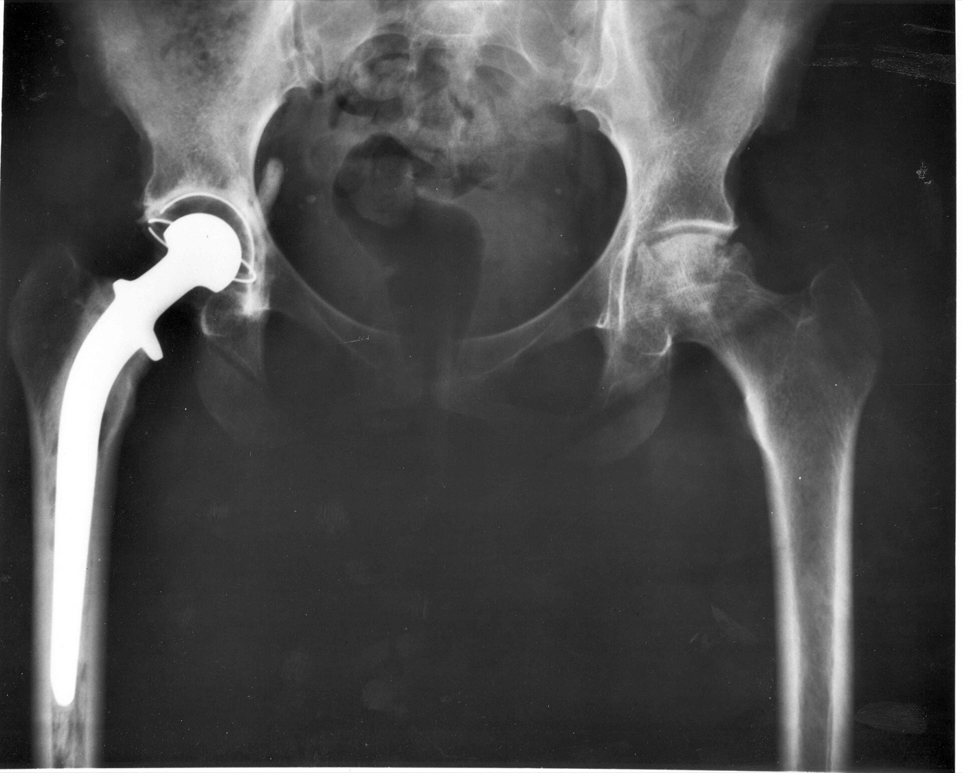
Figure 10. A total hip replacement of the right hip on AP radiograph. National Institutes of Health, public domain, via Wikimedia Commons.
5.5 Timing
Surgery is performed as soon as the patient is fit, within about 24 to 48 hours, on a planned trauma list, because large population studies link earlier surgery to lower mortality, morbidity, and length of stay. A short delay is justified only to correct a reversible medical problem.[38] Multidisciplinary co-management with care-of-the-elderly physicians, and peripheral nerve blocks for analgesia, improve outcomes.[39]
Part VI - Complications
The mortality of a hip fracture is sobering: about 15% in hospital and 30% at one year (four to five times the matched population), reaching 50% at one year in the cognitively impaired.[40] After fixation, the complications are those the anatomy predicts. Avascular necrosis affects about 16% of undisplaced and 27% of displaced fractures (lower in modern series), presents late, most often in the second year, and is treated by hip preservation early or arthroplasty once the head collapses.[41] Nonunion is uncommon after undisplaced fractures (about 7%) but reaches 25% or more after displaced fractures, and together with fixation failure drives the high reoperation rate. Salvage is usually conversion to arthroplasty, with head-preserving options (vascularised grafts, a valgus osteotomy) reserved for the young.[42]
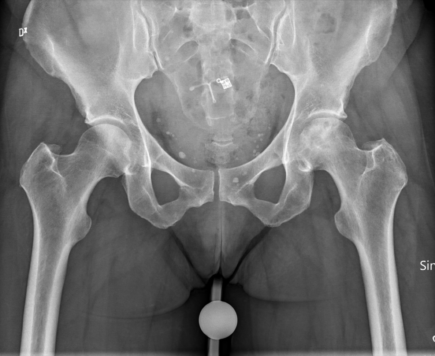
Figure 11. Avascular necrosis of the femoral head on radiograph. Mikael Häggström, CC0, via Wikimedia Commons.
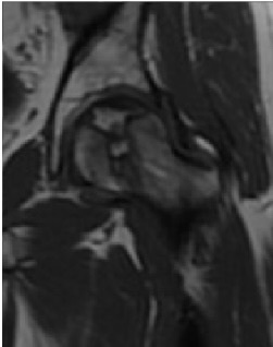
Figure 12. Avascular necrosis of the femoral head on T1-weighted MRI. Ruiz Santiago et al., CC BY 4.0, via Wikimedia Commons.
After arthroplasty the complications are dislocation (about 2-3% after hemiarthroplasty, higher after THA and with a posterior approach), infection (a major problem in the frail elderly, with a one-year mortality of 40-50% for a deep prosthetic joint infection), periprosthetic fracture (higher with uncemented stems), and, after hemiarthroplasty, acetabular erosion.[43] Across all of these patients the risk of venous thromboembolism is high, so mechanical and chemical prophylaxis (low-molecular-weight heparin or a newer oral anticoagulant plus a pneumatic device) is routine. Delirium is the commonest medical complication.[44]
Part VII - A Synthesis: How to Reason About the Femoral Neck Fracture
Reason from the blood supply outward. The neck is intracapsular, has no periosteum, and is fed by retinacular vessels off the medial femoral circumflex artery, so a displaced fracture both heals slowly and starves the head. That is why displacement is the pivotal fact. Read the film for one thing above all, displaced or undisplaced (Garden III-IV versus I-II), and confirm the occult fracture with MRI. Then apply the rule. An undisplaced fracture is fixed in situ at any age with screws or a sliding hip screw. A young patient with a displaced fracture has the head preserved by a prompt, anatomical reduction and fixation (a fixed-angle device for the vertical Pauwels-3 pattern). And a displaced fracture in the older patient is replaced, with a total hip for the fit and independent and a cemented hemiarthroplasty for the frail majority. Operate within a day or two on an optimised patient, co-manage the medicine, and prophylax against thrombosis. The headline numbers are grim and worth remembering: a one-year mortality near 30%, a reoperation rate near 40% if you fix a displaced fracture in an old person, and an avascular-necrosis rate that climbs with displacement. The art of the topic is matching the right hip to the right operation, which is almost always the same question asked twice: is it displaced, and is the patient young?
References
-
Rockwood & Green’s Fractures in Adults, pp.3619, 3649 (the intracapsular neck has no periosteum and heals by endosteal union alone; displaced fractures tear the retinacular vessels and risk AVN/nonunion; the decision rests on displacement, age/physiological status, and bone quality, with undisplaced fractures fixed and displaced fractures in the elderly replaced).
-
Rockwood & Green’s Fractures in Adults, pp.3602-3604 (hip fractures ~20% of the operative orthopaedic-trauma workload; intracapsular fractures 55-59% of hip fractures; lifetime risk 40-50% women / 13-22% men; projected rise 1.66 million in 1990 to 6.26 million by 2050, with a recent levelling/fall in some Western populations).
-
Rockwood & Green’s Fractures in Adults, pp.3602-3604 (hip fractures ~20% of the operative orthopaedic-trauma workload; intracapsular fractures 55-59% of hip fractures; lifetime risk 40-50% women / 13-22% men; projected rise 1.66 million in 1990 to 6.26 million by 2050, with a recent levelling/fall in some Western populations).
-
Rockwood & Green’s Fractures in Adults, pp.3602-3605 (bimodal, predominantly elderly women falling sideways onto the trochanter, ~3% under 60 and usually men from high energy; ~15% undisplaced and the rest displaced; osteoporosis the underlying cause, each 1-SD fall in femoral-neck BMD raising risk ~2.6×); Miller’s Review of Orthopaedics, pp.916-919 (geriatric fragility fracture vs the young high-energy proximal-femur injury).
-
Rockwood & Green’s Fractures in Adults, pp.3602-3605 (bimodal, predominantly elderly women falling sideways onto the trochanter, ~3% under 60 and usually men from high energy; ~15% undisplaced and the rest displaced; osteoporosis the underlying cause, each 1-SD fall in femoral-neck BMD raising risk ~2.6×); Miller’s Review of Orthopaedics, pp.916-919 (geriatric fragility fracture vs the young high-energy proximal-femur injury).
-
Rockwood & Green’s Fractures in Adults, pp.3617-3619 (no periosteum, endosteal union only; three blood-supply sources, the retinacular vessels the most important; the deep branch of the MFCA giving the superior retinacular/ascending vessels that supply the weight-bearing head, ~82% MFCA vs ~18% LFCA, the foveal artery minor; a displaced subcapital fracture tears the retinacular vessels, risking AVN and nonunion). AO Principles of Fracture Management, p.795 names the medial femoral circumflex supply but a figure legend there erroneously attributes dominance to the lateral femoral circumflex artery; the medial femoral circumflex artery is dominant, as in Rockwood and standard teaching.
-
Rockwood & Green’s Fractures in Adults, pp.3617-3619 (no periosteum, endosteal union only; three blood-supply sources, the retinacular vessels the most important; the deep branch of the MFCA giving the superior retinacular/ascending vessels that supply the weight-bearing head, ~82% MFCA vs ~18% LFCA, the foveal artery minor; a displaced subcapital fracture tears the retinacular vessels, risking AVN and nonunion). AO Principles of Fracture Management, p.795 names the medial femoral circumflex supply but a figure legend there erroneously attributes dominance to the lateral femoral circumflex artery; the medial femoral circumflex artery is dominant, as in Rockwood and standard teaching.
-
Rockwood & Green’s Fractures in Adults, pp.3617-3619 (no periosteum, endosteal union only; three blood-supply sources, the retinacular vessels the most important; the deep branch of the MFCA giving the superior retinacular/ascending vessels that supply the weight-bearing head, ~82% MFCA vs ~18% LFCA, the foveal artery minor; a displaced subcapital fracture tears the retinacular vessels, risking AVN and nonunion). AO Principles of Fracture Management, p.795 names the medial femoral circumflex supply but a figure legend there erroneously attributes dominance to the lateral femoral circumflex artery; the medial femoral circumflex artery is dominant, as in Rockwood and standard teaching.
-
Rockwood & Green’s Fractures in Adults, pp.3617-3619 (no periosteum, endosteal union only; three blood-supply sources, the retinacular vessels the most important; the deep branch of the MFCA giving the superior retinacular/ascending vessels that supply the weight-bearing head, ~82% MFCA vs ~18% LFCA, the foveal artery minor; a displaced subcapital fracture tears the retinacular vessels, risking AVN and nonunion). AO Principles of Fracture Management, p.795 names the medial femoral circumflex supply but a figure legend there erroneously attributes dominance to the lateral femoral circumflex artery; the medial femoral circumflex artery is dominant, as in Rockwood and standard teaching.
-
Rockwood & Green’s Fractures in Adults, pp.3616-3620 (the calcar femorale reinforcing the posteroinferior neck; neck-shaft angle ~128-130°, anteversion ~15-25°; the capsule reaching the intertrochanteric line anteriorly but the lateral half of the neck extracapsular posteriorly, the basis of the basicervical distinction; iliopsoas externally rotating the shaft).
-
Rockwood & Green’s Fractures in Adults, pp.3616-3620 (the calcar femorale reinforcing the posteroinferior neck; neck-shaft angle ~128-130°, anteversion ~15-25°; the capsule reaching the intertrochanteric line anteriorly but the lateral half of the neck extracapsular posteriorly, the basis of the basicervical distinction; iliopsoas externally rotating the shaft).
-
Rockwood & Green’s Fractures in Adults, pp.3616-3620 (the calcar femorale reinforcing the posteroinferior neck; neck-shaft angle ~128-130°, anteversion ~15-25°; the capsule reaching the intertrochanteric line anteriorly but the lateral half of the neck extracapsular posteriorly, the basis of the basicervical distinction; iliopsoas externally rotating the shaft).
-
Rockwood & Green’s Fractures in Adults, pp.3605-3610 (90% from a simple fall through the greater trochanter; displaced fracture shortened/externally rotated/painful, indistinguishable from extracapsular; 25-30% cognitively impaired; medical optimisation but not routine cardiac over-investigation which delays surgery; preoperative skin traction no longer recommended).
-
Rockwood & Green’s Fractures in Adults, pp.3605-3610 (90% from a simple fall through the greater trochanter; displaced fracture shortened/externally rotated/painful, indistinguishable from extracapsular; 25-30% cognitively impaired; medical optimisation but not routine cardiac over-investigation which delays surgery; preoperative skin traction no longer recommended).
-
Rockwood & Green’s Fractures in Adults, pp.3605-3610 (90% from a simple fall through the greater trochanter; displaced fracture shortened/externally rotated/painful, indistinguishable from extracapsular; 25-30% cognitively impaired; medical optimisation but not routine cardiac over-investigation which delays surgery; preoperative skin traction no longer recommended).
-
Rockwood & Green’s Fractures in Adults, pp.3606-3608 (AP pelvis and lateral hip identify most fractures; for the occult fracture with normal radiographs, MRI is the current recommended modality, the most sensitive and radiation-free, showing haemarthrosis and soft-tissue causes, with CT an alternative); Miller’s Review of Orthopaedics, p.919 (MRI or bone scan for the occult/stress fracture, MRI most sensitive especially within 24 hours).
-
Rockwood & Green’s Fractures in Adults, pp.3606-3608 (AP pelvis and lateral hip identify most fractures; for the occult fracture with normal radiographs, MRI is the current recommended modality, the most sensitive and radiation-free, showing haemarthrosis and soft-tissue causes, with CT an alternative); Miller’s Review of Orthopaedics, p.919 (MRI or bone scan for the occult/stress fracture, MRI most sensitive especially within 24 hours).
-
Rockwood & Green’s Fractures in Adults, pp.3611-3614 (the decisive distinction is displaced vs undisplaced and intracapsular vs extracapsular; Garden I incomplete valgus-impacted, II complete undisplaced, III incompletely displaced with retinacular attachments partly intact, IV completely displaced; collapsed in practice to nondisplaced I-II vs displaced III-IV, four-grade interobserver agreement only ~22%); Miller’s Review of Orthopaedics, pp.916-917 (Garden I/II nondisplaced vs III/IV displaced).
-
Rockwood & Green’s Fractures in Adults, pp.3611-3614 (the decisive distinction is displaced vs undisplaced and intracapsular vs extracapsular; Garden I incomplete valgus-impacted, II complete undisplaced, III incompletely displaced with retinacular attachments partly intact, IV completely displaced; collapsed in practice to nondisplaced I-II vs displaced III-IV, four-grade interobserver agreement only ~22%); Miller’s Review of Orthopaedics, pp.916-917 (Garden I/II nondisplaced vs III/IV displaced).
-
Rockwood & Green’s Fractures in Adults, pp.3611-3614 (the decisive distinction is displaced vs undisplaced and intracapsular vs extracapsular; Garden I incomplete valgus-impacted, II complete undisplaced, III incompletely displaced with retinacular attachments partly intact, IV completely displaced; collapsed in practice to nondisplaced I-II vs displaced III-IV, four-grade interobserver agreement only ~22%); Miller’s Review of Orthopaedics, pp.916-917 (Garden I/II nondisplaced vs III/IV displaced).
-
Rockwood & Green’s Fractures in Adults, pp.3612-3613 (Pauwels type 1 <30°, type 2 30-50°, type 3 >50°, the vertical Pauwels-3 with more shear and higher failure/nonunion, most relevant in younger high-energy fractures; AO/OTA 31B with B1 subcapital, B2 transcervical, B3 displaced subcapital); AO Principles of Fracture Management, pp.794-795, 802 (AO/OTA 31B; the vertical-shear concept).
-
Rockwood & Green’s Fractures in Adults, pp.3612-3613 (Pauwels type 1 <30°, type 2 30-50°, type 3 >50°, the vertical Pauwels-3 with more shear and higher failure/nonunion, most relevant in younger high-energy fractures; AO/OTA 31B with B1 subcapital, B2 transcervical, B3 displaced subcapital); AO Principles of Fracture Management, pp.794-795, 802 (AO/OTA 31B; the vertical-shear concept).
-
Rockwood & Green’s Fractures in Adults, pp.3620-3621, 3649 (decision by displacement, age, cognition, demand, bone quality; undisplaced fixed at any age, young displaced reduced and fixed, older displaced replaced; the contested “young old” 60-80 group with the trend to arthroplasty from ~60; nonoperative treatment only for the non-ambulator or prohibitive surgical risk, with 19-46% displacement if an undisplaced fracture is treated nonoperatively); Miller’s Review of Orthopaedics, pp.916-919 (the geriatric vs young-adult algorithm).
-
Rockwood & Green’s Fractures in Adults, pp.3620-3621, 3649 (decision by displacement, age, cognition, demand, bone quality; undisplaced fixed at any age, young displaced reduced and fixed, older displaced replaced; the contested “young old” 60-80 group with the trend to arthroplasty from ~60; nonoperative treatment only for the non-ambulator or prohibitive surgical risk, with 19-46% displacement if an undisplaced fracture is treated nonoperatively); Miller’s Review of Orthopaedics, pp.916-919 (the geriatric vs young-adult algorithm).
-
Rockwood & Green’s Fractures in Adults, pp.3620-3621, 3649 (decision by displacement, age, cognition, demand, bone quality; undisplaced fixed at any age, young displaced reduced and fixed, older displaced replaced; the contested “young old” 60-80 group with the trend to arthroplasty from ~60; nonoperative treatment only for the non-ambulator or prohibitive surgical risk, with 19-46% displacement if an undisplaced fracture is treated nonoperatively); Miller’s Review of Orthopaedics, pp.916-919 (the geriatric vs young-adult algorithm).
-
Rockwood & Green’s Fractures in Adults, pp.3622-3625, 3666 (undisplaced fractures fixed in situ with three cannulated screws, often an inverted triangle with the inferior screw on the calcar, or a sliding hip screw; union >90%, nonunion ~7%, late AVN ~5.8%; FAITH trial no difference in 24-month reoperation [~20-22% both], AVN slightly higher with the SHS ~9% vs ~5%; the Targon locking plate an alternative without proven superiority). The currently marketed Femoral Neck System (FNS) is the modern locking-plate device; the mined Rockwood chapter predates that trade name and discusses the Targon plate, so “FNS” is noted as standard current usage rather than cited to the source.
-
Rockwood & Green’s Fractures in Adults, pp.3622-3625, 3666 (undisplaced fractures fixed in situ with three cannulated screws, often an inverted triangle with the inferior screw on the calcar, or a sliding hip screw; union >90%, nonunion ~7%, late AVN ~5.8%; FAITH trial no difference in 24-month reoperation [~20-22% both], AVN slightly higher with the SHS ~9% vs ~5%; the Targon locking plate an alternative without proven superiority). The currently marketed Femoral Neck System (FNS) is the modern locking-plate device; the mined Rockwood chapter predates that trade name and discusses the Targon plate, so “FNS” is noted as standard current usage rather than cited to the source.
-
Rockwood & Green’s Fractures in Adults, pp.3622-3625, 3666 (undisplaced fractures fixed in situ with three cannulated screws, often an inverted triangle with the inferior screw on the calcar, or a sliding hip screw; union >90%, nonunion ~7%, late AVN ~5.8%; FAITH trial no difference in 24-month reoperation [~20-22% both], AVN slightly higher with the SHS ~9% vs ~5%; the Targon locking plate an alternative without proven superiority). The currently marketed Femoral Neck System (FNS) is the modern locking-plate device; the mined Rockwood chapter predates that trade name and discusses the Targon plate, so “FNS” is noted as standard current usage rather than cited to the source.
-
Rockwood & Green’s Fractures in Adults, pp.3622-3625, 3666 (undisplaced fractures fixed in situ with three cannulated screws, often an inverted triangle with the inferior screw on the calcar, or a sliding hip screw; union >90%, nonunion ~7%, late AVN ~5.8%; FAITH trial no difference in 24-month reoperation [~20-22% both], AVN slightly higher with the SHS ~9% vs ~5%; the Targon locking plate an alternative without proven superiority). The currently marketed Femoral Neck System (FNS) is the modern locking-plate device; the mined Rockwood chapter predates that trade name and discusses the Targon plate, so “FNS” is noted as standard current usage rather than cited to the source.
-
Rockwood & Green’s Fractures in Adults, pp.3625-3631 (young displaced fractures reduced, closed or open via Smith-Petersen, and fixed to preserve the head; reduction quality paramount, AVN lowest with anatomical reduction, slight valgus preferred over varus, 20° varus ~55% failure, inferior screw within a few mm of the calcar; Pauwels-3 better held by a fixed-angle device, nonunion ~8% vs ~19%; some evidence earlier reduction lowers AVN/nonunion, but the evidence for routine emergency out-of-hours surgery and for capsulotomy/aspiration to decompress the haematoma is weak/not beneficial); Miller’s Review of Orthopaedics, pp.918-919 (young displaced proximal femur fracture treated urgently with anatomical reduction and fixation).
-
Rockwood & Green’s Fractures in Adults, pp.3625-3631 (young displaced fractures reduced, closed or open via Smith-Petersen, and fixed to preserve the head; reduction quality paramount, AVN lowest with anatomical reduction, slight valgus preferred over varus, 20° varus ~55% failure, inferior screw within a few mm of the calcar; Pauwels-3 better held by a fixed-angle device, nonunion ~8% vs ~19%; some evidence earlier reduction lowers AVN/nonunion, but the evidence for routine emergency out-of-hours surgery and for capsulotomy/aspiration to decompress the haematoma is weak/not beneficial); Miller’s Review of Orthopaedics, pp.918-919 (young displaced proximal femur fracture treated urgently with anatomical reduction and fixation).
-
Rockwood & Green’s Fractures in Adults, pp.3625-3631 (young displaced fractures reduced, closed or open via Smith-Petersen, and fixed to preserve the head; reduction quality paramount, AVN lowest with anatomical reduction, slight valgus preferred over varus, 20° varus ~55% failure, inferior screw within a few mm of the calcar; Pauwels-3 better held by a fixed-angle device, nonunion ~8% vs ~19%; some evidence earlier reduction lowers AVN/nonunion, but the evidence for routine emergency out-of-hours surgery and for capsulotomy/aspiration to decompress the haematoma is weak/not beneficial); Miller’s Review of Orthopaedics, pp.918-919 (young displaced proximal femur fracture treated urgently with anatomical reduction and fixation).
-
Rockwood & Green’s Fractures in Adults, pp.3625-3631 (young displaced fractures reduced, closed or open via Smith-Petersen, and fixed to preserve the head; reduction quality paramount, AVN lowest with anatomical reduction, slight valgus preferred over varus, 20° varus ~55% failure, inferior screw within a few mm of the calcar; Pauwels-3 better held by a fixed-angle device, nonunion ~8% vs ~19%; some evidence earlier reduction lowers AVN/nonunion, but the evidence for routine emergency out-of-hours surgery and for capsulotomy/aspiration to decompress the haematoma is weak/not beneficial); Miller’s Review of Orthopaedics, pp.918-919 (young displaced proximal femur fracture treated urgently with anatomical reduction and fixation).
-
Rockwood & Green’s Fractures in Adults, pp.3640-3651 (displaced fractures in the older patient best treated by arthroplasty, internal fixation reoperation ~30-50% vs <10% after replacement; THA better function and lower revision, hemiarthroplasty lower dislocation; pooled revision 9.6% hemi vs 5.4% THA, dislocation 2.5% hemi vs 7.9% THA, 1-year mortality 20% fixation / 23% hemi / 12.4% THA; THA for the fit independent cognitively intact patient [~25-30% of the population], hemiarthroplasty for the lower-demand majority). The landmark THA-vs-hemiarthroplasty randomised trial is the HEALTH trial; it is not named in the mined Rockwood/Miller extracts, which cover the question through other RCTs (Baker, Blomfeldt, van den Bekerom) and meta-analyses, so HEALTH is cited here as standard current knowledge.
-
Rockwood & Green’s Fractures in Adults, pp.3640-3651 (displaced fractures in the older patient best treated by arthroplasty, internal fixation reoperation ~30-50% vs <10% after replacement; THA better function and lower revision, hemiarthroplasty lower dislocation; pooled revision 9.6% hemi vs 5.4% THA, dislocation 2.5% hemi vs 7.9% THA, 1-year mortality 20% fixation / 23% hemi / 12.4% THA; THA for the fit independent cognitively intact patient [~25-30% of the population], hemiarthroplasty for the lower-demand majority). The landmark THA-vs-hemiarthroplasty randomised trial is the HEALTH trial; it is not named in the mined Rockwood/Miller extracts, which cover the question through other RCTs (Baker, Blomfeldt, van den Bekerom) and meta-analyses, so HEALTH is cited here as standard current knowledge.
-
Rockwood & Green’s Fractures in Adults, pp.3640-3651 (displaced fractures in the older patient best treated by arthroplasty, internal fixation reoperation ~30-50% vs <10% after replacement; THA better function and lower revision, hemiarthroplasty lower dislocation; pooled revision 9.6% hemi vs 5.4% THA, dislocation 2.5% hemi vs 7.9% THA, 1-year mortality 20% fixation / 23% hemi / 12.4% THA; THA for the fit independent cognitively intact patient [~25-30% of the population], hemiarthroplasty for the lower-demand majority). The landmark THA-vs-hemiarthroplasty randomised trial is the HEALTH trial; it is not named in the mined Rockwood/Miller extracts, which cover the question through other RCTs (Baker, Blomfeldt, van den Bekerom) and meta-analyses, so HEALTH is cited here as standard current knowledge.
-
Rockwood & Green’s Fractures in Adults, pp.3634-3644, 3669 (a cemented stem currently favoured because uncemented stems show higher registry revision from periprosthetic fracture, with caution on cement pressurisation in the frail cardiac patient; a posterior approach raises dislocation so anterior/lateral preferred; bipolar heads cause less acetabular erosion than unipolar).
-
Rockwood & Green’s Fractures in Adults, pp.3653-3654, 3663 (surgery within ~24-48 hours on a planned list, large population studies linking earlier surgery to lower mortality/morbidity/length of stay, a short delay only to optimise a reversible problem; multidisciplinary co-management and peripheral nerve blocks improving outcomes); Miller’s Review of Orthopaedics, p.916 (geriatric hip fractures fixed within 48 hours).
-
Rockwood & Green’s Fractures in Adults, pp.3653-3654, 3663 (surgery within ~24-48 hours on a planned list, large population studies linking earlier surgery to lower mortality/morbidity/length of stay, a short delay only to optimise a reversible problem; multidisciplinary co-management and peripheral nerve blocks improving outcomes); Miller’s Review of Orthopaedics, p.916 (geriatric hip fractures fixed within 48 hours).
-
Rockwood & Green’s Fractures in Adults, pp.3653, 3655-3662 (mortality ~15% in hospital and ~30% at 1 year, 4-5× the matched population, 50% at 1 year with cognitive impairment; AVN ~16% undisplaced / ~27% displaced, presenting late in the second year, treated by hip preservation early or arthroplasty after collapse; nonunion ~7% undisplaced but ≥25% displaced, driving reoperation, salvaged usually by arthroplasty with head-preserving options for the young); Miller’s Review of Orthopaedics, pp.916-919 (AVN 10-40%, nonunion 10-30%, fixation failure up to 30%, 1-year mortality ~30%).
-
Rockwood & Green’s Fractures in Adults, pp.3653, 3655-3662 (mortality ~15% in hospital and ~30% at 1 year, 4-5× the matched population, 50% at 1 year with cognitive impairment; AVN ~16% undisplaced / ~27% displaced, presenting late in the second year, treated by hip preservation early or arthroplasty after collapse; nonunion ~7% undisplaced but ≥25% displaced, driving reoperation, salvaged usually by arthroplasty with head-preserving options for the young); Miller’s Review of Orthopaedics, pp.916-919 (AVN 10-40%, nonunion 10-30%, fixation failure up to 30%, 1-year mortality ~30%).
-
Rockwood & Green’s Fractures in Adults, pp.3653, 3655-3662 (mortality ~15% in hospital and ~30% at 1 year, 4-5× the matched population, 50% at 1 year with cognitive impairment; AVN ~16% undisplaced / ~27% displaced, presenting late in the second year, treated by hip preservation early or arthroplasty after collapse; nonunion ~7% undisplaced but ≥25% displaced, driving reoperation, salvaged usually by arthroplasty with head-preserving options for the young); Miller’s Review of Orthopaedics, pp.916-919 (AVN 10-40%, nonunion 10-30%, fixation failure up to 30%, 1-year mortality ~30%).
-
Rockwood & Green’s Fractures in Adults, pp.3654-3655, 3661-3665 (dislocation ~2-3% after hemiarthroplasty and higher after THA/posterior approach; deep prosthetic joint infection a major problem with a 40-50% 1-year mortality; periprosthetic fracture higher with uncemented stems; acetabular erosion after hemiarthroplasty; high VTE risk managed by LMWH or a newer oral anticoagulant plus a mechanical device; delirium the commonest medical complication).
-
Rockwood & Green’s Fractures in Adults, pp.3654-3655, 3661-3665 (dislocation ~2-3% after hemiarthroplasty and higher after THA/posterior approach; deep prosthetic joint infection a major problem with a 40-50% 1-year mortality; periprosthetic fracture higher with uncemented stems; acetabular erosion after hemiarthroplasty; high VTE risk managed by LMWH or a newer oral anticoagulant plus a mechanical device; delirium the commonest medical complication).
-
Rockwood & Green’s Fractures in Adults, pp.3617-3619.
-
Rockwood & Green’s Fractures in Adults, pp.3611-3614.
-
Rockwood & Green’s Fractures in Adults, p.3611.
-
Rockwood & Green’s Fractures in Adults, pp.3612-3613.
-
Rockwood & Green’s Fractures in Adults, pp.3607-3608.
-
Rockwood & Green’s Fractures in Adults, pp.3620, 3649; Miller’s Review of Orthopaedics, pp.916-919.
-
Rockwood & Green’s Fractures in Adults, pp.3622-3624, 3666.
-
Rockwood & Green’s Fractures in Adults, pp.3625-3631.
-
Rockwood & Green’s Fractures in Adults, pp.3640-3651.
-
Rockwood & Green’s Fractures in Adults, pp.3638, 3644.
-
Rockwood & Green’s Fractures in Adults, pp.3653-3654.
-
Rockwood & Green’s Fractures in Adults, p.3653.
-
Rockwood & Green’s Fractures in Adults, pp.3655-3662.
-
Rockwood & Green’s Fractures in Adults, pp.3654-3655, 3661-3665.