Contents
- Orientation: A Joint That Punishes Mistakes with Stiffness
- Part I - Distal Humerus Fractures
- Part II - Olecranon Fractures
- Part III - The Tension-Band (Weber / AO) Osteosynthesis
- Part IV - Radial Head and Neck Fractures
- Part V - Coronoid Fractures and Elbow Dislocation
- Part VI - The Terrible Triad
- Part VII - Monteggia and Transolecranon Fracture-Dislocations
- Part VIII - A Synthesis: How to Reason Around the Elbow
- References
Orientation: A Joint That Punishes Mistakes with Stiffness
The elbow is a highly congruent, three-articulation joint (the hinge-like ulnohumeral, the rotatory radiocapitellar, and the proximal radioulnar joints) whose stability comes in roughly equal measure from its bony architecture and from its ligaments.[1] That congruity is what makes it powerful, and also what makes it unforgiving. More than any other joint, the injured elbow tends to become stiff, so the single thread running through every injury in this chapter is the need to fix the fracture securely enough to permit early motion.[2] The konspekt names two things, “elbow fractures” and the “Weber tension-band,” and the pairing is apt. The tension-band osteosynthesis of the olecranon is the cleanest illustration in the body of converting a distracting muscular force into compression across a fracture, so it earns its own section here.
The functional arc to aim for is 30° to 130° of flexion and about 50° each of pronation and supination; an elbow kept within that arc is functionally useful even if not normal.[3] The stabilisers worth fixing in mind from the outset are the lateral collateral ligament complex (its lateral ulnar collateral ligament, LUCL, the key restraint to posterolateral rotatory instability), the anterior bundle of the medial collateral ligament (the key valgus restraint), the coronoid (the anterior bony buttress), and the radial head (a secondary valgus and a primary axial stabiliser).[4]
Part I - Distal Humerus Fractures
1.1 Anatomy: two columns and a tie-arch
The distal humerus is best understood as a triangle: two diverging columns (medial and lateral) bridged by the intercalary articular segment (the trochlea medially and the capitellum laterally), the whole behaving architecturally as a tie-arch.[5] The olecranon and coronoid fossae hollow out the centre to receive the ulna in extension and flexion, so a screw placed through a fossa blocks motion.[6] The articular surface sits in slight valgus (the 10-17° carrying angle) and is internally rotated and anteriorly translated relative to the shaft. These relationships must be restored.[7] The ulnar nerve runs behind the medial epicondyle in the cubital tunnel and is the nerve most at risk; the radial nerve is endangered by long, proximally-extending lateral plates.[8]
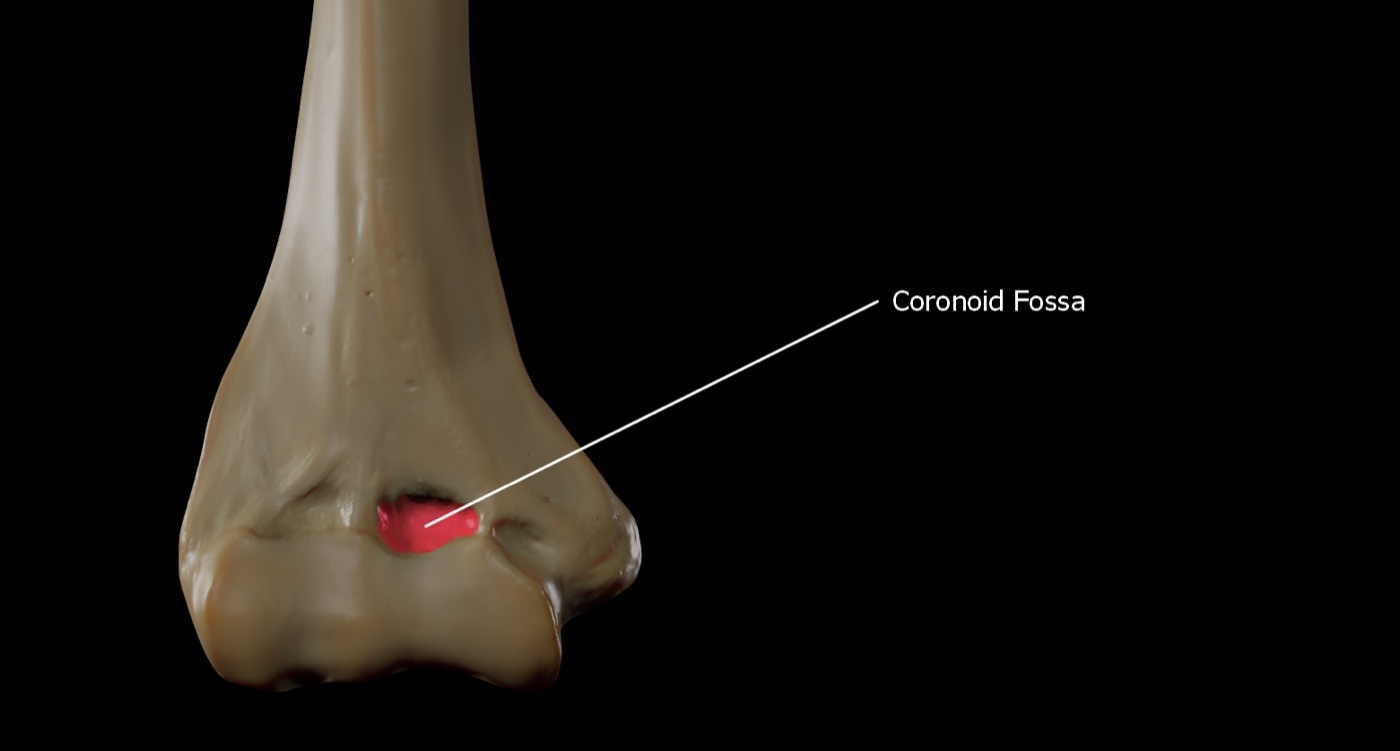
Figure 1. The distal humerus: the medial and lateral columns bridged by the trochlea and capitellum (the coronoid fossa highlighted). Source: Doctor Jana, via Wikimedia Commons, CC BY-SA 4.0.
1.2 Classification
The working scheme is the AO/OTA classification (region 13): type A extra-articular (epicondylar or transcolumn), type B partial articular (a single column, B1 lateral / B2 medial, or B3 a coronal articular shear), and type C complete articular / bicolumnar (C1 simple, C2 with metaphyseal comminution, C3 with both articular and metaphyseal comminution).[9] The older Milch classification of single-column fractures turns on whether the lateral trochlear ridge stays with the shaft (Milch I, stable) or goes with the fracture (Milch II, unstable).[10] The coronal shear (capitellar) fractures carry their own eponymous lineage: the Hahn-Steinthal fracture (a large bony fragment, “type I”), the Kocher-Lorenz fracture (a thin cartilage shell, “type II”), with type III (comminuted) added by Bryan and Morrey and type IV (extension into the lateral trochlea, the “double-arc sign”) added by McKee.[11]
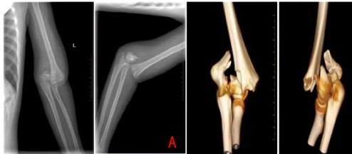
Figure 2. An intercondylar (bicolumnar, AO type-C) distal humerus fracture on radiograph and 3D CT. From Wang et al. (2026), Frontiers in Pediatrics, PMC13006493, CC BY 4.0.
1.3 Treatment
For the frail, low-demand elderly patient who cannot tolerate surgery, the historical non-operative “bag of bones” method (Eastwood; named by Evans) accepts imperfect reduction in a collar-and-cuff, trading anatomy for an acceptable functional arc.[12] For everyone else the standard is open reduction and internal fixation, resting on three principles: anatomical reduction of the articular surface, stable bicolumnar fixation, and early motion.[13] The decisive exposure question is access to the joint surface. The olecranon (chevron) osteotomy through the bare area gives the best articular view and is used for complex type-C fractures, whereas triceps-sparing (paratricipital) approaches preserve the extensor mechanism and are preferred when total elbow arthroplasty is a possible bail-out (an osteotomy must not be made if arthroplasty might be needed).[14]
The bicolumnar fixation is by two plates, and their orientation is a long-running debate: parallel plating (both plates on the medial and lateral columns, with long articular screws crossing to the opposite column, the Sanchez-Sotelo/O’Driscoll principle) versus orthogonal (90-90) plating (a medial plate and a posterolateral plate). The biomechanical studies conflict and no clinical superiority of either has been shown; one-third tubular plates are too weak and must not form the primary construct.[15] The handling of the ulnar nerve (in-situ decompression versus anterior transposition) is similarly unsettled.[16]
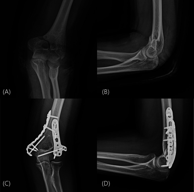
Figure 3. A distal humerus fracture before and after dual-plate (orthogonal) fixation with medial and posterolateral plates. From Lee et al. (2026), J Orthop Traumatol, PMC13287183, CC BY 4.0.
For the elderly patient with an osteoporotic, comminuted, unreconstructable articular fracture, total elbow arthroplasty (TEA) with a linked semiconstrained implant is the alternative. A randomised trial found it gave better outcomes than ORIF for displaced intra-articular fractures in the elderly.[17] Its price is a lifelong lifting restriction (about 5 kg / a few pounds) and the prospect of eventual loosening with a difficult revision, so it is reserved for the low-demand patient.[18] The chief complications of distal humerus fractures are stiffness (the commonest), heterotopic ossification, ulnar neuropathy (present in about a quarter at injury), and supracondylar nonunion.[19]
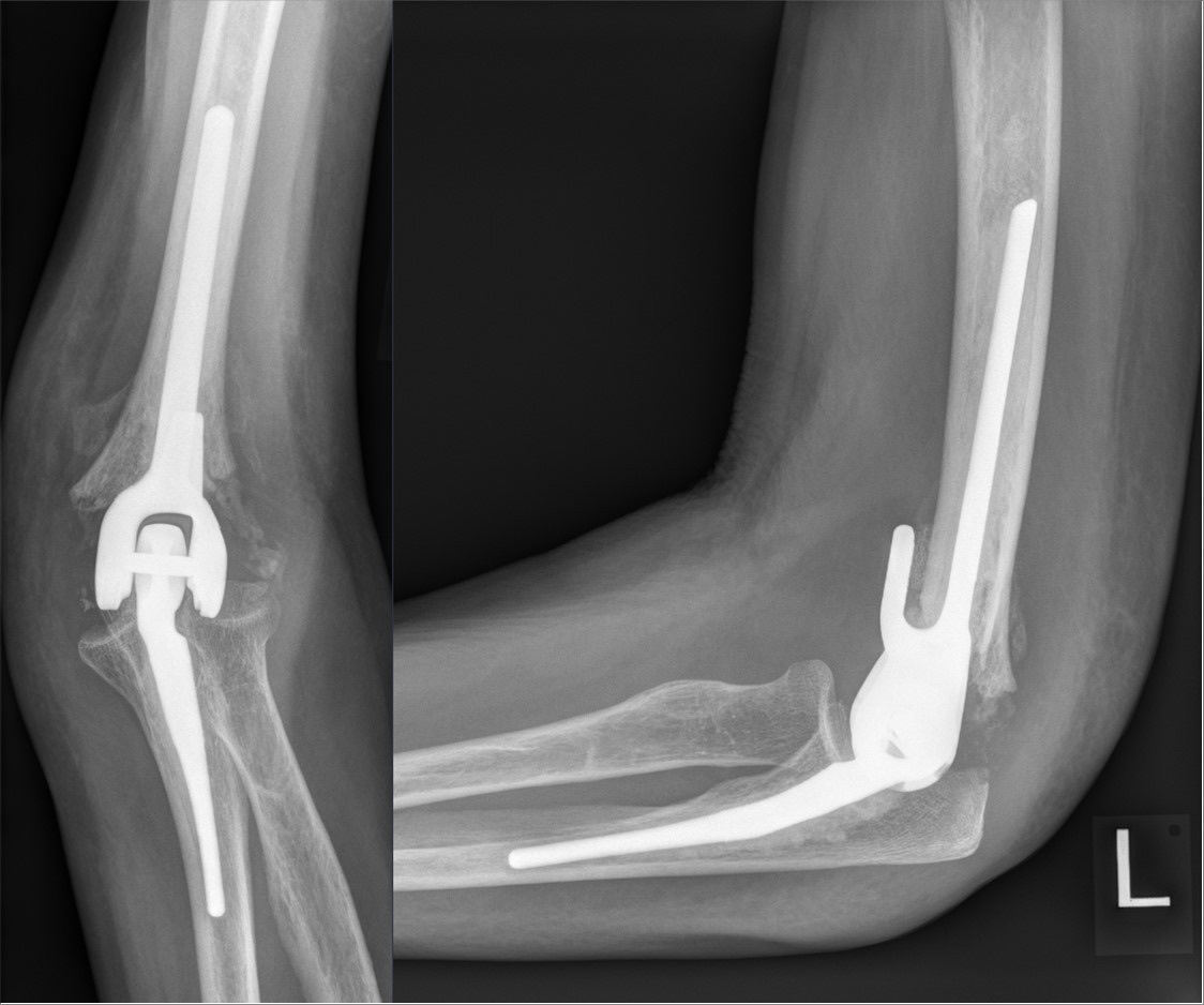
Figure 4. A cemented linked (semiconstrained) total elbow arthroplasty, the alternative to fixation for the unreconstructable fracture in the low-demand elderly. Source: Hellerhoff, via Wikimedia Commons, CC BY-SA 4.0.
Part II - Olecranon Fractures
The olecranon is subcutaneous (so its fractures are often nearly open and present as a tense swelling), it carries the triceps insertion, and it forms, with the coronoid, the trochlear (greater sigmoid) notch that grips the trochlea.[20] The cardinal deforming force is the triceps, which pulls the proximal fragment away and opens a gap at the articular surface, leaving the extensor mechanism incompetent. This is the exact problem the tension band is designed to solve.[21]
The standard classification is the Mayo classification, built on displacement, comminution, and stability: type I undisplaced, type II displaced (with a stable ulnohumeral joint), type III displaced with an unstable ulnohumeral joint, each subdivided into A (noncomminuted) and B (comminuted).[22] Type I fractures are treated non-operatively. Type II and III require surgery, and the modifier matters: the simple, noncomminuted type IIA is the fracture for which tension-band wiring is designed, while comminuted (B) and unstable (III) fractures are plated.[23] In the very low-demand elderly with extensive comminution, fragment excision with triceps advancement is a salvage option (up to roughly half to three-quarters of the olecranon can be excised with intact ligaments, though authors disagree on the exact safe limit).[24]
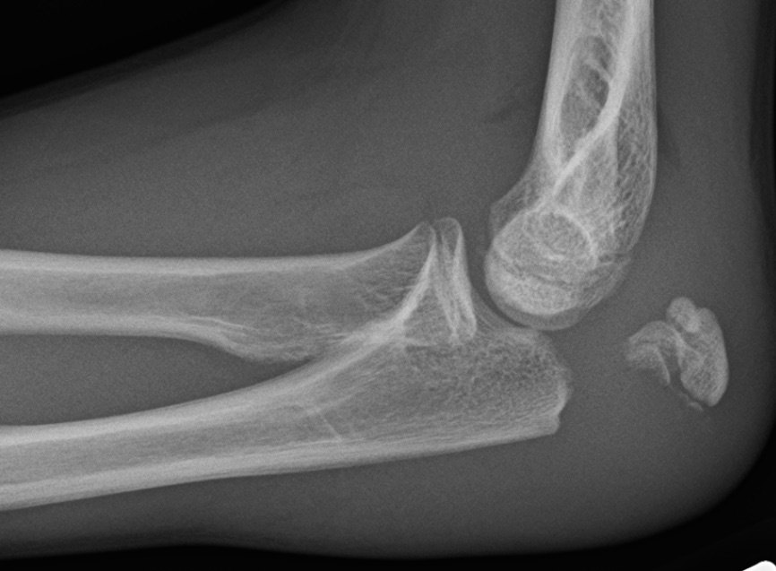
Figure 5. A displaced transverse olecranon fracture on the lateral radiograph: the triceps pulls the proximal fragment away, opening the articular surface. Source: James Heilman, MD, via Wikimedia Commons, CC BY-SA 4.0.
Part III - The Tension-Band (Weber / AO) Osteosynthesis
3.1 The principle
The tension-band is the named centrepiece of this topic. The triceps pulls the olecranon fragment dorsally (the tension side), tending to gape the fracture at the joint. A wire construct placed on the dorsal (tension) surface of the olecranon is meant to catch that tensile pull and convert it into compression at the opposite, articular (volar) cortex as the elbow flexes, so that the very force that would distract the fracture instead presses it together (a dynamic tension band).[25] A caveat appears in both source texts. Recent biomechanical work (Brink) has failed to demonstrate that the distraction force is actually converted to compression through the full range of motion, so the “dynamic” rationale is theoretically sound but unproven. What is not in doubt is the excellent clinical record of the technique in simple fractures with good bone.[26]
3.2 The technique
The classic construct, the AO technique attributed to Weber and Vasey, is two parallel Kirschner wires plus a figure-of-eight tension wire.[27] The steps are:[28]
- Reduce the simple transverse fracture in extension with a pointed reduction clamp.
- Drive two parallel K-wires (about 1.6-1.8 mm) from the proximal olecranon tip across the fracture to engage (penetrate) the anterior ulnar cortex distally.
- Drill a transverse hole in the dorsal ulna about 2 cm distal to the fracture and pass a stainless-steel wire (the AO text specifies 1.0 mm; Rockwood 18-20 gauge) through it.
- Pass the wire as a figure-of-eight deep to the triceps tendon around the proximal ends of the K-wires, and tighten it with a twist on each side so that compression is symmetric.
- Bend the K-wire ends 180° and impact them into the olecranon, deep to the triceps, so they cannot back out during active extension.
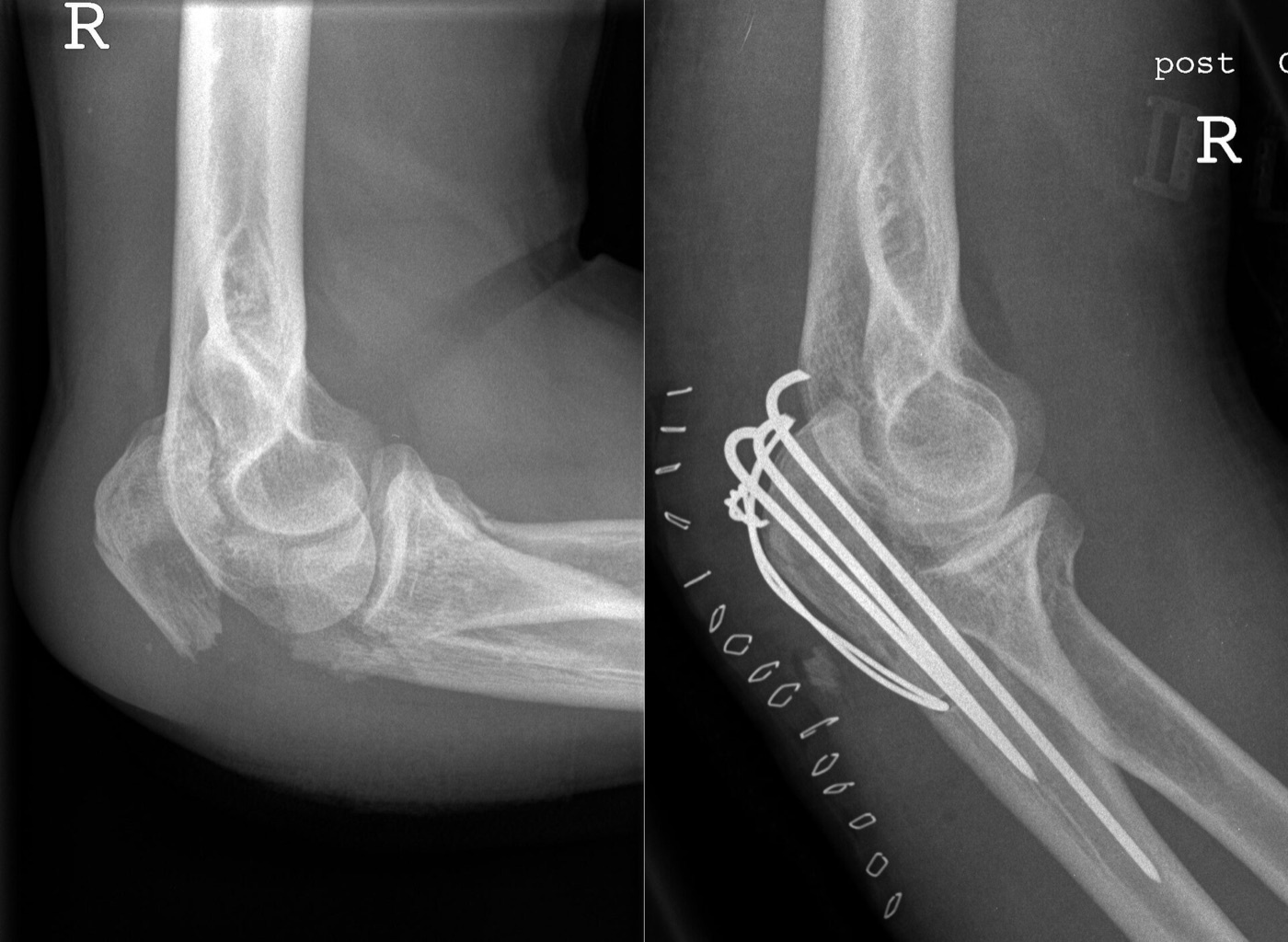
Figure 6. Tension-band (Weber) osteosynthesis: the displaced olecranon fracture (left) and the same elbow after fixation with two parallel K-wires and a figure-of-eight cerclage wire (right). Source: Michael Müller-Hillebrand, via Wikimedia Commons, CC BY 3.0.
3.3 Indications, contraindications, and complications
The tension band is indicated for the simple, transverse (or short oblique) olecranon fracture proximal to the coronoid in a stable elbow (Mayo IIA).[29] Its essential biomechanical prerequisite, shared with every tension band, is an intact opposite (articular) cortex to bear the compression. It is therefore contraindicated in comminuted fractures, unstable (Mayo III) fractures, oblique fractures extending distal to the midpoint of the notch, and fracture-dislocations (Monteggia, transolecranon), all of which are plated instead.[30] The dominant complication is prominent, symptomatic hardware over the subcutaneous olecranon, with hardware-removal rates reported from 15% up to 50-85% (much higher than after plating). K-wire migration and loss of reduction are the others.[31] A randomised trial (Duckworth) found functional outcomes equivalent to plating but a hardware-removal rate of 50% versus 22%, the central trade-off in choosing the technique.[32]
For comminuted, unstable, or fracture-dislocation patterns the implant is a precontoured dorsal locking plate, which itself works in the manner of a tension band and is biomechanically superior for these patterns.[33]
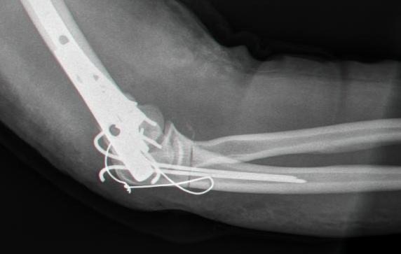
Figure 7. Plate-and-wire fixation of the olecranon, the construct chosen for comminuted, unstable, or fracture-dislocation patterns where a tension band cannot be used. Source: Mehlauge, public domain, via Wikimedia Commons.
Part IV - Radial Head and Neck Fractures
4.1 Function, mechanism, and classification
The radial head is the commonest fracture about the elbow (about 4% of all fractures), typically from a fall on the outstretched hand.[34] Its importance is as a stabiliser. It is a secondary restraint to valgus and posterolateral rotatory forces and, with the interosseous membrane and the distal radioulnar joint, the primary restraint to axial (longitudinal) forearm stability, the axis whose disruption is the Essex-Lopresti lesion (radial head fracture + interosseous membrane tear + DRUJ injury).[35] The Mason classification grades it as type I (nondisplaced/minimally displaced), type II (displaced partial head), and type III (comminuted whole head), with type IV (any radial head fracture with an elbow dislocation) added by Johnston. The Broberg-Morrey modification quantifies type II as more than 2 mm displacement and more than 30% of the articular surface.[36]
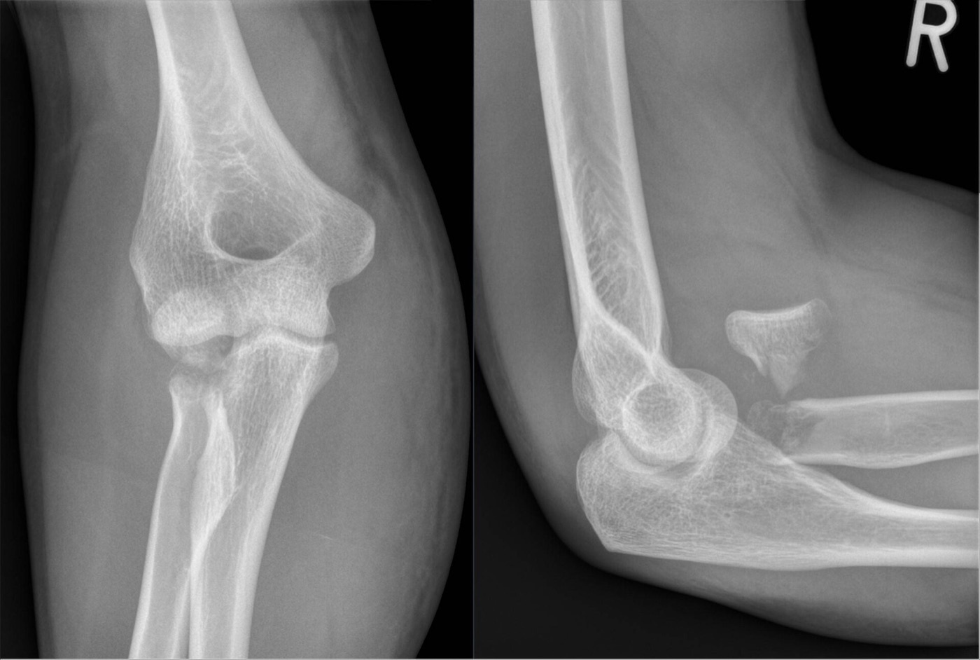
Figure 8. A displaced, rotated radial-head fracture (Mason III), AP and lateral radiographs. Source: Hellerhoff, via Wikimedia Commons, CC BY-SA 4.0.
A practical assessment point: when pain prevents examination, aspirating the haemarthrosis and injecting local anaesthetic allows a reliable test for a mechanical block to forearm rotation, which is itself an indication for surgery. The wrist and DRUJ must be examined for Essex-Lopresti.[37]
4.2 Treatment
Mason I fractures, and isolated Mason II fractures without a mechanical block, are treated non-operatively with brief immobilisation and early motion (immobilisation beyond two weeks loses extension). The management of the displaced Mason II is genuinely contested, with trials divided and the RAMBO trial ongoing.[38] ORIF suits the displaced fracture with three or fewer fragments and good bone, fixed with countersunk headless screws or a plate placed in the lateral “safe zone.”[39] Radial head arthroplasty (replacement) is preferred for the unreconstructable comminuted fracture (more than three fragments), especially with associated instability (the terrible triad, Essex-Lopresti). The cardinal technical error is overstuffing the joint (oversizing the implant, which opens the lateral ulnohumeral joint and causes pain and capitellar wear), so one downsizes rather than oversizes.[40]
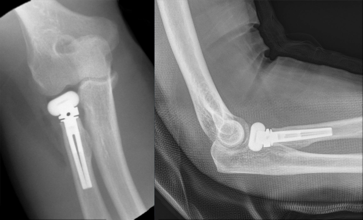
Figure 9. Metallic radial-head arthroplasty after a comminuted (unreconstructable) fracture. Source: Hellerhoff, via Wikimedia Commons, CC BY-SA 4.0.
The cardinal rule of radial head excision is negative and emphatic: the radial head must never be excised in the unstable elbow, that is, in a terrible triad, an Essex-Lopresti lesion, or any setting with collateral ligament or coronoid injury. Excision removes a critical stabiliser and precipitates valgus and axial instability with proximal radial migration. Isolated excision is reserved for the low-demand patient with an isolated comminuted fracture and is rarely done now.[41]
Part V - Coronoid Fractures and Elbow Dislocation
5.1 The coronoid
The coronoid is the anterior bony buttress against posterior subluxation and the primary restraint to varus and posteromedial rotatory forces, its anteromedial facet being the key to that instability.[42] Two classifications are used. The Regan-Morrey classification grades by fragment height: type I a tip, type II ≤50%, type III >50% (with an A/B modifier for the absence/presence of a dislocation).[43] The O’Driscoll classification grades by location and mechanism: the tip (which fails in the terrible triad), the anteromedial facet (which fails in posteromedial rotatory instability), and the base (which fails in olecranon fracture-dislocations).[44] The teaching point is that the coronoid fracture is best understood not in isolation but as the signature of a particular instability pattern.[45]
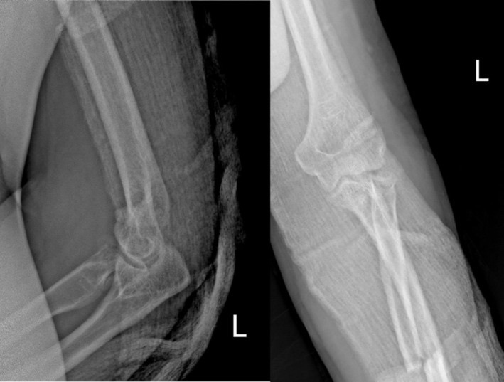
Figure 10. Associated coronoid-process and radial-head fractures, the bony components of a terrible-triad pattern. From Clinical Case Reports (2025), PMC12578567, CC BY 4.0.
5.2 Simple elbow dislocation
The elbow is the second most commonly dislocated major joint (after the shoulder), and a simple dislocation (no fracture) is usually posterolateral, from a fall producing axial load, valgus, and supination.[46] O’Driscoll described the soft-tissue failure as a spectrum progressing from lateral to medial (the LUCL first, then the capsule, then the MCL last), the basis of posterolateral rotatory instability, although newer imaging data suggest some injuries actually begin medially.[47] The modern management is closed reduction, a careful test of stability through the arc of motion, and early mobilisation. Most simple dislocations are stable after reduction and do well, surgery being reserved for those that redislocate before 30° of flexion, the irreducible, the open, and the vascular injury. Immobilisation beyond about three weeks worsens stiffness and outcome.[48]
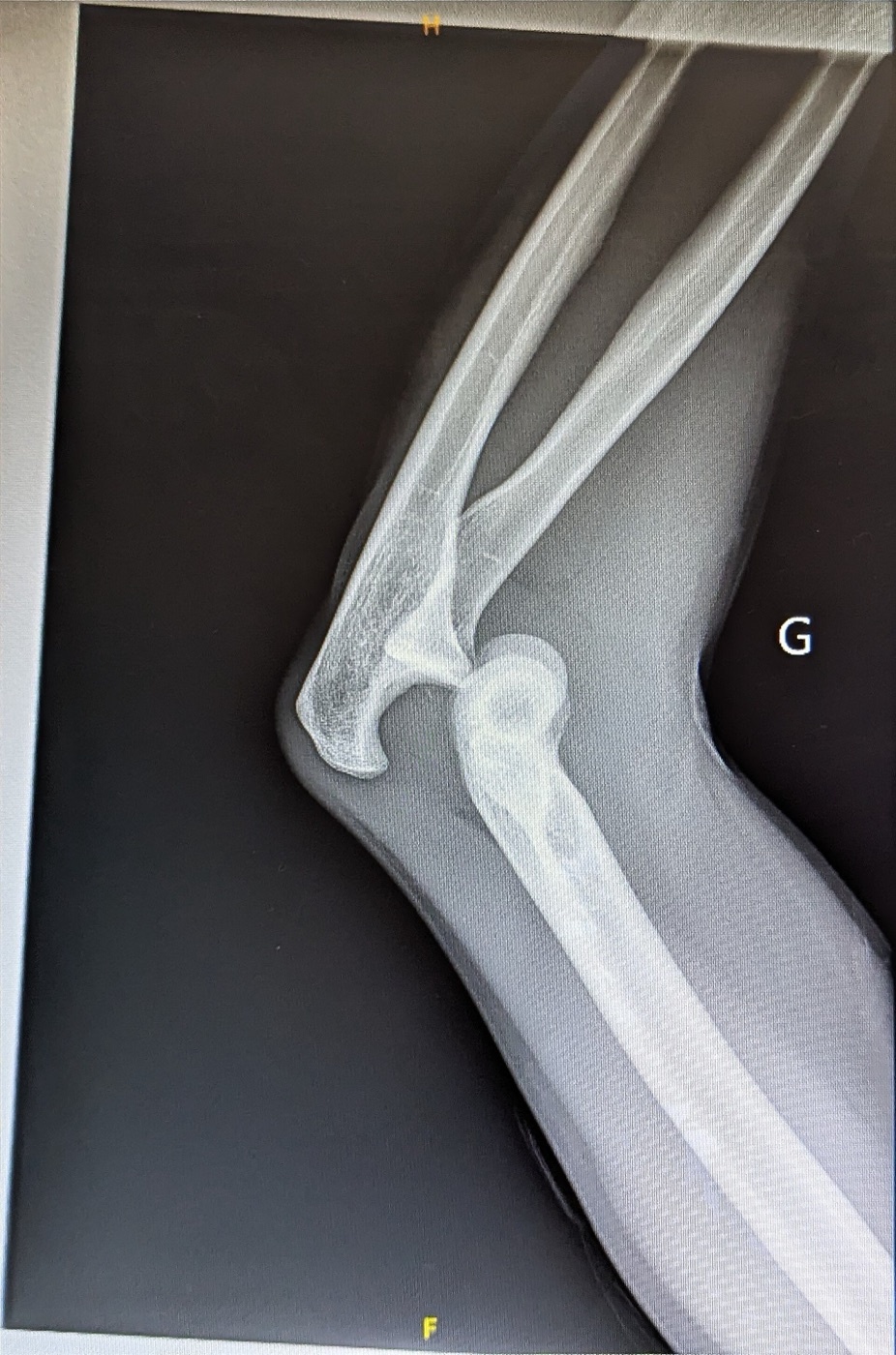
Figure 11. Posterior dislocation of the elbow without fracture (a simple dislocation), lateral radiograph. Source: Flibust1er, via Wikimedia Commons, CC0 1.0.
Part VI - The Terrible Triad
The terrible triad is the combination of an elbow dislocation, a radial head fracture, and a coronoid fracture. It earned its name from the chronic instability, stiffness, and arthrosis that followed its historically poor treatment.[49] Its mechanism is posterolateral rotatory, and the coronoid fracture is usually a small tip (Regan-Morrey I).[50] Outcomes were transformed by a systematic surgical protocol, addressed from deep to superficial through a lateral approach:[51]
- Fix the coronoid first (a screw for larger fragments, or a suture lasso of the anterior capsule for small ones; a tiny tip may be left).
- Fix or replace the radial head, and crucially never excise it.
- Repair the lateral collateral ligament (usually avulsed off the lateral epicondyle) back to the isometric point at the centre of the capitellum.
- Reassess stability; if the elbow is still unstable, repair the medial collateral ligament, and if it remains unstable, apply a hinged or static external fixator (or a temporary bridge plate).
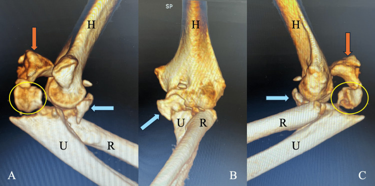
Figure 12. Terrible triad on 3D CT: a radial-head fracture (circle) and a coronoid fracture (arrow) with the dislocation, here also with an olecranon fracture. From Cureus (2026), PMC12999191, CC BY 4.0.
This protocol, with early protected motion afterward, achieves good or excellent results in the majority (Pugh reported 78%).[52] A non-operative trial is permissible only when all of four conditions hold: no mechanical block from the radial head, a small (Regan-Morrey I/II) coronoid, a congruent joint after reduction, and a stable arc to 30° of flexion.[53] A related but distinct pattern is posteromedial rotatory instability, in which an anteromedial coronoid facet fracture occurs (typically without a radial head fracture, the recognition clue) and requires buttress-plate fixation of the facet. A malunited anteromedial facet leads to varus subluxation and arthrosis with no good salvage.[54]
Part VII - Monteggia and Transolecranon Fracture-Dislocations
Two proximal-ulna fracture-dislocations complete the picture, and the distinction between them is examinable. A Monteggia fracture-dislocation is a proximal ulna fracture with dislocation of the radial head (disruption of the proximal radioulnar joint), classified by Bado into four types by the direction of radial head displacement (type I anterior, II posterior, III lateral, IV with a radial shaft fracture), with the Jupiter subdivision of the posterior (Bado II) type by the level of the ulnar fracture.[55] A transolecranon fracture-dislocation, by contrast, is an anterior fracture-dislocation in which the trochlea is driven through the trochlear notch with the proximal radioulnar joint preserved, a primarily bony injury.[56] Both are treated by stable plate fixation, never tension-band wiring. The recurring rule for a failed radial head reduction in a Monteggia is that malreduction of the ulna is almost always the cause.[57]
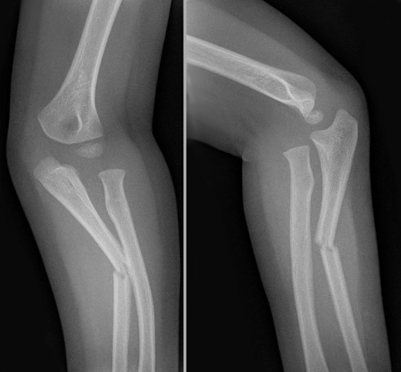
Figure 13. Monteggia fracture-dislocation: an ulnar fracture with dislocation of the radial head (AP and lateral). Source: Hellerhoff, via Wikimedia Commons, CC BY-SA 3.0.
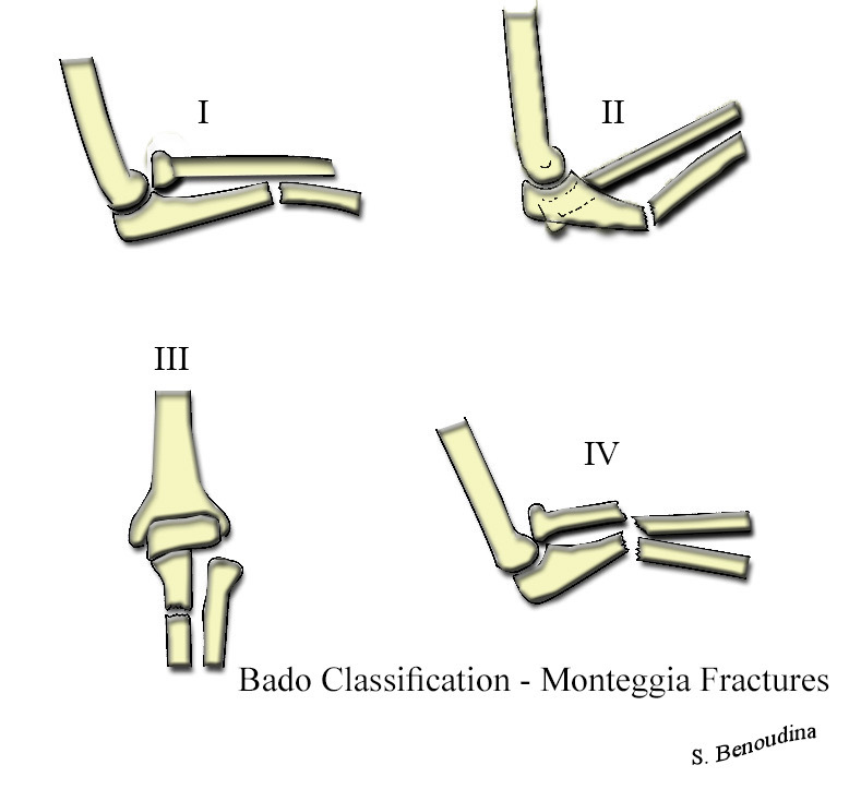
Figure 14. The Bado classification of Monteggia fracture-dislocations (types I to IV), by the direction of radial head displacement. Source: Benoudina Samir, via Wikimedia Commons, CC BY-SA 4.0.
Part VIII - A Synthesis: How to Reason Around the Elbow
The elbow rewards a structural way of thinking. Begin by asking what is broken and what is torn, because the fractures of the elbow are usually the visible signs of an instability pattern: a coronoid tip plus a radial head fracture is a terrible triad, an anteromedial coronoid facet without a radial head fracture is posteromedial rotatory instability, and a proximal ulna fracture with a dislocated radial head is a Monteggia. Then respect the stabilisers. Keep or restore the radial head (never excise it in an unstable elbow), buttress the coronoid, and repair the lateral collateral ligament, in that deep-to-superficial order. Match the implant to the pattern. The simple transverse olecranon is the one fracture made for the tension band, which turns the triceps’ pull into compression. The moment the fracture is comminuted, unstable, or part of a fracture-dislocation, the opposite cortex can no longer bear that compression and a plate is required. In the distal humerus, restore the articular surface and the two columns and move early, choosing arthroplasty over fixation only for the unreconstructable fracture in the low-demand elderly. Through all of this, remember the one complication that defines the joint: the elbow becomes stiff. Every fixation exists to permit early motion, and a stable, slightly stiff elbow always beats a loose, incongruent one.
References
-
Rockwood & Green’s Fractures in Adults, pp.2253, 2344; AO Principles of Fracture Management, p.657.
-
AO Principles of Fracture Management, pp.651, 675 (the elbow, more than any other joint, becomes stiff after injury); Rockwood & Green’s Fractures in Adults, p.2297 (functional arc 30-130°).
-
Rockwood & Green’s Fractures in Adults, p.2297; AO Principles of Fracture Management, p.675.
-
Rockwood & Green’s Fractures in Adults, pp.2344, 2377, 2424.
-
Rockwood & Green’s Fractures in Adults, pp.2251, 2254-2255; AO Principles of Fracture Management, p.644.
-
Rockwood & Green’s Fractures in Adults, pp.2251, 2254-2255; AO Principles of Fracture Management, p.644.
-
Rockwood & Green’s Fractures in Adults, p.2254.
-
Rockwood & Green’s Fractures in Adults, pp.2257, 2296.
-
Rockwood & Green’s Fractures in Adults, pp.2245, 2249-2252; AO Principles of Fracture Management, p.644.
-
Rockwood & Green’s Fractures in Adults, p.2249.
-
Rockwood & Green’s Fractures in Adults, p.2252.
-
Rockwood & Green’s Fractures in Adults, pp.2240, 2258-2259.
-
Rockwood & Green’s Fractures in Adults, pp.2256-2257, 2262; AO Principles of Fracture Management, p.643.
-
Rockwood & Green’s Fractures in Adults, pp.2270-2273; AO Principles of Fracture Management, pp.648-649.
-
Rockwood & Green’s Fractures in Adults, pp.2286-2288, 2293-2294; AO Principles of Fracture Management, pp.651, 653.
-
Rockwood & Green’s Fractures in Adults, p.2271; AO Principles of Fracture Management, pp.647, 654.
-
Rockwood & Green’s Fractures in Adults, p.2299; AO Principles of Fracture Management, p.655 (McKee RCT favouring TEA for elderly C3 fractures).
-
AO Principles of Fracture Management, pp.654-655. The Rockwood narrative’s “~2-5 lb” lifting limit was not in the mined (truncated) extract; the captured figure is AO’s lifelong ~5 kg restriction, consistent with the standard teaching.
-
Rockwood & Green’s Fractures in Adults, pp.2244, 2295-2296; AO Principles of Fracture Management, p.655.
-
Rockwood & Green’s Fractures in Adults, pp.2437-2438; AO Principles of Fracture Management, p.657.
-
Rockwood & Green’s Fractures in Adults, p.2437.
-
Rockwood & Green’s Fractures in Adults, p.2438.
-
Rockwood & Green’s Fractures in Adults, pp.2438, 2440.
-
Rockwood & Green’s Fractures in Adults, pp.2440, 2448 (An et al. >50%; McKeever & Buck up to 80%; Bell <75% to avoid destabilisation).
-
Rockwood & Green’s Fractures in Adults, pp.2437, 2440.
-
Rockwood & Green’s Fractures in Adults, p.2440; AO Principles of Fracture Management, p.665 (Brink et al.: no conversion of distraction to compression through full ROM).
-
The construct is classically the AO tension band of Weber and Vasey; the mined Rockwood and AO texts describe the two-K-wire-plus-figure-of-eight technique in full but do not use the “Weber” eponym verbatim, so the konspekt’s “Weber tension-band” is this AO technique. Rockwood & Green’s Fractures in Adults, pp.2441-2442; AO Principles of Fracture Management, pp.665-667.
-
Rockwood & Green’s Fractures in Adults, pp.2441-2442; AO Principles of Fracture Management, pp.665-667.
-
Rockwood & Green’s Fractures in Adults, p.2440; AO Principles of Fracture Management, p.665.
-
Rockwood & Green’s Fractures in Adults, pp.2440, 2459; AO Principles of Fracture Management, pp.667, 673.
-
Rockwood & Green’s Fractures in Adults, pp.2447-2448.
-
Rockwood & Green’s Fractures in Adults, p.2448 (Duckworth RCT).
-
Rockwood & Green’s Fractures in Adults, pp.2440, 2447; AO Principles of Fracture Management, p.667.
-
Rockwood & Green’s Fractures in Adults, p.2422.
-
Rockwood & Green’s Fractures in Adults, pp.2424, 2436; p.2355.
-
Rockwood & Green’s Fractures in Adults, pp.2423-2424; p.2356. (The type IV/dislocation addition is attributed to Johnston, 1962; it is commonly also credited to Hotchkiss, but the mined texts name Johnston and use Hotchkiss only for the implant “safe zone.”)
-
Rockwood & Green’s Fractures in Adults, pp.2423, 2356.
-
Rockwood & Green’s Fractures in Adults, pp.2425-2426, 2434.
-
Rockwood & Green’s Fractures in Adults, pp.2426, 2436; p.2360.
-
Rockwood & Green’s Fractures in Adults, pp.2426, 2430, 2436; p.2360, 2369.
-
Rockwood & Green’s Fractures in Adults, pp.2426, 2434; p.2363.
-
Rockwood & Green’s Fractures in Adults, pp.2375, 2377.
-
Rockwood & Green’s Fractures in Adults, p.2375.
-
Rockwood & Green’s Fractures in Adults, pp.2375-2376.
-
Rockwood & Green’s Fractures in Adults, pp.2375-2376.
-
Rockwood & Green’s Fractures in Adults, p.2342.
-
Rockwood & Green’s Fractures in Adults, p.2342. The classic lateral-to-medial sequence is O’Driscoll’s three-stage spectrum (sometimes called the “circle of Horii”); the mined text describes the spectrum and cites O’Driscoll but does not use the “circle of Horii” eponym.
-
Rockwood & Green’s Fractures in Adults, pp.2344-2347.
-
Rockwood & Green’s Fractures in Adults, p.2376.
-
Rockwood & Green’s Fractures in Adults, pp.2376-2377.
-
Rockwood & Green’s Fractures in Adults, pp.2379-2382.
-
Rockwood & Green’s Fractures in Adults, pp.2383-2384.
-
Rockwood & Green’s Fractures in Adults, pp.2377-2378.
-
Rockwood & Green’s Fractures in Adults, pp.2385-2392.
-
Rockwood & Green’s Fractures in Adults, pp.2449-2450.
-
Rockwood & Green’s Fractures in Adults, pp.2436, 2449.
-
Rockwood & Green’s Fractures in Adults, pp.2451, 2458-2459; AO Principles of Fracture Management, pp.671-673.
-
Rockwood & Green’s Fractures in Adults, p.2297; AO Principles of Fracture Management, pp.651, 675.
-
Rockwood & Green’s Fractures in Adults, pp.2245-2252, 2256-2257, 2270-2273, 2286-2294.
-
Rockwood & Green’s Fractures in Adults, p.2299; AO Principles of Fracture Management, pp.654-655.
-
Rockwood & Green’s Fractures in Adults, pp.2438, 2440.
-
Rockwood & Green’s Fractures in Adults, pp.2440-2442; AO Principles of Fracture Management, pp.665-667.
-
Rockwood & Green’s Fractures in Adults, pp.2440, 2447-2448, 2459; AO Principles of Fracture Management, pp.665-667.
-
Rockwood & Green’s Fractures in Adults, pp.2423-2426, 2436.
-
Rockwood & Green’s Fractures in Adults, pp.2426, 2434; p.2363.
-
Rockwood & Green’s Fractures in Adults, pp.2376, 2379-2382.
-
Rockwood & Green’s Fractures in Adults, pp.2375-2376.
-
Rockwood & Green’s Fractures in Adults, pp.2436, 2449-2450.
-
Rockwood & Green’s Fractures in Adults, pp.2342, 2344-2347.
-
Rockwood & Green’s Fractures in Adults, pp.2295-2297, 2382; AO Principles of Fracture Management, p.655.
-
Rockwood & Green’s Fractures in Adults, pp.2355, 2372-2373; p.2422.