Contents
- Orientation: The Commonest Fracture, and Why It Is Deceptively Hard
- Part I - Epidemiology and the Fragility-Fracture Significance
- Part II - Applied Anatomy and the Radiographic Parameters
- Part III - The Eponyms
- Part IV - Classification
- Part V - Clinical Assessment and Imaging
- Part VI - Nonoperative Treatment
- Part VII - Operative Treatment
- Part VIII - The Elderly Distal Radius Debate
- Part IX - The DRUJ and Ulnar-Sided Injuries
- Part X - Complications
- Part XI - A Synthesis: How to Reason About the Distal Radius
- References
Orientation: The Commonest Fracture, and Why It Is Deceptively Hard
The distal radius is the most frequently broken bone of the upper limb, accounting for at least one in six of all fractures seen in an emergency department.[1] That frequency makes it easy to dismiss as a simple “wrist fracture,” but it is nothing of the sort. The distal radius carries the greater part of the axial load across the wrist (the load passes chiefly through the lunate fossa), it forms half of the distal radioulnar joint (DRUJ) on which forearm rotation depends, and it presents a small articular surface that must be restored to within a couple of millimetres if the radiocarpal joint is to survive. The injury also comes in two quite different guises. One is a high-energy fracture in a young adult, where the goal is anatomical articular reconstruction. The other is a low-energy fragility fracture in an older woman, where the bone is osteoporotic, the function often forgives radiographic imperfection, and the fracture is a warning sign of skeletal fragility.[2] Holding both of those pictures in mind at once is the key to the topic.
Part I - Epidemiology and the Fragility-Fracture Significance
The distal radius fracture is the second commonest fracture in the elderly and shows a bimodal age distribution: high-energy injuries predominate in young men, while low-energy fragility fractures predominate in older women, the female-to-male incidence being higher by a factor of two to three.[3] US annual prevalence exceeds 600,000, and the worldwide incidence is rising.[4] The mechanism in the vast majority is a fall onto the outstretched hand (FOOSH) from standing height, with the wrist in 40° to 90° of dorsiflexion producing the dorsally displaced pattern, and a fall onto the flexed wrist producing the volar pattern. Fracture severity tracks with poor bone quality, there being a linear correlation between DXA T-scores and early instability and malunion.[5]
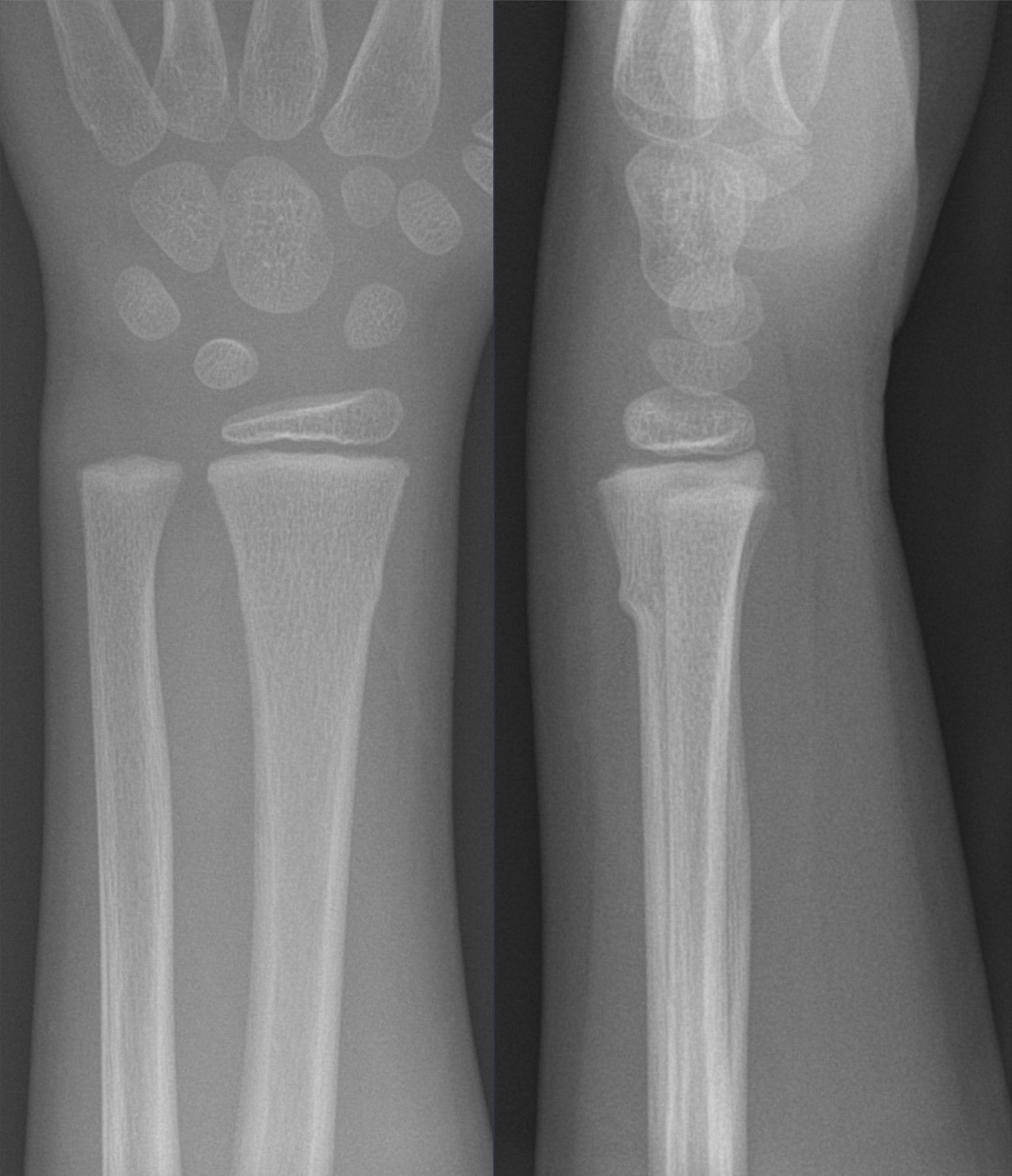
Figure 1. A torus (buckle) fracture of the distal radius in a child, AP and lateral views; the distal radius is the commonest fracture site in children. Hellerhoff, via Wikimedia Commons, CC BY-SA 4.0.
The fragility-fracture dimension deserves emphasis because it is so often neglected. A distal radius fracture in an older patient carries a two- to four-fold increased risk of a future fracture and is a marker of underlying skeletal fragility, yet few such patients are referred for the appropriate bone-health work-up.[6] The orthopaedic surgeon who treats the wrist should ensure follow-up for the underlying metabolic bone disease (prompt DXA, a fracture-liaison service), because low bone mineral density is itself the strongest predictor of the next fracture.[7]
Part II - Applied Anatomy and the Radiographic Parameters
2.1 The three columns and load transmission
A useful biomechanical model (Rikli and Regazzoni) divides the distal forearm into three columns: the radial column (radial styloid and scaphoid fossa), the intermediate column (lunate fossa and the sigmoid notch of the radius), and the ulnar column (the distal ulna with its triangular fibrocartilage complex, the TFCC).[8] The clinically important point is that the lunate fossa carries the large share of the axial load and is therefore the key to the radiocarpal articular surface, while the radial styloid acts mostly as a bony buttress and the anchor for the extrinsic carpal ligaments. The ulna is the stable pivot about which the radius rotates, and the ulnar column, through the TFCC, is the distal end of that pivot.[9]
2.2 The radiographic parameters
Restoring the distal radius means restoring three measurements that every examiner expects you to know. On the posteroanterior (PA) view, the radial height (length) averages about 11 to 12 mm and the radial inclination about 22 to 23°; on a true lateral view, the volar (palmar) tilt averages about 11°.[10] Two further lateral-view measures matter for the articular surface: the teardrop angle, normally about 70° (a value below 45° signals lunate-facet articular incongruity), and ulnar variance, the relative length of the radius and ulna, which is a measure of radial shortening (about 60% of people are “ulnar neutral”).[11] These measurements are all sensitive to forearm rotation, so they must be read off a true lateral with the radius and ulna superimposed; a mere 5° of rotation changes the apparent volar tilt by about 1.6°.[12]
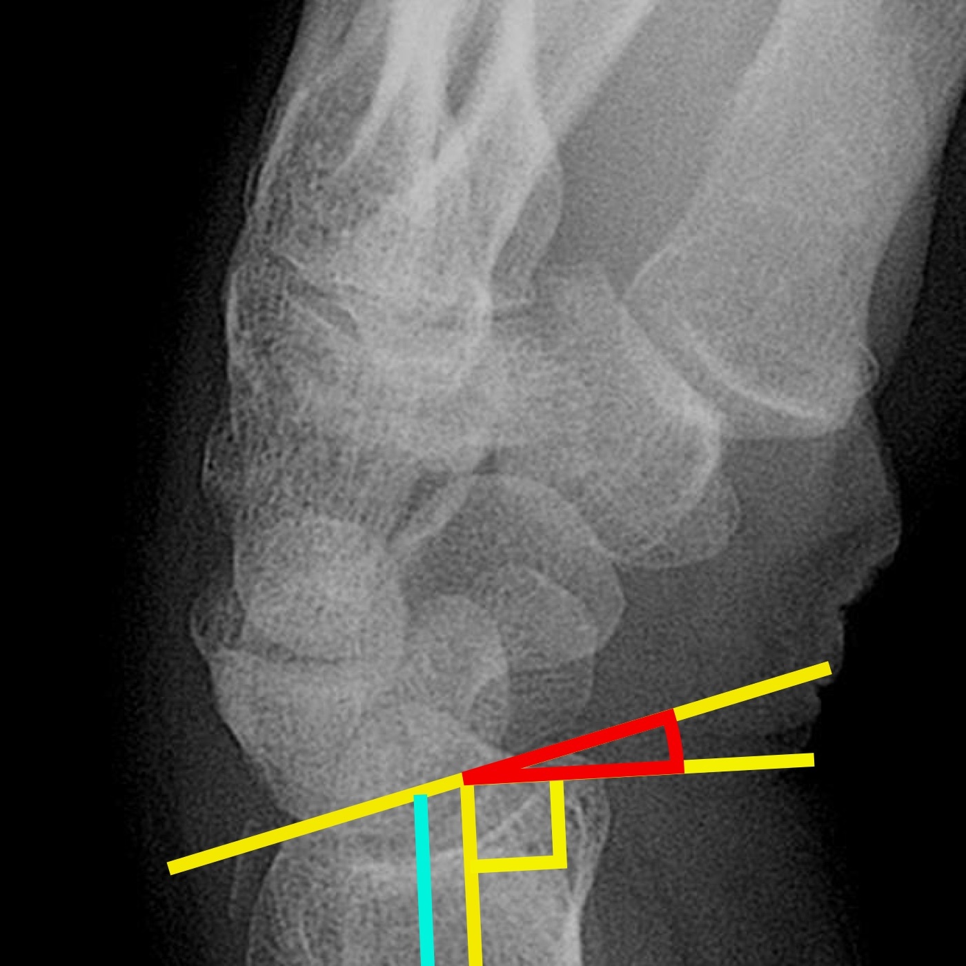
Figure 2. Volar/dorsal tilt of the distal radius on an annotated lateral radiograph. Mikael Häggström, from a base image by Lucien Monfils, via Wikimedia Commons, CC BY-SA 3.0.
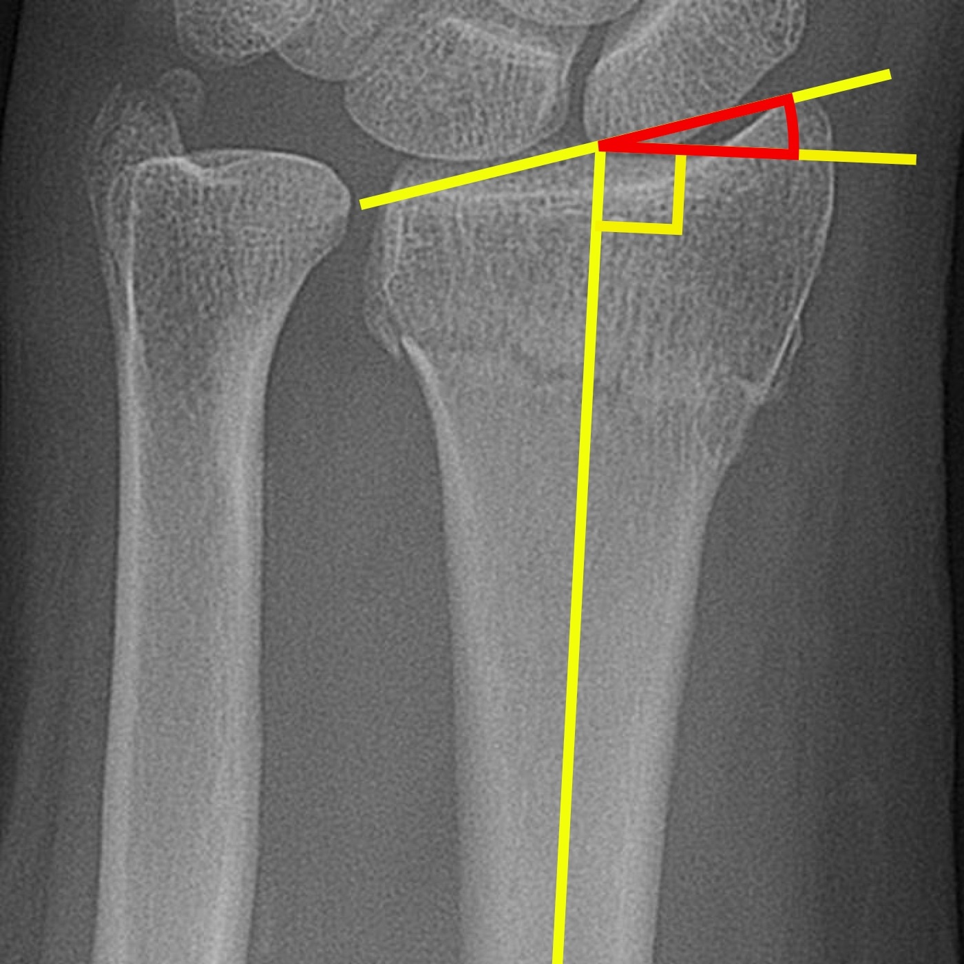
Figure 3. Radial inclination of the distal radius on an annotated AP radiograph. Mikael Häggström, from a base image by Lucien Monfils, via Wikimedia Commons, CC BY-SA 3.0.
2.3 The watershed line, the DRUJ, and the tendons at risk
The volar surface of the distal radius is flat and smooth, covered by the pronator quadratus, which makes it ideal for plate application; the dorsal surface is convex and grooved for the extensor tendons, its most prominent landmark being Lister’s tubercle, around which the extensor pollicis longus (EPL) turns like a pulley.[13] The watershed line is the distal-most ridge of the volar surface, the boundary beyond which the volar extrinsic wrist ligaments attach; a volar plate must sit proximal to the watershed line, because a plate prominent distal to it abrades and ruptures the overlying flexor tendons.[14] On the ulnar side the radius forms the sigmoid notch of the DRUJ, stabilised by the TFCC and, as a secondary restraint, by the distal interosseous membrane (DIOM); an ulnar styloid tip fracture accompanies about 50% of distal radius fractures and usually needs no separate treatment.[15] The median nerve lies in the carpal tunnel just volar to the fracture, which is why acute carpal tunnel syndrome is the injury’s most urgent complication.[16]
Part III - The Eponyms
The wrist carries more eponyms than any other fracture, and the examiner expects each to be defined cleanly:[17]
- Colles fracture (Abraham Colles, 1814, described before radiographs existed): a dorsally angulated, apex-volar metaphyseal fracture of the distal radius, classically extra-articular, producing the “dinner-fork” deformity (dorsal angulation with compensatory carpal flexion). It is the commonest pattern and the one most loosely used as a general label.
- Smith fracture (“reverse Colles”): the opposite, a volarly angulated, apex-dorsal metaphyseal fracture.
- Barton fracture: an intra-articular fracture-dislocation in which a rim fragment displaces with the carpus; the dorsal Barton exits the dorsal cortex with dorsal subluxation, the volar (reverse) Barton exits the volar cortex with volar subluxation.
- Chauffeur’s fracture: an isolated intra-articular fracture of the radial styloid, named because the hand-crank of an early automobile would backfire and strike the wrist. (The synonym “Hutchinson fracture” is widely used but is not the term in the source text.)
- Die-punch fracture: a depressed, impacted fragment of the lunate facet, the articular surface driven down into the metaphysis by the lunate, the conceptual basis of Melone’s “medial complex.”
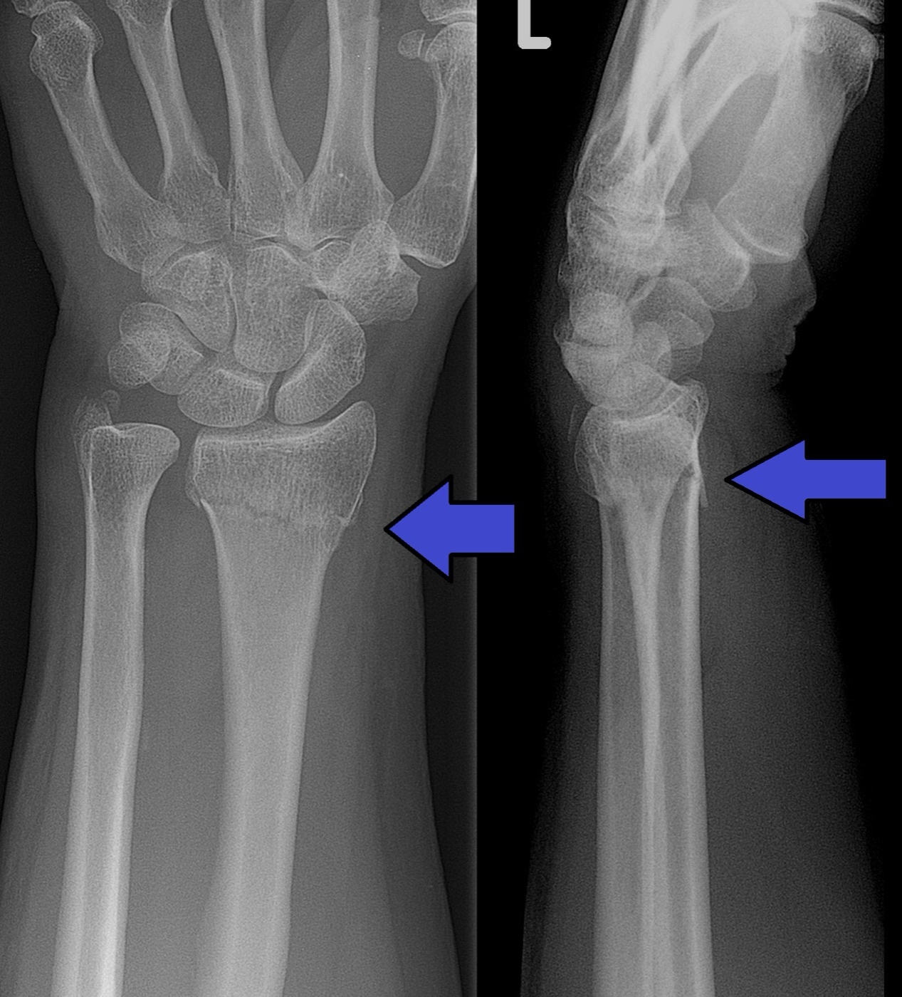
Figure 4. Colles fracture of the distal radius with an associated ulnar styloid fracture (AP and lateral views); the lateral shows the dorsal displacement of the dinner-fork deformity. Lucien Monfils, via Wikimedia Commons, CC BY-SA 3.0.
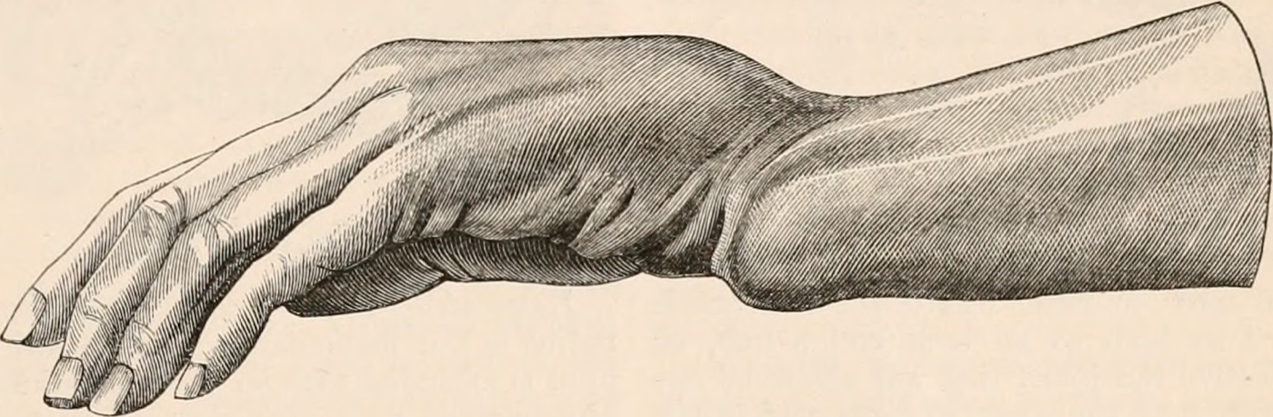
Figure 5. The dinner-fork (silver-fork) deformity of a dorsally displaced distal radius fracture. Engraving from Todd, Cyclopædia of Anatomy and Physiology (1849), public domain.
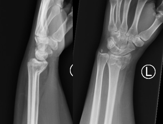
Figure 6. Smith fracture of the distal radius with volar displacement (AP and lateral views), the reverse of a Colles fracture. James Heilman, MD, via Wikimedia Commons, CC BY-SA 4.0.
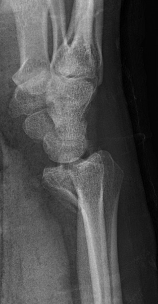
Figure 7. Volar (palmar) Barton fracture-dislocation of the distal radius (lateral view), an intra-articular rim fragment displaced with the carpus. Mikael Häggström, CC0 (public domain).
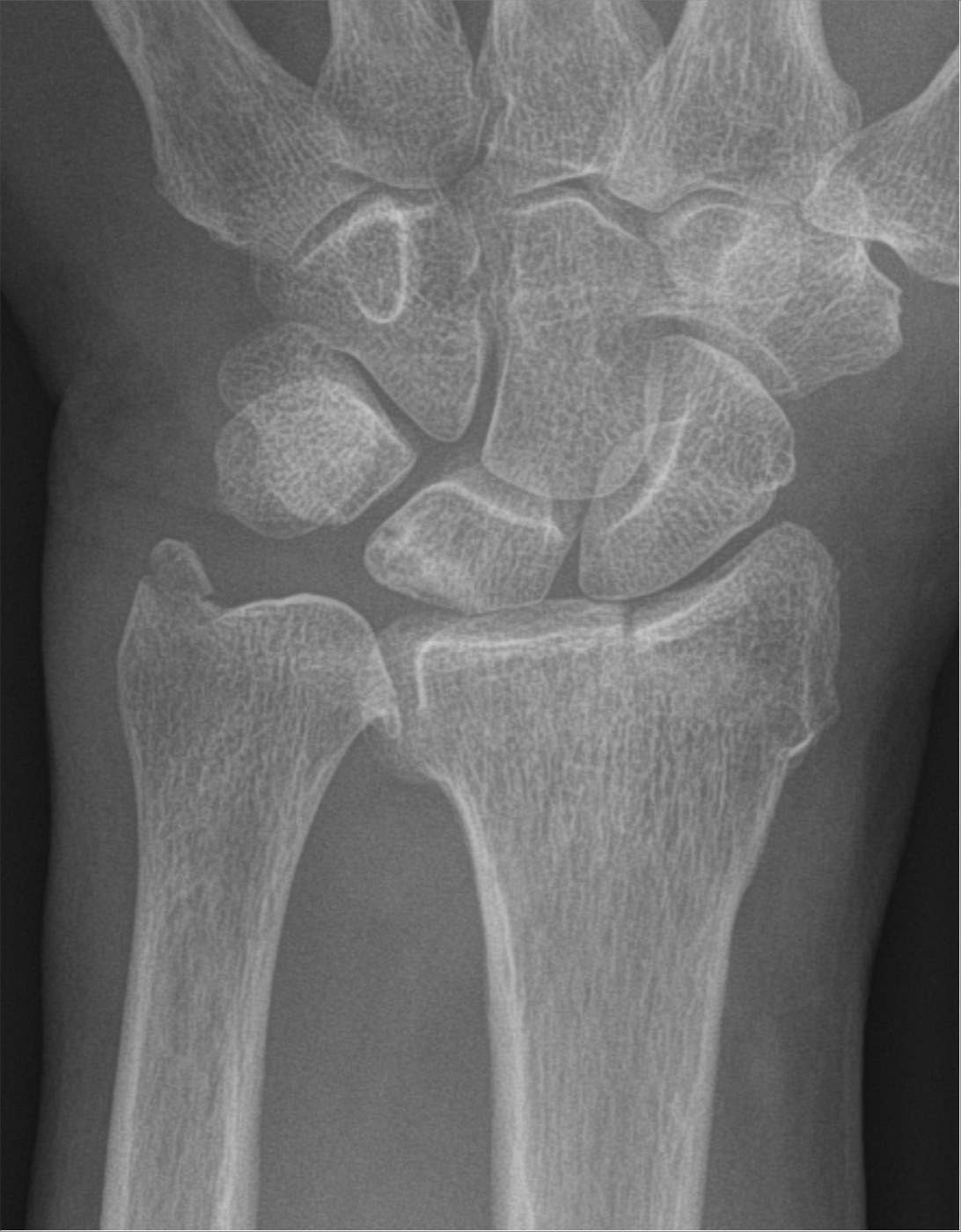
Figure 8. Chauffeur (radial styloid) fracture of the distal radius (AP view). Hellerhoff, via Wikimedia Commons, CC BY-SA 4.0.
Part IV - Classification
The AO/OTA classification is the most detailed and the one used in research. The distal radius is segment 2R3, graded in order of increasing severity into type A extra-articular, type B partial articular, and type C complete articular, each with further groups by comminution and displacement (27 patterns in all).[18] The Fernandez classification is mechanism-based and clinically intuitive, grading by the injuring force from type I (metaphyseal bending) through II (joint-surface shear), III (joint-surface compression/impaction), IV (avulsion / radiocarpal fracture-dislocation) to V (combined high-velocity).[19] Older systems include Frykman (by intra-articular involvement and associated ulnar fracture, but ignoring shortening, comminution, and displacement), Melone (a four-part intra-articular scheme highlighting the lunate-facet “medial complex”), Gartland and Werley (three groups of Colles fractures), and Mayo (by which articular facets are involved).[20] The honest caveat, repeated in both major texts, is that none of these systems is reliable below the level of the three main AO/OTA types (A, B, C), and CT, while it better characterises the articular surface, does not improve reliability or change outcomes.[21]
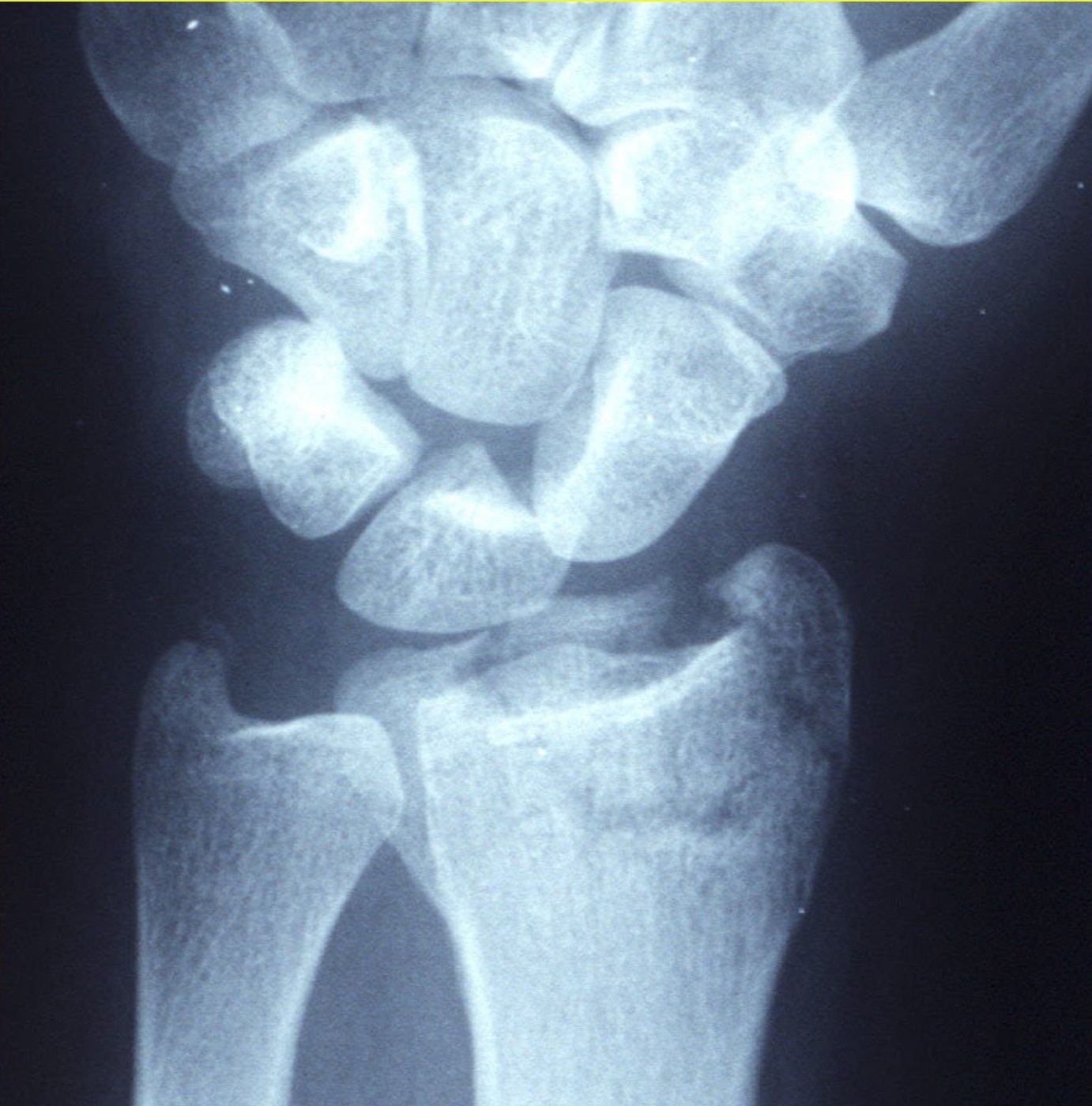
Figure 9. A displaced intra-articular (AO type C) distal radius fracture. Curtishand, via Wikimedia Commons (public domain).
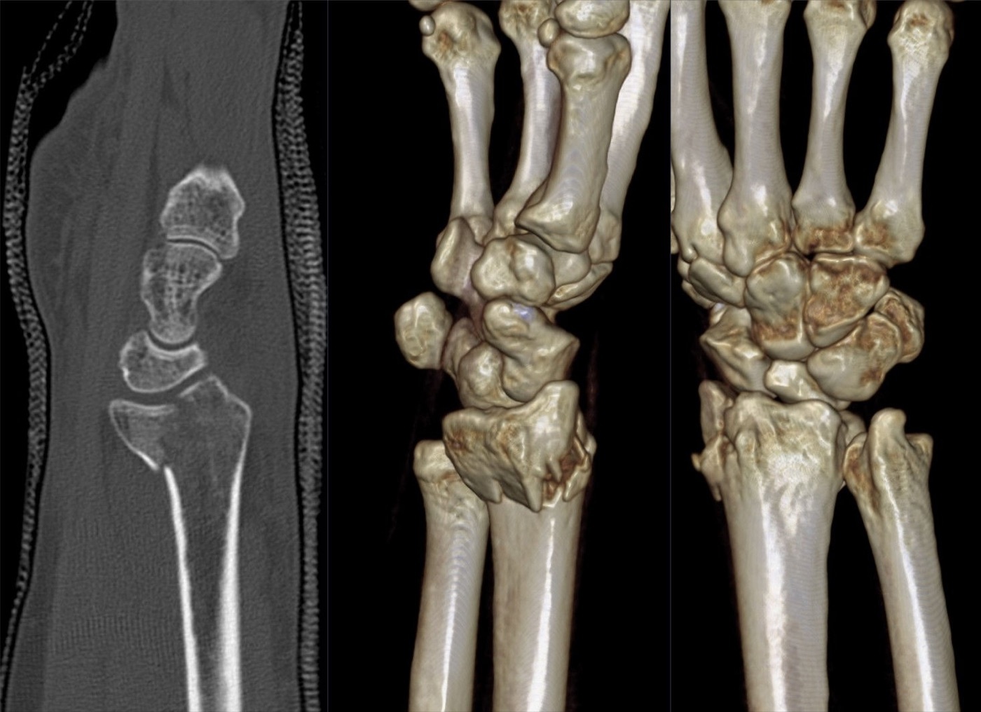
Figure 10. A reverse (dorsal) Barton intra-articular fracture on sagittal CT and 3D reconstructions, the kind of articular detail CT adds to plain films. Hellerhoff, via Wikimedia Commons, CC BY-SA 4.0.
Part V - Clinical Assessment and Imaging
5.1 History, examination, and the median nerve
The patient presents after a fall with a painful, swollen, often visibly deformed wrist. The skin is inspected for an open wound (usually on the volar-ulnar side), and the examiner asks specifically about numbness, because the most urgent associated injury is acute carpal tunnel syndrome (ACTS).[22] Distal radius fractures carry the highest risk of carpal tunnel syndrome of any upper-limb fracture, and the critical clinical task is to distinguish progressive, worsening median-nerve symptoms (acute CTS, which demands urgent release) from the constant, non-progressive numbness of a median-nerve contusion (which is observed and usually improves).[23] Associated soft-tissue injuries are common: scapholunate and lunotriquetral interosseous ligament tears, TFCC tears (39% to 82%), and chondral injuries (up to 32%), though their clinical significance is often unclear.[24]
5.2 Imaging and the criteria for an acceptable reduction
The standard series is a PA, lateral, and oblique view of the wrist; intra-operatively a dorsal tangential (skyline) view detects dorsal cortical screw penetration.[25] CT is reserved for defining articular comminution and depression, especially of the sigmoid notch and lunate facet, although it has no proven functional-outcome benefit.[26] The radiographic limits that constitute an unacceptable reduction (and therefore an indication for surgery in a higher-demand patient) are, by the AAOS guideline: intra-articular step-off greater than 2 mm, dorsal tilt greater than 10°, or radial shortening greater than 3 mm.[27] The AO text frames a parallel set of instability criteria (Ng and McQueen): significant metaphyseal fragmentation, angular deformity over 10°, shortening over 5 mm, articular displacement over 2 mm, and carpal malalignment.[28] (The widely taught Lafontaine five-factor instability predictor, namely age, dorsal comminution, marked initial dorsal tilt, intra-articular involvement, and an associated ulnar fracture, is standard teaching but does not appear in these source extracts.)[29]
Part VI - Nonoperative Treatment
Nonoperative care suits the stable, minimally displaced, or acceptably reducible fracture, and the low-demand or elderly patient in whom surgery offers no proven benefit.[30] A displaced fracture is reduced (under haematoma block, regional block, or sedation) by re-creating and then reversing the deformity under traction, and is then immobilised. The text is explicit that rigid casting is superior to a removable splint for a reduced displaced fracture, that there is no benefit to immobilising above the elbow, and that the metacarpophalangeal joints must be left free with active finger exercises to prevent stiffness; sugar-tong, reverse sugar-tong, Charnley, or fibreglass constructs are all acceptable, none proven superior.[31] The position of immobilisation matters: the historical extreme palmar-flexion and ulnar-deviation “Cotton-Loder” position is abandoned because it compresses the median nerve and stiffens the hand, the wrist now being held closer to neutral. The AO text makes the same point through Böhler’s warning against the “cripple cast,” a too-tight or extreme-position cast that produces swelling, stiffness, and complex regional pain syndrome.[32] Reduced or unstable fractures are followed with weekly radiographs for the first three weeks, and any fracture that slips into unacceptable alignment is treated as a displaced fracture and considered for surgery; low-demand patients are typically immobilised for 3 to 4 weeks.[33]
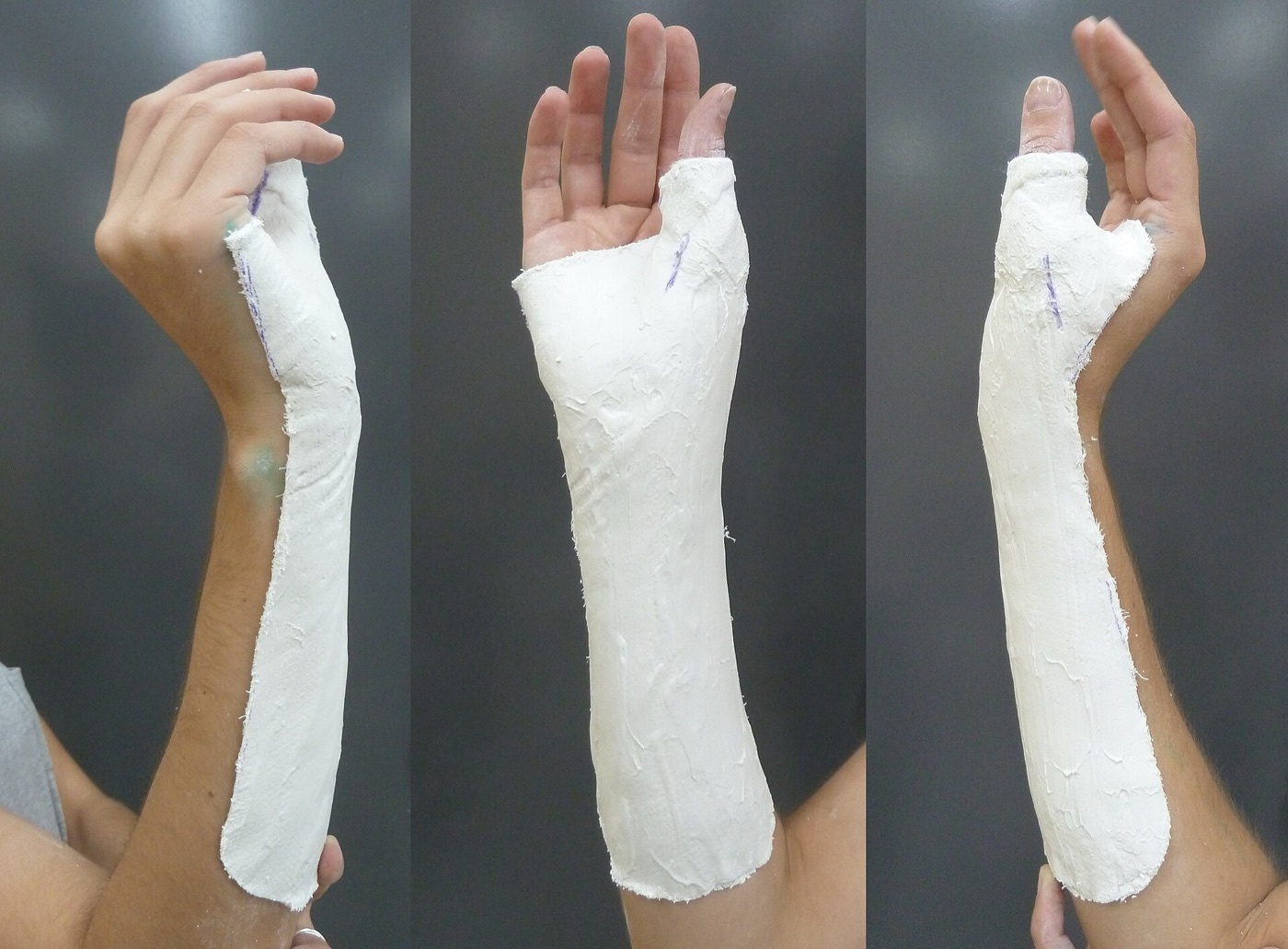
Figure 11. A plaster forearm cast immobilising the wrist after closed treatment of a distal radius fracture. Jackson P\&O, via Wikimedia Commons, CC BY-SA 3.0.
Part VII - Operative Treatment
7.1 The volar locking plate (the workhorse)
The volar locking plate has become the default operative implant, and the reason is biomechanical: in the osteoporotic metaphyseal bone typical of this fracture, conventional screws cannot grip the thin cortex, whereas fixed-angle locking screws or pegs placed into the subchondral bone support the articular surface like the struts of a bridge.[34] The approach is the modified Henry, or flexor carpi radialis (FCR), approach: the incision follows the FCR tendon, the interval is developed between the FCR and the radial artery (protecting the median nerve), the flexor pollicis longus is swept ulnarward, and the pronator quadratus is elevated to expose the fracture.[35] The plate is laid centrally on the flat volar surface, proximal to the watershed line, its built-in distal angle restoring volar tilt as it is brought down onto the shaft; the distal locking screws are placed into subchondral bone and checked fluoroscopically (starting on the ulnar side) to avoid penetrating the joint, the DRUJ, or the dorsal cortex.[36] Early motion is the aim, with immobilisation discontinued between two and four weeks unless the DRUJ is unstable or the fixation is questionable.[37]
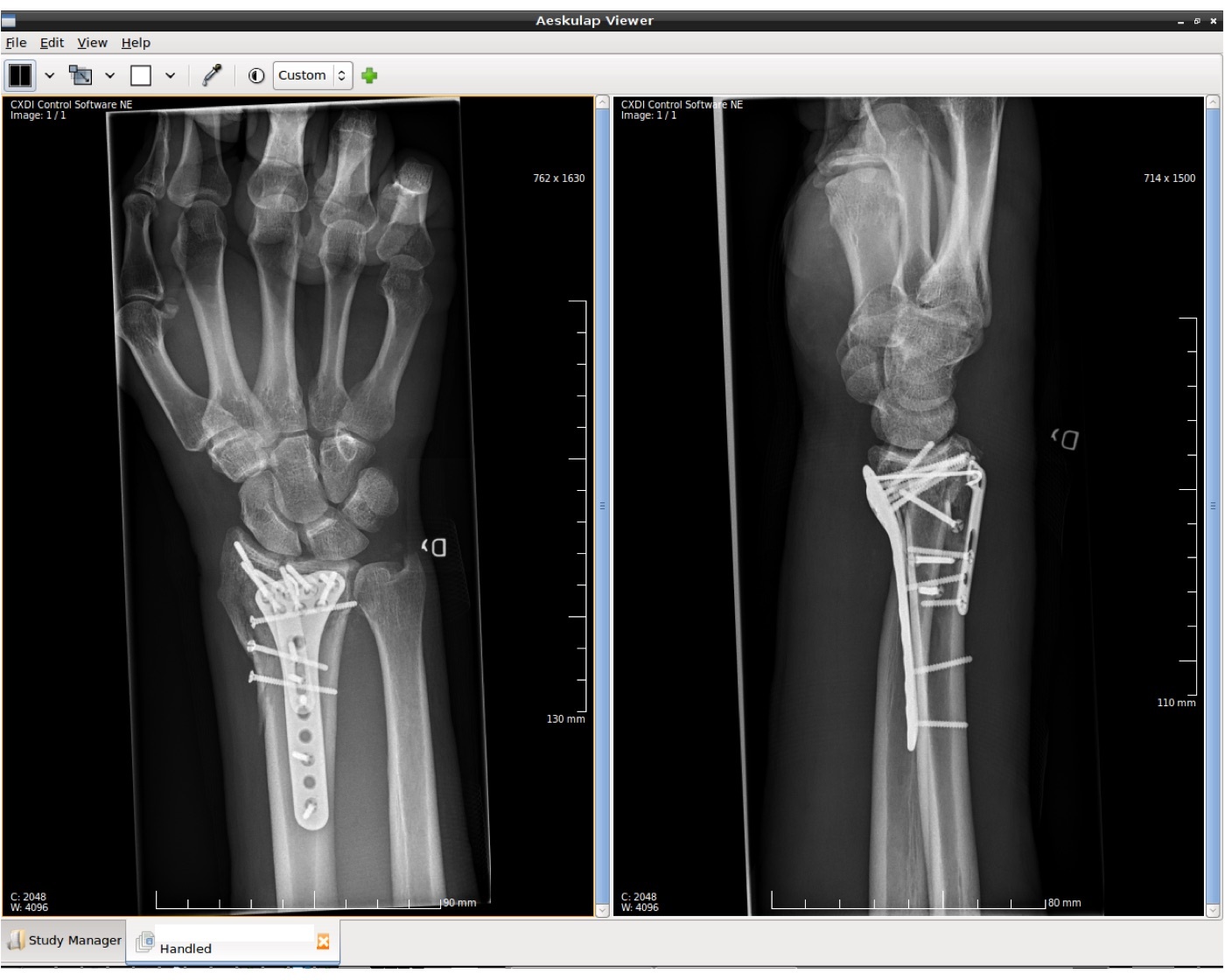
Figure 12. A distal radius fracture after volar plate-and-screw fixation, the workhorse construct (AP and lateral radiographs). DavidIvar, via Wikimedia Commons, CC BY-SA 4.0.
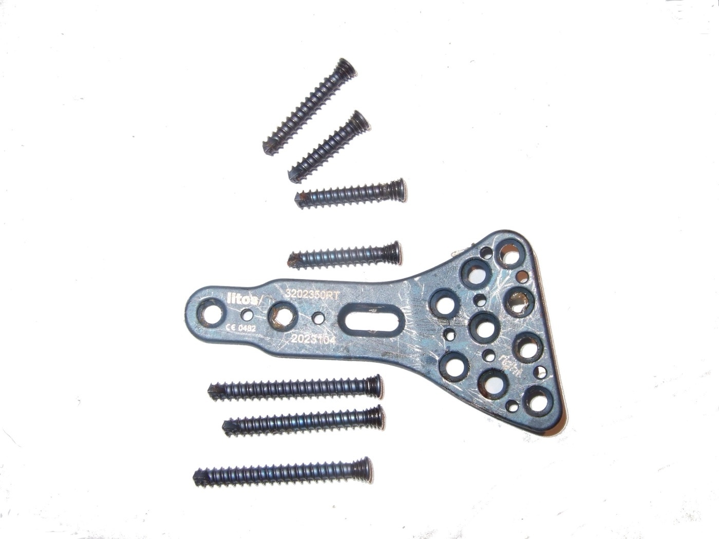
Figure 13. A distal radius (anatomic locking) plate and screws, the implant used for volar fixation. Codc, via Wikimedia Commons, CC BY-SA 4.0.
7.2 Dorsal plating and fragment-specific fixation
Dorsal plating fell out of favour when volar locking plates arrived, because dorsal hardware sits under thin soft tissue and irritates the extensor tendons, but it remains the right choice for dorsal shear fractures, displaced or impacted dorsal/die-punch fragments, and severely comminuted articular fractures where the joint surface must be seen and elevated.[38] The dorsal approach goes through the floor of the third extensor compartment, mobilising and then transposing the EPL out of its groove; the posterior interosseous nerve is excised for pain relief and Lister’s tubercle is taken down for the plate.[39] Fragment-specific fixation (separate small implants for the radial column, the dorsal fragments, and the volar-ulnar fragment) is reserved for rare patterns not addressable from a single approach, because the multiple incisions cause stiffness and, in a randomised comparison, a higher complication rate than volar plating.[40]
7.3 External fixation and ligamentotaxis
External fixation works by ligamentotaxis, the traction across the wrist ligaments pulling the fragments out to length; it is now reserved for severely comminuted fractures not amenable to internal fixation, for grossly contaminated open injuries, and as temporary fixation in the polytrauma patient.[41] A spanning (bridging) frame crosses the radiocarpal joint, with pins in the radial shaft and the second metacarpal; a non-bridging frame, possible when the distal fragment exceeds about 1 cm, captures the distal fragment directly and preserves wrist motion.[42] The cardinal error is over-distraction, which produces digital stiffness and complex regional pain syndrome: some distraction aids reduction, but too much is harmful.[43] Randomised trials show external fixation gives results broadly equivalent to volar plating at one year.[44]
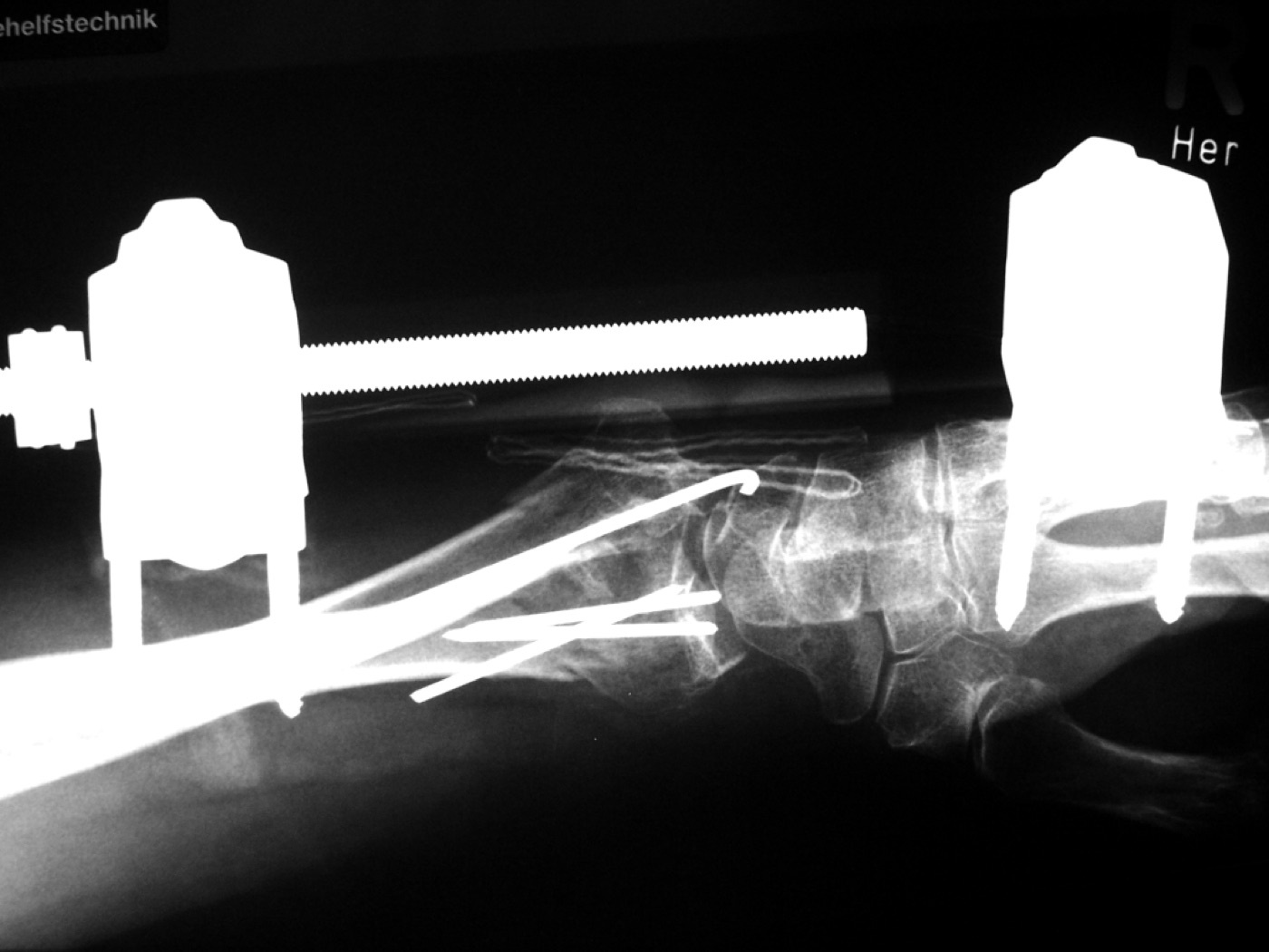
Figure 14. A comminuted distal radius fracture treated with a spanning (bridging) external fixator supplemented by K-wires. THWZ, via Wikimedia Commons, CC BY-SA 3.0.
7.4 Dorsal bridge (“internal fixator”) plating and the bridge plate
A dorsal spanning plate is an internal version of an external fixator: a long plate is tunnelled under the extensor tendons from the radial shaft to the second or third metacarpal, spanning the fracture and relying on ligamentotaxis.[45] Its advantages are that it permits immediate weight-bearing (valuable in the polytrauma patient who must use a walking aid) and is minimally invasive; its constraint is that the reduction must be achieved before the plate is applied, since it cannot be manipulated afterwards. The plate is removed once healing is confirmed, usually at three to four months.[46]
7.5 Percutaneous K-wires
Percutaneous K-wire fixation, including the Kapandji intrafocal technique (wires driven into the fracture site, levered to buttress the cortex, then driven into the far cortex), remains useful for extra-articular and simple fractures, often in younger patients.[47] Smaller, slightly flexible wires (0.045 to 0.054 inch) mould down the canal to give a three-point internal buttress; the main hazards are pin-tract infection and injury to the superficial radial nerve (up to 15% of cases), so wires are placed by open mini-incision down to bone rather than blindly.[48] In severe comminution, K-wires alone are insufficient and are supplemented by an external fixator.[49]
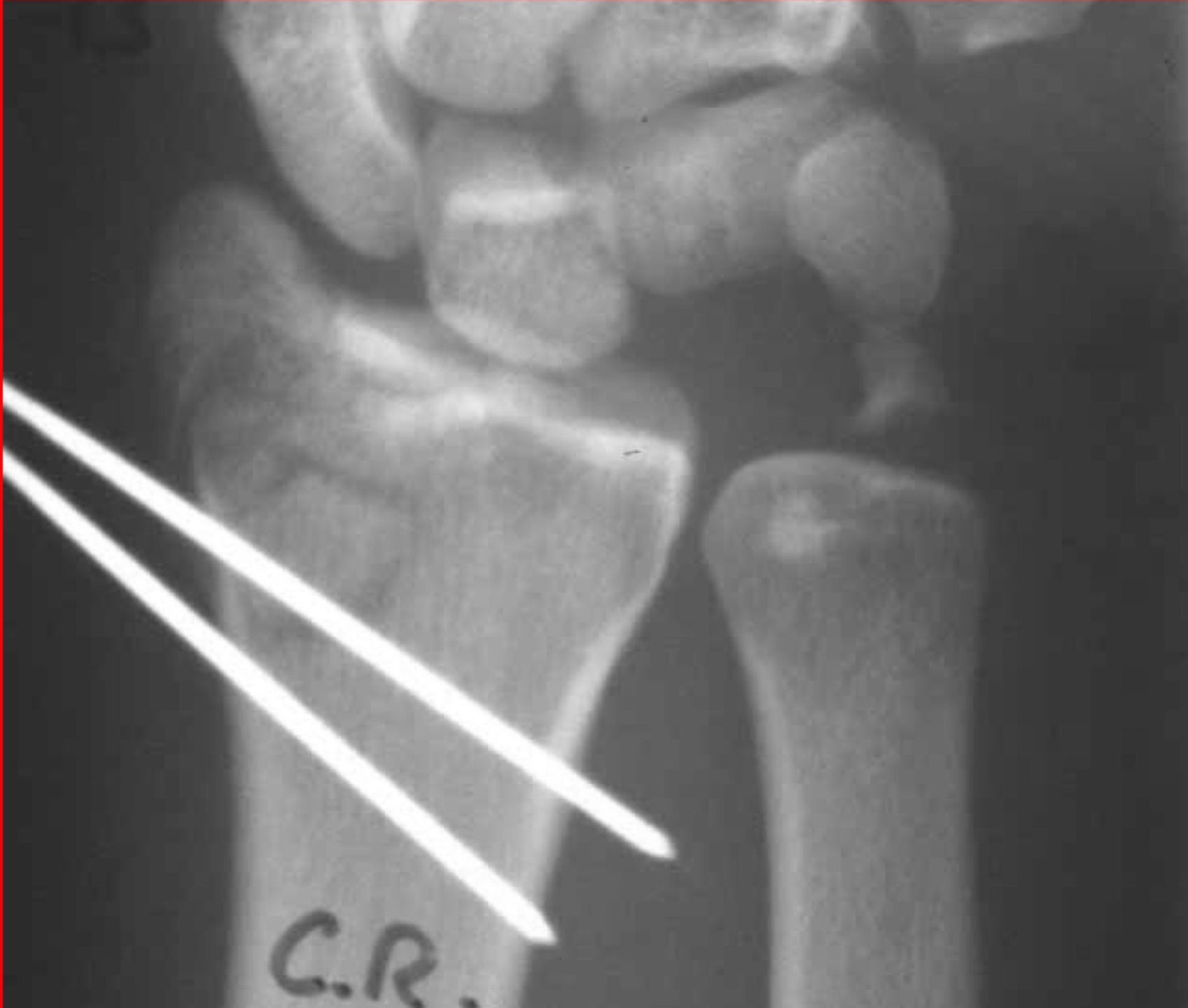
Figure 15. Percutaneous Kirschner wires across a reduced distal radius fracture. Curtishand, via Wikimedia Commons (public domain).
7.6 Choosing a method
No single construct has proven superior in randomised trials; the volar locking plate gives an early advantage in motion and function that has disappeared by one year.[50] The pragmatic algorithm matches the implant to the fracture pattern:[51]
| Fracture pattern | Preferred construct |
|---|---|
| Volar comminution | Volar plate |
| Dorsal shear | Dorsal plate |
| Dorsal comminution / die-punch | Dorsal plate, consider bone graft |
| Distal articular shear | Dorsal spanning plate or external fixator |
| Isolated radial styloid | Lag screws or buttress plate |
| Lunate-facet involvement | Volar plate with ulnar buttress |
The volar plate is the default; the surgeon should be versed in all techniques and choose the one that works best in their hands for the pattern in front of them.[52]
Part VIII - The Elderly Distal Radius Debate
Few questions in trauma are as well studied, or as instructive, as whether the older patient with a displaced distal radius fracture benefits from surgery. The headline evidence is Arora’s randomised trial in patients aged 65 and over, which found no difference in functional outcome at one year between nonoperative treatment and volar locking-plate fixation, despite the better radiographs in the operative group, and a higher complication rate in the operated patients.[53] This is the empirical basis for a principle that recurs throughout the chapter: in the elderly, radiographic malunion correlates poorly with function, so treatment should be driven by the patient’s symptoms and demands, not by the appearance of the film.[54] (The other large trials an examiner may name, the UK DRAFFT trial of K-wires versus volar plate and the multicentre WRIST study, are cited in the source as references but not discussed in detail. In younger patients, Rozental’s trial shows the volar plate’s early functional edge fading by one year.)[55]
Part IX - The DRUJ and Ulnar-Sided Injuries
Displaced fractures of the distal ulna often accompany the radial fracture and are easily overlooked. The governing principle is that they tend to reduce once the distal radius is anatomically fixed, so the ulna itself can often be left alone.[56] The key step is to test DRUJ stability after the radius is stabilised, comparing with the opposite wrist: most associated DRUJ injuries need no separate treatment if the radius (and particularly the sigmoid-notch reduction) is anatomical, because that restores the stabilising DIOM.[57] An ulnar styloid fracture is fixed only when the DRUJ remains unstable despite anatomical radial fixation, not routinely; an untreated styloid fracture does not by itself worsen the outcome.[58] When the distal ulna is genuinely unstable it is fixed (a styloid by tension band or cannulated screw, a head or neck fracture by a mini-fragment T-plate or a small locking plate) through an interval between extensor carpi ulnaris and flexor carpi ulnaris, protecting the dorsal sensory branch of the ulnar nerve.[59]
Part X - Complications
Reported complication rates for distal radius fractures range enormously, from 6% to 80%, the truth lying somewhere between; the three headline categories are nerve injury, tendon injury, and malunion.[60]
Median nerve / carpal tunnel syndrome is among the commonest, and can be acute, subacute, or delayed by as much as 25 years. Acute carpal tunnel syndrome is a progressive median neuropathy that demands urgent surgical release; it must be distinguished from a non-progressive contusion, which is observed.[61] Tendon injury is also common: the EPL is the most frequently ruptured tendon (after both nonoperative and operative treatment, classically as a late attritional rupture around Lister’s tubercle, reconstructed by extensor indicis proprius transfer), while flexor tendon ruptures, most often the flexor pollicis longus, follow a volar plate placed too far distally (distal to the watershed line) or left prominent.[62] Both are prevented by keeping the plate proximal to the watershed line and the dorsal screws from penetrating: the named scheme for grading volar-plate prominence relative to the watershed is the Soong grade, the practical threshold being a plate within 3 mm of the volar rim.[63]
Malunion is the third headline complication, commonest in the elderly nonoperatively treated patient, and again the principle is to treat the symptomatic patient, not the radiograph.[64] There is no agreed classification, but a working definition of malunion is radial inclination under 10°, dorsal (or volar) tilt over 20°, radial height under 10 mm, ulnar variance over 2 mm, or articular incongruity over 2 mm.[65] A symptomatic malunion is treated by corrective osteotomy (increasingly performed early, even at about 6 weeks, when it is technically easier), but the patient is warned that a normal wrist is rarely restored and grip strength rarely exceeds 70% of the other side.[66]
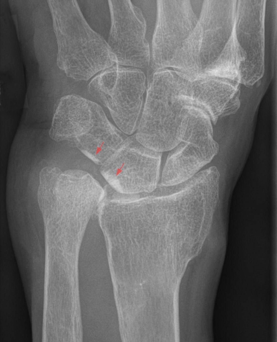
Figure 16. Ulnocarpal impaction after a shortened distal radius malunion (positive ulnar variance; AP view). Hellerhoff, via Wikimedia Commons, CC BY-SA 3.0.
Complex regional pain syndrome (CRPS) is a feared sequela. Vitamin C has been studied as prophylaxis, and here the evidence is genuinely conflicted: the 2009 AAOS guideline gave a moderate recommendation in favour of vitamin C, but a later randomised trial (Ekrol) and a meta-analysis (Evaniew) found no significant benefit (on low-quality evidence), so its use is not universal.[67] CRPS is best prevented by avoiding the over-tight or extreme-position cast (the “cripple cast”) and by early motion. Nonunion of the distal radius is exceedingly rare and confined to patients with major comorbidity.[68]
Part XI - A Synthesis: How to Reason About the Distal Radius
Begin by deciding which patient you are treating. In the older, lower-demand patient with a fragility fracture, the evidence is clear that function usually forgives a degree of radiographic malunion, so the threshold for surgery is high and the most important “treatment” may be the osteoporosis referral that prevents the next fracture. In the younger, higher-demand patient, or any patient whose post-reduction films breach the limits (articular step over 2 mm, dorsal tilt over 10°, shortening over 3 mm), the goal is anatomical reconstruction. Then read the fracture as columns and parameters: restore radial length, inclination, and volar tilt, and above all restore the lunate facet and sigmoid notch, because they carry the load and govern DRUJ congruity. Match the implant to the pattern using the simple table: volar plate for most, dorsal plate for dorsal shear and die-punch, a spanning construct for the unreconstructable comminuted fracture, lag screws for the isolated styloid, and remember that no construct has beaten the others at one year. Respect the soft tissues: keep the plate proximal to the watershed line to save the flexor tendons, keep the dorsal screws short to save the extensors, never force an extreme cast position, and release a progressive acute carpal tunnel syndrome urgently. Finally, test the DRUJ at the end of every case and fix the ulna only if it is truly unstable. The distal radius fracture is common enough to breed complacency and complex enough to punish it; thinking in patients, parameters, and patterns is what keeps the wrist working.
References
-
Rockwood & Green’s Fractures in Adults, p.2563 (the most common fracture of the upper extremity, at least one-sixth of fractures seen in the emergency department); AO Principles of Fracture Management, p.693 (most common fracture of the upper extremity, more than one-sixth of all fractures treated in the ED).
-
Rockwood & Green’s Fractures in Adults, p.2563 (bimodal: high-energy in young men, low-energy in elderly women); AO Principles of Fracture Management, pp.693, 696 (bending vs shear vs compression vs avulsion mechanisms; ~30% require surgery).
-
Rockwood & Green’s Fractures in Adults, p.2563 (second commonest fracture in the elderly; female:male 2-3×; US prevalence
600,000/yr, >640,000 in 2001; incidence rising); AO Principles of Fracture Management, p.693 (highest occurrence in paediatric, ~25% of fractures, and elderly, ~18%, populations).
-
Rockwood & Green’s Fractures in Adults, p.2563 (second commonest fracture in the elderly; female:male 2-3×; US prevalence
600,000/yr, >640,000 in 2001; incidence rising); AO Principles of Fracture Management, p.693 (highest occurrence in paediatric, ~25% of fractures, and elderly, ~18%, populations).
-
Rockwood & Green’s Fractures in Adults, p.2566 (FOOSH from standing height, dorsal pattern with wrist in 40-90° dorsiflexion, volar pattern with the flexed wrist; linear correlation of DXA T-score with instability and malunion); AO Principles of Fracture Management, p.693 (most produced by hyperextension; bending, shear, compression, and avulsion mechanisms).
-
Rockwood & Green’s Fractures in Adults, pp.2563, 2594 (low BMD the main risk factor and a predictor of future fractures; a distal radius fracture carries a 2-4× increased risk of future fractures; few patients receive osteoporosis work-up; surgeons should ensure follow-up via patient education, prompt DXA, and fracture-liaison services).
-
Rockwood & Green’s Fractures in Adults, pp.2563, 2594 (low BMD the main risk factor and a predictor of future fractures; a distal radius fracture carries a 2-4× increased risk of future fractures; few patients receive osteoporosis work-up; surgeons should ensure follow-up via patient education, prompt DXA, and fracture-liaison services).
-
AO Principles of Fracture Management, pp.696-697 (the 3-column concept of Rikli & Regazzoni: radial, intermediate, ulnar columns; the lunate fossa transmits the large share of load and is the key to the radiocarpal surface; the radial styloid is a buttress and ligament anchor; the ulna is the stable pivot, the ulnar column its distal end through the TFCC). The Rockwood chapter does not use the three-column model, working instead from Melone’s four-part “medial complex”; Rockwood & Green’s Fractures in Adults, p.2570.
-
AO Principles of Fracture Management, pp.696-697 (the 3-column concept of Rikli & Regazzoni: radial, intermediate, ulnar columns; the lunate fossa transmits the large share of load and is the key to the radiocarpal surface; the radial styloid is a buttress and ligament anchor; the ulna is the stable pivot, the ulnar column its distal end through the TFCC). The Rockwood chapter does not use the three-column model, working instead from Melone’s four-part “medial complex”; Rockwood & Green’s Fractures in Adults, p.2570.
-
Rockwood & Green’s Fractures in Adults, p.2567 (radial height 11-12 mm, range 8-18; radial inclination 22-23°, range 12-30; volar tilt averaging 11-12° in the body text); AO Principles of Fracture Management, p.694 (radial height average 12 mm, radial inclination 23°, palmar tilt 11-12°). Note an internal discrepancy in the Rockwood source: the body text gives volar tilt 11-12° while the Fig. 42-2 caption gives an average of 11-22° (range 0-28°); the consistently cited value is ~11°.
-
Rockwood & Green’s Fractures in Adults, pp.2566-2568 (teardrop angle ~70°, <45° associated with articular gap/step-off; ulnar variance defined qualitatively as a measure of radial shortening; measurements taken on a true lateral, 5° rotation changes volar tilt by 1.6°, pronation reduces apparent radial length by up to 0.5 mm); AO Principles of Fracture Management, p.694 (teardrop angle average 70°; 60% of the population ulnar neutral).
-
Rockwood & Green’s Fractures in Adults, pp.2566-2568 (teardrop angle ~70°, <45° associated with articular gap/step-off; ulnar variance defined qualitatively as a measure of radial shortening; measurements taken on a true lateral, 5° rotation changes volar tilt by 1.6°, pronation reduces apparent radial length by up to 0.5 mm); AO Principles of Fracture Management, p.694 (teardrop angle average 70°; 60% of the population ulnar neutral).
-
Rockwood & Green’s Fractures in Adults, pp.2573, 2579 (volar surface flat and covered by pronator quadratus, ideal for plating; dorsal surface convex and grooved, Lister’s tubercle a pulley for EPL; plate proximal to the watershed line to avoid tendon complications); AO Principles of Fracture Management, p.699 (the watershed line is the margin between the elevated structures and the volar wrist extrinsic ligaments, which must not be detached; the flat palmar surface auto-corrects fragment malposition).
-
Rockwood & Green’s Fractures in Adults, pp.2573, 2579 (volar surface flat and covered by pronator quadratus, ideal for plating; dorsal surface convex and grooved, Lister’s tubercle a pulley for EPL; plate proximal to the watershed line to avoid tendon complications); AO Principles of Fracture Management, p.699 (the watershed line is the margin between the elevated structures and the volar wrist extrinsic ligaments, which must not be detached; the flat palmar surface auto-corrects fragment malposition).
-
AO Principles of Fracture Management, pp.696, 715 (sigmoid notch and DRUJ; the DIOM as secondary DRUJ stabiliser, from the distal one-sixth of the ulna; ulnar styloid tip fracture present in 50% of distal radius fractures; most DRUJ injuries need no extra surgery if the radius is anatomically and stably fixed); Rockwood & Green’s Fractures in Adults, p.2565 (median nerve and acute carpal tunnel syndrome).
-
AO Principles of Fracture Management, pp.696, 715 (sigmoid notch and DRUJ; the DIOM as secondary DRUJ stabiliser, from the distal one-sixth of the ulna; ulnar styloid tip fracture present in 50% of distal radius fractures; most DRUJ injuries need no extra surgery if the radius is anatomically and stably fixed); Rockwood & Green’s Fractures in Adults, p.2565 (median nerve and acute carpal tunnel syndrome).
-
Rockwood & Green’s Fractures in Adults, pp.2565-2566, 2569-2570 (Colles 1814, dorsally angulated apex-volar metaphyseal, “dinner-fork” deformity; Smith the volarly angulated reverse Colles; Barton intra-articular dorsal rim with dorsal subluxation and the volar/reverse Barton; Chauffeur’s the radial styloid fracture; die-punch the depressed lunate-facet fragment / Melone medial complex). The AO chapter names only Colles, and only historically (AO Principles, p.693); the “Hutchinson” synonym for the chauffeur’s fracture is standard teaching but is not used in the Rockwood extract (Rockwood, p.2569).
-
Rockwood & Green’s Fractures in Adults, pp.2569-2570 (AO classification 1990, AO/OTA 2007; three types A extra-articular, B partial articular, C complete articular, 27 patterns); AO Principles of Fracture Management, p.697 (segment 2R3; A extra-articular, B partial articular, C complete articular, in order of increasing severity; worked codes 2R3A2, 2R3B1, 2R3C1).
-
AO Principles of Fracture Management, p.697 (Fernandez classification by mechanism: I metaphyseal bending, II joint-surface shear, III joint-surface compression, IV avulsion/radiocarpal fracture-dislocation, V combined high-velocity); Rockwood & Green’s Fractures in Adults, pp.2570-2571 (Fernandez mechanism-based, considers associated ligament and ulnar injuries and DRUJ stability).
-
Rockwood & Green’s Fractures in Adults, pp.2570-2571 (Frykman by intra-articular involvement and ulnar fracture; Melone four-part medial complex; Gartland & Werley three groups; Mayo by facet involvement; only AO/OTA A/B/C reliable, subtypes not; CT does not improve reliability or outcomes); AO Principles of Fracture Management, p.697 (“all contemporary classification systems lack intra-rater and inter-rater reliability”).
-
Rockwood & Green’s Fractures in Adults, pp.2570-2571 (Frykman by intra-articular involvement and ulnar fracture; Melone four-part medial complex; Gartland & Werley three groups; Mayo by facet involvement; only AO/OTA A/B/C reliable, subtypes not; CT does not improve reliability or outcomes); AO Principles of Fracture Management, p.697 (“all contemporary classification systems lack intra-rater and inter-rater reliability”).
-
Rockwood & Green’s Fractures in Adults, p.2565 (open wounds usually volar-ulnar; distal radius fractures carry the highest risk of acute and chronic carpal tunnel syndrome; acute CTS must be addressed urgently; distinguish progressive ACTS from the constant numbness of a contusion); AO Principles of Fracture Management, p.693 (assess all wrist fractures for open wounds, usually palmar-ulnar, and for median or ulnar nerve injury).
-
Rockwood & Green’s Fractures in Adults, p.2565 (open wounds usually volar-ulnar; distal radius fractures carry the highest risk of acute and chronic carpal tunnel syndrome; acute CTS must be addressed urgently; distinguish progressive ACTS from the constant numbness of a contusion); AO Principles of Fracture Management, p.693 (assess all wrist fractures for open wounds, usually palmar-ulnar, and for median or ulnar nerve injury).
-
Rockwood & Green’s Fractures in Adults, pp.2564-2565 (scapholunate injury 4.7-46%, lunotriquetral 12-34%, TFCC 39-82%, chondral up to 32%; significance often controversial); AO Principles of Fracture Management, p.696 (~30-40% of distal radius fractures have associated soft-tissue injuries, most often TFCC, scapholunate more common with intra-articular fractures).
-
Rockwood & Green’s Fractures in Adults, pp.2566, 2568-2569 (standard PA, lateral, oblique; intra-op dorsal tangential and radial-incline views; CT for articular comminution, sigmoid notch and lunate facet, no proven outcome benefit); AO Principles of Fracture Management, pp.693-694, 696 (AP and lateral mandatory, oblique and contralateral views helpful; CT for sigmoid-notch/lunate-facet congruity, 3-D reconstruction).
-
Rockwood & Green’s Fractures in Adults, pp.2566, 2568-2569 (standard PA, lateral, oblique; intra-op dorsal tangential and radial-incline views; CT for articular comminution, sigmoid notch and lunate facet, no proven outcome benefit); AO Principles of Fracture Management, pp.693-694, 696 (AP and lateral mandatory, oblique and contralateral views helpful; CT for sigmoid-notch/lunate-facet congruity, 3-D reconstruction).
-
Rockwood & Green’s Fractures in Adults, p.2574 (AAOS 2009 CPG: surgery suggested for post-reduction intra-articular step-off >2 mm, dorsal tilt >10°, radial shortening >3 mm); AO Principles of Fracture Management, pp.694, 698 (instability criteria of Ng & McQueen: metaphyseal fragmentation, angular deformity >10°, shortening >5 mm, articular displacement >2 mm, carpal malalignment; surgical thresholds in high-demand patients >3 mm shortening, >10° dorsal tilt, >2 mm articular displacement).
-
Rockwood & Green’s Fractures in Adults, p.2574 (AAOS 2009 CPG: surgery suggested for post-reduction intra-articular step-off >2 mm, dorsal tilt >10°, radial shortening >3 mm); AO Principles of Fracture Management, pp.694, 698 (instability criteria of Ng & McQueen: metaphyseal fragmentation, angular deformity >10°, shortening >5 mm, articular displacement >2 mm, carpal malalignment; surgical thresholds in high-demand patients >3 mm shortening, >10° dorsal tilt, >2 mm articular displacement).
-
The Lafontaine instability/redisplacement criteria (age, dorsal comminution, initial dorsal angulation, intra-articular involvement, associated ulnar fracture) are standard teaching for predicting loss of reduction after closed treatment but are not present in the mined Rockwood or AO extracts (Rockwood, p.2574, confirms no named instability checklist is given; Lafontaine is cited only as a reference). Stated as established teaching, not page-cited to these sources.
-
Rockwood & Green’s Fractures in Adults, p.2574 (nonoperative for minimally displaced fractures, patients unable to tolerate casting, and low-demand patients; AAOS: no difference between casting and surgery at age ≥55); AO Principles of Fracture Management, p.693 (most distal radius fractures, especially dorsally displaced extra-articular fractures in the elderly, treated nonoperatively; ~30% need surgery).
-
Rockwood & Green’s Fractures in Adults, pp.2573-2574 (rigid cast superior to removable splint for reduced displaced fractures; no benefit to above-elbow immobilisation; MCP joints left free with digit exercises; sugar-tong, reverse sugar-tong, Charnley, or fibreglass options, none superior). The closed-reduction manoeuvre and anaesthesia detail precede the mined extract; the manoeuvre (re-create then reverse the deformity under traction) and the haematoma-block option are standard teaching consistent with the source’s reference list (Rockwood refs 49, 84, 109).
-
AO Principles of Fracture Management, pp.714-715 (CRPS from swelling in a too-tight cast or extreme wrist positioning, Böhler’s “cripple cast”/“krüppel gips,” which is forbidden; extreme palmar or radioulnar positioning to force reduction causes stiffness and CRPS). The “Cotton-Loder” eponym for the abandoned extreme-flexion position is standard teaching; the Rockwood and AO extracts describe the avoidance of extreme flexion (the cripple cast) without using the eponym.
-
Rockwood & Green’s Fractures in Adults, p.2574 (weekly radiographs for 3 consecutive weeks in active or questionably stable fractures; unacceptable alignment at follow-up treated as a displaced fracture and considered for surgery; low-demand patients splinted/cast for 3-4 weeks; Roth: 0 of 82 truly nondisplaced fractures displaced).
-
AO Principles of Fracture Management, pp.699, 704 (thin metaphyseal cortex and osteoporotic bone defeat conventional screws; fixed-angle locking plates give subchondral support and metaphyseal fixation, restore length/inclination/tilt, make dorsal bone grafting unnecessary, and allow early mobilisation with less CRPS); Rockwood & Green’s Fractures in Adults, p.2577 (volar locked plating has risen rapidly in popularity, treats unstable extra-articular, volar shear, and simple articular fractures, and with variable-angle plates more complex intra-articular patterns).
-
Rockwood & Green’s Fractures in Adults, pp.2578-2579 (FCR/modified Henry approach: incision over FCR, interval between radial artery and FCR, elevate pronator quadratus; plate central and proximal to the watershed line; distal locking screws into subchondral bone; capture the volar lunate-facet fragment); AO Principles of Fracture Management, pp.699-702 (FCR approach between FCR and radial artery, protect the median nerve, elevate pronator quadratus to the fibrous transition zone proximal to the watershed line; the plate’s built-in offset angle restores palmar inclination; start fixation on the ulnar side and verify under image intensification to avoid joint/DRUJ/dorsal penetration).
-
Rockwood & Green’s Fractures in Adults, pp.2578-2579 (FCR/modified Henry approach: incision over FCR, interval between radial artery and FCR, elevate pronator quadratus; plate central and proximal to the watershed line; distal locking screws into subchondral bone; capture the volar lunate-facet fragment); AO Principles of Fracture Management, pp.699-702 (FCR approach between FCR and radial artery, protect the median nerve, elevate pronator quadratus to the fibrous transition zone proximal to the watershed line; the plate’s built-in offset angle restores palmar inclination; start fixation on the ulnar side and verify under image intensification to avoid joint/DRUJ/dorsal penetration).
-
Rockwood & Green’s Fractures in Adults, p.2580 (MCP joints free, immediate finger motion, immobilisation discontinued at 2-4 weeks, 6 weeks if DRUJ unstable or fixation questionable); AO Principles of Fracture Management, p.714 (early functional use of the hand and forearm rotation mandatory, sling for only a few days).
-
Rockwood & Green’s Fractures in Adults, pp.2580-2581 (dorsal plating fell out of favour for extensor irritation but is used for dorsal shear, die-punch, and severely comminuted articular fractures; approach through the third extensor compartment, EPL transposed, PIN excised, Lister’s tubercle taken down); AO Principles of Fracture Management, pp.704-706 (dorsal approach via the third compartment for dorsal shear 2R3B2, displaced dorsal lunate-facet, and impacted central fragments; EPL transposed over the repaired retinaculum; dorsal locking plates buttress the radial and intermediate columns).
-
Rockwood & Green’s Fractures in Adults, pp.2580-2581 (dorsal plating fell out of favour for extensor irritation but is used for dorsal shear, die-punch, and severely comminuted articular fractures; approach through the third extensor compartment, EPL transposed, PIN excised, Lister’s tubercle taken down); AO Principles of Fracture Management, pp.704-706 (dorsal approach via the third compartment for dorsal shear 2R3B2, displaced dorsal lunate-facet, and impacted central fragments; EPL transposed over the repaired retinaculum; dorsal locking plates buttress the radial and intermediate columns).
-
Rockwood & Green’s Fractures in Adults, pp.2582-2583, 2588-2589 (fragment-specific fixation for rare patterns not addressable volarly or dorsally; multiple incisions cause stiffness; Landgren RCT showed no outcome difference but a higher complication rate than volar plating).
-
Rockwood & Green’s Fractures in Adults, pp.2583-2585 (external fixation by ligamentotaxis, described 1944 by Anderson & O’Neill, “ligamentary taxis” coined by Vidal 1977; reserved for severe comminution; spanning frame with radial-shaft and second-metacarpal pins, non-bridging for distal fragments >1 cm); AO Principles of Fracture Management, pp.706-708 (external fixator for severe open/contaminated, temporary high-energy, polytrauma, or as a neutralising device; proximal Schanz pins in the radial shaft, distal pins in the second metacarpal at 30-40°).
-
Rockwood & Green’s Fractures in Adults, pp.2583-2585 (external fixation by ligamentotaxis, described 1944 by Anderson & O’Neill, “ligamentary taxis” coined by Vidal 1977; reserved for severe comminution; spanning frame with radial-shaft and second-metacarpal pins, non-bridging for distal fragments >1 cm); AO Principles of Fracture Management, pp.706-708 (external fixator for severe open/contaminated, temporary high-energy, polytrauma, or as a neutralising device; proximal Schanz pins in the radial shaft, distal pins in the second metacarpal at 30-40°).
-
Rockwood & Green’s Fractures in Adults, pp.2586, 2588 (over-distraction increases digital stiffness and CRPS, some distraction beneficial but too much harmful; Egol RCT external fixation vs volar plating equivalent at 1 year); AO Principles of Fracture Management, pp.708, 715 (avoid over-distraction and extreme wrist positioning, both causing stiffness and CRPS; palmar locking plate and external fixator give similar results, Karantana).
-
Rockwood & Green’s Fractures in Adults, pp.2586, 2588 (over-distraction increases digital stiffness and CRPS, some distraction beneficial but too much harmful; Egol RCT external fixation vs volar plating equivalent at 1 year); AO Principles of Fracture Management, pp.708, 715 (avoid over-distraction and extreme wrist positioning, both causing stiffness and CRPS; palmar locking plate and external fixator give similar results, Karantana).
-
Rockwood & Green’s Fractures in Adults, pp.2586-2587, 2590 (dorsal spanning “internal fixator” plate tunnelled to the second/third metacarpal, relies on ligamentotaxis, allows immediate weight-bearing, reduction must precede application; low rates of extensor irritation and digit stiffness); AO Principles of Fracture Management, p.714 (joint-spanning bridge plate for extreme comminution using the proximal carpal row as a template, MIPO technique, removed at 3-4 months).
-
Rockwood & Green’s Fractures in Adults, pp.2586-2587, 2590 (dorsal spanning “internal fixator” plate tunnelled to the second/third metacarpal, relies on ligamentotaxis, allows immediate weight-bearing, reduction must precede application; low rates of extensor irritation and digit stiffness); AO Principles of Fracture Management, p.714 (joint-spanning bridge plate for extreme comminution using the proximal carpal row as a template, MIPO technique, removed at 3-4 months).
-
Rockwood & Green’s Fractures in Adults, pp.2576-2577 (percutaneous K-wires and the Kapandji intrafocal technique, three wires raised to buttress the cortex; 0.045-0.054-in wires give an internal three-point mould; pin-tract infection and superficial radial nerve injury up to 15%, place under direct vision); AO Principles of Fracture Management, pp.706-707 (K-wires for provisional/temporary fixation, insufficient alone in severe comminution and supplemented by an external fixator).
-
Rockwood & Green’s Fractures in Adults, pp.2576-2577 (percutaneous K-wires and the Kapandji intrafocal technique, three wires raised to buttress the cortex; 0.045-0.054-in wires give an internal three-point mould; pin-tract infection and superficial radial nerve injury up to 15%, place under direct vision); AO Principles of Fracture Management, pp.706-707 (K-wires for provisional/temporary fixation, insufficient alone in severe comminution and supplemented by an external fixator).
-
Rockwood & Green’s Fractures in Adults, pp.2576-2577 (percutaneous K-wires and the Kapandji intrafocal technique, three wires raised to buttress the cortex; 0.045-0.054-in wires give an internal three-point mould; pin-tract infection and superficial radial nerve injury up to 15%, place under direct vision); AO Principles of Fracture Management, pp.706-707 (K-wires for provisional/temporary fixation, insufficient alone in severe comminution and supplemented by an external fixator).
-
Rockwood & Green’s Fractures in Adults, pp.2588-2589 (“no specific method has been shown to be significantly better than any other”; early benefit to volar plating with no long-term difference); AO Principles of Fracture Management, p.715 (volar locking plate gives better early ROM/grip/function than K-wires at 3 and 6 months, insignificant by 12 months; palmar plate and external fixator similar).
-
Rockwood & Green’s Fractures in Adults, pp.2589-2590 (Table 42-1, choosing fixation method; volar locked plate the default except where another method gives better fragment control or the fracture is too comminuted; surgeon should be well versed in all techniques).
-
Rockwood & Green’s Fractures in Adults, pp.2589-2590 (Table 42-1, choosing fixation method; volar locked plate the default except where another method gives better fragment control or the fracture is too comminuted; surgeon should be well versed in all techniques).
-
AO Principles of Fracture Management, p.715 (Arora RCT, dorsally displaced fractures in patients >65: no difference in outcome at 12 months, anatomical reconstruction did not improve ROM or ADLs, significantly higher complication rate in the operative group); Rockwood & Green’s Fractures in Adults, pp.2592, 2595 (radiographic outcomes do not consistently correlate with function in the elderly; Arora RCT annotated as showing no functional difference between nonoperative management and volar plating in patients ≥65).
-
AO Principles of Fracture Management, p.715 (Arora RCT, dorsally displaced fractures in patients >65: no difference in outcome at 12 months, anatomical reconstruction did not improve ROM or ADLs, significantly higher complication rate in the operative group); Rockwood & Green’s Fractures in Adults, pp.2592, 2595 (radiographic outcomes do not consistently correlate with function in the elderly; Arora RCT annotated as showing no functional difference between nonoperative management and volar plating in patients ≥65).
-
Rockwood & Green’s Fractures in Adults, pp.2588, 2595 (Rozental: volar plating better DASH at 6-12 weeks, no difference at 1 year; UK DRAFFT (Costa) and the WRIST study cited as references). DRAFFT2 and ORCHID are not present in the source extracts; DRAFFT and WRIST appear as reference titles only, so their detailed results are noted here as standard teaching rather than page-cited findings.
-
Rockwood & Green’s Fractures in Adults, p.2590 (distal ulna fractures often accompany and are overlooked, tend to reduce with radial fixation, fixation often avoidable; test DRUJ stability after stabilising the radius and compare with the opposite wrist); AO Principles of Fracture Management, p.715 (most DRUJ injuries need no extra surgery if anatomical, stable radial fixation is achieved, especially sigmoid-notch reduction restoring the DIOM).
-
Rockwood & Green’s Fractures in Adults, p.2590 (distal ulna fractures often accompany and are overlooked, tend to reduce with radial fixation, fixation often avoidable; test DRUJ stability after stabilising the radius and compare with the opposite wrist); AO Principles of Fracture Management, p.715 (most DRUJ injuries need no extra surgery if anatomical, stable radial fixation is achieved, especially sigmoid-notch reduction restoring the DIOM).
-
Rockwood & Green’s Fractures in Adults, pp.2590-2591 (fix the ulnar styloid only if the DRUJ is unstable in its presence; untreated styloid fractures do not worsen outcomes; interval between ECU and FCU, protect the dorsal sensory branch of the ulnar nerve; styloid by cannulated screw or tension band, distal metaphysis by mini-fragment T-plate); AO Principles of Fracture Management, p.716 (DRUJ instability test with a palpable “clunk”; displaced distal ulnar shaft fracture may need ORIF with a 2.7 locking compression plate).
-
Rockwood & Green’s Fractures in Adults, pp.2590-2591 (fix the ulnar styloid only if the DRUJ is unstable in its presence; untreated styloid fractures do not worsen outcomes; interval between ECU and FCU, protect the dorsal sensory branch of the ulnar nerve; styloid by cannulated screw or tension band, distal metaphysis by mini-fragment T-plate); AO Principles of Fracture Management, p.716 (DRUJ instability test with a palpable “clunk”; displaced distal ulnar shaft fracture may need ORIF with a 2.7 locking compression plate).
-
Rockwood & Green’s Fractures in Adults, p.2591 (overall complication rates 6-80%; the three headlined categories: nerve injury, tendon injury, malunion).
-
Rockwood & Green’s Fractures in Adults, pp.2591-2592 (carpal tunnel syndrome one of the most common complications, acute/subacute/delayed up to 25 years; acute CTS progressive and needs urgent release, distinguish from a non-progressive contusion that improves); AO Principles of Fracture Management, p.714 (acute carpal tunnel syndrome may follow injury or inappropriate cast positioning, examine before and after reduction).
-
Rockwood & Green’s Fractures in Adults, pp.2580, 2592 (EPL the most commonly ruptured tendon in both nonoperative and operative patients, reconstructed by EIP transfer; flexor ruptures, most often FPL, from plate irritation; keep the plate proximal to the watershed line and avoid prominent dorsal screws; some advocate removing plates within 3 mm of the volar rim); AO Principles of Fracture Management, p.714 (extensor rupture from dorsal screw penetration, flexor rupture from a palmar plate too far distal and uncovered by pronator quadratus, EPL rupture after cast treatment). The “Soong grade” for volar-plate prominence is the named system in common use; the source describes the prominence threshold (within 3 mm of the volar rim) without naming Soong in the mined narrative.
-
Rockwood & Green’s Fractures in Adults, pp.2580, 2592 (EPL the most commonly ruptured tendon in both nonoperative and operative patients, reconstructed by EIP transfer; flexor ruptures, most often FPL, from plate irritation; keep the plate proximal to the watershed line and avoid prominent dorsal screws; some advocate removing plates within 3 mm of the volar rim); AO Principles of Fracture Management, p.714 (extensor rupture from dorsal screw penetration, flexor rupture from a palmar plate too far distal and uncovered by pronator quadratus, EPL rupture after cast treatment). The “Soong grade” for volar-plate prominence is the named system in common use; the source describes the prominence threshold (within 3 mm of the volar rim) without naming Soong in the mined narrative.
-
Rockwood & Green’s Fractures in Adults, pp.2592-2593 (malunion commonest in elderly nonoperative patients, treat the symptomatic patient not the radiograph; working definition radial inclination <10°, tilt >20°, radial height <10 mm, ulnar variance >2 mm, articular incongruity >2 mm; corrective osteotomy increasingly performed early, ~6 weeks; grip rarely exceeds 70% of the contralateral side).
-
Rockwood & Green’s Fractures in Adults, pp.2592-2593 (malunion commonest in elderly nonoperative patients, treat the symptomatic patient not the radiograph; working definition radial inclination <10°, tilt >20°, radial height <10 mm, ulnar variance >2 mm, articular incongruity >2 mm; corrective osteotomy increasingly performed early, ~6 weeks; grip rarely exceeds 70% of the contralateral side).
-
Rockwood & Green’s Fractures in Adults, pp.2592-2593 (malunion commonest in elderly nonoperative patients, treat the symptomatic patient not the radiograph; working definition radial inclination <10°, tilt >20°, radial height <10 mm, ulnar variance >2 mm, articular incongruity >2 mm; corrective osteotomy increasingly performed early, ~6 weeks; grip rarely exceeds 70% of the contralateral side).
-
Rockwood & Green’s Fractures in Adults, p.2594 (vitamin C for CRPS: 2009 AAOS moderate recommendation for, Zollinger; later Ekrol RCT and Evaniew meta-analysis found no significant benefit on low-quality evidence; not routine practice; nonunion exceedingly rare); AO Principles of Fracture Management, pp.714-715 (CRPS from a too-tight or extreme-position “cripple cast”, prevented by good cast technique and early motion).
-
Rockwood & Green’s Fractures in Adults, p.2594 (vitamin C for CRPS: 2009 AAOS moderate recommendation for, Zollinger; later Ekrol RCT and Evaniew meta-analysis found no significant benefit on low-quality evidence; not routine practice; nonunion exceedingly rare); AO Principles of Fracture Management, pp.714-715 (CRPS from a too-tight or extreme-position “cripple cast”, prevented by good cast technique and early motion).
-
Rockwood & Green’s Fractures in Adults, pp.2563, 2567-2568; AO Principles of Fracture Management, pp.693, 696.
-
Rockwood & Green’s Fractures in Adults, pp.2566-2568; AO Principles of Fracture Management, p.694.
-
Rockwood & Green’s Fractures in Adults, pp.2565-2566, 2569-2570.
-
Rockwood & Green’s Fractures in Adults, pp.2569-2571; AO Principles of Fracture Management, p.697.
-
Rockwood & Green’s Fractures in Adults, pp.2565, 2591-2592; AO Principles of Fracture Management, p.714.
-
Rockwood & Green’s Fractures in Adults, p.2574; AO Principles of Fracture Management, pp.694, 698.
-
Rockwood & Green’s Fractures in Adults, pp.2573-2574; AO Principles of Fracture Management, pp.714-715.
-
Rockwood & Green’s Fractures in Adults, pp.2577-2579; AO Principles of Fracture Management, pp.699-702.
-
Rockwood & Green’s Fractures in Adults, pp.2579, 2592; AO Principles of Fracture Management, pp.699, 714.
-
Rockwood & Green’s Fractures in Adults, pp.2576-2587; AO Principles of Fracture Management, pp.704-708, 714.
-
AO Principles of Fracture Management, p.715; Rockwood & Green’s Fractures in Adults, pp.2592, 2595.
-
Rockwood & Green’s Fractures in Adults, pp.2590-2591; AO Principles of Fracture Management, pp.715-716.
-
Rockwood & Green’s Fractures in Adults, pp.2580, 2592; AO Principles of Fracture Management, p.714.
-
Rockwood & Green’s Fractures in Adults, p.2594; AO Principles of Fracture Management, pp.714-715.