Contents
- Orientation: An Articular Metaphyseal Fracture Pulled into Recurvatum and Valgus
- Part I - Definition, Epidemiology, and the Geriatric Problem
- Part II - Applied Anatomy and the Deforming Forces
- Part III - Classification
- Part IV - Assessment, Associated Injuries, and Imaging
- Part V - Treatment Principles and Nonoperative Care
- Part VI - Operative Treatment
- Part VII - Periprosthetic Distal Femur Fractures About a Total Knee Replacement
- Part VIII - Complications
- Part IX - Paediatric Distal Femoral Physeal Fractures
- Part X - A Synthesis: How to Reason About the Distal Femur Fracture
- References
Orientation: An Articular Metaphyseal Fracture Pulled into Recurvatum and Valgus
The distal femur fracture is a problem of two zones and two populations. The two zones are the articular block, which must be reconstructed anatomically and held with absolute stability, and the metaphysis above it, which is bridged with relative stability so that it heals by callus; getting the relationship between these two zones right, without leaving the limb in valgus, recurvatum, or varus collapse, is the whole technical task.[1] The two populations are the high-energy young patient (a dashboard or axial-load injury, often comminuted, intra-articular, and open) and the low-energy elderly patient with osteoporotic bone, in whom the fracture behaves like a hip fracture, carrying a comparable one-year mortality and demanding early surgery and early weight bearing. A third, growing group sits within the elderly: the periprosthetic fracture above a total knee replacement, where the implant both causes the fracture (through stress shielding and anterior notching) and constrains its fixation. The deforming forces are characteristic and worth fixing in mind from the start. The gastrocnemius pulls the distal condylar fragment into extension (apex-posterior, recurvatum) while the quadriceps, hamstrings, and adductors shorten the limb and pull it into varus, so the surgeon flexes the knee to relax the gastrocnemius and works hard to avoid the valgus and recurvatum that the anatomy invites.[2]
Part I - Definition, Epidemiology, and the Geriatric Problem
The supracondylar region of the femur is the zone between the femoral condyles and the junction of the metaphysis with the diaphysis, roughly the distal 15 cm measured from the articular surface.[3] Distal femur fractures make up about 6 % of all femoral fractures and show a bimodal distribution: high-energy injuries in young patients (motor-vehicle and motorcycle crashes, axial loading on a flexed knee, frequently comminuted, intra-articular, and open) and low-energy injuries in the osteoporotic elderly (a ground-level fall on a flexed knee). One-third of the younger patients have multisystem trauma and only one-fifth present as an isolated injury, and almost 50 % of high-energy intra-articular distal femur fractures are open.[4]
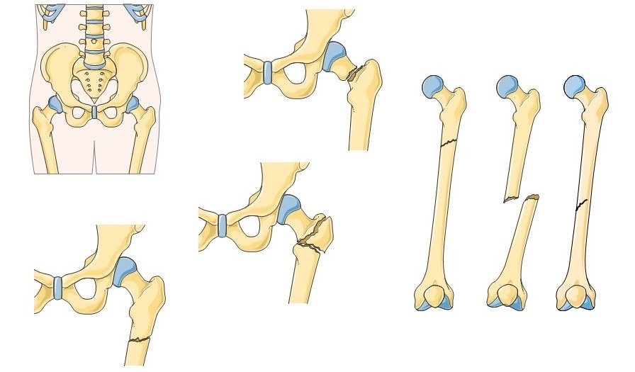
Figure 1. Common fracture locations of the femur, including the supracondylar (distal) region. Illustration by Servier Medical Art, CC BY-SA 3.0, via Wikimedia Commons.
The geriatric fracture is a hip-fracture-equivalent.[5] In frail elderly patients with a low-energy distal femur fracture, mortality rates approach those of the hip-fracture population, and a surgical delay beyond four days increases six-month and one-year mortality; brief delays for genuine medical optimisation do not. The principle is to treat these patients like hip-fracture patients: early surgery once optimised, orthogeriatric co-management, and a fixation construct that permits early weight bearing. The periprosthetic-fracture cohort makes the point starkly, with mortality of about 6 % at 30 days, 18 % at 6 months, and 25 % at 1 year, and total knee arthroplasty itself is an independent risk factor for reduced survival.[6]
Part II - Applied Anatomy and the Deforming Forces
Three anatomical facts govern fixation.[7] First, viewed end-on the distal femur is a trapezoid, wider posteriorly than anteriorly, with the medial surface sloping about 25° and the lateral surface about 10° (AO) to 15° (Rockwood); a lateral plate must sit flat on this lateral slope, and a condylar screw that looks the right length on the AP view may be too long and protrude medially, causing painful irritation. Second, the limb carries an anatomical valgus of about 6-7° (range 2-10°) at the knee, expressed by the AO as a lateral distal femoral angle of 80-84°, and the contralateral limb is the template for restoring it. Third, the cruciate ligaments occupy the intercondylar notch, so a misdirected screw can violate the notch and damage them, a particular hazard with variable-angle locking plates.[8]
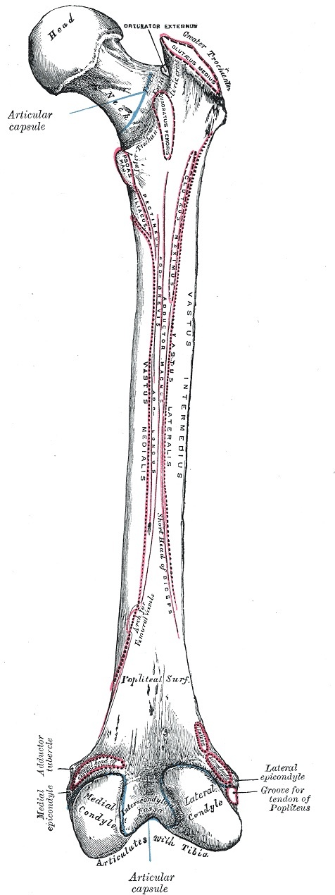
Figure 2. Posterior surface of the right femur showing the medial and lateral condyles, the intercondylar fossa, and the epicondyles. Gray’s Anatomy (Henry Vandyke Carter), public domain, via Wikimedia Commons.
The popliteal artery is the key soft-tissue relation.[9] The femoral vessels pass through the adductor (Hunter’s) canal and pierce the adductor magnus about 10-12 cm above the knee to become the popliteal artery and vein in the popliteal fossa, where the artery lies deep and medial to the vein and tibial nerve. The bundle is tethered proximally at the adductor hiatus and distally by the soleal arch, which leaves little room for displacement but, paradoxically, keeps the rate of popliteal injury relatively low after an isolated supracondylar fracture. The deforming forces then produce the characteristic deformity: the gastrocnemius, arising from the posterior aspect of both condyles, flexes the distal fragment into apex-posterior (recurvatum) angulation and posterior displacement, while the quadriceps, hamstrings, and adductors shorten the limb and pull it into varus; the classic picture is a shortened limb with the proximal fragment piercing the quadriceps anteriorly and the distal fragment flexed, varus, and rotated posteriorly. The surgical corollary is to flex the knee 30-60° over a bolster to relax the gastrocnemius and prevent recurvatum.[10]
Part III - Classification
The preferred system is the AO/OTA classification of the distal femur, region 33 (older texts use the region code 43).[11] It divides fractures into three types by their relationship to the joint: 33A, extra-articular; 33B, partial articular (part of the condyle is fractured while the rest remains in continuity with the shaft); and 33C, complete articular (the articular surface is both disrupted and separated from the shaft). Within these, the standard compendium subgroups, which the source extracts name only in passing, are: A1 simple, A2 metaphyseal wedge, A3 metaphyseal complex; B1 lateral condyle (sagittal), B2 medial condyle (sagittal), B3 coronal-plane (the Hoffa fragment); and C1 articular-simple with metaphyseal-simple, C2 articular-simple with metaphyseal-multifragmentary, C3 articular-multifragmentary. The complete bicondylar split is the classic “T- or Y-condylar” fracture, corresponding to 33C.[12]
The high-yield partial-articular entity is the Hoffa fracture, a coronal-plane fracture of a posterior femoral condyle (AO 33B3).[13] It is frequently overlooked on plain radiographs and is the principal reason that CT is mandatory for any intra-articular distal femur fracture: one study found a 40 % rate of coronal-plane (Hoffa) fractures accompanying intercondylar fractures, many invisible on plain films. The lateral condyle is more often involved than the medial, the fragment is largely articular (which makes fixation demanding), and recognising it preoperatively changes the surgical plan.[14]
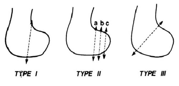
Figure 3. Letenneur classification of the Hoffa fracture (coronal-plane posterior femoral condyle), Types I-III. Image by Kapoor et al., CC BY-SA 1.0, via Wikimedia Commons.
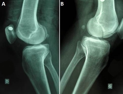
Figure 4. Hoffa fracture of the femoral condyle on the lateral radiograph, the view on which this coronal-plane fracture is best seen. Image by Koné et al., CC BY-SA 2.0, via Wikimedia Commons.
Part IV - Assessment, Associated Injuries, and Imaging
The assessment centres on the soft tissues and the neurovascular examination.[15] Vascular injury occurs in about 3 % and nerve injury in about 1 % of distal femur fractures; although the tethered popliteal artery is injured less often than after a knee dislocation, the consequence is limb-threatening, so pulses and an ankle-ankle index are checked, a value within 10 % of the other side making arterial injury unlikely, and a diminished index, expanding haematoma, or bruit prompting CT angiography. Distal perfusion present means the fracture is stabilised first; severe ischaemia or a delay beyond 6 hours means circulation is restored first, with an external fixator or shunt to hold length before vascular repair (repairing a vessel across an unreduced, shortened fracture is the classic avoidable error). Associated injuries include knee ligament and meniscal injury (the ACL is the most commonly injured ligament; menisci and osteochondral fractures in 8-12 %, patella fractures in about 15 %), and proximal diaphyseal extension in up to 50 % of high-energy cases. Knee ligaments are examined after fixation, not before.[16]
Imaging is AP and lateral radiographs of the knee and the whole femur, including the hip, with traction views helpful when shortening obscures the pattern.[17] The crucial investigation is CT with axial, coronal, and sagittal reconstruction for any intra-articular extension, both to map the articular fragments and, above all, to detect the coronal Hoffa fracture that plain films miss. The contralateral limb is templated for length, alignment (the LDFA), and rotation. A useful aside is that visible osteoporosis on a plain radiograph already implies a loss of 40 % or more of bone density.[18]
Part V - Treatment Principles and Nonoperative Care
Surgery is the default for virtually all displaced distal femur fractures in physiologically stable adults; nonoperative treatment is reserved for nondisplaced fractures in a reliable patient, the nonambulatory or unfit patient, and austere settings.[19] When chosen, it uses skeletal traction (a proximal-tibial or distal-femoral pin, 20-30 lb) followed by a fracture brace applied in extension, external rotation, and slight valgus to counter the varus that is the commonest casting complication. The accepted limits are coronal malalignment no more than 7°, sagittal no more than 7-10°, shortening of 1-1.5 cm, and an articular step of no more than 2 mm. The case for surgery in the elderly is decisive: Butt’s comparison showed operative fixation threefold reduced the complications of recumbency (deep-vein thrombosis, urinary infection, pressure sores, pneumonia) and cut the risk of a poor result by a third.[20]
The operative goals are anatomic reduction of the articular surface, restoration of length, alignment, and rotation of the metaphysis, stable fixation, and early knee motion.[21] This is delivered by the dual-stability principle: the articular block is reconstructed under direct vision and compressed with lag screws (absolute stability), using a position screw rather than a lag screw where there is bone loss, so as not to over-compress and narrow the condyles, and the metaphysis is then bridged to the shaft with relative stability, the extended-approach, fully-stripped open reduction of the metaphysis having been abandoned in favour of biological (indirect) plating, now the gold standard. Reduction tools restore length and counter the deforming forces: a femoral distractor, a bolster under the supracondylar fragment to control the sagittal recurvatum, Schanz-pin joysticks, and complete muscle paralysis. The deformities to avoid are named and predictable: valgus (the commonest coronal malalignment after lateral plating, prevented by setting the distal screws parallel to the joint), recurvatum/the “golf-club” deformity (from the gastrocnemius, prevented by flexing the knee), varus collapse (the failure mode of old non-locking plates and of a blade plate inserted too posteriorly), external malrotation, and shortening.[22]
Part VI - Operative Treatment
6.1 Lateral locked plating - the workhorse
The lateral locked plate (the LISS or the distal-femoral locking compression plate) is the workhorse, an “internal fixator” applied submuscularly so that it does not compress the periosteum, with a fixed-angle distal cluster of locking screws that resists the varus collapse of older non-locking plates.[23] It is placed through a lateral or lateral-parapatellar approach (the parapatellar arthrotomy giving the articular access needed for intercondylar fractures), reducing and lag-screwing the articular block first. Two technical rules recur: “lag before you lock” (a non-locked cortical screw must be placed in a fragment before any locking screw in it, or the lag fixation is compromised), and the distal locking screws or guidewire must be set parallel to the joint line so the plate’s built-in 5-8° of valgus is reproduced rather than exaggerated. Longer plates with well-spaced screws are preferred, aiming for at least eight cortices (or holes) proximal to the fracture, with bicortical locking screws (monocortical-only LISS fixation fails at a high rate).[24]
The biomechanical lesson of distal femur plating is that an implant can be too stiff.[25] A lateral-only locked construct that is excessively rigid produces an asymmetric strain environment, suppresses callus on the far (medial) cortex, and predisposes to medial metaphyseal nonunion and varus collapse; this is why surgeons modulate stiffness with far-cortical locking screws, longer working lengths, and the practice of filling no more than about half the screw holes. Where there is severe medial metaphyseal comminution, a lateral plate alone may be insufficient and a second, anteromedial plate (dual plating) is added, or a nail-plate combination is used for the “exploded femur.” The historical fixed-angle devices, the 95° blade plate and the dynamic condylar screw, gave excellent angular stability and are still useful for nonunion and malunion reconstruction, but they are technically demanding (the blade must be set correctly in three planes simultaneously) and have largely been replaced by anatomical locking plates.[26]
6.2 The Hoffa (coronal, 33B3) fracture
The Hoffa fragment is a coronal shear of a posterior condyle, largely articular, and best fixed with two or more carefully measured lag screws (2.7-, 3.5-, or 4.0-mm) inserted anterior-to-posterior and countersunk beneath the articular cartilage, supplemented by an antiglide (buttress) plate when a nonarticular spike extends superiorly from the fragment.[27] A medial Hoffa is approached through a medial subvastus approach. Posterior-to-anterior screws and headless compression screws are common alternatives in standard practice, though the mined sources specify the anterior-to-posterior countersunk technique.[28]
6.3 Retrograde intramedullary nailing
The retrograde nail suits extra-articular fractures (33A) and simple articular patterns (33C1, 33C2), and is valuable in the osteoporotic, obese, or polytrauma patient, provided any articular split is lag-screwed first, outside the nail path.[29] It is entered through a short medial-parapatellar or tendon-splitting approach with the knee flexed about 30°, the start point taken anterior to the intercondylar notch, respecting the origin of the posterior cruciate ligament and avoiding Blumensaat’s line. Because the distal medullary canal is wide relative to the nail, blocking (Poller) screws are often needed to centralise the nail and prevent malalignment, and the distal fragment is captured with multiple distal interlocking screws or a spiral blade; the nail should reach above the lesser trochanter and provides enough stability for early weight bearing. Where a single implant cannot control a very comminuted or periprosthetic pattern, a combined nail-and-plate construct is used.[30]
6.4 External fixation and distal femoral replacement
A knee-spanning external fixator is the damage-control option for the polytrauma patient, the severe open fracture, and the limb with vascular injury, restoring length, alignment, and rotation to make later definitive fixation easier.[31] The AO sequence is to apply tibial Schanz screws and restore length first, then place the femoral Schanz screws well anterior and away from the future surgical field (femoral screws placed through the quadriceps before length is restored will hinder it). For the elderly patient with an unreconstructable, comminuted, or osteoporotic distal articular fracture, or a fracture about a loose knee prosthesis, a distal femoral replacement (megaprosthesis) is an established option whose decisive advantage is immediate weight bearing, at the cost of reduced longevity compared with internal fixation; this is standard teaching, supported in the sources mainly through the periprosthetic and arthroplasty discussions rather than as a primary acute-trauma chapter.[32]
Part VII - Periprosthetic Distal Femur Fractures About a Total Knee Replacement
The supracondylar fracture above a total knee replacement is the commonest periprosthetic fracture about the knee and a high-yield exam topic.[33] It complicates 0.3-2.5 % of primary and 1.7-38 % of revision arthroplasties, and its risk factors are anterior femoral notching (a notched femur fails at 18 % less force in bending and 39 % less in torsion), periprosthetic osteopenia (bone density in the distal femur falls 19-44 % in the year after arthroplasty through stress shielding), rheumatoid arthritis, corticosteroids, and revision surgery. Two classifications are used together. The Lewis-Rorabeck classification combines displacement and implant fixation: Type I, nondisplaced with a stable implant; Type II, displaced with a well-fixed implant; Type III, any displacement with a loose or failing implant. The Su classification adds the location relative to the femoral component (Type I proximal to it, Type II at its proximal end, Type III extending distal to its proximal border).[34]
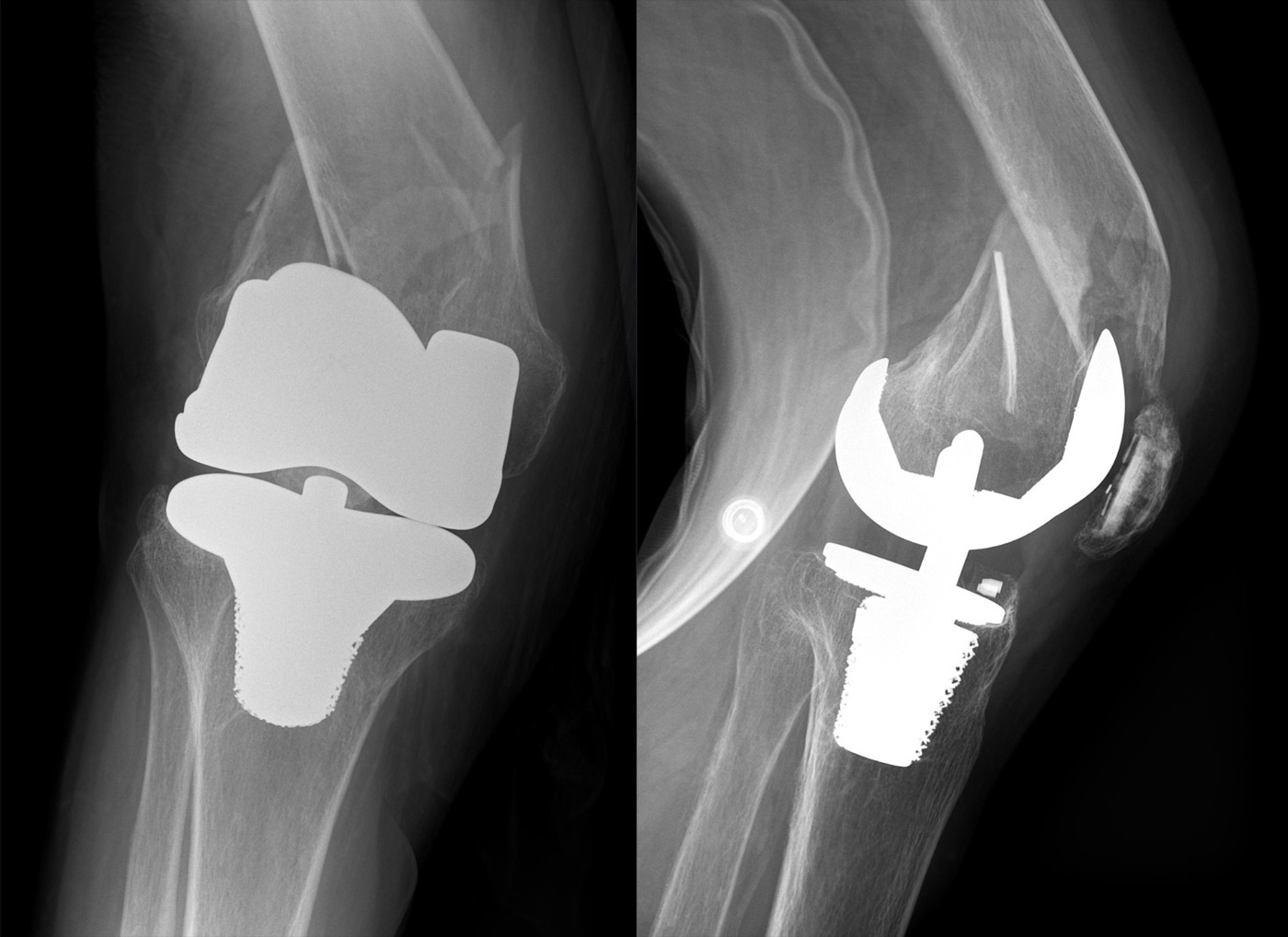
Figure 5. Periprosthetic distal femur fracture immediately above a total knee arthroplasty (AP and lateral radiographs). Image by Hellerhoff, CC BY-SA 4.0, via Wikimedia Commons.
The treatment algorithm follows the classification.[35] A nondisplaced fracture with a stable implant may be treated nonoperatively or with ORIF; a displaced fracture with a well-fixed implant (Lewis-Rorabeck II) is fixed, with lateral locked plating the workhorse because its fixed-angle distal screws resist varus collapse even in a short distal segment. A retrograde nail is an excellent alternative that allows early weight bearing, but only if the femoral component is “open-box” with an accessible intercondylar notch to admit the nail; a closed or narrow box (as in many posterior-stabilised designs) precludes or limits it, so the surgeon confirms an open box with an intraoperative notch view and is prepared to abort to a lateral plate if the box is closed. A loose or failing implant (Lewis-Rorabeck III), or bone too deficient for stable fixation, calls for revision arthroplasty or a distal femoral replacement. Comparative data favour modern fixed-angle constructs over old non-locking plates (a 415-case review gave retrograde nailing an 87 % relative reduction in nonunion versus non-locked plating), but nonunion and varus collapse remain real, with reported nonunion rates around 14-22 % after locked plating; obese, diabetic patients are the classic failures, and the geriatric mortality (25 % at one year) frames every decision toward early surgery and immediate weight bearing.[36]
Part VIII - Complications
Nonunion is the signature complication, occurring at the metaphyseal-diaphyseal junction and reported in about 20 % of distal femur fractures even with modern lateral locked plating.[37] Its drivers are an open fracture, diabetes, obesity, infection, a stainless-steel implant, and an over-stiff construct that suppresses callus and gaps the medial cortex; the prevention is a biologically respectful reduction and a stiffness-modulated construct, and the treatment is revision fixation, exchange to a nail, a 95° blade plate for compression, and biologic augmentation with cancellous graft or bone morphogenetic protein. Malunion is usually valgus in the coronal plane and apex-posterior (recurvatum/“golf-club”) in the sagittal plane, with malalignment somewhat more frequent after nailing than plating. Infection should not exceed 3-5 % in closed fractures and is treated by débridement with retention of stable implants. Knee stiffness is common, since immobilisation beyond three weeks tends to leave permanent stiffness, so the target is 90° of flexion by four weeks, with arthroscopic lysis or quadricepsplasty for the established case. Post-traumatic arthritis follows articular incongruity (about 50 % radiographically in one long-term series), and hardware irritation arises from the iliotibial band over the plate and from over-long medial condylar screws, the latter avoided with a 20-25° rollover fluoroscopic view. The commonest mode of fixation loss is varus collapse.[38]
Part IX - Paediatric Distal Femoral Physeal Fractures
The distal femoral physeal separation makes up about 5 % of physeal fractures and 1-2 % of all paediatric fractures, and it matters out of proportion to its frequency because the distal femoral physis is the largest and fastest-growing physis in the body (the standard teaching figure of roughly 70 % of femoral and nearly 40 % of lower-limb growth is not stated in the source but explains the consequences it describes).[39] The physis has a complex, undulating contour that resists shear, so a fracture here signals that the knee was exposed to very high force, and the same contour predisposes it to growth arrest, reported in up to 50 % of cases even after anatomic reduction, with the risk rising with the severity of displacement and worsened by metaphyseal comminution in Salter-Harris II fractures. Most fractures are Salter-Harris I and II; a Salter-Harris III of the medial condyle results from a valgus force and may carry cruciate-ligament and osteochondral injury. The mechanism maps to the displacement direction: hyperextension drives the epiphysis anteriorly (threatening the popliteal artery and peroneal nerve as the metaphysis displaces into the popliteal fossa; standard teaching), valgus drives it medially, varus laterally, and a flexed-knee impact posteriorly.[40]
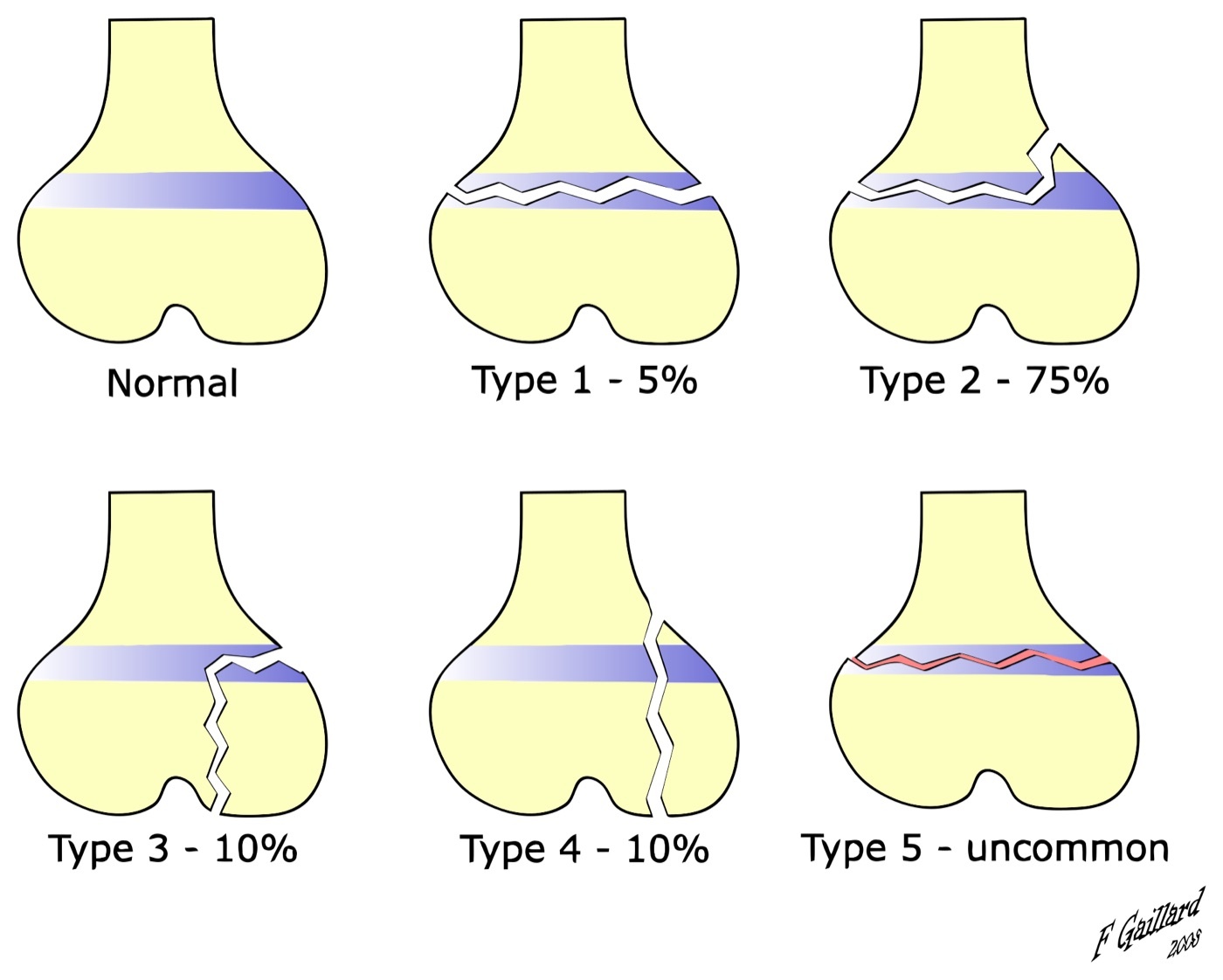
Figure 6. Salter-Harris classification of physeal (growth-plate) fractures, illustrated on a distal-femur-type epiphysis; distal femoral physeal fractures are mostly Types I and II. Image by Dr Frank Gaillard, CC BY-SA 3.0, via Wikimedia Commons.
Treatment is graded by displacement and Salter-Harris type.[41] A stable, nondisplaced fracture is cast for 3-4 weeks with close early radiographs to catch displacement. A displaced separation is reduced gently under anaesthesia and held with crossed percutaneous (smooth) pins to prevent redisplacement; a large Salter-Harris II Thurston-Holland metaphyseal fragment can take one or two cancellous screws (with a supplemental pin if only one screw is used). Displaced Salter-Harris III and IV fractures, an open fracture, neurovascular disruption, or entrapped soft tissue blocking reduction require open reduction, the articular and physeal surfaces restored precisely and fixed with two cannulated 6.5- or 7.3-mm screws placed to spare the physis. The dominant message is that growth arrest is common despite perfect reduction, so the family is counselled at the outset, the child is followed with serial radiographs (and MRI at 4-6 months to detect an early physeal bar) to skeletal maturity, and an established arrest is managed by bar resection or, for the resulting deformity, osteotomy and, for limb-length discrepancy, contralateral epiphysiodesis.[42]
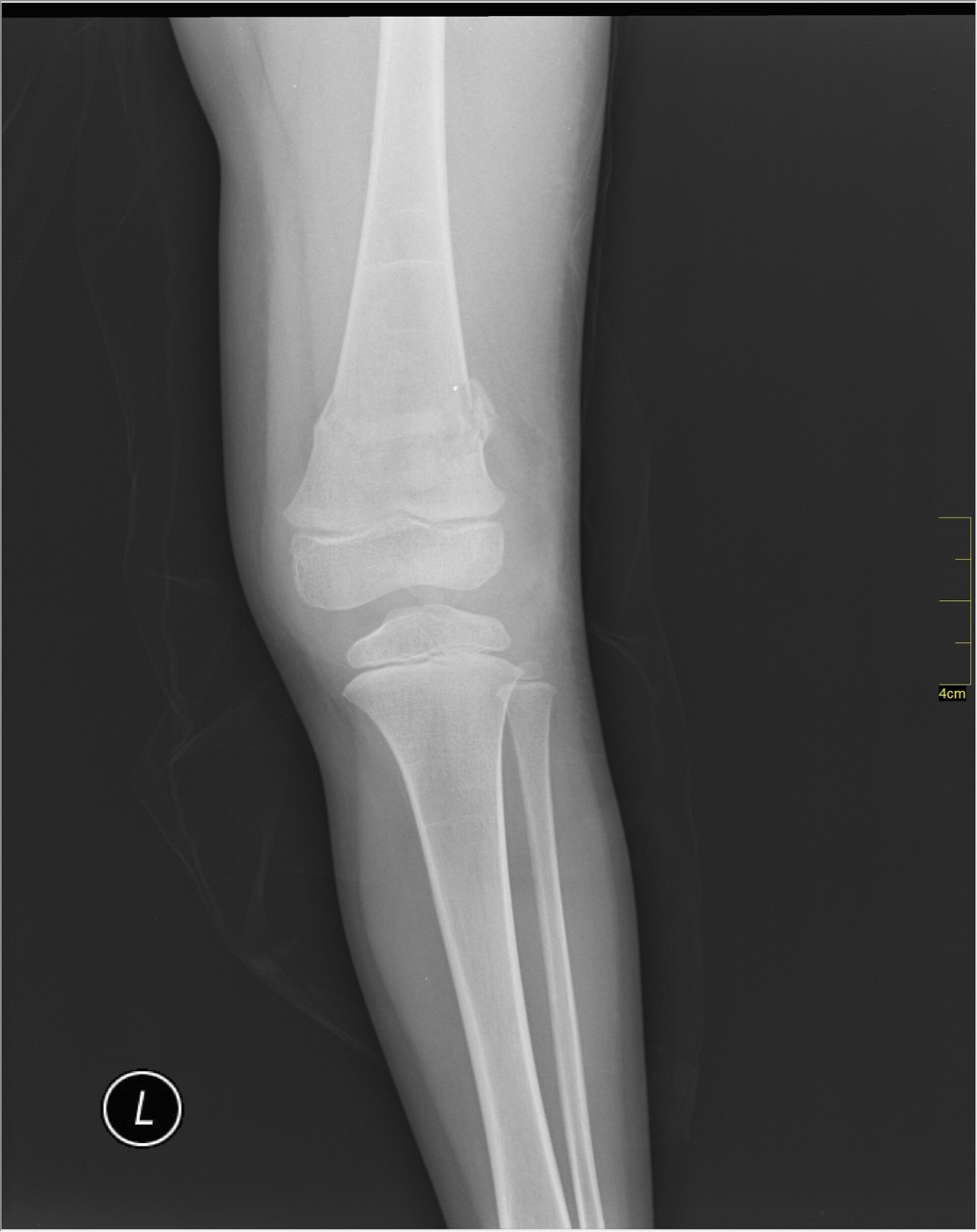
Figure 7. Distal femoral fracture in a skeletally immature patient with open physes, AP radiograph. Image by Nevit Dilmen, CC BY-SA 3.0, via Wikimedia Commons.
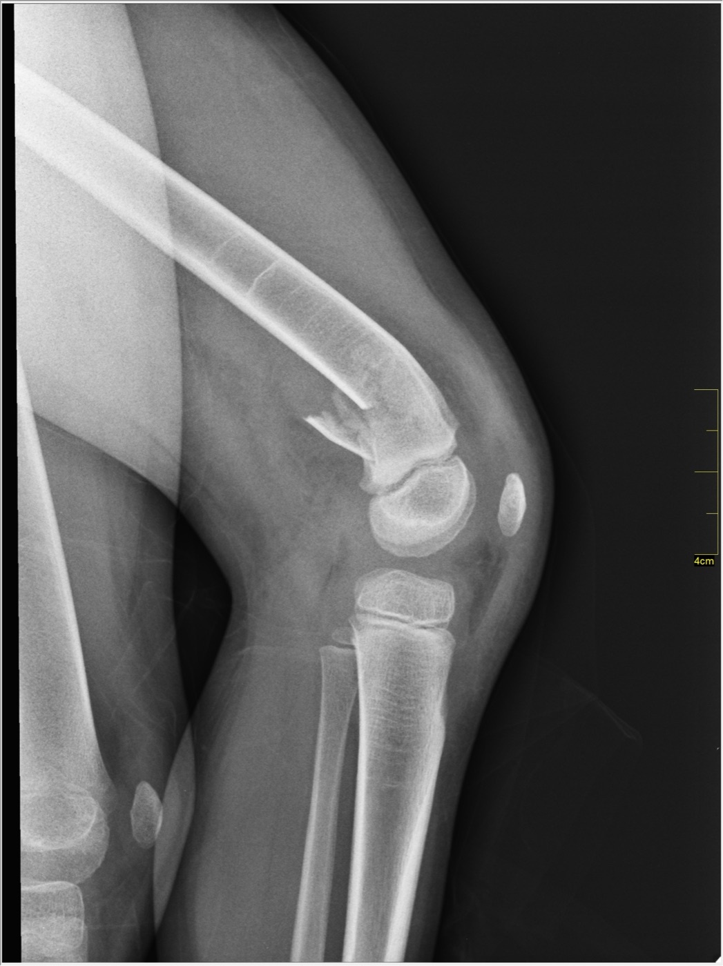
Figure 8. The same paediatric distal femoral fracture on the lateral radiograph, the distal fragment displaced posteriorly. Image by Nevit Dilmen, CC BY-SA 3.0, via Wikimedia Commons.
Part X - A Synthesis: How to Reason About the Distal Femur Fracture
Reason about this fracture as two zones in two patients. The articular block is reconstructed anatomically and held with absolute stability, lag-screwed (or position-screwed where bone is lost), and the metaphysis is bridged to the shaft with relative stability and a biologically respectful, indirect technique, the whole construct tuned so it is neither too loose nor, the commoner error, too stiff. The deformities the anatomy invites are valgus, recurvatum, and varus collapse, so set the distal screws parallel to the joint, flex the knee to silence the gastrocnemius, and use a fixed-angle distal cluster. The young patient has a high-energy, often open and comminuted injury that may need staged external fixation and a nail-plate or dual-plate construct; the elderly patient has a hip-fracture-equivalent that demands early surgery, immediate weight bearing, and, when the bone or a loose knee prosthesis cannot be reconstructed, a distal femoral replacement. The lateral locked plate is the workhorse and the retrograde nail its partner, the latter only through an open-box prosthesis. Always get the CT to find the Hoffa fragment, always check the popliteal artery, and in the child remember that the distal femoral physis is the body’s fastest grower, so growth arrest is common even after a perfect reduction and the family must be told and the child followed to maturity.
References
-
AO Principles of Fracture Management, pp.837, 838, 841 (the dual-zone strategy of absolute stability for the articular block and relative stability/bridge plating for the metaphysis as the gold standard; the gastrocnemius flexing the distal fragment into recurvatum, the quadriceps/hamstrings/adductors causing shortening and varus, and knee flexion to relax the gastrocnemius); Rockwood & Green’s Fractures in Adults, pp.3942, 3943, 3973 (the bimodal high-energy-young / low-energy-elderly distribution, the geriatric mortality comparable to hip fracture, and articular reduction first then metaphyseal stabilisation).
-
AO Principles of Fracture Management, pp.837, 838, 841 (the dual-zone strategy of absolute stability for the articular block and relative stability/bridge plating for the metaphysis as the gold standard; the gastrocnemius flexing the distal fragment into recurvatum, the quadriceps/hamstrings/adductors causing shortening and varus, and knee flexion to relax the gastrocnemius); Rockwood & Green’s Fractures in Adults, pp.3942, 3943, 3973 (the bimodal high-energy-young / low-energy-elderly distribution, the geriatric mortality comparable to hip fracture, and articular reduction first then metaphyseal stabilisation).
-
Rockwood & Green’s Fractures in Adults, pp.3941, 3942, 3948 (supracondylar zone = distal 15 cm; up to 6 % of femoral fractures; bimodal distribution; axial-load and flexed-knee mechanisms); AO Principles of Fracture Management, p.835 (about 6 % of femoral fractures; high-energy young vs low-energy osteoporotic elderly; one-third polytrauma and one-fifth isolated; ~50 % of high-energy intra-articular fractures open).
-
Rockwood & Green’s Fractures in Adults, pp.3941, 3942, 3948 (supracondylar zone = distal 15 cm; up to 6 % of femoral fractures; bimodal distribution; axial-load and flexed-knee mechanisms); AO Principles of Fracture Management, p.835 (about 6 % of femoral fractures; high-energy young vs low-energy osteoporotic elderly; one-third polytrauma and one-fifth isolated; ~50 % of high-energy intra-articular fractures open).
-
Rockwood & Green’s Fractures in Adults, pp.3943, 3948, 4056 (geriatric mortality comparable to hip fracture; surgical delay beyond four days worsens six-month and one-year mortality while brief optimisation delays do not; periprosthetic distal femur mortality 6 %/18 %/25 % at 30 days/6 months/1 year with TKA an independent risk factor); Miller’s Review of Orthopaedics, p.923 (high one-year mortality, reduced if ORIF is performed within 48 hours).
-
Rockwood & Green’s Fractures in Adults, pp.3943, 3948, 4056 (geriatric mortality comparable to hip fracture; surgical delay beyond four days worsens six-month and one-year mortality while brief optimisation delays do not; periprosthetic distal femur mortality 6 %/18 %/25 % at 30 days/6 months/1 year with TKA an independent risk factor); Miller’s Review of Orthopaedics, p.923 (high one-year mortality, reduced if ORIF is performed within 48 hours).
-
Rockwood & Green’s Fractures in Adults, pp.3949, 3950 (trapezoidal distal femur, medial slope ~25° and lateral slope ~15°, the screw-length pitfall; anatomical valgus 6-7°, range 2-10°); AO Principles of Fracture Management, pp.835, 837 (trapezoid with ~25° medial and ~10° lateral slope, plate must lie flat on the lateral slope; LDFA 80-84° with the contralateral limb as reference; cruciate ligaments in the intercondylar notch and screw-violation risk, especially with variable-angle locking plates).
-
Rockwood & Green’s Fractures in Adults, pp.3949, 3950 (trapezoidal distal femur, medial slope ~25° and lateral slope ~15°, the screw-length pitfall; anatomical valgus 6-7°, range 2-10°); AO Principles of Fracture Management, pp.835, 837 (trapezoid with ~25° medial and ~10° lateral slope, plate must lie flat on the lateral slope; LDFA 80-84° with the contralateral limb as reference; cruciate ligaments in the intercondylar notch and screw-violation risk, especially with variable-angle locking plates).
-
Rockwood & Green’s Fractures in Adults, pp.3942, 3944, 3950, 3961 (femoral vessels piercing the adductor magnus 10-12 cm above the knee [p.3961], the popliteal artery deep and medial in the fossa, the proximal adductor-hiatus and distal soleal-arch tethering explaining the low popliteal-injury rate; the gastrocnemius producing apex-posterior angulation and the quadriceps/hamstrings/adductors causing shortening and varus); AO Principles of Fracture Management, pp.837, 841, 854 (gastrocnemius flexion of the distal fragment, the typical shortened-anteriorly-pierced-quadriceps deformity, and knee flexion 30-45° intra-operatively and 60° during surgery to prevent recurvatum).
-
Rockwood & Green’s Fractures in Adults, pp.3942, 3944, 3950, 3961 (femoral vessels piercing the adductor magnus 10-12 cm above the knee [p.3961], the popliteal artery deep and medial in the fossa, the proximal adductor-hiatus and distal soleal-arch tethering explaining the low popliteal-injury rate; the gastrocnemius producing apex-posterior angulation and the quadriceps/hamstrings/adductors causing shortening and varus); AO Principles of Fracture Management, pp.837, 841, 854 (gastrocnemius flexion of the distal fragment, the typical shortened-anteriorly-pierced-quadriceps deformity, and knee flexion 30-45° intra-operatively and 60° during surgery to prevent recurvatum).
-
Rockwood & Green’s Fractures in Adults, p.3947 (the OTA classification preferred, distinguishing extra-articular type A, partial articular type B, and complete articular type C, labelled region 43); AO Principles of Fracture Management, p.837 (region 33: 33A extra-articular, 33B partial articular, 33C complete articular, with 33A3/33C2/33C3 being the metaphyseal-multifragmentary patterns and 33C1/33C2 the simple-articular patterns suitable for retrograde nailing). The full A1-C3 subgroup definitions and the “T-/Y-condylar” eponym are standard AO/OTA-compendium teaching, named only in passing in these extracts.
-
Rockwood & Green’s Fractures in Adults, p.3947 (the OTA classification preferred, distinguishing extra-articular type A, partial articular type B, and complete articular type C, labelled region 43); AO Principles of Fracture Management, p.837 (region 33: 33A extra-articular, 33B partial articular, 33C complete articular, with 33A3/33C2/33C3 being the metaphyseal-multifragmentary patterns and 33C1/33C2 the simple-articular patterns suitable for retrograde nailing). The full A1-C3 subgroup definitions and the “T-/Y-condylar” eponym are standard AO/OTA-compendium teaching, named only in passing in these extracts.
-
Rockwood & Green’s Fractures in Adults, p.3946 (Hoffa = coronal-plane posterior-condyle fracture, the 40 % association with intercondylar fractures, often missed on plain films, CT recommended); AO Principles of Fracture Management, p.835 (coronal fractures of the posterior condyles - Hoffa fractures - missed on plain x-rays, so CT with 2-D and 3-D reconstruction is recommended for intra-articular fractures).
-
Rockwood & Green’s Fractures in Adults, p.3946 (Hoffa = coronal-plane posterior-condyle fracture, the 40 % association with intercondylar fractures, often missed on plain films, CT recommended); AO Principles of Fracture Management, p.835 (coronal fractures of the posterior condyles - Hoffa fractures - missed on plain x-rays, so CT with 2-D and 3-D reconstruction is recommended for intra-articular fractures).
-
AO Principles of Fracture Management, p.837 (vascular injury ~3 %, nerve injury ~1 %, menisci/osteochondral lesions 8-12 %, patella fractures ~15 %; examine ligaments after stabilisation); Rockwood & Green’s Fractures in Adults, pp.3943, 3944, 3945, 3946 (ankle-ankle index within 10 % makes vascular injury unlikely, the indications for angiography, the 6-hour ischaemia threshold and external-fixator/shunt-before-repair principle; the ACL most commonly injured; proximal diaphyseal extension in up to 50 %; open fractures 5-10 % overall).
-
AO Principles of Fracture Management, p.837 (vascular injury ~3 %, nerve injury ~1 %, menisci/osteochondral lesions 8-12 %, patella fractures ~15 %; examine ligaments after stabilisation); Rockwood & Green’s Fractures in Adults, pp.3943, 3944, 3945, 3946 (ankle-ankle index within 10 % makes vascular injury unlikely, the indications for angiography, the 6-hour ischaemia threshold and external-fixator/shunt-before-repair principle; the ACL most commonly injured; proximal diaphyseal extension in up to 50 %; open fractures 5-10 % overall).
-
Rockwood & Green’s Fractures in Adults, p.3946 (AP/lateral of knee and femur plus hip, traction views, CT with axial/coronal/sagittal reconstruction for intra-articular extension, the 40 % bone-density-loss rule); AO Principles of Fracture Management, p.835 (radiographs including the adjacent joints, traction views, CT with 2-D/3-D reconstruction especially for Hoffa fractures, MRI not essential acutely, contralateral LDFA as reference).
-
Rockwood & Green’s Fractures in Adults, p.3946 (AP/lateral of knee and femur plus hip, traction views, CT with axial/coronal/sagittal reconstruction for intra-articular extension, the 40 % bone-density-loss rule); AO Principles of Fracture Management, p.835 (radiographs including the adjacent joints, traction views, CT with 2-D/3-D reconstruction especially for Hoffa fractures, MRI not essential acutely, contralateral LDFA as reference).
-
Rockwood & Green’s Fractures in Adults, pp.3951, 3952 (operative treatment the default; nonoperative indications; skeletal traction 20-30 lb and a fracture brace in extension/external rotation/slight valgus; acceptable nonoperative alignment - coronal ≤7°, sagittal 7-10°, shortening 1-1.5 cm, articular step ≤2 mm; Butt’s data favouring operative treatment); AO Principles of Fracture Management, p.838 (nonoperative treatment justified only in impacted nondisplaced extra-articular fractures or the nonambulatory/inoperable patient).
-
Rockwood & Green’s Fractures in Adults, pp.3951, 3952 (operative treatment the default; nonoperative indications; skeletal traction 20-30 lb and a fracture brace in extension/external rotation/slight valgus; acceptable nonoperative alignment - coronal ≤7°, sagittal 7-10°, shortening 1-1.5 cm, articular step ≤2 mm; Butt’s data favouring operative treatment); AO Principles of Fracture Management, p.838 (nonoperative treatment justified only in impacted nondisplaced extra-articular fractures or the nonambulatory/inoperable patient).
-
Rockwood & Green’s Fractures in Adults, pp.3952, 3962, 3966, 3977 (operative goals; femoral distractor, bolster, and Schanz-pin joysticks; valgus the usual coronal malalignment with the distal screws set parallel to the joint; rotational malalignment usually external and length usually short); AO Principles of Fracture Management, pp.838, 848, 854 (biological plating the gold standard; articular block absolute stability with lag or position screws, metaphysis relative stability/bridge plating; genu recurvatum from the gastrocnemius; varus and malrotation more frequent with the blade plate when inserted too posteriorly; over-stiff implants producing the wrong strain environment).
-
Rockwood & Green’s Fractures in Adults, pp.3952, 3962, 3966, 3977 (operative goals; femoral distractor, bolster, and Schanz-pin joysticks; valgus the usual coronal malalignment with the distal screws set parallel to the joint; rotational malalignment usually external and length usually short); AO Principles of Fracture Management, pp.838, 848, 854 (biological plating the gold standard; articular block absolute stability with lag or position screws, metaphysis relative stability/bridge plating; genu recurvatum from the gastrocnemius; varus and malrotation more frequent with the blade plate when inserted too posteriorly; over-stiff implants producing the wrong strain environment).
-
Rockwood & Green’s Fractures in Adults, pp.3954, 3962, 3963, 3966 (the LISS/locking plate as an internal fixator applied submuscularly; lateral and lateral-parapatellar approaches; “lag before you lock”; distal screws parallel to the joint to reproduce the built-in 5-8° valgus; longer plates with ≥8 holes proximally); AO Principles of Fracture Management, pp.849, 850 (LISS and LCP-DF, bicortical locking screws, three to four bicortical proximal screws, never monocortical alone, the plate seated on the lateral slope, the reference pin parallel to the joint line).
-
Rockwood & Green’s Fractures in Adults, pp.3954, 3962, 3963, 3966 (the LISS/locking plate as an internal fixator applied submuscularly; lateral and lateral-parapatellar approaches; “lag before you lock”; distal screws parallel to the joint to reproduce the built-in 5-8° valgus; longer plates with ≥8 holes proximally); AO Principles of Fracture Management, pp.849, 850 (LISS and LCP-DF, bicortical locking screws, three to four bicortical proximal screws, never monocortical alone, the plate seated on the lateral slope, the reference pin parallel to the joint line).
-
AO Principles of Fracture Management, pp.850, 854 (a lateral plate alone insufficient for severe, especially medial, metaphyseal comminution - add an anteromedial 3.5 plate; over-stiff implants producing the wrong strain and nonunion); Rockwood & Green’s Fractures in Adults, pp.3966, 3967, 3975 (far-cortical locking to reduce stiffness and balance callus, filling no more than ~50 % of holes; dual plating and nail-plate combination for the unstable/“exploded” femur; the 95° blade plate and dynamic condylar screw now largely reserved for nonunion/malunion).
-
AO Principles of Fracture Management, pp.850, 854 (a lateral plate alone insufficient for severe, especially medial, metaphyseal comminution - add an anteromedial 3.5 plate; over-stiff implants producing the wrong strain and nonunion); Rockwood & Green’s Fractures in Adults, pp.3966, 3967, 3975 (far-cortical locking to reduce stiffness and balance callus, filling no more than ~50 % of holes; dual plating and nail-plate combination for the unstable/“exploded” femur; the 95° blade plate and dynamic condylar screw now largely reserved for nonunion/malunion).
-
Rockwood & Green’s Fractures in Adults, p.3972 (Hoffa fixation with two or more 2.7/3.5/4.0-mm lag screws anterior-to-posterior, countersunk beneath the articular surface, with an antiglide plate on a superior spike); AO Principles of Fracture Management, p.844 (medial Hoffa via the medial subvastus approach). Posterior-to-anterior and headless compression screws are standard-teaching alternatives not specified in these extracts.
-
Rockwood & Green’s Fractures in Adults, p.3972 (Hoffa fixation with two or more 2.7/3.5/4.0-mm lag screws anterior-to-posterior, countersunk beneath the articular surface, with an antiglide plate on a superior spike); AO Principles of Fracture Management, p.844 (medial Hoffa via the medial subvastus approach). Posterior-to-anterior and headless compression screws are standard-teaching alternatives not specified in these extracts.
-
AO Principles of Fracture Management, pp.845, 846, 850, 851 (retrograde nailing for 33A and simple 33C1/33C2; entry anterior to the notch respecting the PCL origin; the wide distal canal requiring Poller/blocking screws; distal locking with interlocking screws or a spiral blade; the nail providing enough stability for early weight bearing); Rockwood & Green’s Fractures in Adults, pp.3970, 3974, 3975, 4068 (nails for extra-articular and simple fractures, articular fragments fixed first outside the nail path, the mini-open medial-parapatellar entry, and the nail-plate combination for difficult and periprosthetic patterns; the long nail reaching above the lesser trochanter per the periprosthetic retrograde-nailing technique, p.4068).
-
AO Principles of Fracture Management, pp.845, 846, 850, 851 (retrograde nailing for 33A and simple 33C1/33C2; entry anterior to the notch respecting the PCL origin; the wide distal canal requiring Poller/blocking screws; distal locking with interlocking screws or a spiral blade; the nail providing enough stability for early weight bearing); Rockwood & Green’s Fractures in Adults, pp.3970, 3974, 3975, 4068 (nails for extra-articular and simple fractures, articular fragments fixed first outside the nail path, the mini-open medial-parapatellar entry, and the nail-plate combination for difficult and periprosthetic patterns; the long nail reaching above the lesser trochanter per the periprosthetic retrograde-nailing technique, p.4068).
-
AO Principles of Fracture Management, pp.838, 851 (knee-spanning external fixation for polytrauma, severe open fractures, and vascular injury; restore length before placing femoral Schanz screws, kept anterior and out of the future surgical field); Rockwood & Green’s Fractures in Adults, pp.3957, 4057, 4072 and Miller’s Review of Orthopaedics, p.922 (distal femoral replacement for unreconstructable or loose-prosthesis fractures in the elderly, allowing immediate weight bearing with reduced longevity versus internal fixation). The role of primary distal femoral replacement in acute geriatric trauma is largely standard teaching, the extracts describing it chiefly in the periprosthetic and arthroplasty context.
-
AO Principles of Fracture Management, pp.838, 851 (knee-spanning external fixation for polytrauma, severe open fractures, and vascular injury; restore length before placing femoral Schanz screws, kept anterior and out of the future surgical field); Rockwood & Green’s Fractures in Adults, pp.3957, 4057, 4072 and Miller’s Review of Orthopaedics, p.922 (distal femoral replacement for unreconstructable or loose-prosthesis fractures in the elderly, allowing immediate weight bearing with reduced longevity versus internal fixation). The role of primary distal femoral replacement in acute geriatric trauma is largely standard teaching, the extracts describing it chiefly in the periprosthetic and arthroplasty context.
-
Rockwood & Green’s Fractures in Adults, pp.4053, 4054, 4055 (commonest periprosthetic fracture about the knee; incidence 0.3-2.5 % primary and 1.7-38 % revision; anterior notching reducing bending strength 18 % and torsional strength 39 %; periprosthetic bone density falling 19-44 %; the Lewis-Rorabeck Types I/II/III by displacement and implant stability and the Su classification by location).
-
Rockwood & Green’s Fractures in Adults, pp.4053, 4054, 4055 (commonest periprosthetic fracture about the knee; incidence 0.3-2.5 % primary and 1.7-38 % revision; anterior notching reducing bending strength 18 % and torsional strength 39 %; periprosthetic bone density falling 19-44 %; the Lewis-Rorabeck Types I/II/III by displacement and implant stability and the Su classification by location).
-
Rockwood & Green’s Fractures in Adults, pp.4054, 4057, 4065, 4066, 4067, 4072 (nonoperative for nondisplaced/stable; lateral locked plating the workhorse with fixed-angle distal screws against varus collapse; retrograde nailing only with an open-box component, confirmed by an intraoperative notch view, aborting to a plate if closed; revision/distal femoral replacement for a loose implant or deficient bone; the 415-case systematic review with retrograde nailing’s 87 % relative nonunion reduction; locked-plating nonunion 14-22 % with diabetic/obese failures); AO Principles of Fracture Management, pp.864, 866 (a loose implant requires revision and cannot be treated by internal fixation; a stable open-box prosthesis allows a retrograde nail with early weight bearing); Miller’s Review of Orthopaedics, pp.922, 923 (arthroplasty/distal femoral replacement for unachievable fixation or pre-existing arthropathy; high one-year mortality reduced by ORIF within 48 hours).
-
Rockwood & Green’s Fractures in Adults, pp.4054, 4057, 4065, 4066, 4067, 4072 (nonoperative for nondisplaced/stable; lateral locked plating the workhorse with fixed-angle distal screws against varus collapse; retrograde nailing only with an open-box component, confirmed by an intraoperative notch view, aborting to a plate if closed; revision/distal femoral replacement for a loose implant or deficient bone; the 415-case systematic review with retrograde nailing’s 87 % relative nonunion reduction; locked-plating nonunion 14-22 % with diabetic/obese failures); AO Principles of Fracture Management, pp.864, 866 (a loose implant requires revision and cannot be treated by internal fixation; a stable open-box prosthesis allows a retrograde nail with early weight bearing); Miller’s Review of Orthopaedics, pp.922, 923 (arthroplasty/distal femoral replacement for unachievable fixation or pre-existing arthropathy; high one-year mortality reduced by ORIF within 48 hours).
-
Rockwood & Green’s Fractures in Adults, pp.3989, 3990, 3991, 3992, 3993 (nonunion ~20 % with lateral locked plating, the risk factors open/diabetes/obesity/infection/stainless steel and over-stiff constructs, treated by revision/exchange-to-nail/95° blade plate/BMP; malunion valgus and apex-posterior; infection ≤3-5 % closed; immobilisation beyond three weeks causing permanent stiffness with a 90°-by-four-weeks target; ~50 % radiographic post-traumatic arthritis; hardware irritation from the iliotibial band and over-long medial screws, the rollover view); Miller’s Review of Orthopaedics, pp.922, 923 (valgus the commonest coronal and hyperextension the commonest sagittal malreduction, malalignment more common with nails; varus collapse the commonest loss of fixation; prominent medial screws to be avoided).
-
Rockwood & Green’s Fractures in Adults, pp.3989, 3990, 3991, 3992, 3993 (nonunion ~20 % with lateral locked plating, the risk factors open/diabetes/obesity/infection/stainless steel and over-stiff constructs, treated by revision/exchange-to-nail/95° blade plate/BMP; malunion valgus and apex-posterior; infection ≤3-5 % closed; immobilisation beyond three weeks causing permanent stiffness with a 90°-by-four-weeks target; ~50 % radiographic post-traumatic arthritis; hardware irritation from the iliotibial band and over-long medial screws, the rollover view); Miller’s Review of Orthopaedics, pp.922, 923 (valgus the commonest coronal and hyperextension the commonest sagittal malreduction, malalignment more common with nails; varus collapse the commonest loss of fixation; prominent medial screws to be avoided).
-
Lovell & Winter’s Pediatric Orthopaedics, pp.5691, 5692 (~5 % of physeal fractures and 1-2 % of paediatric fractures; the physis resisting very high shear so a fracture signals high force; growth arrest up to 50 % even with anatomic reduction, rising with displacement and worsened by metaphyseal comminution; predominance of Salter-Harris I and II; the SH III medial-condyle valgus mechanism with cruciate/osteochondral injury; the mechanism-to-displacement map). That this is the largest/fastest-growing physis (~70 % of femoral growth) and that anterior/hyperextension displacement threatens the popliteal artery and peroneal nerve are standard teaching consistent with the source, which states the consequences without the growth-percentage figures or the femur-specific neurovascular anatomy.
-
Lovell & Winter’s Pediatric Orthopaedics, pp.5691, 5692 (~5 % of physeal fractures and 1-2 % of paediatric fractures; the physis resisting very high shear so a fracture signals high force; growth arrest up to 50 % even with anatomic reduction, rising with displacement and worsened by metaphyseal comminution; predominance of Salter-Harris I and II; the SH III medial-condyle valgus mechanism with cruciate/osteochondral injury; the mechanism-to-displacement map). That this is the largest/fastest-growing physis (~70 % of femoral growth) and that anterior/hyperextension displacement threatens the popliteal artery and peroneal nerve are standard teaching consistent with the source, which states the consequences without the growth-percentage figures or the femur-specific neurovascular anatomy.
-
Lovell & Winter’s Pediatric Orthopaedics, pp.5691, 5692, 5693, 5697 (nondisplaced cast 3-4 weeks with early radiographs; displaced separations reduced under anaesthesia and held with crossed percutaneous pins; a large Thurston-Holland fragment fixed with one or two cancellous screws plus a supplemental pin if single; open reduction for displaced SH III/IV, open injury, neurovascular disruption, or entrapped tissue, fixed with two cannulated 6.5/7.3-mm screws; counselling the family, serial radiographs and MRI at 4-6 months for early arrest); growth-arrest management by physeal-bar resection, osteotomy for deformity, and contralateral epiphysiodesis for limb-length discrepancy is standard teaching (the source names contralateral epiphysiodesis and osteotomy in its general paediatric-femur section, p.5690), as are smooth K-wires and the explicit “follow to maturity” instruction.
-
Lovell & Winter’s Pediatric Orthopaedics, pp.5691, 5692, 5693, 5697 (nondisplaced cast 3-4 weeks with early radiographs; displaced separations reduced under anaesthesia and held with crossed percutaneous pins; a large Thurston-Holland fragment fixed with one or two cancellous screws plus a supplemental pin if single; open reduction for displaced SH III/IV, open injury, neurovascular disruption, or entrapped tissue, fixed with two cannulated 6.5/7.3-mm screws; counselling the family, serial radiographs and MRI at 4-6 months for early arrest); growth-arrest management by physeal-bar resection, osteotomy for deformity, and contralateral epiphysiodesis for limb-length discrepancy is standard teaching (the source names contralateral epiphysiodesis and osteotomy in its general paediatric-femur section, p.5690), as are smooth K-wires and the explicit “follow to maturity” instruction.
-
Rockwood & Green’s Fractures in Adults, pp.3942, 3948; AO Principles of Fracture Management, p.837.
-
AO Principles of Fracture Management, p.837; Rockwood & Green’s Fractures in Adults, p.3947.
-
Rockwood & Green’s Fractures in Adults, pp.3946, 3972; AO Principles of Fracture Management, p.835.
-
AO Principles of Fracture Management, pp.838, 848; Rockwood & Green’s Fractures in Adults, p.3973.
-
AO Principles of Fracture Management, pp.850, 854; Rockwood & Green’s Fractures in Adults, pp.3966, 3975.
-
AO Principles of Fracture Management, pp.845, 866; Rockwood & Green’s Fractures in Adults, pp.3970, 4067.
-
Rockwood & Green’s Fractures in Adults, pp.4053, 4057, 4072.
-
Rockwood & Green’s Fractures in Adults, pp.3943, 4056; Miller’s Review of Orthopaedics, pp.922, 923.
-
Rockwood & Green’s Fractures in Adults, pp.3962, 3977; AO Principles of Fracture Management, pp.841, 854.
-
Rockwood & Green’s Fractures in Adults, pp.3944, 3945, 3946; AO Principles of Fracture Management, p.837.
-
Lovell & Winter’s Pediatric Orthopaedics, pp.5691, 5692, 5697.
-
Rockwood & Green’s Fractures in Adults, pp.3990, 3991, 3992; Miller’s Review of Orthopaedics, p.922.