Contents
- Part I - A Framework for the Complications of Fracture Treatment
- Part II - The Local Complications: An Overview
- Part III - Venous Thromboembolism: Scope and Mechanism
- Part IV - Risk Factors and Diagnosis of VTE
- Part V - Prophylaxis Against VTE
- Part VI - Treatment of Established VTE
- Part VII - Fat Embolism Syndrome
- References
Part I - A Framework for the Complications of Fracture Treatment
A complication of fracture treatment is best defined simply as “an undesired turn of events in the treatment of a fracture,” with the honest caveat (“undesired by whom?”) that patient, surgeon and insurer may judge the same outcome differently.[1] An important conceptual point frames the whole subject: many complications are intrinsic to fracture care and are part of the natural history of fractures rather than markers that something went wrong.[2] Every fracture poses two linked problems, a biological one and a mechanical one, and most complications can be traced to a failure of one or the other.[3]
Complications are organised along two axes. The first is local versus systemic (general): the three major systemic disturbances of fracture care are fat embolism syndrome, thromboembolic disease (DVT/PE), and multiple-organ dysfunction, while the local complications group into soft-tissue/vascular problems, post-traumatic arthrosis, peripheral-nerve injury and complex regional pain syndrome.[4] The second axis is temporal: immediate, delayed (early), or late:[5]
- Immediate complications (hours to days) are largely the consequences of the injury itself rather than its treatment: skin demarcation and sloughing, ecchymosis, thrombosed vessels, and the post-operative haematoma.
- Delayed (early) complications include complex regional pain syndrome and the disuse atrophy of “fracture disease.”
- Late complications are the long-term, often permanent results (malunion and post-traumatic arthrosis) and the insidious disturbances of fracture healing (delayed and non-union) that declare themselves over weeks to months.
With multiple injuries the complication rates become “more than additive,” which is exactly why the Injury Severity Score sums the squares of the regional injuries.[6] This topic frames that whole landscape and then treats its two great systemic complications, thromboembolism and fat embolism, in depth; the bone-healing failures (non-union, malunion, CRPS, Volkmann, periprosthetic fracture) are the subject of Topic 8 and bone infection of Topic 9.
Part II - The Local Complications: An Overview
Vascular injury
Arterial injury complicates about 2-6% of civilian fractures or dislocations and under 1% of isolated injuries, but roughly one third of war-related long-bone injuries, and a posterior knee dislocation carries about a 30% risk of popliteal artery damage.[7] It presents acutely (haemorrhage, ischaemia) or late (pseudoaneurysm, arteriovenous fistula); CT angiography has largely replaced formal angiography for diagnosis.[8] The prognosis is sobering: a popliteal or infra-trifurcation injury may be unreconstructable, a good functional result follows revascularisation in only about a quarter of cases, and amputation after infection sets in is performed at a higher level than if done at the outset.[9] Prolonged limb ischaemia is the route to Volkmann ischaemic contracture (covered with the operative complications of Topic 8).
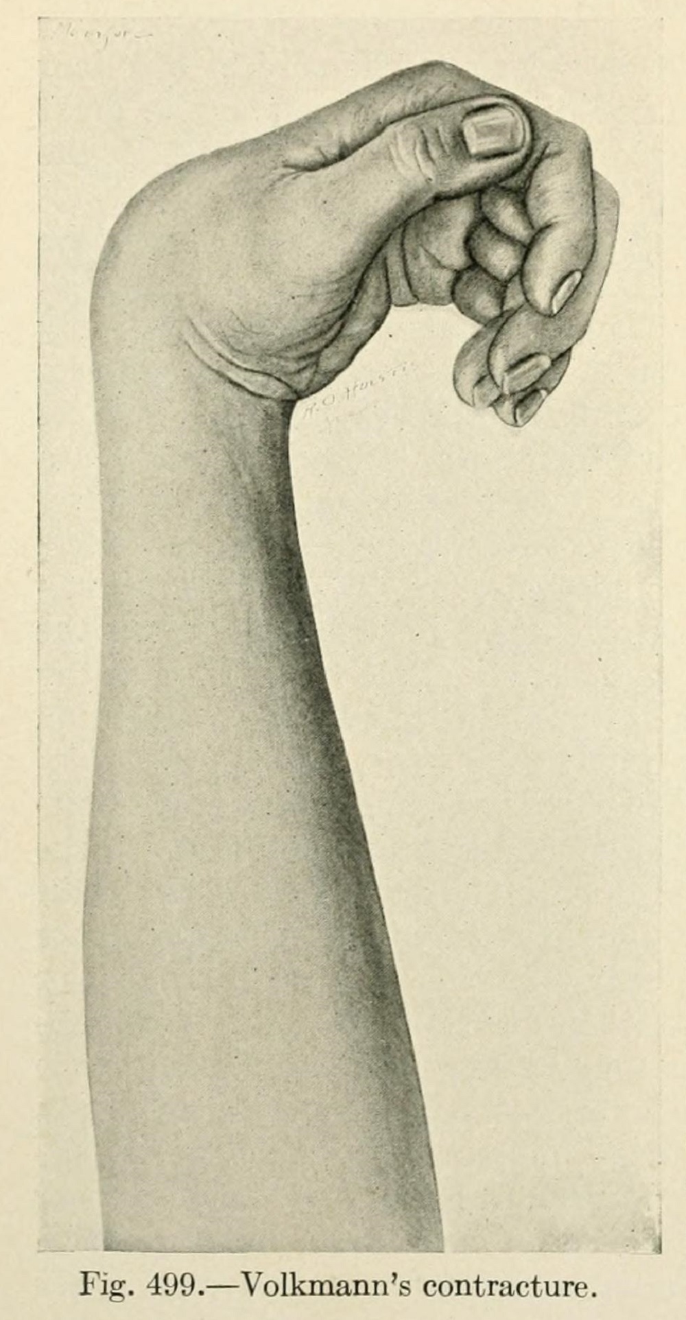
Figure 1. Volkmann ischaemic contracture of the forearm and hand, the end-result of untreated compartment ischaemia. Source: Mumford, The Practice of Surgery (1910), public domain, via Wikimedia Commons / Internet Archive.
Peripheral-nerve injury
Nerve injury is graded by two complementary schemes. Seddon describes three grades and Sunderland five:[10]
- Neurapraxia (Sunderland 1): a reversible conduction block from local ischaemia and selective demyelination, with no Wallerian degeneration; recovery is invariably complete. Good prognosis.
- Axonotmesis (Sunderland 2): the axon is disrupted but the endoneurium and connective sheaths are intact, so Wallerian degeneration occurs distally but the nerve regenerates along its tubes. Fair prognosis.
- Neurotmesis (Sunderland 3-5): complete disruption of the nerve including the endoneurium; recovery requires surgery. Poor prognosis.
Electromyography is most informative about 10-14 days after injury, once Wallerian degeneration has had time to occur.[11] Particular fractures carry characteristic nerve injuries: radial nerve palsy in 12-19% of humeral midshaft fractures (radial 60% of all fracture-associated nerve injuries) and neurological injury in 46% of double-vertical pelvic fractures, while overall 95% of fracture-associated nerve injuries are in the upper limb.[12] The outlook is generally good: about 83% of fracture-associated nerve injuries recover spontaneously, and radial palsy with a humeral fracture recovers in roughly 90%, so most are observed (with an advancing Tinel sign as the marker of regeneration) before exploration is considered.[13]
Post-traumatic osteoarthritis
Post-traumatic arthrosis is the cardinal late local complication of an intra-articular fracture, driven by articular incongruity, the cartilage damage delivered at impact, malalignment, malorientation of the joint, and repetitive overload.[14] Its frequency is striking and rises with the energy and articular involvement of the injury: acetabular fractures 6.5-56%, ankle fractures 20-40%, bicondylar tibial plateau fractures 42%, and a posterior hip dislocation reduced after six hours 76% (versus 30% if reduced earlier).[15] It is not inevitably progressive, and because cartilage is largely aneural the degeneration may be radiographic before it is symptomatic.[16]
Complex regional pain syndrome (Sudeck’s atrophy)
Formerly called reflex sympathetic dystrophy or Sudeck’s atrophy, complex regional pain syndrome (CRPS) is now divided into type I (no identifiable nerve injury, the old RSD) and type II (causalgia, with a documented nerve injury).[17] It is defined by continuing pain with allodynia or hyperalgesia disproportionate to the inciting event, accompanied by oedema and changes in skin blood flow and sweating, with no better explanation.[18] Trauma is the commonest cause; it follows about 1-5% of nerve injuries, 28% of Colles fractures and 30% of tibial fractures, and classically evolves through three stages (acute/warm → dystrophic → atrophic/cold-stiff).[19] (The remaining late bone complications, namely avascular necrosis of the femoral head, scaphoid and talus, heterotopic ossification, joint stiffness and growth-plate arrest, are covered with the relevant regional injuries; they are recognised local complications of fracture treatment.[20])
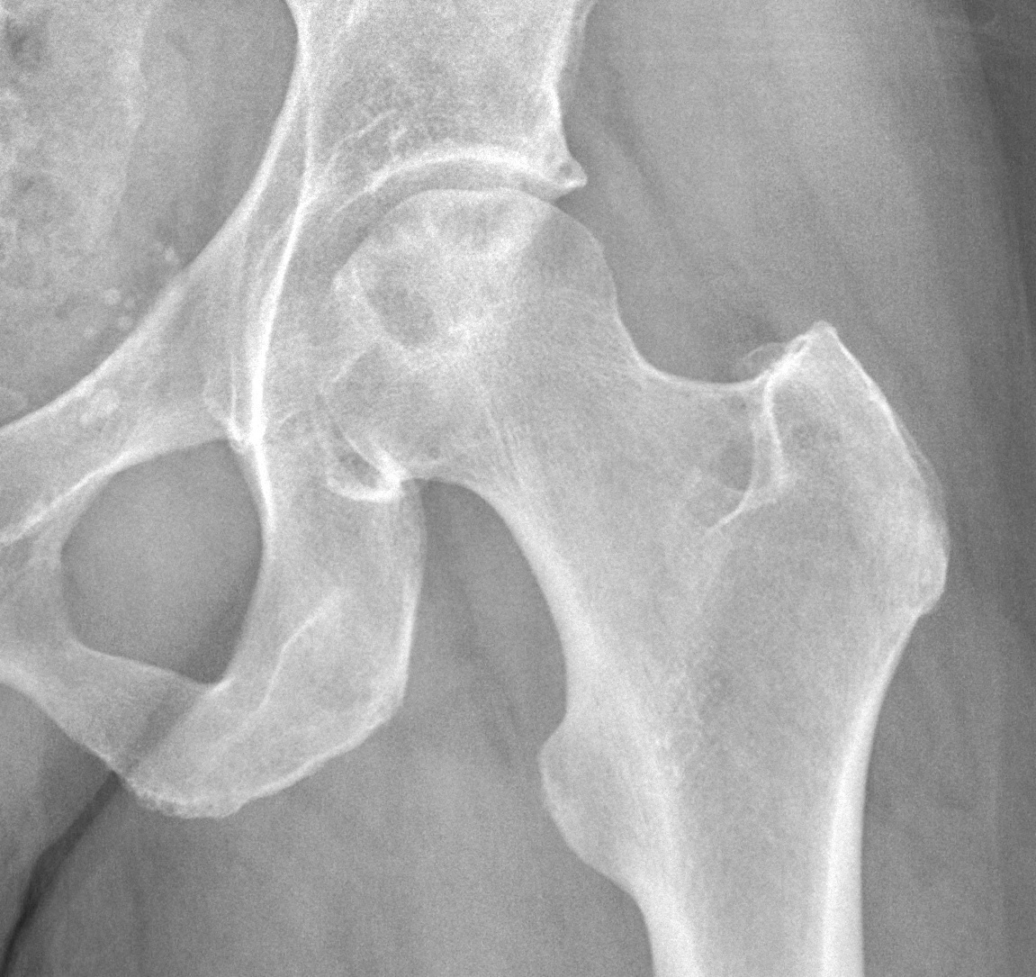
Figure 2. Avascular necrosis of the femoral head on a hip radiograph (subchondral lucency and an irregular head surface), a recognised late complication of a femoral-neck fracture. Source: Mikael Häggström, M.D., via Wikimedia Commons, CC0 1.0.
Part III - Venous Thromboembolism: Scope and Mechanism
Venous thromboembolism (VTE), meaning deep vein thrombosis (DVT) and its lethal sequel, pulmonary embolism (PE), is the most important preventable systemic complication of skeletal trauma. Its scale without prophylaxis is large: in Geerts’ landmark venographic study of injured patients, 58% developed a DVT (18% proximal), rising to 69% in those with a lower-limb orthopaedic injury, and more than half of polytrauma patients thrombose.[21] An untreated hip fracture carries a 50% DVT rate (25% proximal), and the fatal-PE rate after emergency hip surgery without prophylaxis reaches 7.5-10%.[22] Even with prophylaxis the residual DVT rate can be as high as 12%.[23] PE is a leading cause of post-injury death, and “two-thirds of patients with a fatal PE die within thirty minutes,” which is why prevention, not treatment, dominates the subject.[24]
The mechanism is Virchow’s triad (venous stasis, endothelial [vessel-wall] injury, and hypercoagulability), every limb of which is aggravated by trauma.[25] Injury exposes tissue factor and collagen, antithrombin activity falls (it is subnormal in 61% of the critically injured), and fibrinolysis is suppressed by raised plasminogen-activator-inhibitor-1. This hypercoagulable state begins at the moment of injury, which is why “prophylaxis” is really better called DVT protection.[26] The dangerous clots are those at or above the popliteal fossa: distal calf thrombi carry a PE risk under 5%, but about 20-30% propagate above the knee and then become as dangerous as any thigh clot, and roughly 90% of acute PEs arise from thrombus proximal to the calf.[27]
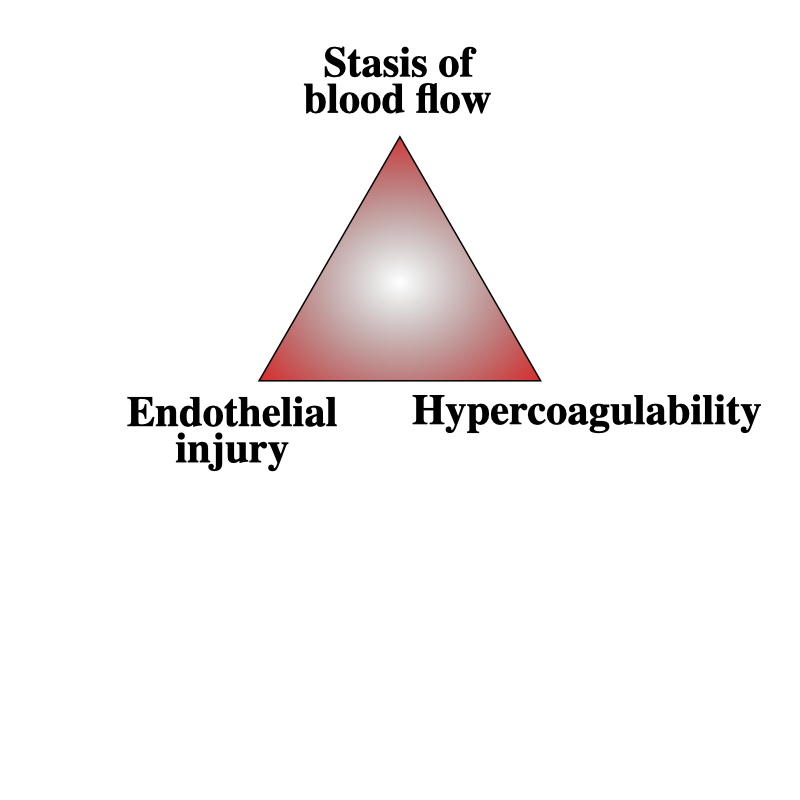
Figure 3. Virchow’s triad: venous stasis, endothelial injury and hypercoagulability, every limb of which is aggravated by trauma. Source: Rudolf.hellmuth, via Wikimedia Commons, CC BY-SA 3.0.
Part IV - Risk Factors and Diagnosis of VTE
Risk factors and risk assessment
The risk factors divide into patient factors (age over 55, obesity, previous VTE, active cancer, the genetic thrombophilias such as factor V Leiden, pregnancy and oestrogen therapy) and injury factors (pelvic, lower-limb and hip fractures, acute spinal cord injury, a rising Injury Severity Score, head injury, prolonged ventilation and shock).[28] Spinal cord injury is the standout: it brings a 500-fold increase in PE-related death in the first month, and a meta-analysis found that, of all the conventional factors, only spinal fracture (2×) and cord injury (3×) reached statistical significance, a genuine challenge to the assumed independent risk of pelvic and long-bone fractures.[29] Formal stratification uses the Caprini score (a value above 3 warrants prophylaxis, and a major lower-limb fracture is “highest-risk” by definition), though many units, including the cited authors, simply apply the ACCP guidelines from the history and examination.[30]
Diagnosis
DVT is clinically silent in two-thirds of cases, and its signs (limb swelling, a positive Homan’s sign) are unreliable; the best of them, a calf-diameter increase over 2 cm, has a likelihood ratio of only 1.8.[31] The investigations are:[32]
- Compression duplex (colour-flow Doppler) ultrasound: the standard test for DVT (sensitivity ~83%, specificity ~98%), though operator-dependent and insensitive to isolated calf thrombi.
- CT pulmonary angiography (CTPA): the gold standard for PE (sensitivity ~83%, specificity ~96%), having displaced the V/Q scan.
- Venography: the old gold standard, now abandoned (invasive, can itself cause phlebitis).
- MR/CT venography: useful for pelvic DVT after pelvic or acetabular trauma, but with a high false-positive rate.
- D-dimer: useful to exclude DVT in low-risk outpatients, but not useful in trauma, where it is almost always raised regardless.
The Wells score stratifies pre-test probability of DVT or PE, but in skeletal trauma it is only an adjunct, because the injuries themselves place the patient in a moderate-to-high probability band.[33] Routine surveillance ultrasound is not cost-effective and is recommended against; targeted screening is reserved for at-risk patients who have not received adequate prophylaxis.[34]
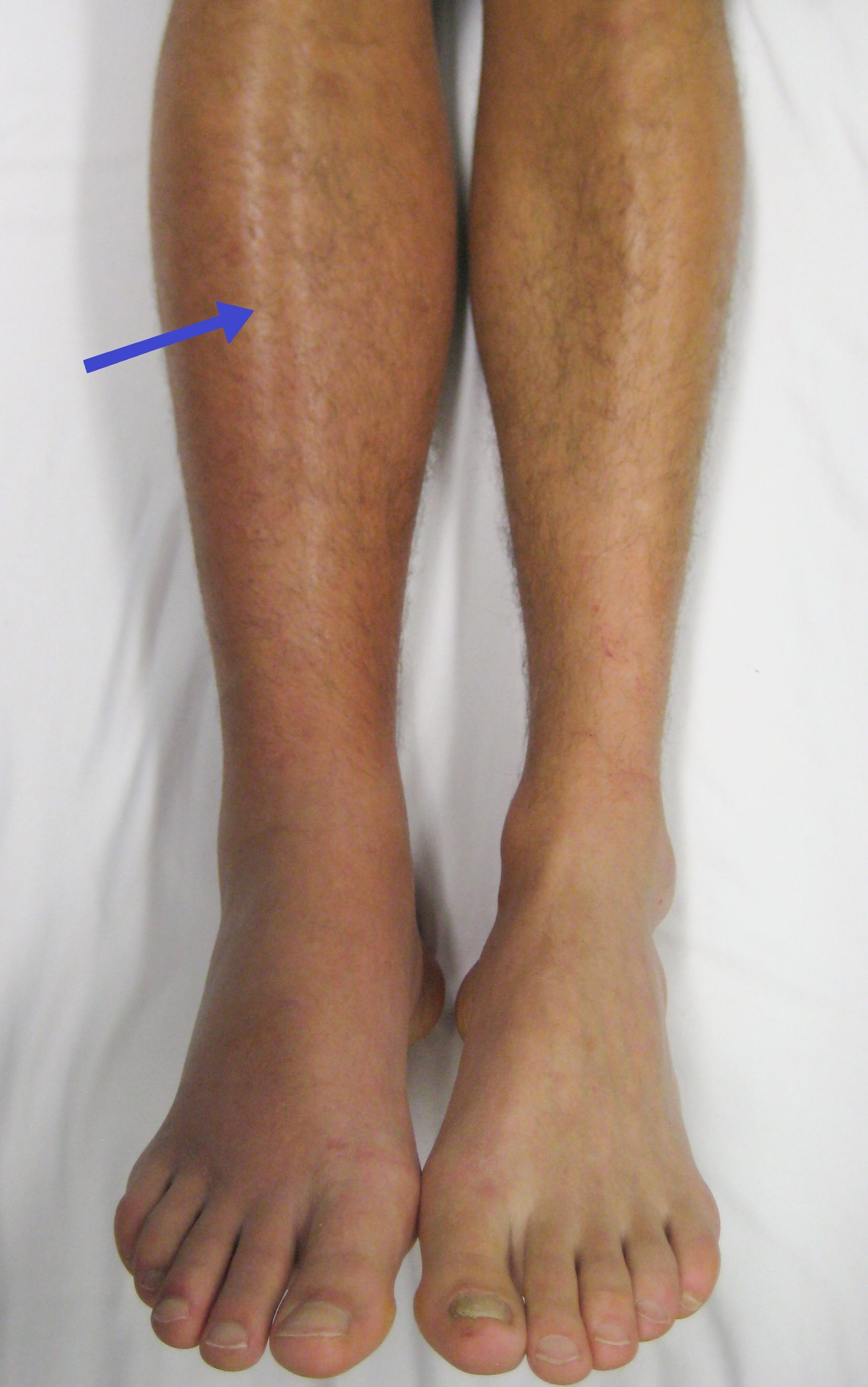
Figure 4. Deep vein thrombosis of the right leg: a swollen, erythematous calf. Source: James Heilman, MD, via Wikimedia Commons, CC BY-SA 3.0.
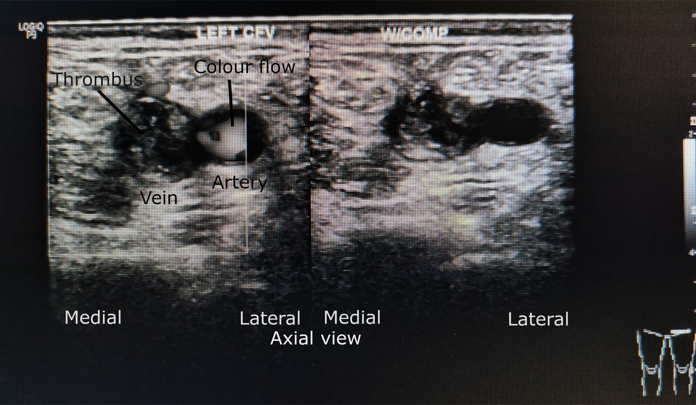
Figure 5. Compression duplex ultrasound of the common femoral vein, the standard test for DVT: echogenic thrombus filling the vein, which remains non-compressible. Source: Cerevisae, via Wikimedia Commons, CC BY-SA 4.0.
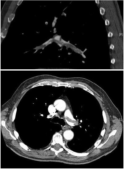
Figure 6. Pulmonary embolism on CT pulmonary angiography, the gold standard for PE: a saddle embolus straddling the pulmonary-artery bifurcation. Source: Myat & Ahsan, Thrombosis Journal 2007, via Wikimedia Commons, CC BY 2.0.
Part V - Prophylaxis Against VTE
Mechanical prophylaxis
Graduated compression stockings and intermittent pneumatic compression (IPC) reduce stasis and lower DVT and PE rates, but mechanical methods alone do not match chemical prophylaxis.[35] Their value is as the prophylaxis of choice when anticoagulation is contraindicated (coagulopathy, an immediate high bleeding risk), in low-risk patients, and as an adjunct to chemical prophylaxis in the high-risk patient: combined prophylaxis lowers DVT and PE further without added complications.[36] Soft-tissue injury often simply prevents the device being applied.[37]
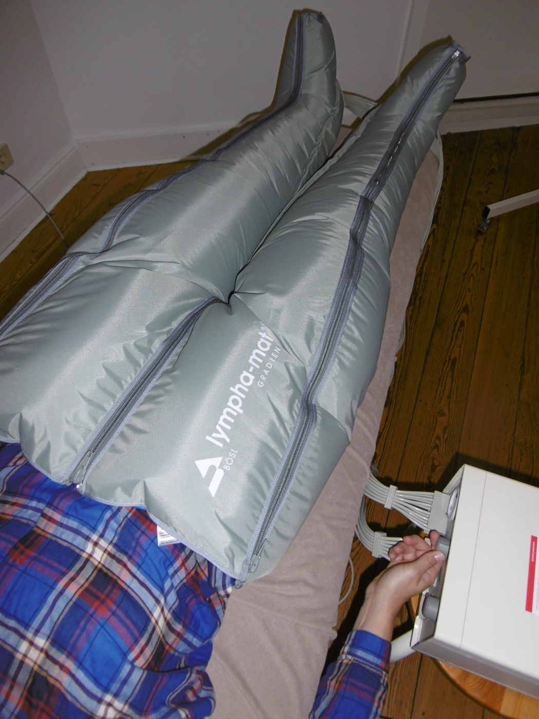
Figure 7. Intermittent pneumatic compression sleeves connected to an air pump, mechanical VTE prophylaxis. Source: “Enter”, via Wikimedia Commons, CC BY-SA 4.0.
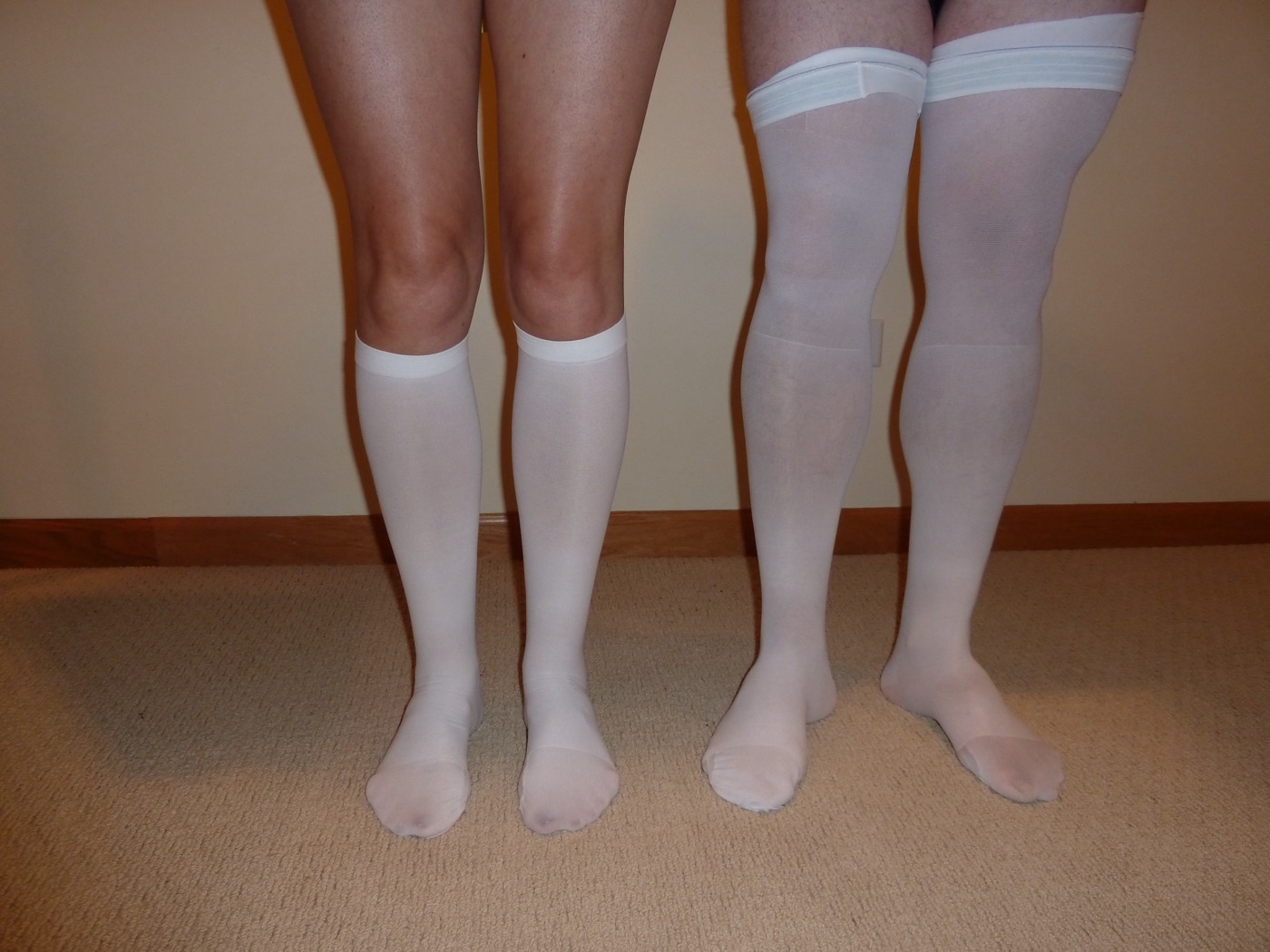
Figure 8. Graduated (anti-embolism, TED) compression stockings. Source: Lentpjuve, via Wikimedia Commons, CC BY-SA 4.0.
Chemical prophylaxis
The available agents, with the doses the sources give:[38]
- Low-molecular-weight heparin (LMWH, e.g. enoxaparin 30 mg twice daily or 40 mg once daily): the workhorse “gold-standard” agent, being subcutaneous, predictable, free of routine monitoring, and reversible with protamine.
- Unfractionated heparin: cheaper, short-acting and fully reversible (a useful perioperative bridge), but needs monitoring and carries the risk of heparin-induced thrombocytopenia.
- Fondaparinux: a selective factor-Xa inhibitor (one hip-fracture trial: VTE 8.3% versus 19.1% for enoxaparin); contraindicated if creatinine clearance is below 30.
- Warfarin: for prophylaxis a target INR of about 1.5-2.0 (the texts cite 1.4-1.6 and 1.5-1.8), needing monitoring.
- Direct oral anticoagulants: rivaroxaban, apixaban (prophylactic dose 2.5 mg twice daily), edoxaban and dabigatran; convenient and effective in arthroplasty but with limited trauma-population data and the drawback of irreversibility and unreliable absorption in the acute patient.
- Aspirin (81-325 mg): labelled the “least effective” agent yet increasingly favoured for its safety, and now recommended by the AAOS and ACCP after arthroplasty.
Timing balances thrombosis against bleeding: the cited authors start chemical prophylaxis on the day of admission (within 12-24 hours) once major haemorrhage has been excluded, and within 72 hours even after a haemorrhagic head injury; mechanical prophylaxis bridges any delay.[39] Duration runs to a minimum of 10-14 days and often longer: at least 28 days after a geriatric hip fracture, and about 6 weeks for an isolated pelvic, femoral or tibial fracture.[40] An inferior vena cava (IVC) filter is reserved for the patient at high risk who cannot be anticoagulated, or who re-embolises despite anticoagulation; routine “primary-prophylaxis” use is discouraged (the PREPIC trials showed filters reduce PE but increase DVT without changing mortality), and retrievable filters should be removed early.[41]
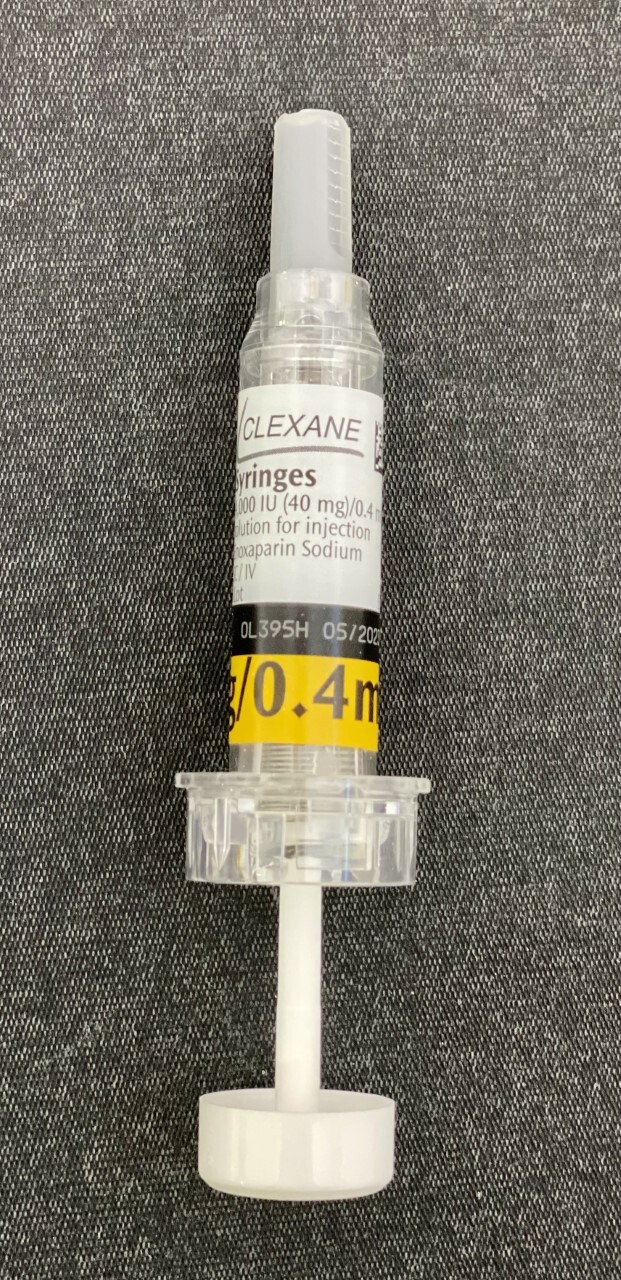
Figure 9. A low-molecular-weight heparin (enoxaparin) pre-filled syringe, the workhorse of chemical thromboprophylaxis. Source: Whispyhistory, via Wikimedia Commons, CC BY-SA 4.0.
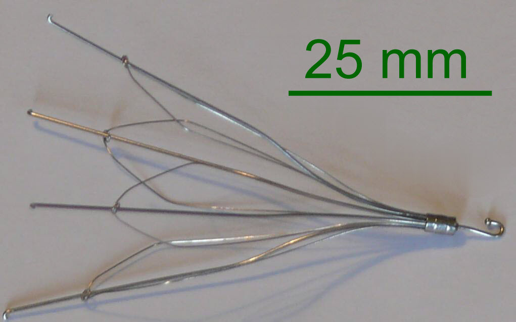
Figure 10. An inferior vena cava filter, reserved for the high-risk patient who cannot be anticoagulated or who re-embolises despite it. Source: BozMo, via Wikimedia Commons, CC BY-SA 3.0.
Part VI - Treatment of Established VTE
Once VTE is confirmed the goals are to stop the clot propagating and embolising and to prevent recurrence.[42] For a proximal DVT or PE without cancer, the standard is at least three months of anticoagulation, and the current preference is a direct oral anticoagulant over warfarin or LMWH for most patients.[43] An isolated distal (calf) DVT may be treated for three months or simply followed with serial ultrasound for two weeks if it is asymptomatic and not extending, and an incidental subsegmental PE without a proximal DVT may be watched rather than anticoagulated.[44] A massive PE with haemodynamic instability (systolic pressure below 90 mmHg) warrants thrombolysis, or catheter-based clot removal when the bleeding risk is too high.[45] Anticoagulation itself carries about a 3% haemorrhage risk, weighed against a 25% risk of recurrent VTE if it is withheld.[46]
Part VII - Fat Embolism Syndrome
Definition, incidence and pathophysiology
Fat embolism, meaning fat globules in the circulation, is common and usually subclinical after a long-bone fracture; fat embolism syndrome (FES) is the clinical illness, “the occurrence of hypoxia, confusion, and petechiae a few days or even hours after a long-bone fracture.”[47] Finding fat globules in blood, urine or sputum is too sensitive to be diagnostic; it is the syndrome, a diagnosis of exclusion once pulmonary embolism, contusion, aspiration and the rest are ruled out, that matters.[48] Overt FES complicates about 8.75% of fracture patients (mortality ~2.5%), rising to 35% with multiple fractures, though other series report 0.9-3.5% for long-bone fractures.[49] Characteristically it strikes the young, follows lower-limb rather than upper-limb fractures, and is more frequent with closed fractures.[50]
Two theories explain it, and both probably operate.[51] The mechanical theory holds that marrow fat globules (most 20-40 µm) enter torn marrow veins and obstruct the pulmonary capillaries, reaching the brain and skin through a patent foramen ovale or precapillary shunts. The biochemical theory holds that mediators from the fracture coalesce circulating chylomicrons, and that serum lipase hydrolyses the embolised neutral fat into free fatty acids that are toxic to the pulmonary endothelium, a chemical phase that neatly explains the latent interval before the lungs fail.[52] Inflammatory mediators (thromboxane, vasoactive amines) add pulmonary vasospasm and increased permeability, and thrombocytopenia is so consistent that it serves as a diagnostic criterion.[53]
Timing, features and diagnosis
The syndrome begins after a latent (“lucid”) interval, typically 1-2 days: 60% of cases appear within 24 hours and 90% within 72 hours.[54] The classic triad is respiratory (hypoxaemia, where a PaO₂ below 60 mmHg is an early warning), cerebral (confusion, drowsiness; neurological change in up to 80%, from petechial haemorrhages in the brain rather than hypoxia alone, so it persists despite oxygen), and cutaneous (the transient petechial rash over the conjunctivae, cheek, neck, axillae and palate).[55]
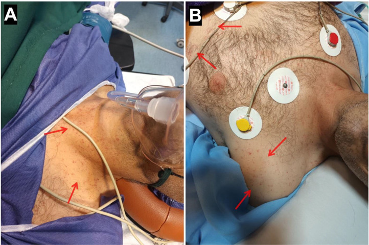
Figure 11. The petechial rash of fat embolism syndrome over the chest and axilla. From a case report (2024), PMC11625105, CC BY 4.0.
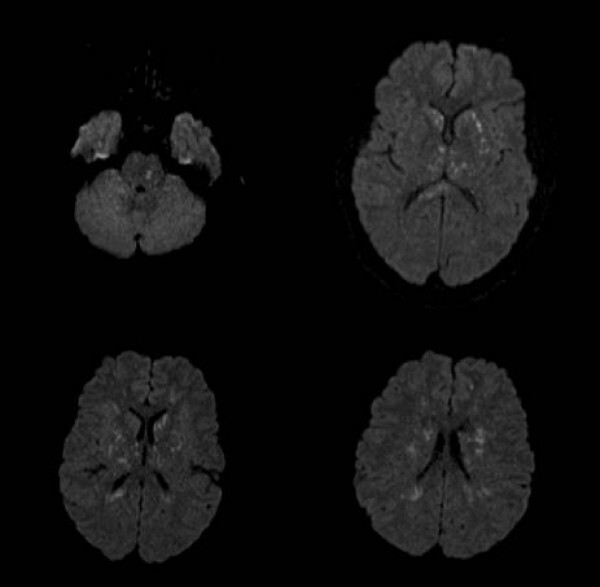
Figure 12. Cerebral fat embolism on diffusion-weighted MRI: scattered punctate hyperintensities, the “starfield” pattern. From Parizel et al. (2009), Cases Journal 2:212, PMC2783161, CC BY 2.0.
Diagnosis uses Gurd and Wilson’s criteria: one major plus four minor signs (with fat macroglobulinaemia):[56]
- Major: respiratory insufficiency/hypoxaemia, cerebral involvement, petechial rash (the table adds pulmonary oedema).
- Minor: tachycardia (> 110), pyrexia (> 38.3 °C), retinal emboli, fat in urine or sputum, thrombocytopenia, falling haematocrit, jaundice, renal changes.
Lindeque’s criteria offer a respiratory-only alternative in which a single one suffices: a PaCO₂ above 55 mmHg or pH below 7.3, a sustained respiratory rate above 35, or dyspnoea with tachycardia and anxiety.[57]
Prevention and treatment
Prevention rests on three measures: proper splinting and prompt transport, oxygen in the post-injury period, and early operative stabilisation of long-bone fractures.[58] Reaming during intramedullary nailing raises intramedullary pressure and embolises marrow fat, a real concern, especially with a coexisting chest injury, but stabilising the fracture also removes the source of ongoing embolisation, so the balance favours early fixation, individualised to the patient’s pulmonary state.[59] Treatment is mainly supportive, because “fat embolism syndrome is primarily a disease of the respiratory system”; oxygen and meticulous mechanical ventilation are the mainstay.[60] Corticosteroids are genuinely controversial: prophylactic methylprednisolone reduces post-traumatic hypoxaemia and probably FES by limiting free-fatty-acid endothelial damage, but the risks mean steroids are “not routinely employed.”[61] Fluid loading, hypertonic glucose, alcohol, heparin, dextran and aspirin have all been shown to have no effect on the rate of FES.[62]
References
-
Skeletal Trauma 5e, ch.23 (Diagnosis and Treatment of Complications), p.689.
-
Skeletal Trauma 5e p.692.
-
Skeletal Trauma 5e p.689.
-
Skeletal Trauma 5e p.692.
-
Skeletal Trauma 5e pp.675-677.
-
Skeletal Trauma 5e p.677.
-
Skeletal Trauma 5e p.676.
-
Skeletal Trauma 5e p.676.
-
Skeletal Trauma 5e p.676.
-
Skeletal Trauma 5e pp.680-681, Table 23-8.
-
Skeletal Trauma 5e pp.681-683.
-
Skeletal Trauma 5e p.681, Table 23-9.
-
Skeletal Trauma 5e p.684.
-
Skeletal Trauma 5e pp.677-679.
-
Skeletal Trauma 5e p.680, Table 23-7.
-
Skeletal Trauma 5e p.679.
-
Skeletal Trauma 5e pp.684-685, Table 23-12.
-
Skeletal Trauma 5e p.685.
-
Skeletal Trauma 5e pp.684-686.
-
Avascular necrosis, heterotopic ossification and physeal arrest are standard fracture complications cross-referenced to the regional chapters; they are not detailed in the mined Skeletal Trauma local-complications extract.
-
Rockwood & Green 9e, ch.25 (Ahsan & Firoozabadi), p.1275; Skeletal Trauma 5e p.667.
-
Rockwood 9e pp.1276, 1289; Skeletal Trauma 5e p.668.
-
Rockwood 9e p.1276.
-
Skeletal Trauma 5e p.668; Rockwood 9e p.1276.
-
Rockwood 9e p.1272; Skeletal Trauma 5e p.667.
-
Rockwood 9e p.1277; Skeletal Trauma 5e pp.667, 669.
-
Rockwood 9e pp.1276, 1293.
-
Rockwood 9e pp.1277-1278, Table 25-1.
-
Rockwood 9e p.1276; Skeletal Trauma 5e p.667.
-
Rockwood 9e pp.1279-1281, Table 25-2.
-
Rockwood 9e p.1289; Skeletal Trauma 5e p.668.
-
Rockwood 9e pp.1290-1292, Table 25-6; Skeletal Trauma 5e pp.668-669.
-
Rockwood 9e pp.1290-1291, Table 25-5.
-
Rockwood 9e pp.1292-1293; Skeletal Trauma 5e p.669.
-
Rockwood 9e pp.1285-1286.
-
Rockwood 9e p.1286.
-
Skeletal Trauma 5e p.671.
-
Rockwood 9e pp.1281-1285, Tables 25-3 to 25-4.
-
Rockwood 9e pp.1284, 1286.
-
Rockwood 9e pp.1287-1289.
-
Rockwood 9e p.1286; Skeletal Trauma 5e pp.669-670.
-
Rockwood 9e p.1293.
-
Rockwood 9e p.1293.
-
Rockwood 9e p.1293.
-
Rockwood 9e pp.1293-1294.
-
Rockwood 9e p.1293.
-
Skeletal Trauma 5e p.663.
-
Skeletal Trauma 5e p.664.
-
Skeletal Trauma 5e pp.663-664.
-
Skeletal Trauma 5e p.664.
-
Skeletal Trauma 5e p.665.
-
Skeletal Trauma 5e p.665.
-
Skeletal Trauma 5e p.665.
-
Skeletal Trauma 5e p.663.
-
Skeletal Trauma 5e pp.663-664.
-
Skeletal Trauma 5e pp.663-664, Table 23-1.
-
Skeletal Trauma 5e p.664.
-
Skeletal Trauma 5e p.666.
-
Skeletal Trauma 5e pp.665-666.
-
Skeletal Trauma 5e p.667.
-
Skeletal Trauma 5e pp.666-667.
-
Skeletal Trauma 5e p.666.
-
Skeletal Trauma 5e pp.675-677, 692.
-
Skeletal Trauma 5e pp.680-684.
-
Rockwood 9e pp.1272, 1277.
-
Rockwood 9e pp.1276, 1290-1292.
-
Rockwood 9e pp.1284-1289.
-
Rockwood 9e pp.1293-1294.
-
Skeletal Trauma 5e pp.663-665.
-
Skeletal Trauma 5e pp.663-664.
-
Skeletal Trauma 5e pp.666-667.
-
Skeletal Trauma 5e pp.677-680.