Contents
- Orientation: A Ring Around the Lunate, and a Hand That Punishes Stiffness
- Part I - Carpal Anatomy and Kinematics
- Part II - Scaphoid Fractures
- Part III - Perilunate and Lunate Dislocations
- Part IV - Scapholunate and Lunotriquetral Dissociation
- Part V - The Other Carpal Fractures
- Part VI - Metacarpal Fractures
- Part VII - The Thumb Metacarpal Base: Bennett and Rolando
- Part VIII - Carpometacarpal (CMC) Dislocations
- Part IX - Principles of Hand-Fracture Fixation, and the Phalanges
- Part X - A Synthesis: How to Reason About the Carpus and Hand
- References
Orientation: A Ring Around the Lunate, and a Hand That Punishes Stiffness
Two ideas organise this large topic. The first is that the carpus behaves as a ring of bones articulating around the lunate, so that a single force, applied to the outstretched hand, can travel around that ring and break or dislocate it in a predictable sequence. Understanding that sequence (the Mayfield progression) makes sense of the scaphoid fracture, the scapholunate tear, and the perilunate dislocation all at once: they are points on one spectrum.[1] The second idea is that the hand, like the elbow, punishes stiffness, so that the goal of treating any metacarpal or phalangeal fracture is not radiographic union but the return of function through early, controlled motion. As the AO text puts it, nowhere in the body does function follow form as closely as in the hand, and the outcome of a hand fracture is judged on movement, not on the radiograph.[2] Two anatomical facts dominate the clinical reasoning throughout: the retrograde blood supply of the scaphoid (which makes its proximal pole prone to avascular necrosis and nonunion) and the intolerance of the fingers to malrotation (which scissor on flexion if a metacarpal heals twisted).
Part I - Carpal Anatomy and Kinematics
The carpus is two rows of bones. The proximal row, from radial to ulnar, is the scaphoid, lunate, and triquetrum; the distal row is the trapezium, trapezoid, capitate, and hamate (the pisiform is a sesamoid within the flexor carpi ulnaris tendon).[3] The proximal row is the “intercalated segment”: it has no tendon attachments of its own and moves passively, its position dictated by the shapes of the bones and the ligaments. The lunate is the keystone, seated in the lunate fossa of the radius and tethered on each side by the scapholunate (SL) and lunotriquetral (LT) interosseous ligaments, the two most important intrinsic ligaments of the wrist.[4] Within the SL ligament the dorsal portion is the thick, strong part (the primary restraint); within the LT ligament the strong part is palmar.[5] The extrinsic ligaments span from the radius to the carpus, and a V-shaped weak interval over the capitolunate joint, the space of Poirier, is where the lunate escapes into the carpal tunnel in a dislocation.[6]
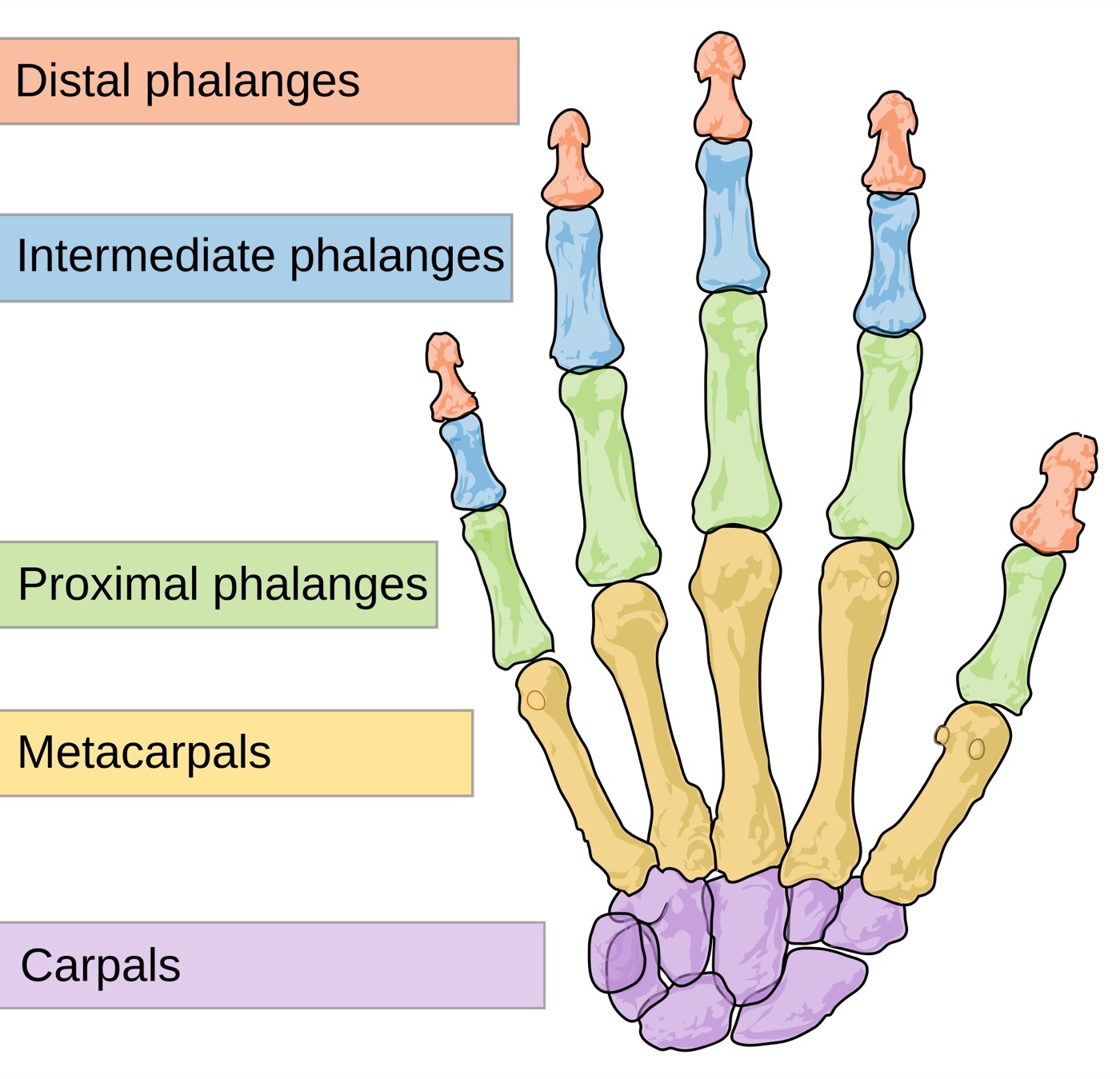
Figure 1. The skeleton of the hand: the carpus, the metacarpals, and the phalanges. Diagram by Mariana Ruiz Villarreal (LadyofHats), public domain, via Wikimedia Commons.
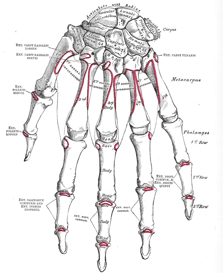
Figure 2. The bones of the wrist named (dorsal view): the proximal row (scaphoid, lunate, triquetrum) and the distal row (trapezium, trapezoid, capitate, hamate). Gray’s Anatomy (1918), public domain, via Wikimedia Commons.
The scaphoid is the mechanical link between the two rows, lying obliquely at about 45° and flexing under load; it transmits a flexion moment to the lunate, which is held in balance against the extension tendency of the triquetrum.[7] When that balance is lost, the lunate tilts into one of two patterns seen on the lateral radiograph: dorsal intercalated segment instability (DISI), where the lunate extends (scapholunate angle over 60°, classically from a scapholunate injury or scaphoid nonunion), or volar intercalated segment instability (VISI), where the lunate flexes (scapholunate angle under 30°, classically from a lunotriquetral injury).[8] The normal scapholunate angle is about 30° to 60°.[9] On the posteroanterior film, carpal alignment is checked against Gilula’s arcs, three smooth lines along the proximal and distal joint surfaces of the proximal row; a break in these arcs signals a dislocation or dissociation.[10]
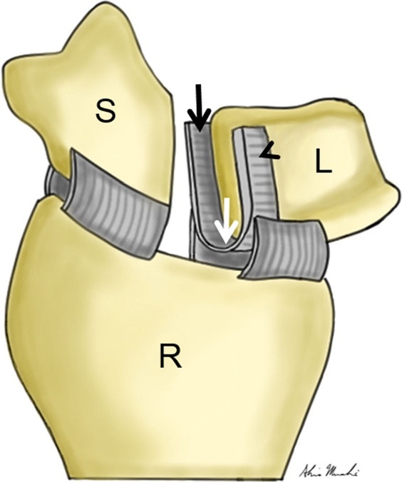
Figure 3. The scapholunate interosseous ligament between the scaphoid (S) and lunate (L) on the radius (R), the primary stabiliser of the scapholunate joint. Tischler et al., CC BY 4.0, via Wikimedia Commons.
Part II - Scaphoid Fractures
2.1 Why the scaphoid is special: the retrograde blood supply
The scaphoid is the most commonly fractured carpal bone, accounting for 60% to 80% of all carpal fractures and about 2% to 3% of all fractures, typically in young men after a fall on the outstretched hand.[11] What makes it dangerous is its blood supply. The scaphoid is over 80% covered by articular cartilage, leaving little surface for periosteal healing, and it is fed by a largely retrograde supply: the dorsal carpal branch of the radial artery enters distally at the dorsal ridge and supplies 70% to 80% of the bone, including the entire proximal pole, while a smaller volar branch supplies the distal 20% to 30%.[12] The consequence is the central fact of the topic: a fracture through the waist or proximal pole interrupts the retrograde flow to the proximal fragment, so that proximal-pole and displaced waist fractures are prone to avascular necrosis and nonunion, while distal-pole and tubercle fractures heal readily.[13]
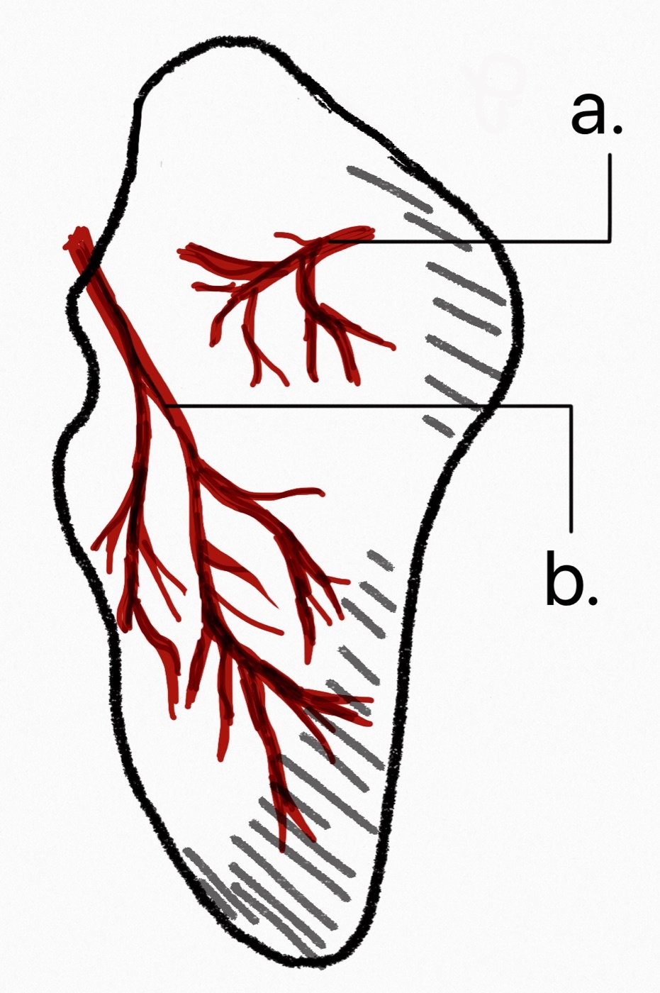
Figure 4. The retrograde blood supply of the scaphoid: the dorsal branch of the radial artery enters distally and supplies the proximal pole, which is therefore prone to avascular necrosis. Iiibalesiii, CC BY-SA 4.0, via Wikimedia Commons.
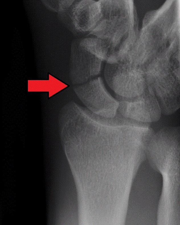
Figure 5. A scaphoid waist fracture on the PA wrist radiograph (arrow). Gilo1969, CC BY-SA 3.0, via Wikimedia Commons.
2.2 Assessment and the occult fracture
The classic sign is tenderness in the anatomical snuffbox, supported by scaphoid-tubercle tenderness and pain on axial compression of the thumb; no single sign is both sensitive and specific, so they are combined.[14] The recurring clinical problem is the occult (clinically suspected) fracture: up to 30% to 40% of scaphoid fractures are invisible on the initial radiographs, yet missing one risks nonunion in an active young patient.[15] The safe strategy is to immobilise any clinically suspected fracture and re-image, with MRI the most sensitive investigation for the occult fracture (and increasingly the most cost-effective first study), while CT best demonstrates displacement, anatomy, and union.[16]
2.3 Classification
Scaphoid fractures are classified first by location (distal pole/tubercle, waist [the commonest], proximal pole) and then by stability.[17] The Herbert classification is the working scheme: type A stable acute (incomplete/tubercle), type B unstable acute (any complete/bicortical fracture, B1 distal oblique, B2 complete waist [the commonest single pattern], B3 proximal pole, B4 trans-scaphoid perilunate fracture-dislocation, B5 comminuted), type C delayed union, and type D nonunion (D1 fibrous, D2 sclerotic).[18] A fracture is considered displaced/unstable with more than 1 mm of displacement, a lateral intrascaphoid angle over 35°, comminution, proximal-pole location, or a DISI deformity; all displaced fractures are unstable.[19]
2.4 Treatment
A nondisplaced fracture is treated nonoperatively in a below-elbow cast, with union rates of about 95% to 99%; the long-standing debates over above- versus below-elbow casting and thumb-spica versus Colles casting have been settled by trials showing no advantage to the longer or thumb-including cast, so a below-elbow forearm cast is advocated.[20] Operative treatment with a percutaneous headless compression screw (the Herbert-type variable-pitch screw, placed volar or dorsal, neither approach proven superior) is indicated for displaced, unstable, and proximal-pole fractures, and is increasingly offered as a “fast-track” to athletes and manual workers because it shortens time to union and return to work without clearly changing the union rate (the trade-off being a higher complication rate).[21] Proximal-pole fractures are usually fixed through a small dorsal approach because of their high nonunion risk.[22]
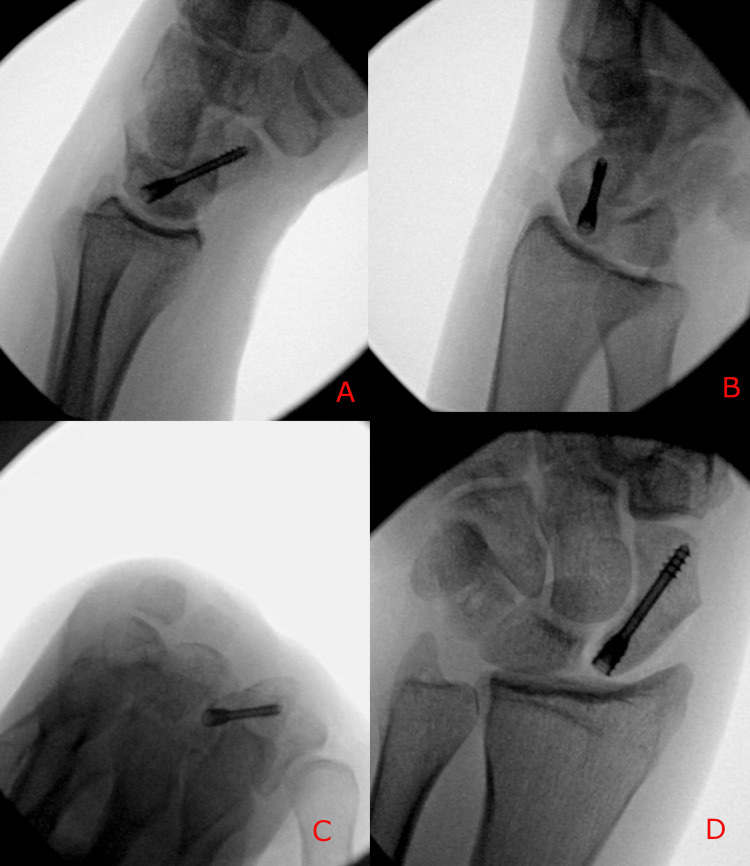
Figure 6. A scaphoid fracture fixed with a headless compression screw (intra-operative fluoroscopy). Mannan & Kent, Cureus 2026, CC BY 4.0.
2.5 Complications: malunion, nonunion, SNAC, and AVN
A displaced scaphoid heals in the humpback deformity (a flexed, foreshortened scaphoid with a DISI of the carpus), which limits extension and grip.[23] Nonunion affects about 10% of all waist fractures (near zero for well-treated nondisplaced fractures, but up to 50% for displaced fractures), and an untreated nonunion follows a predictable degenerative path, the scaphoid nonunion advanced collapse (SNAC) wrist, beginning at the radial styloid and progressing to pan-carpal arthritis (the analogue of the SLAC wrist after a scapholunate injury).[24] Nonunion is managed by resecting the fibrous tissue, correcting the deformity with a bone graft (non-vascularised, or vascularised from the distal radius such as a 1,2-intercompartmental supraretinacular artery pedicle, or from the medial femoral condyle for the avascular proximal pole), and stable screw fixation.[25] Avascular necrosis of the proximal pole is the third complication, especially after proximal-pole fractures; it is confirmed definitively only by the absence of punctate bleeding from the bone at surgery, and is treated with a vascularised graft.[26]
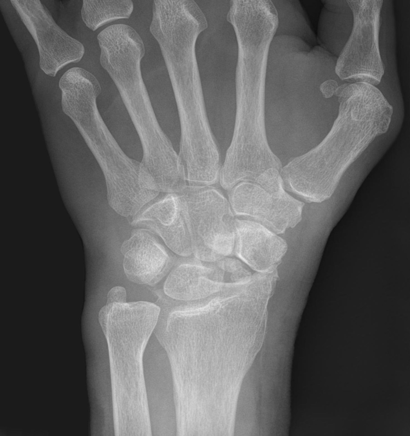
Figure 7. Scaphoid nonunion with advanced collapse (SNAC) on the PA radiograph. Hellerhoff, CC BY-SA 4.0, via Wikimedia Commons.
Part III - Perilunate and Lunate Dislocations
These are the high-energy end of the carpal-injury spectrum, the commonest true wrist dislocation, in young men, and they are missed in 16% to 25% of cases at presentation.[27] They are understood through the Mayfield progression of perilunar instability, in which a force travels around the lunate from radial to ulnar in four stages: stage I, scapholunate failure (a scaphoid fracture or scapholunate dissociation); stage II, capitolunate dislocation (the capitate displaces dorsally through the space of Poirier); stage III, lunotriquetral disruption (a perilunate dislocation); and stage IV, lunate dislocation, in which the lunate is extruded volarly into the carpal tunnel, hinging on its intact palmar ligaments.[28] Injuries are divided into lesser-arc (purely ligamentous, around the lunate) and greater-arc (with a fracture of one or more surrounding bones); the commonest pattern overall is the trans-scaphoid perilunate fracture-dislocation, a greater-arc injury.[29]
The diagnosis rests on the lateral and PA radiographs: disruption of Gilula’s arcs and a triangular-appearing lunate on the PA, and on the lateral the “spilled teacup” sign, the volarly rotated lunate (the source text writes “spilled teapot”).[30] Because the median nerve is at risk in the carpal tunnel, the injury demands emergent closed reduction (the Tavernier manoeuvre), which decompresses the nerve; but closed treatment alone is unreliable, with loss of reduction in around 60%, so nearly all of these injuries require open reduction, ligament repair, and fixation (a dorsal or combined dorsal-volar approach, reducing in reverse-Mayfield order from the lunotriquetral joint).[31] Even with good treatment the prognosis is guarded, with post-traumatic arthritis in a high proportion.[32]
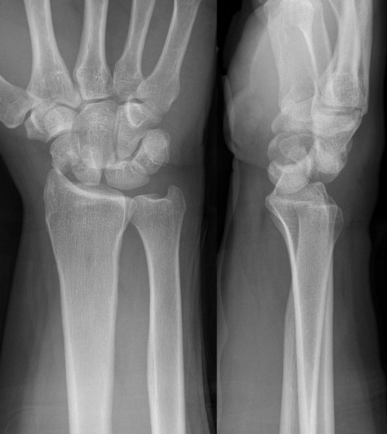
Figure 8. A trans-scaphoid perilunate fracture-dislocation, PA and lateral radiographs, the commonest greater-arc injury. Hellerhoff, CC BY-SA 3.0, via Wikimedia Commons.
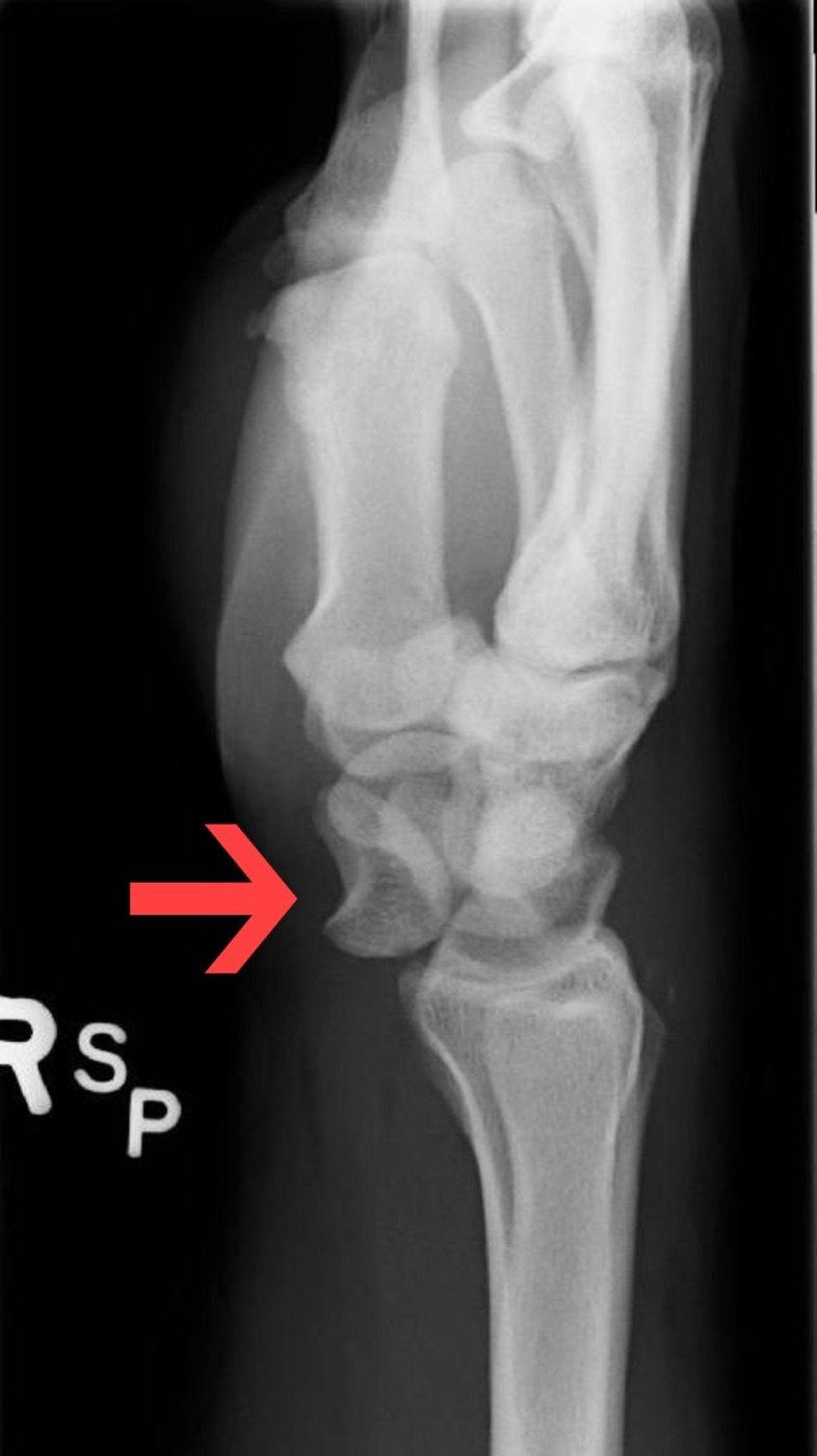
Figure 9. Lunate dislocation on the lateral radiograph: the lunate is tipped volarly (the “spilled teacup”), the end stage of the Mayfield sequence. James Heilman, MD, CC BY-SA 3.0, via Wikimedia Commons.
Part IV - Scapholunate and Lunotriquetral Dissociation
4.1 Scapholunate dissociation
Scapholunate dissociation is the commonest carpal ligament injury. It is suspected clinically by the Watson (scaphoid shift) test and confirmed radiographically by the signs of a flexed, dissociated scaphoid: the Terry-Thomas sign (a scapholunate gap, suggestive over 3 mm, diagnostic over 5 mm), the cortical ring sign (the flexed scaphoid tubercle seen end-on), a scapholunate angle over 60° (diagnostic over 80°), and a DISI on the lateral.[33] Untreated, it follows the degenerative cascade of the scapholunate advanced collapse (SLAC) wrist.[34] Treatment depends on chronicity and tissue quality: an acute tear is repaired directly (with capsulodesis) and pinned; a chronic, reducible injury is reconstructed (for example a three-ligament tenodesis); and a fixed or arthritic wrist is salvaged by partial fusion or proximal row carpectomy.[35]
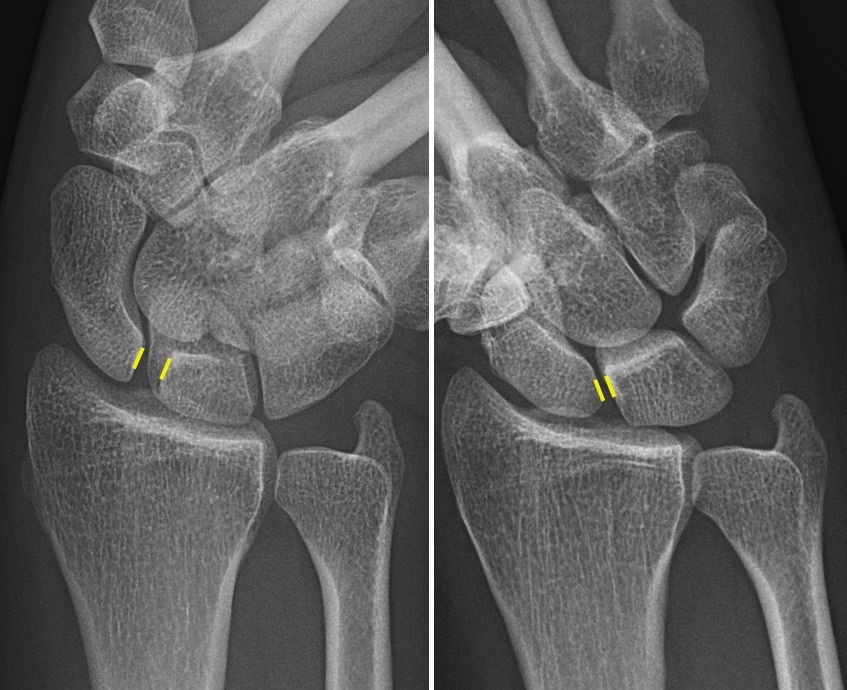
Figure 10. Scapholunate dissociation: a widened scapholunate interval (the Terry-Thomas sign), annotated radiographs. Mikael Häggström, CC0, via Wikimedia Commons.
4.2 Lunotriquetral dissociation
Lunotriquetral dissociation is less common than scapholunate injury and presents with ulnar-sided wrist pain and a clunk on deviation; it is tested by ballottement and the lunotriquetral shear test and may produce a VISI deformity.[36] Most acute injuries are treated in a cast or by pinning, with reconstruction or lunotriquetral fusion reserved for chronic, symptomatic cases.[37]
Part V - The Other Carpal Fractures
After the scaphoid, the carpal fractures in rough order of frequency are:[38]
- Triquetrum: the second commonest carpal fracture, usually a benign dorsal cortical chip (avulsion) seen best on the oblique/lateral view, treated symptomatically.
- Trapezium: body or ridge fractures, often associated with a thumb CMC fracture-dislocation or a hook-of-hamate fracture.
- Hamate: the hook of the hamate fracture, typical of racquet sports and golf, which threatens the ulnar nerve and the small-finger flexor tendons, is poorly seen on plain films (carpal-tunnel view or CT), and is treated by excision or fixation of the displaced or symptomatic hook.
- Capitate: uncommon, often part of a perilunate injury; the scaphocapitate syndrome is a fracture of both the scaphoid and the capitate in which the capitate fragment rotates up to 180°, at risk of avascular necrosis.
- Pisiform: usually a direct-blow body fracture, near the ulnar nerve, treated in a cast or by excision if painful.
- Lunate: rare in isolation, but the seat of Kienböck’s disease (idiopathic avascular necrosis of the lunate), classically associated with an ulnar-minus variant.
- Trapezoid: the least common carpal fracture, requiring high energy because of its strong ligamentous bracing.
Most isolated, undisplaced non-scaphoid carpal fractures do well in a short cast; displacement or an associated dislocation calls for reduction and fixation.[39]
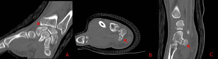
Figure 11. Nonunion of the hook of the hamate on CT (axial, coronal, and sagittal panels). Mannan & Kent, Cureus 2026, CC BY 4.0.
Part VI - Metacarpal Fractures
6.1 The principles: rotation and the cascade
Metacarpal fractures are the third commonest fracture of the upper limb (after phalangeal and distal radius fractures).[40] The metacarpals form a cascade like a Roman arch stabilised by the deep transverse intermetacarpal ligaments, with the rigid central index and middle rays and the mobile border (ring and little) rays; the central rays tolerate the least deformity, the border rays the most.[41] The cardinal clinical task is to detect malrotation: a metacarpal that heals rotated causes the finger to scissor (cross over the adjacent finger) on flexion, the least-tolerated of all hand deformities, corrected for any rotation over about 10°.[42] Significant apex-dorsal angulation produces compensatory pseudoclawing (MCP hyperextension), itself an indication for surgery.[43]
6.2 By location
The neck fracture is typified by the “boxer’s fracture” of the little-finger metacarpal neck, the commonest metacarpal fracture, sustained by punching; the mobile little finger tolerates a large amount of angulation (up to about 50°), so most are treated closed, reduced by the Jahss manoeuvre when needed.[44] In the shaft, the acceptable apex-dorsal angulation increases from radial to ulnar (about 10° in the index and middle, 25° in the ring, and up to 45° in the little finger); shortening is tolerated to about 5 to 6 mm (each 2 mm of shortening costs roughly 7° of extensor lag), but rotation is never accepted.[45] Head fractures are intra-articular and fixed anatomically with headless or small lag screws; base fractures are frequently associated with a carpometacarpal dislocation.[46]
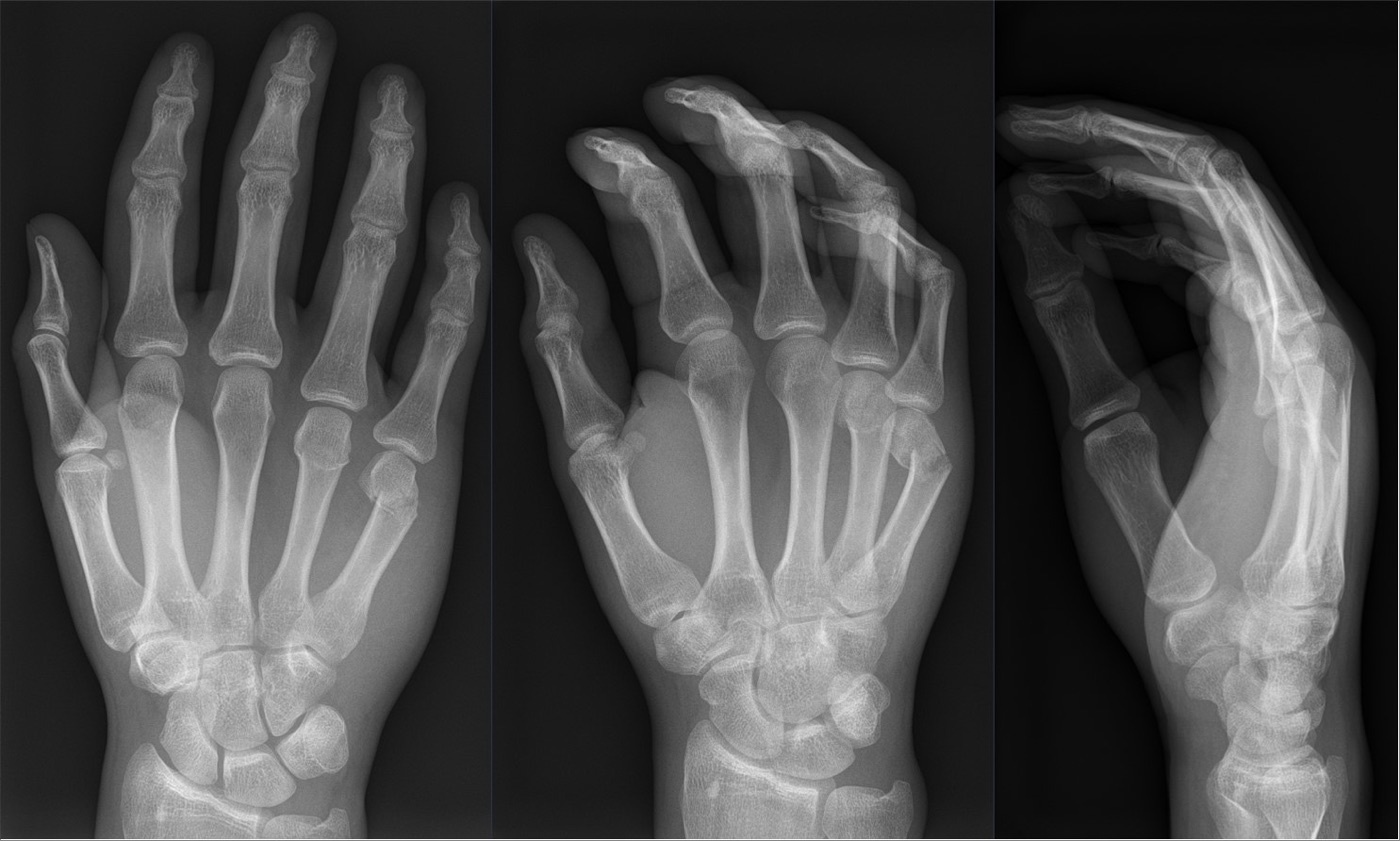
Figure 12. A boxer’s fracture: the little-finger metacarpal neck with apex-dorsal angulation, three views. Hellerhoff, CC BY-SA 4.0, via Wikimedia Commons.
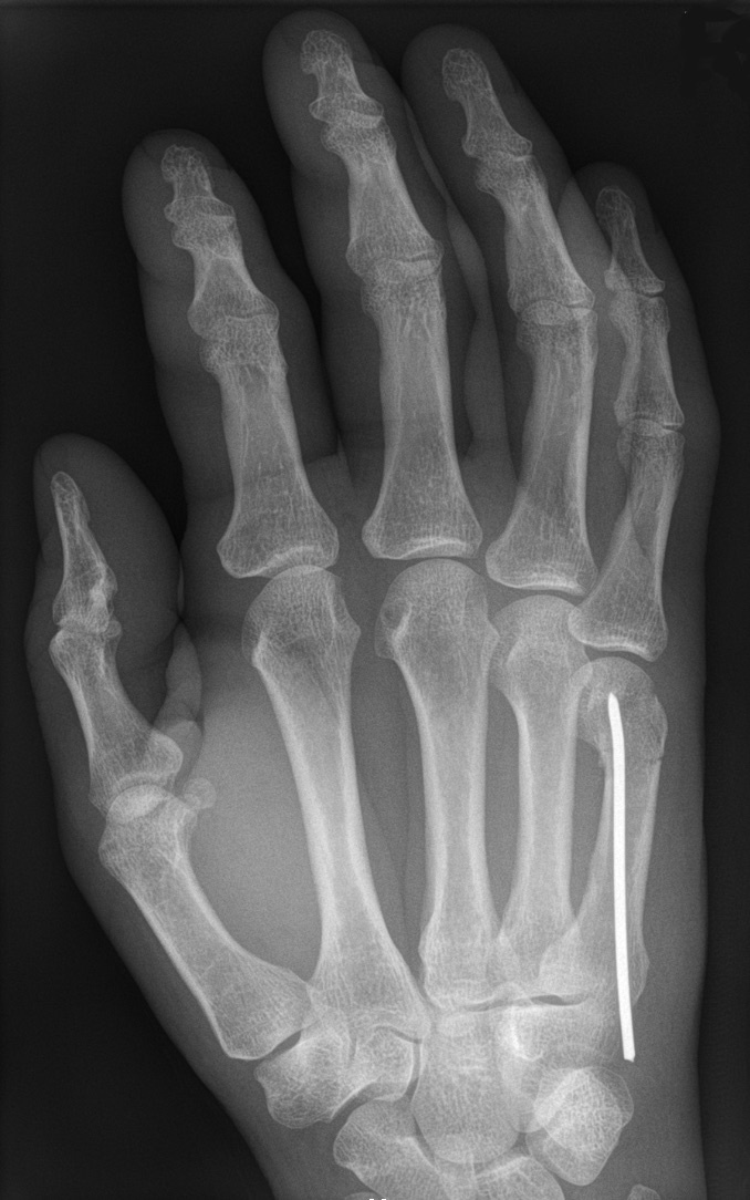
Figure 13. A little-finger metacarpal fracture after intramedullary K-wire fixation. Hellerhoff, CC BY-SA 3.0, via Wikimedia Commons.
6.3 Treatment
The great majority of metacarpal fractures are stable and treated nonoperatively with a brief period in an intrinsic-plus splint (MCP at 90°) followed by buddy taping and early motion.[47] Operative fixation is reserved for malrotation, multiple fractures, open injuries, intra-articular fractures, significant shortening, or unacceptable angulation, and the options are closed reduction and percutaneous K-wiring (the mainstay, including the antegrade intramedullary “bouquet” technique for neck fractures), lag screws for long oblique/spiral fractures, and plating for unstable or multiple fractures that need immediate motion; randomised data show pinning gives better motion than plating with no functional difference.[48]
Part VII - The Thumb Metacarpal Base: Bennett and Rolando
Thumb metacarpal base fractures account for the large majority of thumb metacarpal injuries and are mostly intra-articular fracture-dislocations that need surgery.[49] The Bennett fracture is a two-part intra-articular fracture-dislocation of the base of the thumb metacarpal: a volar-ulnar fragment stays reduced, held to the trapezium by the anterior oblique (“beak”) ligament, while the metacarpal shaft subluxates proximally, dorsally, and radially, pulled proximally by the abductor pollicis longus and into adduction and supination by the adductor pollicis.[50] Because these deforming forces make it unstable, it always requires operative reduction and stabilisation, usually by closed reduction and percutaneous K-wiring (the thumb metacarpal pronated, with a pin to the index metacarpal or trapezium), reserving open reduction and lag-screw fixation for fragments involving more than about 25% of the joint surface.[51]
The Rolando fracture is the more severe, higher-energy injury: a comminuted, complete intra-articular (Y- or T-shaped) fracture of the thumb metacarpal base, in which the entire articular surface is separated from the shaft.[52] Its prognosis is worse, and treatment is dictated by the comminution: lag-screw or T-plate fixation for a reconstructable pattern, or K-wires with a bridging external fixator from the trapezium for severe comminution.[53]
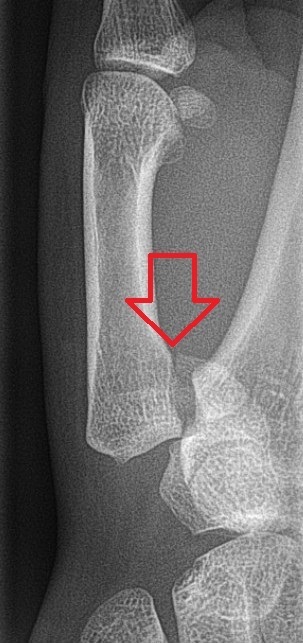
Figure 14. A Bennett fracture: the intra-articular fracture-dislocation of the thumb metacarpal base (arrow). Hellerhoff, CC BY-SA 3.0, via Wikimedia Commons.
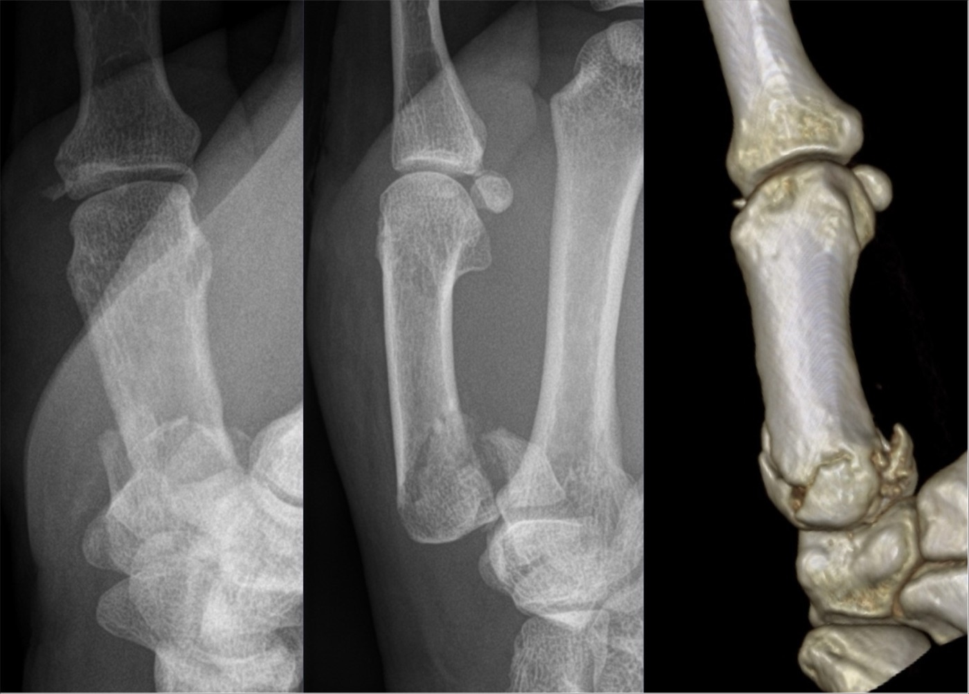
Figure 15. A Rolando fracture: the comminuted Y/T-shaped intra-articular fracture of the thumb metacarpal base, radiographs and 3D CT. Hellerhoff, CC BY-SA 4.0, via Wikimedia Commons.
Part VIII - Carpometacarpal (CMC) Dislocations
Carpometacarpal dislocations are easily missed and most often involve the little and ring fingers, usually dorsal, from an axial blow on a clenched fist.[54] They are frequently fracture-dislocations: a higher-energy injury producing a base fracture of the little-finger metacarpal (the “reverse Bennett” fracture) or a hamate articular fracture, and they threaten the ulnar motor nerve at the small-finger CMC.[55] The diagnostic trap is that overlap on standard films hides the injury, so a 30°-pronated oblique (and, for the thumb, the Roberts) view, or CT, is needed; the lost clear joint space is the clue.[56] Treatment is closed reduction and percutaneous pinning for most, with open reduction for irreducible or large intra-articular fragments; a missed dislocation leads to post-traumatic arthritis.[57]
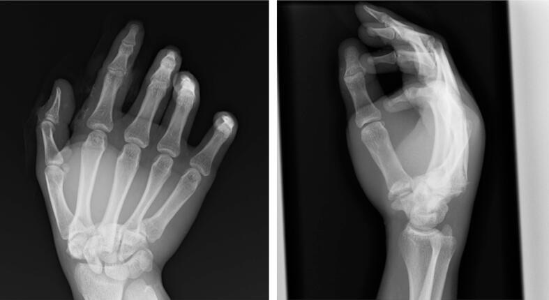
Figure 16. Dorsal carpometacarpal dislocation (3rd and 4th CMC joints), PA and lateral radiographs. Mogi et al., Trauma Case Reports 2026, CC BY 4.0.
Part IX - Principles of Hand-Fracture Fixation, and the Phalanges
The AO principles for the hand follow from “function follows form”: the goals are to restore articular anatomy, correct angular and rotational deformity, achieve stability sufficient for early motion, and use an approach that spares the gliding tendons.[58] Because the phalanges are wrapped in gliding extensor and flexor tendons, a bulky plate or an unnecessary approach causes adhesions and permanent stiffness, so fixation is chosen to permit early, controlled, active mobilisation within 2 to 3 days, the hand splinted in the position of safety (wrist extended, MCP joints flexed 90°, interphalangeal joints in full extension).[59] The implants are a graded set of small screws (about 2.4 mm at the thumb metacarpal, 2.0 mm at the finger metacarpals, 1.5 mm and smaller at the phalanges), used as lag screws (the fracture line at least twice, and for spiral shafts three times, the bone’s diameter) or low-profile plates; flexor tendons are never split.[60]
Phalangeal fractures, the commonest hand fractures, follow the same logic: most are stable and treated by buddy taping and early motion, while displaced, intra-articular, or malrotated fractures are fixed by K-wires, lag screws, or low-profile plates, condylar fractures needing two points of fixation to control rotation.[61] Two named entities round out the topic. The mallet finger is an avulsion of the terminal extensor tendon from the dorsal base of the distal phalanx (a bony or tendinous mallet), treated in most cases by extension splinting of the distal interphalangeal joint. The volar plate / pilon injury at the base of the middle phalanx (the PIP fracture-dislocation) is notoriously difficult and is managed by extension-block splinting, dynamic traction, or open reduction depending on the size of the articular fragment.[62]
Part X - A Synthesis: How to Reason About the Carpus and Hand
Approach the wrist as a ring around the lunate. A fall on the outstretched hand sends a force around that ring, and where it lands tells you the injury: a scaphoid fracture, a scapholunate tear, a perilunate dislocation, and a lunate dislocation are the successive stages of one mechanism (Mayfield), so finding one should make you look for the others. Respect the scaphoid’s retrograde blood supply: treat the displaced, proximal, or occult fracture seriously, because the price of complacency is a humpback nonunion and a SNAC wrist. Read every wrist radiograph along Gilula’s arcs and the scapholunate interval, and never let a perilunate dislocation, with its threatened median nerve, go unreduced. Approach the hand with the opposite worry: not blood supply but stiffness. Check rotation in every metacarpal fracture, because a scissoring finger is a functional disaster that a perfect radiograph will not reveal; tolerate angulation generously in the mobile border rays and barely at all in the rigid central ones; and remember the two thumb-base eponyms, Bennett (the two-part fracture-subluxation held by the beak ligament) and Rolando (its comminuted, worse-prognosis cousin). Through all of it, fix only as much as you must, and move the hand as early as you can, because in the hand the radiograph is never the goal, the working finger is.
References
-
Rockwood & Green’s Fractures in Adults, pp.2628-2630 (carpal instability occurs predominantly around the lunate, the carpal keystone; the Mayfield progressive perilunar instability sequence).
-
AO Principles of Fracture Management, p.719 (“Nowhere in the body does function follow form as closely as in the hand”; the outcome is judged more on the return of function than on skeletal union).
-
Rockwood & Green’s Fractures in Adults, pp.2611-2616 (two rows of eight bones; proximal row scaphoid/lunate/triquetrum, the intercalated segment with no tendon attachments; pisiform a sesamoid in FCU; lunate the keystone; SL and LT the two most important intrinsic ligaments, the SL dorsal portion and LT palmar portion the strong parts).
-
Rockwood & Green’s Fractures in Adults, pp.2611-2616 (two rows of eight bones; proximal row scaphoid/lunate/triquetrum, the intercalated segment with no tendon attachments; pisiform a sesamoid in FCU; lunate the keystone; SL and LT the two most important intrinsic ligaments, the SL dorsal portion and LT palmar portion the strong parts).
-
Rockwood & Green’s Fractures in Adults, pp.2611-2616 (two rows of eight bones; proximal row scaphoid/lunate/triquetrum, the intercalated segment with no tendon attachments; pisiform a sesamoid in FCU; lunate the keystone; SL and LT the two most important intrinsic ligaments, the SL dorsal portion and LT palmar portion the strong parts).
-
Rockwood & Green’s Fractures in Adults, p.2615 (the space of Poirier, a V-shaped interval of capsular weakness over the capitolunate joint through which the lunate displaces into the carpal canal during dorsal dislocations).
-
Rockwood & Green’s Fractures in Adults, pp.2621-2628 (the scaphoid as the oblique link between rows, flexing under compression; DISI = lunate extended, SL angle >60°, capitolunate >15°; VISI = lunate flexed, SL angle <30°; normal SL angle ~30-60°; dissociative vs nondissociative, static vs dynamic instability).
-
Rockwood & Green’s Fractures in Adults, pp.2621-2628 (the scaphoid as the oblique link between rows, flexing under compression; DISI = lunate extended, SL angle >60°, capitolunate >15°; VISI = lunate flexed, SL angle <30°; normal SL angle ~30-60°; dissociative vs nondissociative, static vs dynamic instability).
-
Rockwood & Green’s Fractures in Adults, pp.2621-2628 (the scaphoid as the oblique link between rows, flexing under compression; DISI = lunate extended, SL angle >60°, capitolunate >15°; VISI = lunate flexed, SL angle <30°; normal SL angle ~30-60°; dissociative vs nondissociative, static vs dynamic instability).
-
Rockwood & Green’s Fractures in Adults, pp.2687, 2701 (disruption of Gilula’s lines on the PA view as a sign of perilunate dislocation and dissociation). The three discrete carpal arcs are standard radiographic teaching; the mined extract refers to “Gilula’s lines” and the figure rather than enumerating the three arcs.
-
Rockwood & Green’s Fractures in Adults, pp.2642-2643 (scaphoid fractures = 60-80% of all carpal fractures, 2-3% of all fractures; mean age 25-35, male:female ~2.5:1, FOOSH/hyperextension in ~90%).
-
Rockwood & Green’s Fractures in Adults, pp.2641-2642 (over 80% covered by articular cartilage, reduced periosteal healing; retrograde supply, dorsal branch supplies 70-80% including the proximal pole, volar branch the distal 20-30%; proximal-pole fractures inexorably disrupt the proximal blood supply).
-
Rockwood & Green’s Fractures in Adults, pp.2641-2642 (over 80% covered by articular cartilage, reduced periosteal healing; retrograde supply, dorsal branch supplies 70-80% including the proximal pole, volar branch the distal 20-30%; proximal-pole fractures inexorably disrupt the proximal blood supply).
-
Rockwood & Green’s Fractures in Adults, pp.2643-2644 (anatomical snuffbox tenderness, scaphoid-tubercle tenderness, axial thumb compression; no single sign adequately sensitive and specific; 30-40% not seen on initial radiographs = suspected/occult fractures).
-
Rockwood & Green’s Fractures in Adults, pp.2643-2644 (anatomical snuffbox tenderness, scaphoid-tubercle tenderness, axial thumb compression; no single sign adequately sensitive and specific; 30-40% not seen on initial radiographs = suspected/occult fractures).
-
Rockwood & Green’s Fractures in Adults, pp.2645-2647 (MRI the most sensitive for the occult fracture and argued the best investigation, CT best for displacement/anatomy/union; immobilise-and-re-image vs early MRI strategies).
-
Rockwood & Green’s Fractures in Adults, pp.2650-2651, 2673 (location distal/waist/proximal; Herbert & Fisher A stable acute, B unstable acute with B1-B5 subtypes [B2 complete waist 36% the commonest], C delayed union, D nonunion D1 fibrous/D2 sclerotic; Russe by fracture-line inclination, AO by location).
-
Rockwood & Green’s Fractures in Adults, pp.2650-2651, 2673 (location distal/waist/proximal; Herbert & Fisher A stable acute, B unstable acute with B1-B5 subtypes [B2 complete waist 36% the commonest], C delayed union, D nonunion D1 fibrous/D2 sclerotic; Russe by fracture-line inclination, AO by location).
-
Rockwood & Green’s Fractures in Adults, pp.2651, 2654 (Mayo instability criteria: >1 mm displacement, lateral intrascaphoid angle >35°, comminution, malalignment, proximal-pole, DISI; all displaced fractures are unstable; translation >1 mm raises nonunion odds, gap >3 mm odds ratio ~9.9).
-
Rockwood & Green’s Fractures in Adults, pp.2655-2657 (nondisplaced fractures cast-treated, union 95-99%; systematic reviews and RCTs show no advantage to above-elbow or thumb-spica casts, below-elbow forearm cast advocated; most stable fractures unite in 6-8 weeks).
-
Rockwood & Green’s Fractures in Adults, pp.2657-2666 (percutaneous headless compression screw, Herbert-type, volar or dorsal with neither superior; operative indications displaced/unstable/proximal pole and the fast-track for athletes; operative gives faster union/return but higher complications and no clear union-rate benefit for nondisplaced fractures; proximal-pole fractures via a dorsal approach). The “Acutrak” trade name is standard usage; the source names the Herbert screw and generic headless variable-pitch cannulated screws.
-
Rockwood & Green’s Fractures in Adults, pp.2657-2666 (percutaneous headless compression screw, Herbert-type, volar or dorsal with neither superior; operative indications displaced/unstable/proximal pole and the fast-track for athletes; operative gives faster union/return but higher complications and no clear union-rate benefit for nondisplaced fractures; proximal-pole fractures via a dorsal approach). The “Acutrak” trade name is standard usage; the source names the Herbert screw and generic headless variable-pitch cannulated screws.
-
Rockwood & Green’s Fractures in Adults, pp.2669-2673 (humpback deformity with DISI; nonunion ~10% of waist fractures, near zero for well-treated nondisplaced fractures, up to 50% for displaced; SNAC, the scaphoid-nonunion analogue of SLAC, progressing from the radial styloid to pan-carpal arthritis).
-
Rockwood & Green’s Fractures in Adults, pp.2669-2673 (humpback deformity with DISI; nonunion ~10% of waist fractures, near zero for well-treated nondisplaced fractures, up to 50% for displaced; SNAC, the scaphoid-nonunion analogue of SLAC, progressing from the radial styloid to pan-carpal arthritis).
-
Rockwood & Green’s Fractures in Adults, pp.2673-2679 (nonunion managed by resection, deformity correction with non-vascularised or vascularised bone graft, and screw fixation; vascularised distal-radius and medial femoral condyle grafts for the avascular proximal pole; AVN confirmed only by absence of punctate bleeding at surgery). The “1,2-ICSRA” pedicle name is standard teaching; the source describes distal-radius vascularised pedicle grafts and the second-dorsal-compartment harvest without that eponym.
-
Rockwood & Green’s Fractures in Adults, pp.2673-2679 (nonunion managed by resection, deformity correction with non-vascularised or vascularised bone graft, and screw fixation; vascularised distal-radius and medial femoral condyle grafts for the avascular proximal pole; AVN confirmed only by absence of punctate bleeding at surgery). The “1,2-ICSRA” pedicle name is standard teaching; the source describes distal-radius vascularised pedicle grafts and the second-dorsal-compartment harvest without that eponym.
-
Rockwood & Green’s Fractures in Adults, pp.2628-2630, 2698-2701 (commonest wrist dislocation, in young males, missed in 16-25%; Mayfield stages I scapholunate, II capitolunate via the space of Poirier, III lunotriquetral, IV lunate dislocated volarly into the carpal tunnel hinging on palmar ligaments; ~16% have median nerve symptoms at presentation).
-
Rockwood & Green’s Fractures in Adults, pp.2628-2630, 2698-2701 (commonest wrist dislocation, in young males, missed in 16-25%; Mayfield stages I scapholunate, II capitolunate via the space of Poirier, III lunotriquetral, IV lunate dislocated volarly into the carpal tunnel hinging on palmar ligaments; ~16% have median nerve symptoms at presentation).
-
Rockwood & Green’s Fractures in Adults, pp.2628, 2699-2700 (lesser-arc = purely ligamentous perilunate; greater-arc = with a fracture; trans-scaphoid perilunate fracture-dislocation the commonest, 61% of Herzberg’s series).
-
Rockwood & Green’s Fractures in Adults, pp.2701-2705 (PA: disruption of Gilula’s lines, triangular lunate; lateral: the volarly rotated lunate, the source’s “spilled teapot sign”; emergent closed reduction by the Tavernier manoeuvre decompresses the median nerve; loss of reduction ~59% with casting, so open reduction with ligament repair and fixation for most, dorsal or combined approach reducing in reverse-Mayfield order; post-traumatic arthrosis mean ~38%). The classic eponym for this lateral-view appearance is the “spilled teacup” sign; the mined Rockwood text uses “spilled teapot.”
-
Rockwood & Green’s Fractures in Adults, pp.2701-2705 (PA: disruption of Gilula’s lines, triangular lunate; lateral: the volarly rotated lunate, the source’s “spilled teapot sign”; emergent closed reduction by the Tavernier manoeuvre decompresses the median nerve; loss of reduction ~59% with casting, so open reduction with ligament repair and fixation for most, dorsal or combined approach reducing in reverse-Mayfield order; post-traumatic arthrosis mean ~38%). The classic eponym for this lateral-view appearance is the “spilled teacup” sign; the mined Rockwood text uses “spilled teapot.”
-
Rockwood & Green’s Fractures in Adults, pp.2701-2705 (PA: disruption of Gilula’s lines, triangular lunate; lateral: the volarly rotated lunate, the source’s “spilled teapot sign”; emergent closed reduction by the Tavernier manoeuvre decompresses the median nerve; loss of reduction ~59% with casting, so open reduction with ligament repair and fixation for most, dorsal or combined approach reducing in reverse-Mayfield order; post-traumatic arthrosis mean ~38%). The classic eponym for this lateral-view appearance is the “spilled teacup” sign; the mined Rockwood text uses “spilled teapot.”
-
Rockwood & Green’s Fractures in Adults, pp.2685-2689 (SLD the commonest carpal ligament injury; Watson scaphoid-shift test; Terry-Thomas sign SL gap >3 mm suggestive, >5 mm diagnostic; cortical ring sign of the flexed scaphoid; SL angle >60° suggestive, >80° diagnostic; DISI; SLAC progression as stage V of the Kuo-Wolfe scheme).
-
Rockwood & Green’s Fractures in Adults, pp.2685-2689 (SLD the commonest carpal ligament injury; Watson scaphoid-shift test; Terry-Thomas sign SL gap >3 mm suggestive, >5 mm diagnostic; cortical ring sign of the flexed scaphoid; SL angle >60° suggestive, >80° diagnostic; DISI; SLAC progression as stage V of the Kuo-Wolfe scheme).
-
Rockwood & Green’s Fractures in Adults, pp.2690-2695 (acute repair with capsulodesis and pinning; chronic reducible injury reconstructed, e.g. three-ligament tenodesis; salvage by partial fusion, four-corner fusion, or proximal row carpectomy). The classic SLAC stage I-III progression (radial styloid → radioscaphoid → capitolunate) is standard teaching; the mined extract presents SLAC only as the end stage of the Kuo-Wolfe classification.
-
Rockwood & Green’s Fractures in Adults, pp.2696-2698 (LT dissociation less frequent than SL, ulnar-sided pain and clunk, ballottement and the lunotriquetral shear test the most sensitive, VISI; treated by cast or pinning acutely, reconstruction or LT fusion for chronic cases). The “Reagan” ballottement eponym is standard teaching; the source describes the ballottement and shear tests without that name.
-
Rockwood & Green’s Fractures in Adults, pp.2696-2698 (LT dissociation less frequent than SL, ulnar-sided pain and clunk, ballottement and the lunotriquetral shear test the most sensitive, VISI; treated by cast or pinning acutely, reconstruction or LT fusion for chronic cases). The “Reagan” ballottement eponym is standard teaching; the source describes the ballottement and shear tests without that name.
-
Rockwood & Green’s Fractures in Adults, pp.2680-2685, 2625 (triquetrum the 2nd commonest, dorsal chip avulsion >90%; trapezium body/ridge fractures associated with thumb CMC and hook-of-hamate fractures; hamate hook fracture in racquet/club sports, threatening the ulnar nerve and little-finger flexors, diagnosed by carpal-tunnel view/CT, treated by excision or ORIF; capitate fractures often perilunate, the scaphocapitate syndrome with the capitate rotated up to 180°; pisiform direct-blow fractures near the ulnar nerve; lunate fractures rare, the site of Kienböck’s idiopathic AVN associated with ulnar-minus variance; trapezoid the least common; most isolated undisplaced carpal fractures treated in a cast).
-
Rockwood & Green’s Fractures in Adults, pp.2680-2685, 2625 (triquetrum the 2nd commonest, dorsal chip avulsion >90%; trapezium body/ridge fractures associated with thumb CMC and hook-of-hamate fractures; hamate hook fracture in racquet/club sports, threatening the ulnar nerve and little-finger flexors, diagnosed by carpal-tunnel view/CT, treated by excision or ORIF; capitate fractures often perilunate, the scaphocapitate syndrome with the capitate rotated up to 180°; pisiform direct-blow fractures near the ulnar nerve; lunate fractures rare, the site of Kienböck’s idiopathic AVN associated with ulnar-minus variance; trapezoid the least common; most isolated undisplaced carpal fractures treated in a cast).
-
Rockwood & Green’s Fractures in Adults, pp.2840-2848 (metacarpal fractures 3rd commonest UL fracture, ~70% in the 2nd-5th decades; the Roman-arch cascade with the deep transverse intermetacarpal ligaments, rigid central and mobile border rays; malrotation and scissoring the least-tolerated deformity, corrected for >10°; pseudoclawing from apex-dorsal angulation an operative indication; AO Principles, pp.719, 723, 727: rotation must always be corrected).
-
Rockwood & Green’s Fractures in Adults, pp.2840-2848 (metacarpal fractures 3rd commonest UL fracture, ~70% in the 2nd-5th decades; the Roman-arch cascade with the deep transverse intermetacarpal ligaments, rigid central and mobile border rays; malrotation and scissoring the least-tolerated deformity, corrected for >10°; pseudoclawing from apex-dorsal angulation an operative indication; AO Principles, pp.719, 723, 727: rotation must always be corrected).
-
Rockwood & Green’s Fractures in Adults, pp.2840-2848 (metacarpal fractures 3rd commonest UL fracture, ~70% in the 2nd-5th decades; the Roman-arch cascade with the deep transverse intermetacarpal ligaments, rigid central and mobile border rays; malrotation and scissoring the least-tolerated deformity, corrected for >10°; pseudoclawing from apex-dorsal angulation an operative indication; AO Principles, pp.719, 723, 727: rotation must always be corrected).
-
Rockwood & Green’s Fractures in Adults, pp.2840-2848 (metacarpal fractures 3rd commonest UL fracture, ~70% in the 2nd-5th decades; the Roman-arch cascade with the deep transverse intermetacarpal ligaments, rigid central and mobile border rays; malrotation and scissoring the least-tolerated deformity, corrected for >10°; pseudoclawing from apex-dorsal angulation an operative indication; AO Principles, pp.719, 723, 727: rotation must always be corrected).
-
Rockwood & Green’s Fractures in Adults, pp.2844-2855 (boxer’s = little-finger neck fracture, the commonest, tolerating up to ~50° angulation, the Jahss reduction manoeuvre; shaft acceptable angulation index/middle ~10°, ring ~25°, little ~45°, shortening tolerated to 5-6 mm with ~7° extensor lag per 2 mm, rotation never accepted; intra-articular head fractures fixed with headless/lag screws; base fractures often with CMC dislocation).
-
Rockwood & Green’s Fractures in Adults, pp.2844-2855 (boxer’s = little-finger neck fracture, the commonest, tolerating up to ~50° angulation, the Jahss reduction manoeuvre; shaft acceptable angulation index/middle ~10°, ring ~25°, little ~45°, shortening tolerated to 5-6 mm with ~7° extensor lag per 2 mm, rotation never accepted; intra-articular head fractures fixed with headless/lag screws; base fractures often with CMC dislocation).
-
Rockwood & Green’s Fractures in Adults, pp.2844-2855 (boxer’s = little-finger neck fracture, the commonest, tolerating up to ~50° angulation, the Jahss reduction manoeuvre; shaft acceptable angulation index/middle ~10°, ring ~25°, little ~45°, shortening tolerated to 5-6 mm with ~7° extensor lag per 2 mm, rotation never accepted; intra-articular head fractures fixed with headless/lag screws; base fractures often with CMC dislocation).
-
Rockwood & Green’s Fractures in Adults, pp.2845-2857 (most metacarpal fractures stable and nonoperative, intrinsic-plus splint then buddy taping and early motion; operative for malrotation, multiple, open, intra-articular, shortening >5-6 mm, or angulation beyond ray thresholds; CRPP the mainstay including the antegrade bouquet technique, lag screws for long oblique/spiral, plates for unstable/multiple; Melamed meta-analysis pinning better ROM, no functional difference); AO Principles, pp.727-728 (rotation correction, anatomical metacarpal-neck plate, LC-DCP 2.0 for transverse/oblique shafts, lag screws for spiral shafts >3× bone diameter).
-
Rockwood & Green’s Fractures in Adults, pp.2845-2857 (most metacarpal fractures stable and nonoperative, intrinsic-plus splint then buddy taping and early motion; operative for malrotation, multiple, open, intra-articular, shortening >5-6 mm, or angulation beyond ray thresholds; CRPP the mainstay including the antegrade bouquet technique, lag screws for long oblique/spiral, plates for unstable/multiple; Melamed meta-analysis pinning better ROM, no functional difference); AO Principles, pp.727-728 (rotation correction, anatomical metacarpal-neck plate, LC-DCP 2.0 for transverse/oblique shafts, lag screws for spiral shafts >3× bone diameter).
-
Rockwood & Green’s Fractures in Adults, pp.2858-2862 (thumb base fractures ~80% of thumb CMC injuries; Bennett = volar-ulnar intra-articular fragment held by the palmar oblique ligament, the shaft subluxating proximally/radially/dorsally, abductor pollicis longus driving proximal migration and the adductor pollicis adduction/supination); AO Principles, pp.724-725 (the articular fragment held by the anterior oblique ligament, the base subluxing radially/dorsally/proximally, the injury always requiring operative treatment).
-
Rockwood & Green’s Fractures in Adults, pp.2858-2862 (thumb base fractures ~80% of thumb CMC injuries; Bennett = volar-ulnar intra-articular fragment held by the palmar oblique ligament, the shaft subluxating proximally/radially/dorsally, abductor pollicis longus driving proximal migration and the adductor pollicis adduction/supination); AO Principles, pp.724-725 (the articular fragment held by the anterior oblique ligament, the base subluxing radially/dorsally/proximally, the injury always requiring operative treatment).
-
Rockwood & Green’s Fractures in Adults, pp.2864, 2872-2873 (Bennett treated by CRPP with the metacarpal pronated and a pin to the index metacarpal/trapezium, ORIF with a lag screw for fragments
25% of the surface); AO Principles, p.725 (indirect reduction by traction, pronation, and pressure; percutaneous transarticular K-wire, or the “open-book” lag-screw technique for fragments >25%).
-
Rockwood & Green’s Fractures in Adults, pp.2859, 2873-2874 (Rolando = a higher-energy comminuted Y/T-shaped complete intra-articular base fracture, worse prognosis, treated by ORIF with lag screws/T-plate or by K-wires plus a bridging external fixator from the trapezium for severe comminution); AO Principles, p.726 (Rolando simple articular fixed by K-wires or a T-plate 2.0, complex articular by a spanning mini external fixator from trapezium to metacarpal).
-
Rockwood & Green’s Fractures in Adults, pp.2859, 2873-2874 (Rolando = a higher-energy comminuted Y/T-shaped complete intra-articular base fracture, worse prognosis, treated by ORIF with lag screws/T-plate or by K-wires plus a bridging external fixator from the trapezium for severe comminution); AO Principles, p.726 (Rolando simple articular fixed by K-wires or a T-plate 2.0, complex articular by a spanning mini external fixator from trapezium to metacarpal).
-
Rockwood & Green’s Fractures in Adults, pp.2857-2876 (CMC dislocations most often little/ring fingers, usually dorsal, from axial load on a clenched fist; the reverse-Bennett base fracture of the little finger and hamate fractures; ulnar motor branch at risk; the 30°-pronated oblique and Roberts views and CT needed because overlap hides the injury; CRPP for most, ORIF for irreducible/large fragments; missed dislocation causes post-traumatic arthritis); AO Principles, p.726 (70% involve the little metacarpal-hamate joint; the semi-pronated lateral view; K-wires across the CMC joint, ORIF with lag screws/plate for larger fragments).
-
Rockwood & Green’s Fractures in Adults, pp.2857-2876 (CMC dislocations most often little/ring fingers, usually dorsal, from axial load on a clenched fist; the reverse-Bennett base fracture of the little finger and hamate fractures; ulnar motor branch at risk; the 30°-pronated oblique and Roberts views and CT needed because overlap hides the injury; CRPP for most, ORIF for irreducible/large fragments; missed dislocation causes post-traumatic arthritis); AO Principles, p.726 (70% involve the little metacarpal-hamate joint; the semi-pronated lateral view; K-wires across the CMC joint, ORIF with lag screws/plate for larger fragments).
-
Rockwood & Green’s Fractures in Adults, pp.2857-2876 (CMC dislocations most often little/ring fingers, usually dorsal, from axial load on a clenched fist; the reverse-Bennett base fracture of the little finger and hamate fractures; ulnar motor branch at risk; the 30°-pronated oblique and Roberts views and CT needed because overlap hides the injury; CRPP for most, ORIF for irreducible/large fragments; missed dislocation causes post-traumatic arthritis); AO Principles, p.726 (70% involve the little metacarpal-hamate joint; the semi-pronated lateral view; K-wires across the CMC joint, ORIF with lag screws/plate for larger fragments).
-
Rockwood & Green’s Fractures in Adults, pp.2857-2876 (CMC dislocations most often little/ring fingers, usually dorsal, from axial load on a clenched fist; the reverse-Bennett base fracture of the little finger and hamate fractures; ulnar motor branch at risk; the 30°-pronated oblique and Roberts views and CT needed because overlap hides the injury; CRPP for most, ORIF for irreducible/large fragments; missed dislocation causes post-traumatic arthritis); AO Principles, p.726 (70% involve the little metacarpal-hamate joint; the semi-pronated lateral view; K-wires across the CMC joint, ORIF with lag screws/plate for larger fragments).
-
AO Principles of Fracture Management, pp.719-734 (goals: restore articular anatomy, correct angular/rotational deformity, stabilise, spare hand function, mobilise rapidly; the phalanges enveloped by gliding tendons predispose to adhesion and stiffness; early active mobilisation within 2-3 days, the position of safety wrist-extended/MCP-90°/IP-extended; screw sizes 2.4 mm thumb metacarpal, 2.0 mm metacarpals, 1.5 mm phalanges; lag-screw fracture line ≥2× bone diameter, spiral shaft >3×; flexor tendons never split).
-
AO Principles of Fracture Management, pp.719-734 (goals: restore articular anatomy, correct angular/rotational deformity, stabilise, spare hand function, mobilise rapidly; the phalanges enveloped by gliding tendons predispose to adhesion and stiffness; early active mobilisation within 2-3 days, the position of safety wrist-extended/MCP-90°/IP-extended; screw sizes 2.4 mm thumb metacarpal, 2.0 mm metacarpals, 1.5 mm phalanges; lag-screw fracture line ≥2× bone diameter, spiral shaft >3×; flexor tendons never split).
-
AO Principles of Fracture Management, pp.719-734 (goals: restore articular anatomy, correct angular/rotational deformity, stabilise, spare hand function, mobilise rapidly; the phalanges enveloped by gliding tendons predispose to adhesion and stiffness; early active mobilisation within 2-3 days, the position of safety wrist-extended/MCP-90°/IP-extended; screw sizes 2.4 mm thumb metacarpal, 2.0 mm metacarpals, 1.5 mm phalanges; lag-screw fracture line ≥2× bone diameter, spiral shaft >3×; flexor tendons never split).
-
AO Principles of Fracture Management, pp.729-734 (phalangeal diaphyseal fractures fixed by K-wires or 1.5-2.0 mm lag screws/plates, condylar fractures needing two screws to control rotation; depressed volar-base middle-phalanx [PIP] fractures by distraction frames or lag screws and a palmar buttress plate, “notoriously difficult”). The mallet finger (terminal extensor avulsion from the dorsal distal-phalanx base, treated by extension splinting) is standard teaching; it is not covered in the mined AO hand chapter, which omits the distal phalanx, nor in the mined carpal/metacarpal extracts.
-
AO Principles of Fracture Management, pp.729-734 (phalangeal diaphyseal fractures fixed by K-wires or 1.5-2.0 mm lag screws/plates, condylar fractures needing two screws to control rotation; depressed volar-base middle-phalanx [PIP] fractures by distraction frames or lag screws and a palmar buttress plate, “notoriously difficult”). The mallet finger (terminal extensor avulsion from the dorsal distal-phalanx base, treated by extension splinting) is standard teaching; it is not covered in the mined AO hand chapter, which omits the distal phalanx, nor in the mined carpal/metacarpal extracts.
-
Rockwood & Green’s Fractures in Adults, pp.2641-2642.
-
Rockwood & Green’s Fractures in Adults, pp.2644-2647.
-
Rockwood & Green’s Fractures in Adults, pp.2650-2666.
-
Rockwood & Green’s Fractures in Adults, pp.2628-2630.
-
Rockwood & Green’s Fractures in Adults, pp.2628, 2699-2700.
-
Rockwood & Green’s Fractures in Adults, pp.2701-2705.
-
Rockwood & Green’s Fractures in Adults, pp.2685-2689.
-
Rockwood & Green’s Fractures in Adults, pp.2680-2685.
-
Rockwood & Green’s Fractures in Adults, pp.2844, 2853.
-
Rockwood & Green’s Fractures in Adults, pp.2842, 2847-2848.
-
Rockwood & Green’s Fractures in Adults, pp.2859-2873; AO Principles of Fracture Management, pp.724-725.
-
Rockwood & Green’s Fractures in Adults, pp.2859, 2873-2874; AO Principles of Fracture Management, p.726.
-
Rockwood & Green’s Fractures in Adults, pp.2857-2876.
-
AO Principles of Fracture Management, pp.719-734.