Contents
- Orientation
- Part I - Two Injuries, One Region: Epidemiology and the Energy Distinction
- Part II - Applied Anatomy of the Ankle Mortise
- Part III - The Stability Concept in Malleolar Fractures
- Part IV - Classification of Malleolar Fractures
- Part V - Assessment and Imaging of Ankle Fractures
- Part VI - Treatment of Malleolar Fractures
- Part VII - Tibial Pilon Fractures: Mechanism, Anatomy, Assessment and Classification
- Part VIII - Tibial Pilon Fractures: Staged Treatment, Approaches, Fixation and Outcomes
- Part IX - Complications Common to Both Injuries
- Part X - Paediatric Distal Tibial Physeal and Transitional Fractures
- References
Orientation
Two very different injuries share the distal end of the tibia, and the state examination expects a candidate to keep them clearly apart while understanding why they belong together. The malleolar (ankle) fracture is the prototypical low-energy rotational injury: a twist of the planted foot drives the talus against the ring of bone and ligament that surrounds it, and the bone fails in a sequence that can be read backwards from the radiograph. The tibial pilon (plafond) fracture is its high-energy mirror image. An axial load drives the talus upward like a pestle into a mortar, exploding the weight-bearing articular surface and, no less importantly, wrecking the thin soft-tissue envelope that must heal before any reconstruction can succeed. The first injury teaches mechanics and classification; the second teaches soft-tissue respect and timing.[1]
This summary follows the official konspekt sequence and draws its facts from Rockwood & Green’s Fractures in Adults (9th ed., chapters 63 and 64), the AO Principles of Fracture Management (3rd ed., sections 6.8.3 and 6.9), Miller’s Review of Orthopaedics (9th ed.) for high-yield examination framing, and Lovell & Winter’s Pediatric Orthopaedics (8th ed.) for the paediatric transitional fractures. Every substantive claim is page-cited in its footnote. Where a fact is genuinely established teaching but absent from these particular sources, it is flagged as standard teaching rather than given a false citation. A Bulgarian glossary and a viva-voce appendix close the document.
The single thread that ties everything together is congruity. The ankle tolerates incongruity poorly. A classic cadaveric study found that one millimetre of lateral talar shift reduces the tibiotalar contact area by 42 per cent, concentrating load on a thin layer of cartilage and predisposing to arthritis.[2] Whether the surgeon is restoring fibular length in a Weber C fracture or rafting a comminuted plafond, the goal is the same: put the talus back under the tibia, and keep it there until the bone heals.
Part I - Two Injuries, One Region: Epidemiology and the Energy Distinction
The malleolar fracture: common, bimodal, rotational
Ankle fractures are among the most frequent injuries an orthopaedic surgeon treats. They account for roughly 10 per cent of all fractures, with an incidence near 137 per 100,000 population per year, and they are the second most common lower-limb fracture after hip fractures.[3] The mean age at injury is about 45 years, and the distribution is classically bimodal: peaks in younger men and in older women separated by some fifty years, though the bimalleolar and trimalleolar subset peaks only in elderly women.[4] The dominant mechanism is low-energy and rotational, a simple fall or sporting twist rather than an axial blow, and the position of the foot at the moment of injury largely dictates the fracture pattern that results.[5]
The pilon fracture: uncommon, high-energy, axial
The tibial pilon fracture is, by contrast, an uncommon injury, accounting for less than 1 per cent of lower-limb injuries and only 3 to 10 per cent of tibial fractures, most often in men in their fourth decade.[6] Its defining feature is the mechanism: axial loading from a fall from height or a motor-vehicle crash drives the talus proximally into the distal tibia, producing the “explosion” fracture of the articular surface. The contrast with the rotational ankle fracture is the single most important conceptual distinction in this topic, and the comparison repays memorisation.[7]
The energy distinction explains everything downstream. The rotational ankle fracture can usually be fixed promptly with plates and screws through skin that tolerates surgery. The axial pilon fracture cannot, because the same energy that comminuted the joint also devitalised the skin; here the soft tissues, not the bone, dictate the timing and frequently the outcome.[8] The fibular fracture pattern is a reliable clinical clue to the mechanism: compression failure produces valgus, tension failure produces varus, and an intact fibula points to a pure axial load (and, paradoxically, a high prevalence of severe partial-articular plafond injuries).[9]
Part II - Applied Anatomy of the Ankle Mortise
The mortise as a three-bone joint
The ankle functions as a mortise: the body of the talus articulates with the tibial plafond above, the medial malleolus medially, the posterior malleolus behind, and the distal fibula laterally.[10] Several features of this anatomy are mechanically decisive. The fibula carries about one sixth of the load across the joint. The joint axis sits in roughly 15 degrees of external rotation because the medial malleolus is shorter and more anterior than the lateral. And the talar dome is shaped like a frustum, wider anteriorly than posteriorly, so that in dorsiflexion the talus is driven into the mortise and the joint becomes close-packed and most stable.[11]
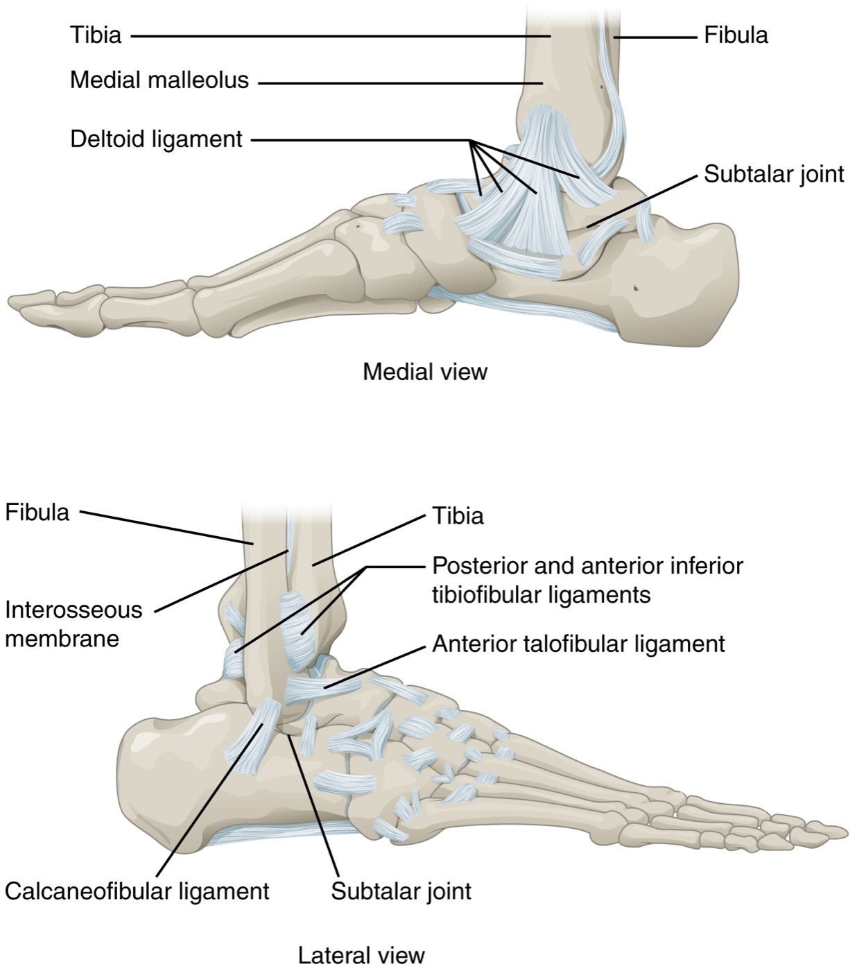
Figure 1. Ligamentous anatomy of the ankle: the medial (deltoid) and lateral collateral complexes and the distal tibiofibular syndesmosis with its interosseous membrane, arranged around the talus within the bony mortise. Illustration by OpenStax College, CC BY 3.0, via Wikimedia Commons.
The three ligamentous complexes
Stability rests on three ligamentous complexes arranged around the bony mortise, and the single most useful simplification in the whole topic is that two of the three must be intact for the ankle to be stable.[12] The syndesmosis (the inferior tibiofibular complex) binds the fibula into the tibial incisura through the anterior inferior tibiofibular ligament (AITFL, running from the tubercle of Chaput on the tibia to the tubercle of Wagstaffe on the fibula), the stronger posterior inferior tibiofibular ligament (PITFL, attaching to Volkmann’s tubercle of the posterior malleolus), and the stout interosseous ligament continuous with the interosseous membrane above.[13] Medially, the deltoid ligament stabilises the joint through a superficial layer that runs from the anterior colliculus and fans to talus, navicular and sustentaculum, and a deep intra-articular layer from the posterior colliculus to the talar body that is the principal restraint against lateral talar shift.[14] The lateral collateral complex (anterior talofibular, calcaneofibular and posterior talofibular ligaments) prevents varus tilt; the anterior talofibular is the weakest and the one torn in the common sprain.[15]
Surgical anatomy for approaches
The distal tibia is subcutaneous along its anteromedial border, which is why direct medial incisions through swollen or contused skin carry an unacceptably high rate of wound breakdown, a fact that dominates pilon surgery.[16] The nerves at risk in the standard exposures repay rehearsal. The superficial peroneal nerve lies subcutaneously over the anterolateral ankle (a branch sits within 5 mm of the fibula in half of people at 10 cm above the tip) and is the structure most often injured in the lateral approach. The sural nerve accompanies the short saphenous vein behind the lateral malleolus and is at risk in posterolateral work. The saphenous nerve and great saphenous vein cross just anterior to the medial malleolus.[17] On the posteromedial side, the mnemonic “Tom, Dick, and a Very Nervous Harry” orders the tarsal-tunnel structures behind the medial malleolus: tibialis posterior, flexor digitorum longus, posterior tibial artery and vein, tibial nerve, and flexor hallucis longus.[18]
Part III - The Stability Concept in Malleolar Fractures
Stability is the organising principle of ankle-fracture treatment. A stable, congruent ankle does well almost regardless of how it is treated, whereas an unstable one left subluxed does badly almost regardless of treatment.[19] The clinical question that recurs in every isolated lateral malleolar fracture is whether the medial side is competent. A truly isolated lateral malleolar fracture, with an intact deltoid, is stable; cadaveric work shows it does not produce abnormal kinematics, and long-term cohorts are excellent (no arthritis in 98 per cent at 30 years in one series, good function in 95 per cent at 21 years in another).[20] The same fibular fracture combined with deltoid rupture is a supination-external rotation stage IV equivalent, is unstable, and generally needs fixation.
The level of the fibular fracture gives a first approximation of stability, and this is the practical value of the Weber/AO scheme. A Weber A (infrasyndesmotic) fracture is usually stable and rarely needs surgery; a Weber B (transsyndesmotic) fracture is variably unstable and must be tested; a Weber C (suprasyndesmotic) fracture is inherently unstable and usually requires fixation.[21] Two structures deserve special mention in the stability calculus. The posterior malleolus contributes disproportionately to syndesmotic stability: fixing it restores about 70 per cent of that stability, and displacement of its articular surface by more than 2 mm worsens one-year outcomes regardless of fragment size.[22] The deltoid, when ruptured, makes the ankle behave as if the medial malleolus were broken, which is the reasoning behind the “bimalleolar-equivalent” label that converts an apparently isolated fibular fracture into a surgical one.[23]
Part IV - Classification of Malleolar Fractures
Danis-Weber / AO: by the level of the fibular fracture
The Danis-Weber classification, adopted into the AO/OTA system, sorts ankle fractures by the level of the fibular fracture relative to the syndesmosis, and its great virtue is simplicity: even a tired surgeon at four in the morning can apply it.[24] Type A is below the syndesmosis (infrasyndesmotic), implies an adduction mechanism with the fibula failing in tension and an intact syndesmosis, and is usually stable. Type B is at the level of the syndesmosis (transsyndesmotic), arises from supination-external rotation, produces the characteristic oblique fibular fracture with a long posterior spike, may partially disrupt the AITFL, and is of variable stability. Type C is above the syndesmosis (suprasyndesmotic), implies disruption of both the medial and the syndesmotic complexes with an interosseous membrane tear up to the level of the fracture, and is inherently unstable.[25] The typical distribution is roughly A 38 per cent, B 52 per cent, C 10 per cent. The malleolar segment carries the AO/OTA region code 44, an exception to the rest of the system that exists specifically to separate rotational malleolar injuries from axial pilon injuries (coded 43).[26]
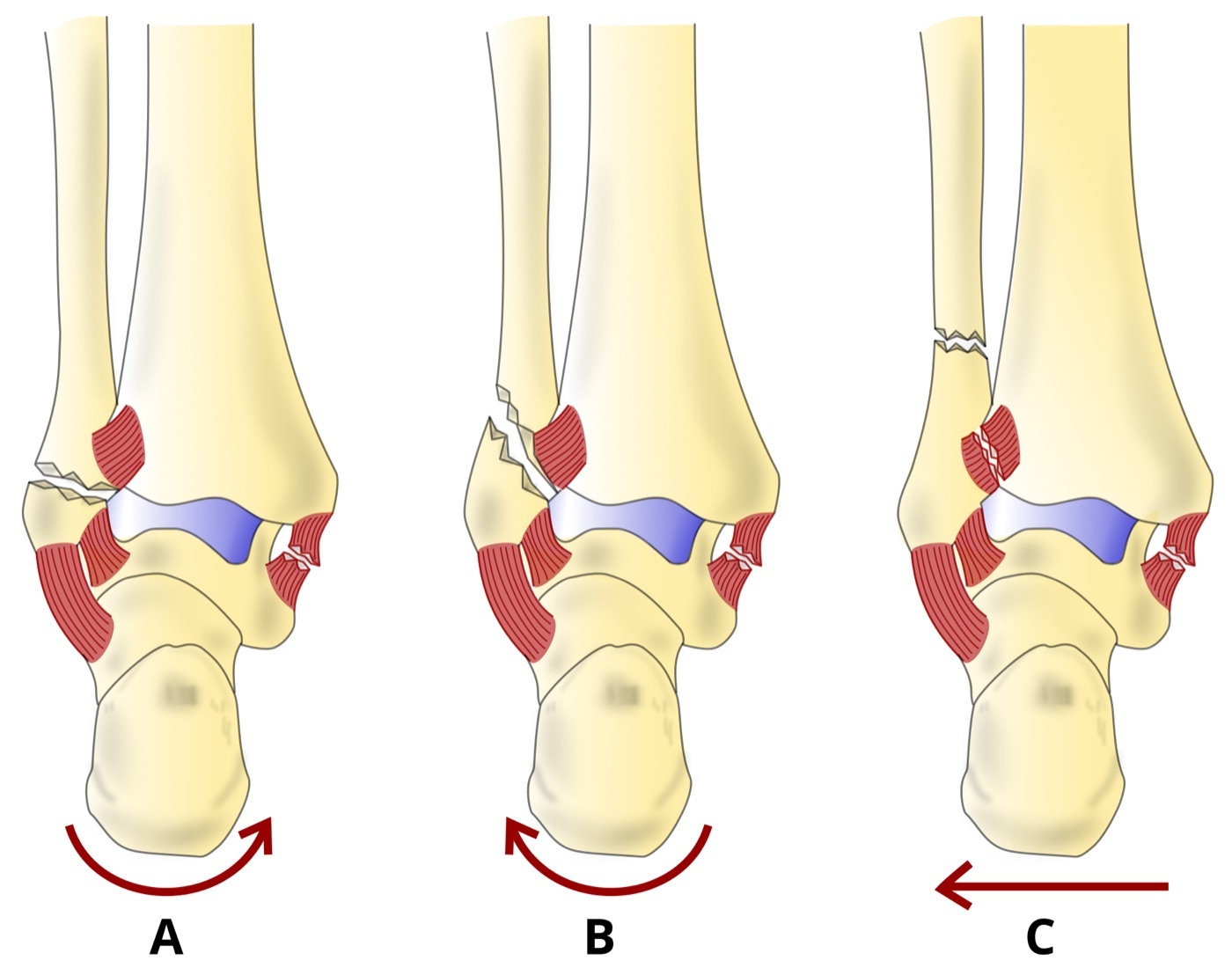
Figure 2. The Danis-Weber classification by the level of the fibular fracture relative to the syndesmosis: type A below (infrasyndesmotic), type B at (transsyndesmotic), and type C above (suprasyndesmotic) the joint line. Diagram by DrFO.Tn, CC BY 4.0, via Wikimedia Commons.
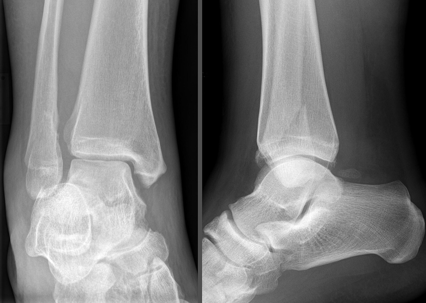
Figure 3. A Weber B fracture of the lateral malleolus: the oblique distal fibular fracture at the level of the syndesmosis, with its characteristic long posterior spike, on AP and lateral views. Radiograph by Hellerhoff, CC BY-SA 3.0, via Wikimedia Commons.
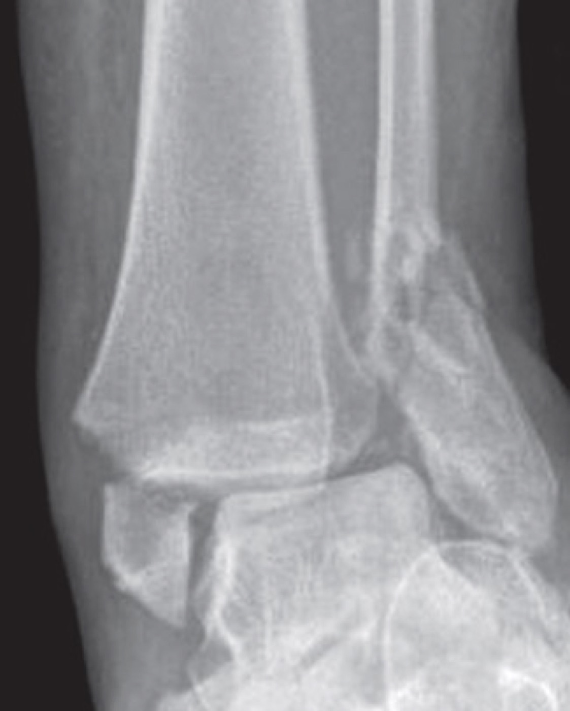
Figure 4. A bimalleolar ankle fracture: combined fractures of the lateral and medial malleoli on the AP view, an inherently unstable pattern. Radiograph by Tellisi, Abdulkareem and Giannoudis, CC BY 4.0, via Wikimedia Commons.
Lauge-Hansen: by mechanism
The Lauge-Hansen classification reconstructs the injury mechanism from the foot position and the deforming force, naming each pattern with two words and a stage number: the first word is the position of the foot (supination or pronation), the second the deforming force, and the number the stage of progression.[27] Foot position governs which structures are taut and therefore fail first, so supination tightens the lateral structures, pronation tightens the medial structures. The four patterns and their approximate frequencies are supination-external rotation (SER, about 60 per cent, the commonest), supination-adduction (SAD, about 20 per cent), pronation-abduction (PAB, about 8 per cent), and pronation-external rotation (PER, about 12 per cent).[28]
The staged sequences repay memorisation because they predict which structures are injured even when these are not obvious on the film. In SER, the commonest pattern, the sequence runs: stage I, AITFL rupture; stage II, the oblique distal fibular fracture at the joint line (the Weber B equivalent, still stable); stage III, PITFL rupture or a posterior malleolar fracture; stage IV, medial failure (deltoid rupture or a transverse medial malleolar fracture), at which point the ankle becomes unstable.[29] In SAD, only two stages occur: stage I is a transverse avulsion of the distal fibula at or below the joint (the Weber A equivalent), and stage II is a near-vertical shear fracture of the medial malleolus, which in up to half of cases carries medial plafond impaction that must be sought and addressed.[30] PAB runs through medial failure, then AITFL or Chaput avulsion, to a comminuted fibular fracture at or above the syndesmosis, the mark of a large lateral translatory force.[31] PER begins with medial failure, then AITFL or Chaput injury, then a high spiral suprasyndesmotic fibular fracture (the Weber C equivalent, with the long spike running anteriorly and proximally), then PITFL or posterior malleolar injury; its classic variant is the Maisonneuve fracture.[32]
Eponyms and special patterns
Several eponymous patterns are examination favourites. The Maisonneuve fracture is a fracture of the proximal fibula with a medial malleolar fracture or deltoid injury and a torn syndesmosis; it accounts for about 5 per cent of ankle fractures, is highly unstable, and the ankle radiograph may look deceptively normal, so the proximal fibula must always be palpated and imaged when tender.[33] Volkmann’s fracture is the posterior malleolar fragment (Volkmann’s triangle), to which the PITFL attaches. The Bosworth fracture-dislocation traps the distal fibula behind the tibial incisura and resists closed reduction because the interosseous membrane stays intact. The “log-splitter” injury drives the talus into the syndesmotic space and carries a 54 to 85 per cent risk of arthritis. The posterior pilon variant adds comminution of the posterior malleolus and is inherently unstable from syndesmotic involvement.[34]
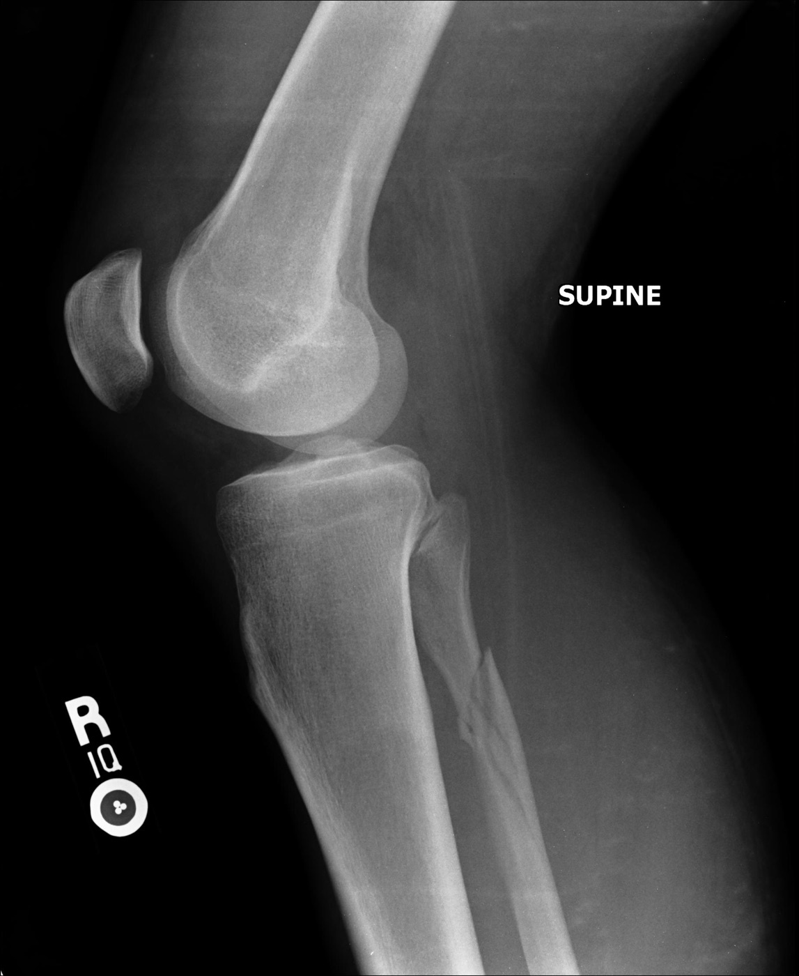
Figure 5. Maisonneuve fracture: the high fracture of the proximal fibula that accompanies a medial-side injury and a torn syndesmosis, often with a deceptively normal-looking ankle film. Radiograph by RotorMotor2, CC BY-SA 3.0, via Wikimedia Commons.
Part V - Assessment and Imaging of Ankle Fractures
History, examination, and the Ottawa rules
Assessment begins with the mechanism and the comorbidities that will shape treatment: diabetes, neuropathy, smoking, alcohol and vascular disease all matter, because each raises the wound-complication rate.[35] Examination proceeds systematically from the proximal fibula down to both malleoli and across the foot, since associated injuries (a proximal-fibular Maisonneuve, a fifth-metatarsal base, a lateral talar process, a midfoot disruption) are easy to miss; skin discolouration or blanching over a displaced fragment calls for prompt reduction.[36] The Ottawa ankle rules identify which injuries warrant radiographs: pain near a malleolus plus any one of age over 55, inability to bear weight, or bone tenderness over the posterior edge or tip of either malleolus.[37]
Standard radiographs and the mortise view
Three radiographic views are standard: AP, lateral, and mortise. The mortise view is taken with the leg internally rotated (about 15 degrees in RG, 20 degrees in AO) so that the transmalleolar axis lies parallel to the plate and the frustral talus no longer overlaps the malleoli.[38] Several measurements quantify congruity and fibular length, and the candidate should know the normal values. The medial clear space should equal the superior tibiotalar space and stay below about 4 to 5 mm; widening of the medial clear space is the radiographic signature of talar shift. The tibiofibular clear space, measured 10 mm above the joint, should be under about 6 mm and is the measurement most predictive of syndesmotic disruption. The talocrural angle is about 83 degrees and, compared with the opposite side, is a sensitive index of fibular length.[39]
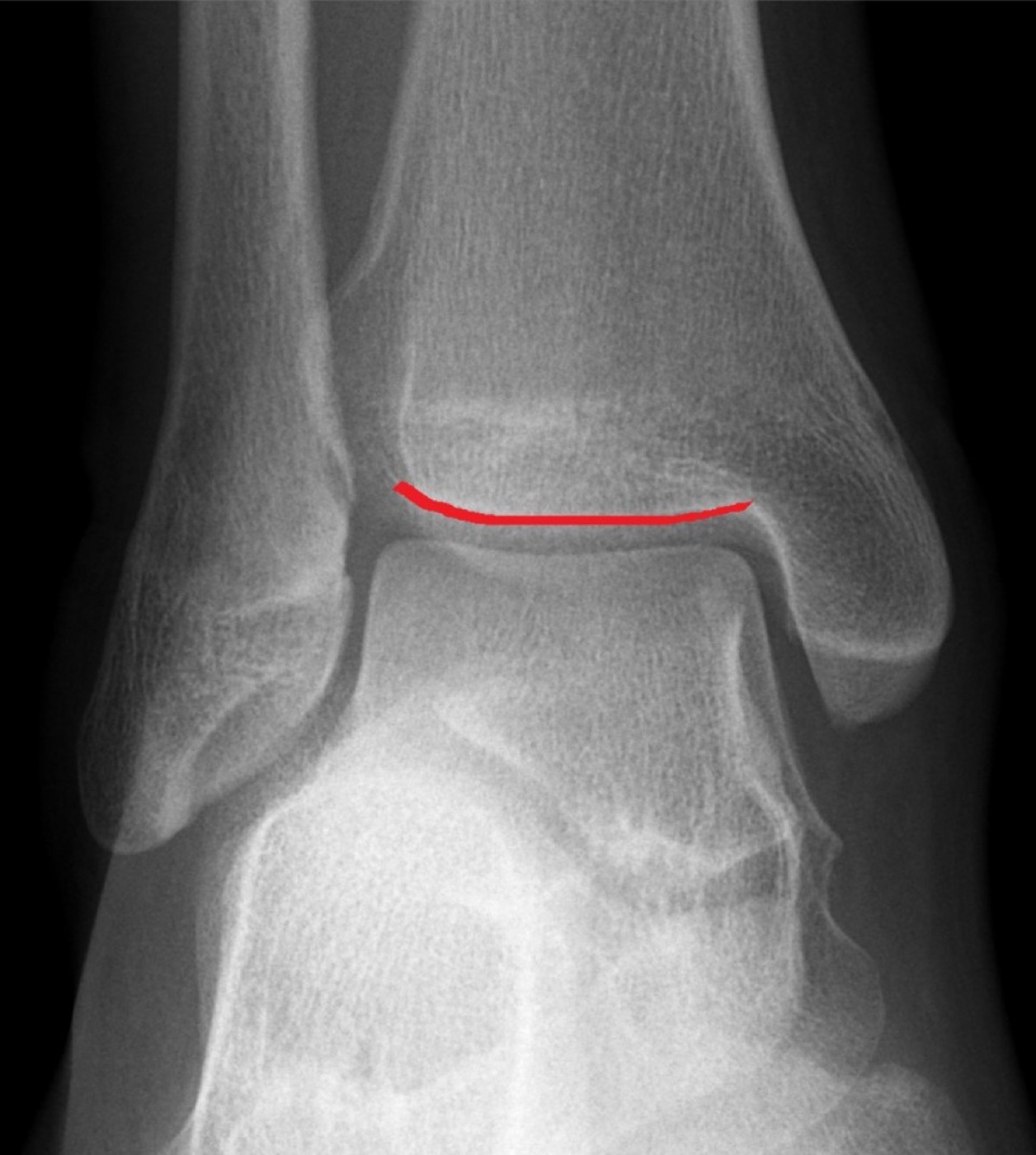
Figure 6. Mortise radiograph of the ankle with the tibial plafond marked: the reference view for the medial clear space, the tibiofibular clear space and overlap, and the congruent position of the talus beneath the plafond. Radiograph by Hellerhoff, CC BY-SA 4.0, via Wikimedia Commons.
Stress views: deciding the borderline Weber B fracture
The central diagnostic problem is the apparently isolated lateral malleolar fracture with a congruent mortise on the resting film: is the deltoid competent (a stable SER II, treated nonoperatively) or incompetent (an occult SER IV, usually operative)? Two stress techniques address it. The manual external-rotation stress view is taken in the mortise position with the ankle plantigrade (never plantarflexed, which falsely narrows the joint) while a firm external-rotation force is applied. The gravity stress view lays the patient on the injured side with the foot hanging free, letting gravity supply the force, and gives equivalent results with less radiation and discomfort. Widening of the medial clear space implies deep deltoid disruption and an unstable pattern.[40]
The crucial caveat, and a favourite examination point, is that a positive stress test does not equate to clinical instability or a poor outcome. Multiple cohorts (Egol, Tornetta, Koval, and the COTS randomised trial) found that many ankles with a “positive” stress test (medial clear space of 4 to 5 mm or more) did perfectly well treated nonoperatively, that the 4 mm cut-off is too stringent, and that surgery imposed on them merely added wound complications.[41] Many units accordingly use a pragmatic “walking test”: the patient is given a removable boot, allowed to bear weight, and re-radiographed at one week; if the mortise stays reduced, the fracture is stable, and if it displaces, surgery is offered.[42]
Computed tomography
CT is not routine but earns its place in three settings: sizing and characterising the posterior malleolar fragment (whose severity is regularly underestimated on plain films), planning posterior pilon and SAD impaction patterns, and confirming the accuracy of syndesmotic reduction postoperatively, since two-dimensional fluoroscopy cannot detect fibular rotation within the incisura of up to 30 degrees.[43]
Part VI - Treatment of Malleolar Fractures
The goal and the nonoperative option
The aim of treatment is a congruent, stable joint, and the corollary is that indiscriminate surgery does not improve outcomes while exposing the patient to wound and hardware complications.[44] Stable isolated lateral malleolar fractures (Weber A, SER II, SAD I) and minimally displaced patterns that pass the stress or walking test are treated nonoperatively in a below-knee weight-bearing cast, boot or brace, with no clear advantage of one device over another beyond three months. Isolated medial malleolar fractures can generally also be managed nonoperatively, with low nonunion rates despite some displacement.[45] Even unstable bimalleolar and trimalleolar fractures can be treated in a cast in the elderly or unfit, though the Willett trial is the cautionary counterpoint: close-contact casting matched fixation at six months in patients over 65, but at the cost of a 25 per cent treatment-failure rate (remanipulation or conversion) and a further 15 per cent rate of malunion.[46]
ORIF: lateral malleolus
The fifty-year gold standard for the fibula is a lag screw protected by a laterally applied one-third tubular neutralisation plate, restoring the length and rotation of the fibula, which is the key to relocating the talus.[47] Several variations exist. The posterior antiglide (buttress) plate allows posterior-to-anterior screws that avoid the joint and a more posterior incision that spares the superficial peroneal nerve, and it is biomechanically strong, though it irritates the peroneal tendons in up to 43 per cent of cases.[48] In comminuted Weber C fractures the plate itself becomes a reduction tool, restoring length by the “push” technique against a proximal screw, and reduction is confirmed against the talocrural angle of the uninjured side.[49] For the elderly osteoporotic ankle or the limb with a precarious soft-tissue envelope, the fibular intramedullary nail is increasingly attractive; the modern designs that permit a syndesmotic screw (such as the Acumed nail) control length, angulation and rotation, and a randomised trial in patients over 65 showed a significant wound-complication advantage over plating.[50]
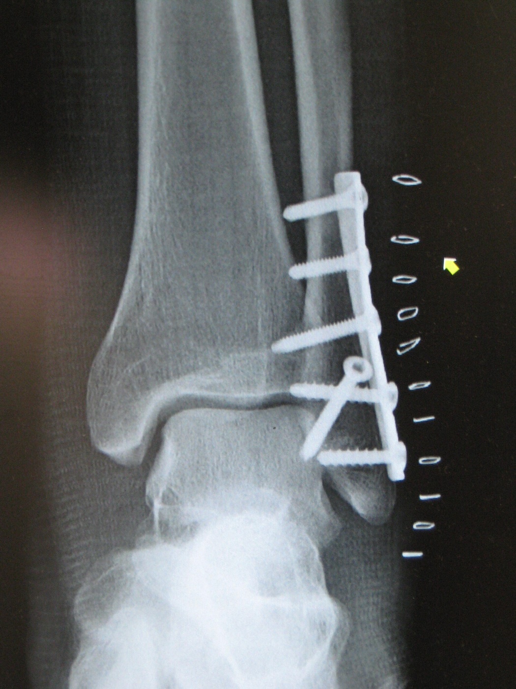
Figure 7. Postoperative ankle ORIF: a lateral (distal fibular) plate with screws and medial-side screw fixation, restoring fibular length and a congruent mortise. Radiograph by James Heilman, MD, CC BY-SA 3.0, via Wikimedia Commons.
ORIF: medial and posterior malleolus
The medial malleolus is usually fixed with two parallel partially threaded cancellous screws perpendicular to the fracture, or a single screw and an anti-rotation K-wire when the fragment is small; tension-band wiring is the better choice for small or osteoporotic fragments, and the vertical shear (SAD) fracture demands a buttress plate or screws parallel to the plafond, plus elevation and grafting of any plafond impaction.[51] The posterior malleolus has been the subject of a long-running debate. The old size thresholds (fix if more than 25, 33 or 50 per cent of the articular surface) are now read as proxies for the tendency to posterior subluxation rather than absolute indications, and the modern rationale for fixation is syndesmotic stability: because the PITFL stays attached to the posterior fragment, fixing it can stabilise the syndesmosis more reliably than a syndesmotic screw, as Gardner’s cadaveric work showed.[52]
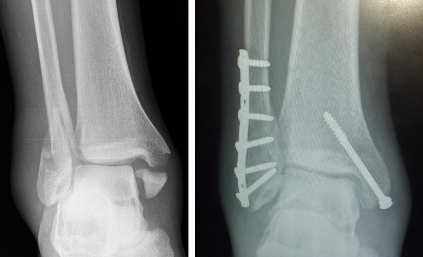
Figure 8. An ankle fracture before and after surgery: the displaced malleolar fracture (left) and ORIF with a lateral fibular plate and a medial malleolar screw (right). Radiographs by Chaim Mintz, CC BY-SA 4.0, via Wikimedia Commons.
The syndesmosis
A syndesmotic diastasis requires rupture of three ligaments plus the interosseous membrane and is a substantial insult that does poorly if missed.[53] Instability is sought intraoperatively once the fibula and medial side are fixed, most popularly with the hook (Cotton) test (pulling the fibula laterally) or an external-rotation stress view, with medial-space widening of more than 2 mm taken as positive.[54] The dominant clinical problem is malreduction, which significantly worsens outcomes and is alarmingly common: roughly half of closed syndesmotic reductions and about 15 per cent of open ones are malreduced, and fibular rotation is invisible on plain fluoroscopy, so direct visualisation or postoperative CT is often needed.[55]
Fixation is by a position (set) screw or a suture button. The screw runs from fibula into tibia, directed about 25 to 30 degrees posterior-to-anterior and parallel to the plafond, placed about 2 cm above the joint; crucially it is not a lag screw, so both fibula and tibia are tapped and the fibula is held in anatomical position without compression, since compressing it would widen the mortise once the screw is removed.[56] Routine screw removal lacks supporting evidence, and a meta-analysis found no role for it; AO permits removal at 12 to 16 weeks before return to full activity.[57] The suture-button (dynamic) construct is the main alternative, semirigid and possibly more physiological; randomised data on whether it reduces more accurately than a screw are contradictory (Naqvi favoured it, Kortekangas found no difference), with neither showing a functional difference at two years.[58] Whichever is used, fibular length and rotation must be correct before fixation, and the deltoid does not require routine repair.[59]
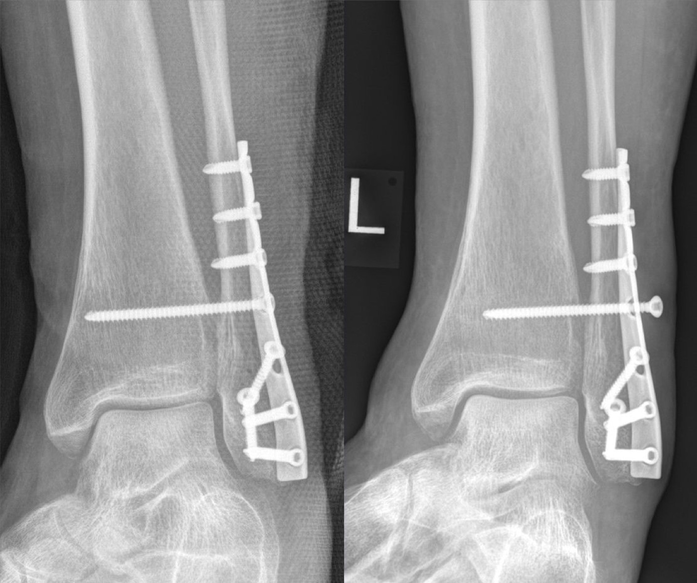
Figure 9. ORIF of a Weber C ankle fracture: fibular plating with a transsyndesmotic positioning (set) screw stabilising the distal tibiofibular joint above the plafond. Radiograph by Hellerhoff, CC BY-SA 4.0, via Wikimedia Commons.
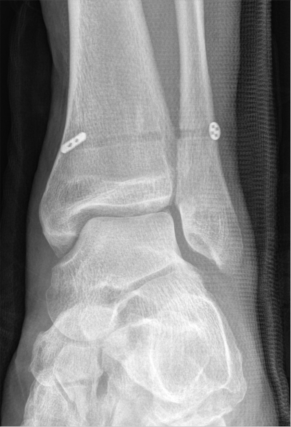
Figure 10. Suture-button (TightRope) fixation of a syndesmotic rupture, the dynamic alternative to a positioning screw, with paired cortical buttons on the medial tibia and lateral fibula. Radiograph by Hellerhoff, CC BY-SA 4.0, via Wikimedia Commons.
Special situations and aftercare
The diabetic and Charcot ankle is the most dangerous setting: wound infection rates reach 32 per cent, open diabetic ankle fractures carry a 64 per cent infection and 42 per cent amputation rate, and neuroarthropathy demands stronger constructs (superconstructs with extended fixation, transarticular Steinmann pins) and a prolonged period (8 to 10 weeks) of protected weight bearing.[60] Weight bearing in plaster after fixation is generally safe for the compliant patient, though it should be withheld in dementia, neuropathy, and substance abuse; after syndesmotic fixation a period of non-weight-bearing (RG prefers eight weeks, AO six to eight) is usual; and braking reaction time returns to normal at about nine weeks after surgery.[61]
Part VII - Tibial Pilon Fractures: Mechanism, Anatomy, Assessment and Classification
A short history that explains current practice
The modern management of the pilon fracture is best understood through its history. Rüedi and Allgöwer in the late 1960s showed that principled open reduction could give durable results with few complications, and ORIF became the North American standard by the early 1990s.[62] When that approach was applied to high-energy North American injuries, the deep infection and wound-breakdown rates proved catastrophic (deep infection in 37 per cent of comminuted pilons in one series, major complications in 40 per cent in another). Sirkin and, separately, Patterson and Cole crystallised the decisive realisation in 1999: these disasters came from operating through swollen, compromised soft tissues, and the Rüedi cohort had been mostly low-energy skiing injuries operated on the day of injury.[63] The result is today’s staged protocol, the single most important concept in pilon management.
Mechanism, anatomy, and the constant fragments
The pilon is an axial-load explosion of the plafond, and the foot position at impact dictates the pattern: dorsiflexion drives anterior comminution, plantarflexion drives the rarer posterior pattern, and a neutral foot produces central impaction.[64] What matters for reconstruction is the pattern of constant fragments. Because the major ankle ligaments usually stay intact, the plafond breaks into three predictable, ligament-bearing pieces: the medial malleolar fragment (deltoid), the anterolateral Chaput fragment (AITFL), and the posterolateral Volkmann fragment (PITFL), with a zone of central comminution and die-punch impaction at their intersection.[65] These fragments are what make reduction systematic, and the strongest cancellous bone lies within 3 cm of the subchondral plate, which tells the surgeon where fixation will hold.[66]
Assessment: soft tissues are paramount
The examination centres on the soft-tissue envelope, assessed circumferentially for swelling, contusion, abrasions, blisters, open wounds and compartment syndrome. Grossly displaced or dislocated fragments must be reduced and splinted immediately, before radiographs, to relieve skin tension and protect circulation.[67] Fracture blisters come in two forms with prognostic meaning: clear-filled blisters are superficial and re-epithelialise quickly, whereas blood-filled blisters mark a deeper dermal injury, heal slowly, and should not be incised through.[68] The “wrinkle sign” (return of skin wrinkles as the shiny oedema settles, usually within 7 to 17 days) signals that the soft tissues will now tolerate definitive surgery.[69] These are high-energy injuries, so a full trauma survey is needed; occult vascular abnormalities turned up on CT angiography in 52 per cent of high-energy plafond fractures even though all limbs had palpable or Doppler-detectable pedal pulses.[70]
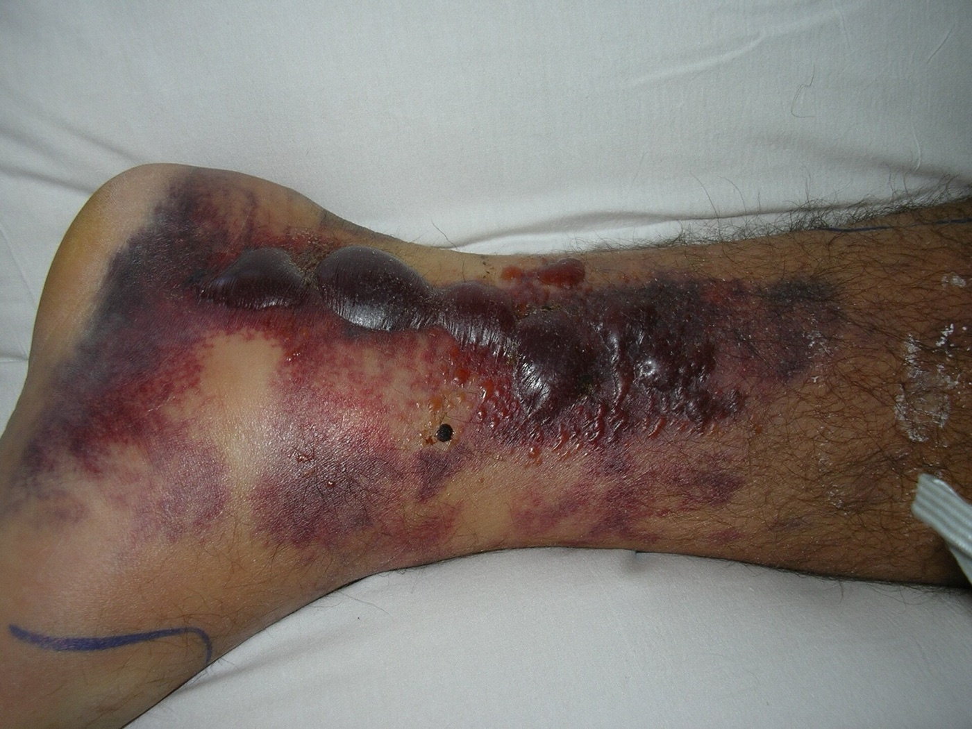
Figure 11. Fracture blisters and soft-tissue swelling over the distal leg: the kind of soft-tissue compromise that mandates staged management, with spanning external fixation before any definitive internal fixation. Photograph by Cindy L. Budge, public domain, via Wikimedia Commons.
Imaging and classification
The diagnosis is made on AP, mortise and lateral radiographs plus full-length tibia/fibula films, but CT is mandatory, and it is best obtained after a spanning external fixator has restored length and axis, because traction disimpacts the talus and lets the articular fragments be seen and counted; CT changed the operative plan in 64 per cent of patients in Tornetta and Gorup’s study.[71] Two classifications dominate. The Rüedi-Allgöwer system grades by articular displacement and comminution (type I non-displaced, type II displaced without comminution, type III displaced with comminution), and the AO/OTA 43 system grades by articular involvement (43A extra-articular, 43B partial articular, 43C complete articular). Both have only moderate reliability and, as Miller bluntly notes, describe comminution without much help for operative planning, where the CT fragment map is what guides the surgeon.[72]
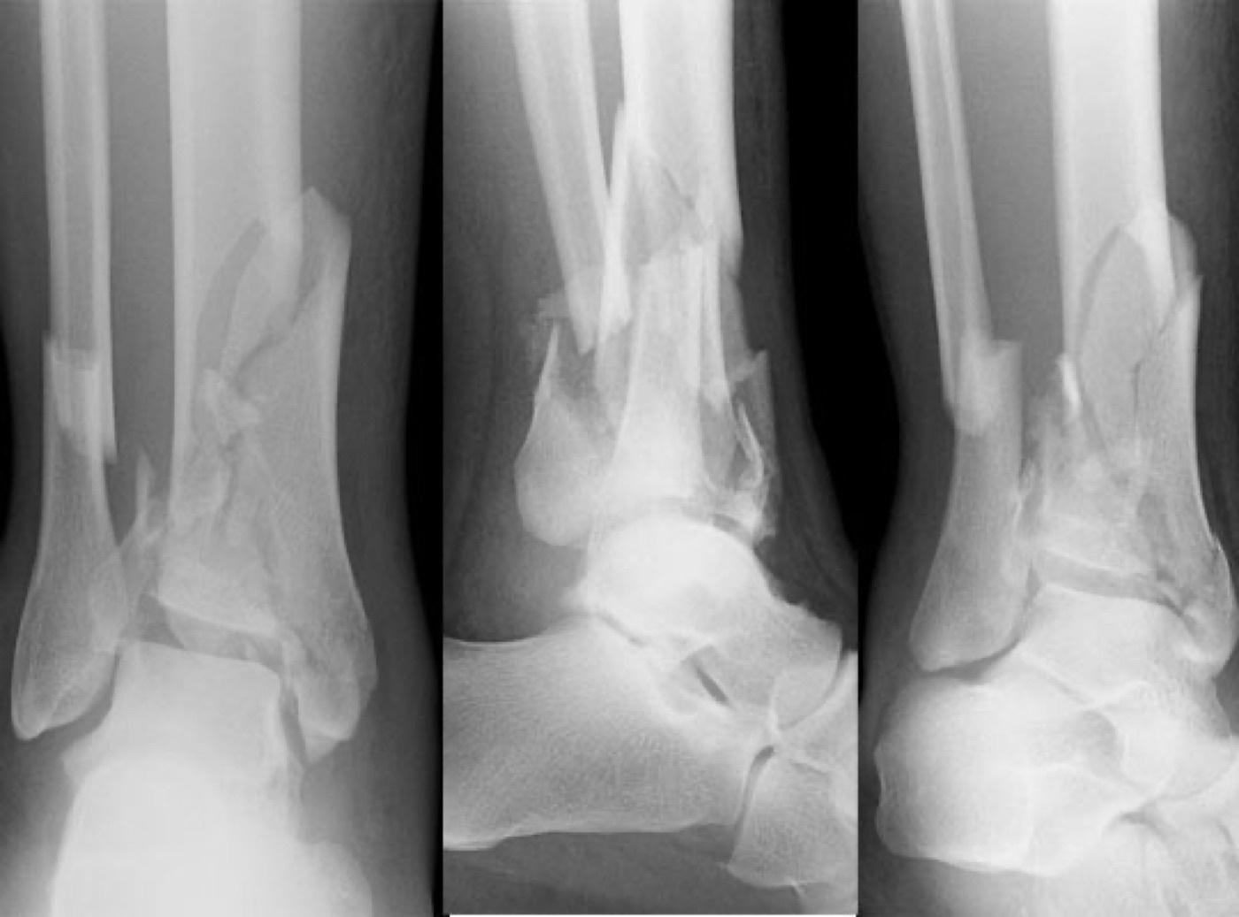
Figure 12. Tibial pilon (plafond) fracture: a comminuted, intra-articular fracture of the distal tibial weight-bearing surface produced by high-energy axial loading. Radiograph by Elhehir, CC BY-SA 4.0, via Wikimedia Commons.
Part VIII - Tibial Pilon Fractures: Staged Treatment, Approaches, Fixation and Outcomes
The staged protocol: “span, scan, plan”
The contemporary standard for a displaced, high-energy pilon with compromised soft tissues is the two-stage protocol captured in the AO mnemonic “span, scan, plan.” In stage one, performed acutely, a joint-spanning external fixator (a simple delta frame outside the zone of injury, often with a metatarsal pin to prevent equinus) restores length, alignment and rotation by ligamentotaxis and lets the soft tissues recover; the fibula is frequently fixed acutely at the same sitting if it is fractured.[73] The fixator stays until the wrinkle sign returns, then a CT (scan) is obtained in the reduced position, and definitive ORIF (the plan) follows once the soft tissues are reparative, typically after 7 to 21 days.[74] Sirkin’s protocol cut the deep-infection rate dramatically (only three deep infections in 46 fractures), and Patterson and Cole reported no wound complications in 21 patients.[75]
Operative principles, sequence and approaches
The operative indications are an articular step over 2 mm, valgus over 5 degrees, any varus, open fracture, compartment syndrome, vascular injury or polytrauma, and the priority of the whole operation is accurate articular reduction.[76] The reconstruction proceeds, in effect, from back to front and from the joint outward. The posterolateral Volkmann fragment (the “constant” fragment, often reduced indirectly by fixing the fibula) is restored first, then the central impaction is disimpacted and grafted, then the medial and anterolateral fragments are reduced, and finally the reconstructed articular block is fixed to the diaphysis.[77] A recurring strategic idea is to convert a complete (C-type) injury into a partial (B-type) or even extra-articular (A-type) one by anatomically reducing a minimally comminuted column early, which simplifies the rest of the reconstruction.[78]
The fibula is usually addressed first because the lateral and posterolateral skin tolerates surgery better, and a correctly reduced fibula indirectly realigns the plafond and provides a fulcrum for the frame; a rigidly fixed malreduced fibula, by contrast, makes the tibial reduction much harder, so the sequence is occasionally reversed.[79] Approach selection follows the fragments and, above all, the soft tissues, with the anteromedial and anterolateral approaches the workhorses; a minimum skin bridge of 5 to 6 cm is respected, incisions are planned along the angiosomes, and full-thickness flaps are raised only once swelling has settled.[80] Posterior approaches give direct access to posterior fragments but add dissection and have been associated with more nonunions.[81]
Fixation options and aftercare
Definitive fixation today uses anatomically contoured, low-profile periarticular locking plates (anteromedial, anterolateral, anterior and fibular), with the plate acting as a buttress on the side of failure; most pilons need both a medial and an anterolateral plate, and the articular surface is supported by rafting screws beneath the subchondral bone.[82] External fixation remains a definitive option, either ankle-spanning or ankle-sparing (hybrid or Ilizarov ring), particularly when the soft tissues are too poor for plating; fine wires are kept out of the joint capsule to avoid septic arthritis.[83] Primary ankle arthrodesis is reserved as a salvage for an unreconstructable plafond with more than 50 per cent cartilage loss.[84] Aftercare is prolonged. The foot is splinted plantigrade, gentle motion begins within days, weight bearing is limited (toe-touch initially, full weight bearing usually only after about three months), and smoking is discouraged because delayed healing is common.[85]
Outcomes and the ORIF-versus-external-fixation debate
Pilon outcomes are sobering. Even with optimal treatment they are often unfavourable, because the chondral injury sustained at impact is irreversible; Pollak’s landmark study found that pilon patients scored worse on the SF-36 than patients with several chronic illnesses, and that 43 per cent of those previously employed remained out of work; ankle function, meanwhile, continues to improve for over two years (Marsh).[86] Post-traumatic arthritis is the most common long-term complication and is frequently radiographically severe (Blauth found arthritic change in 94 per cent), yet the radiographic arthritis correlates only weakly with clinical outcome, and relatively few patients come to late arthrodesis (roughly 7 to 12 per cent within ten years).[87] The long-running comparison of ORIF against external fixation has no clear winner: external fixation tends to give fewer deep wound complications, while ORIF tends to give fewer union problems, with the better retrospective and prospective cohorts (Pollak, Richards) suggesting an advantage for ORIF in union, function and general health, even as a meta-analysis found no overall difference.[88]
Part IX - Complications Common to Both Injuries
The complications of these two injuries overlap but differ in emphasis. After malleolar fractures, most patients do well (about 88 per cent pain-free at one year), and the dominant late problem is post-traumatic arthritis, whose chief modifiable determinant is failure to achieve an anatomical mortise reduction; the mean interval from fracture to end-stage arthritis is about 21 years, and syndesmotic malreduction is a frequently missed driver of a poor result.[89] Wound infection (1 to 10 per cent, far higher in diabetics), symptomatic hardware (effective when removed only about half the time), nonunion (commonest after nonoperative treatment), and cutaneous neuromata of the superficial peroneal, sural or saphenous nerves complete the list.[90]
After pilon fractures, the soft-tissue envelope is the primary driver, so the complications cluster around the wound. Superficial wound necrosis and partial-thickness slough is the most common wound complication (5 to 17 per cent), usually managed expectantly; deep infection is the feared one, demanding urgent debridement, retention of stable implants, removal of loose ones, and frequently negative-pressure dressings and free-tissue coverage, because there is little local soft tissue at the distal tibia.[91] Nonunion is almost always metaphyseal (intra-articular nonunion is rare) and is more frequent after external fixation than ORIF; malunion, especially varus near the ankle, is poorly tolerated; and a significant minority of deep pilon infections ultimately come to below-knee amputation.[92]
Part X - Paediatric Distal Tibial Physeal and Transitional Fractures
Low-risk and high-risk physeal fractures
Paediatric ankle fractures are conventionally divided, after Vahvanen and Aalto, into low-risk (Salter-Harris I and II, and avulsions not involving the physis) and high-risk (Salter-Harris III and IV) groups, with the transitional fractures best treated as a separate third category because of their distinct pathoanatomy.[93] Low-risk fractures are managed by closed reduction and casting; a residual physeal gap of more than 3 mm after reduction points to interposed soft tissue (usually a flap of periosteum, occasionally tendon or neurovascular structures), and minor displacement is acceptable, especially under 8 years, since these injuries are extra-articular with a good growth prognosis.[94] High-risk Salter-Harris III and IV fractures are intra-articular and unstable; closed reduction rarely succeeds, the reduction must be “perfect” to restore the joint and minimise growth arrest, and fixation is by intraepiphyseal smooth wires or cannulated screws that avoid crossing the physis when more than two years of growth remain. The governing rule is that restoration of articular integrity outranks preservation of growth at the ankle, where the physis contributes only 5 to 7 mm of length per year.[95]
The transitional fractures: Tillaux and triplane
The transitional fractures occur in adolescents as the distal tibial physis is closing, so growth disturbance is not the concern; articular congruity is.[96] The Tillaux fracture is a Salter-Harris III avulsion of the anterolateral distal tibial epiphysis by the anterior inferior tibiofibular ligament, produced by external rotation; it can be subtle on plain films, often reduces closed by internal rotation, and is fixed (anterolateral approach, partially threaded cancellous screw) when the articular step exceeds 2 mm.[97] The triplane fracture is a multiplanar injury that classically looks like a Salter-Harris III on the AP film and a Salter-Harris II on the lateral; CT defines whether it is a two-, three- or four-part fracture (three radiating lines on the axial cut give the “Mercedes sign”) and confirms reduction. It is fixed for incongruity over 2 mm, late presentation, or failed closed reduction, using an anterolateral approach for two-part fractures and adding a posteromedial exposure for three- and four-part patterns.[98]
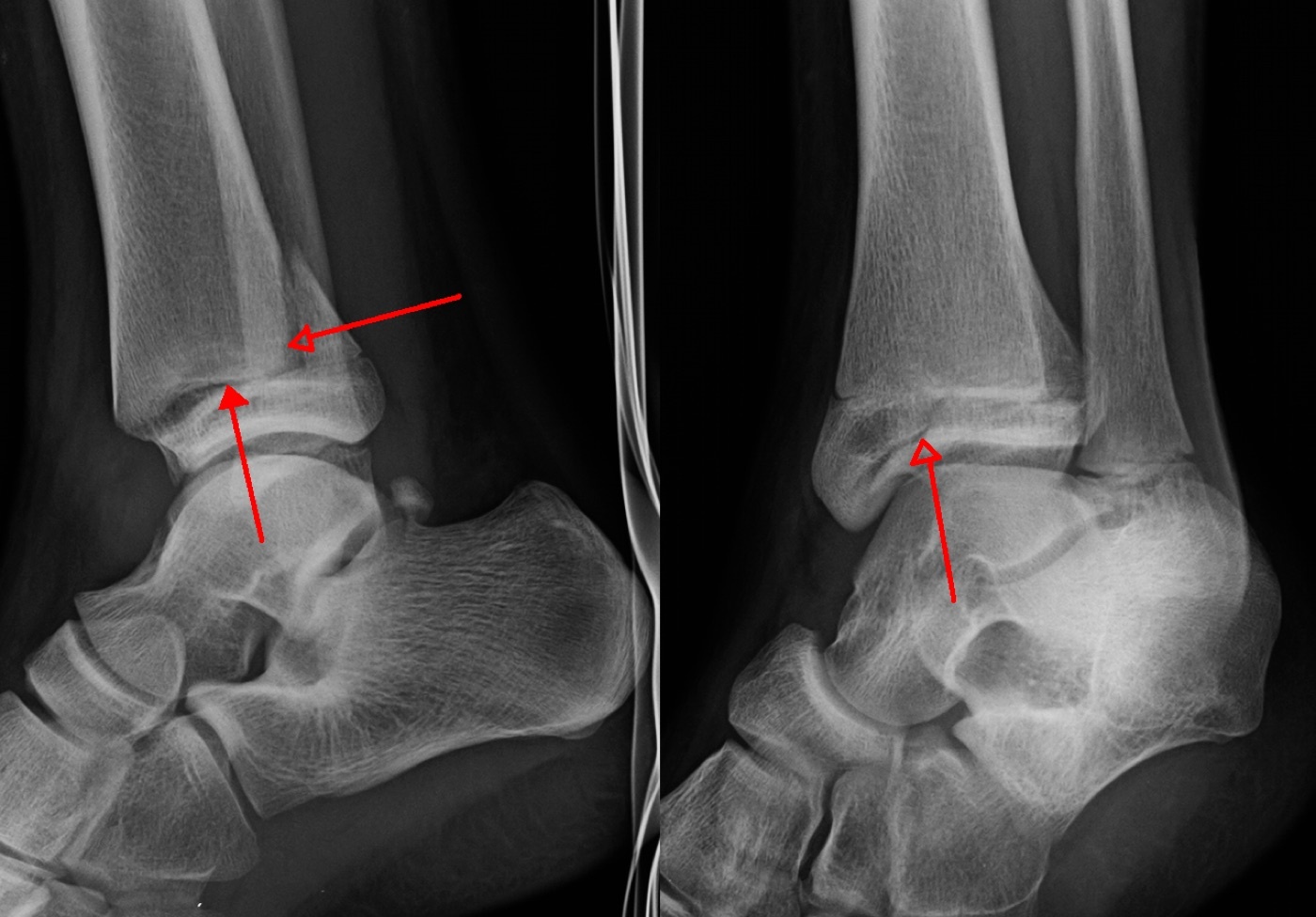
Figure 13. Paediatric triplane fracture of the distal tibia on plain radiographs (arrows); the open distal tibial physis is evident. Radiograph by James Heilman, MD, CC BY-SA 3.0, via Wikimedia Commons.
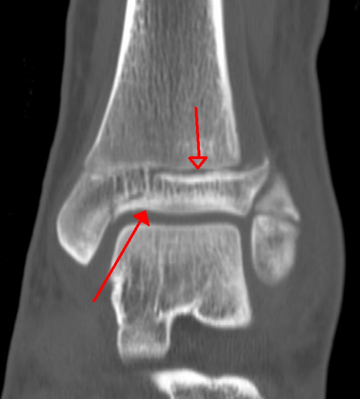
Figure 14. Triplane fracture on coronal CT, showing the multiplanar epiphyseal and metaphyseal components crossing the closing physis; CT defines the part-count and the articular step. Image by James Heilman, MD, CC BY-SA 3.0, via Wikimedia Commons.
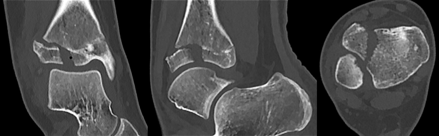
Figure 15. Juvenile Tillaux fracture on CT (sagittal, coronal and axial): the isolated anterolateral distal tibial epiphyseal fragment avulsed by the anterior inferior tibiofibular ligament. Image by Hellerhoff, CC BY-SA 3.0, via Wikimedia Commons.
References
-
The shared vocabulary is anatomical. “Pilon” is French for a pestle, and “plafond” is French for ceiling; both names capture the image of the talus hammering the roof of the ankle joint (RG p.4447; AO p.933). Malleolar fractures, by contrast, are named for the malleoli that fail around an essentially intact plafond. The unifying anatomical structure is the mortise, the box formed by the tibial plafond, the medial malleolus, the posterior malleolus, and the distal fibula, into which the talus fits (RG p.4554).
-
Ramsey and Hamilton’s contact-area study is the most cited single fact in this field and is repeated across all three adult sources (RG p.4555; AO p.958; Miller p.628). It is the mechanical justification for anatomical reduction of every displaced ankle fracture and for the intolerance of malreduction.
-
RG p.4552; the AO text concurs that ankle fractures are “second in frequency only to proximal femoral fractures” (AO p.953).
-
RG p.4552-4553. Kannus and colleagues documented a 319 per cent rise in low-energy elderly ankle fractures over the three decades to 2000 and predicted the number could triple by 2030, the increase concentrated in women (RG p.4552). These are best regarded as true osteoporotic fractures even though systemic bone mineral density is often normal, because the local distal-tibial trabecular architecture is depleted (RG p.4553).
-
RG p.4552; AO p.953; Miller p.625. Obesity (unstable fractures in 29 per cent of the obese versus 4 per cent of stable fractures) and alcohol (consumed within four hours by 29 per cent of patients) are recognised risk factors (RG p.4553).
-
AO p.933; Miller p.631. Pilon fractures may occur in isolation but are frequently part of polytrauma, and their incidence is rising as improved vehicle restraints and trauma care let occupants survive crashes whose energy is still transmitted to the lower limbs (RG p.4450).
-
RG p.4449. Rotational injury applies load slowly, releases little energy at the yield point, produces mainly translational talar displacement with little comminution, and inflicts minimal soft-tissue injury. Axial loading applies load rapidly; because bone is viscoelastic it absorbs more energy before failing, and at failure that energy is released into the soft-tissue envelope, producing comminution, articular impaction, and severe swelling and blistering even without an open wound (RG p.4449, Table 63-1).
-
RG p.4447; AO p.933. The phrase to internalise is that restoration of bony anatomy “while ignoring the often-traumatized soft tissue envelope” is what produced the historically catastrophic results of early pilon surgery (RG p.4447).
-
AO p.934; RG p.4462. The same logic carries into the operative plan: the side that fails in compression is the side that needs the buttress plate.
-
RG p.4554; AO p.956. The talar surface is 70 per cent covered in articular cartilage, has no muscular attachments, and depends on a tenuous retrograde blood supply (RG p.4555).
-
RG p.4554-4555. In dorsiflexion the wide anterior talus forces the fibula to rotate externally and translate posterolaterally, a normal movement at the syndesmosis that is essential to ankle function (AO p.957-958). This is why the syndesmotic ligaments must permit a small, controlled widening of the joint.
-
RG p.4556. The three complexes are the medial (deltoid), the lateral collateral, and the syndesmosis. AO frames the same idea as progressively increasing instability from Weber A to C (AO p.958). Because ligaments do not appear on radiographs, the surgeon must infer ligamentous injury from the bony fracture pattern (AO p.958).
-
RG p.4554-4555; AO p.956. A useful surgical corollary: because the posterior malleolus carries the PITFL, reducing the fibula tends to reduce the posterior malleolus, and stabilising the posterior malleolus tends to restore fibular stability (RG p.4555).
-
RG p.4556. The deep deltoid is the structure whose competence decides whether an isolated lateral malleolar fracture is stable. When the deltoid is incompetent, it functions as a “medial malleolus equivalent,” and the fracture behaves as a bimalleolar-equivalent injury (Miller p.628).
-
RG p.4556; AO p.957; Miller p.634.
-
RG p.4458; AO p.947. The medial skin of the distal tibia is the most vulnerable to blisters, necrosis and breakdown, which is why minimally invasive and low-profile plating is favoured medially.
-
RG p.4558-4559; AO p.968.
-
RG p.4558. The posteromedial and posterolateral approaches both exploit the interval around flexor hallucis longus, whose muscle belly protects the neurovascular bundle.
-
RG p.4576. The satisfactory outcome is defined by two words: congruent (the talus sits anatomically under the plafond) and stable (it stays there until healing) (RG p.4576).
-
RG p.4576. Surgery for a genuinely stable isolated lateral malleolar fracture can actually worsen outcomes by adding wound and hardware complications; a randomised trial confirmed no benefit (RG p.4576-4577).
-
Miller p.627-628; AO p.958, p.965. The Weber B fracture is the diagnostic problem case, and the rest of Part V is largely about how to test it.
-
Miller p.628. The corollary at operation is that in a pronation injury with medial, posterior and lateral involvement, posterior malleolar fixation is the key to restoring syndesmotic stability (Miller p.630).
-
Miller p.628; RG p.4567-4569.
-
RG p.4560; AO p.958; Miller p.627. The chief limitations are that the level of the fibular fracture does not reliably predict the level or presence of syndesmotic injury, and that the scheme says nothing about the medial side (RG p.4560).
-
AO p.958-961; Miller p.627-628. A caveat worth knowing: a spiral fibular fracture above the syndesmosis can arise from pure external rotation that tears only the AITFL, leaving a relatively stable pattern on an intact interosseous membrane and PITFL (AO p.961).
-
RG p.4560; AO p.958, p.963.
-
RG p.4564; AO p.963. The scheme was derived by Lauge-Hansen from cadaveric experiments in 1950, building on Ashhurst and Bromer. Its reproducibility is only modest (interobserver agreement 43-60 per cent) and later workers could not always replicate the staged sequence, but the degree of articular damage does correlate with stage, giving it prognostic value (RG p.4570).
-
RG p.4564. Between 83 and 99 per cent of ankle fractures can be classified by the system (RG p.4564).
-
RG p.4567; Miller p.626-627. The radiographic signature of SER II is the oblique fibular fracture with a long posterior spike; the stress test exists chiefly to distinguish the stable SER II from the unstable SER IV.
-
RG p.4568; Miller p.626. The vertical medial malleolar line and the medial plafond impaction are the two things never to miss in a SAD fracture; SAD injuries are also associated with a second orthopaedic injury (Miller p.626).
-
RG p.4569; Miller p.627. Note that the sources differ slightly on whether PAB is a three- or four-stage mechanism; RG describes three stages, Miller adds a fourth (PITFL or posterior malleolar) (RG p.4565; Miller p.627).
-
RG p.4569; AO p.961; Miller p.627. The PER spike runs in the opposite direction to the SER spike, which is a quick way to distinguish the two high fibular fractures on the lateral film.
-
RG p.4569-4570. A Maisonneuve injury whose proximal fibula is not fixed still needs at least one syndesmotic screw to control length and rotation (Miller p.630).
-
RG p.4570; Miller p.628. The three constant ligament-bearing fragments common to both ankle and pilon injuries are worth fixing in memory now, because they reappear in the pilon section: the medial fragment carries the deltoid, the Volkmann (posterolateral) fragment carries the PITFL, and the Chaput (anterolateral) fragment carries the AITFL (Miller p.631).
-
RG p.4570-4571; AO p.953. The neurovascular examination should mark the dorsalis pedis and posterior tibial pulses at presentation.
-
RG p.4571. Palpate the Achilles and perform the Simmonds (Thompson) test to exclude a coincident rupture.
-
RG p.4571, Table 64-1. The rules are highly sensitive and cost-effective, though their applicability is questioned in some groups such as diabetics.
-
RG p.4571; AO p.954; Miller p.625. A tender proximal fibula mandates full-length tibia/fibula films to exclude a Maisonneuve injury (RG p.4570).
-
RG p.4572-4573, Table 64-2; AO p.954; Miller p.625-626. Fibular length is best judged by the continuity of the subchondral bone line across the joint (the “ball sign” or “dime sign”); shortening produces lateral and valgus talar subluxation. The medial clear space is a trap because it more than doubles with limb rotation, so comparison with the uninjured side is wise (RG p.4572).
-
RG p.4578; Miller p.625; AO p.955. AO adds that stress views are only reliable in the fully anaesthetised patient and when compared with the normal side (AO p.955).
-
RG p.4579-4580. In Egol’s series, the subgroup with a positive stress test but no medial clinical signs did equally well whether treated functionally or operatively, and none became unstable. Koval’s MRI work showed that most ankles with an abnormal stress clear space had only partial deep deltoid tears, not the complete rupture that would mandate surgery (RG p.4580).
-
RG p.4580-4581. AO frames the same idea as repeat radiographs after a short period (about one week) of weight bearing (AO p.965).
-
RG p.4574, p.4588; AO p.954-955; Miller p.625. The point that two-dimensional fluoroscopy cannot detect fibular rotation up to 30 degrees is made at RG p.4588. MRI can assess the deep deltoid but is not common practice and often reveals incidental osteochondral lesions of uncertain importance.
-
RG p.4576. AO frames the decision to operate as based not on fracture pattern alone but on the soft tissues, which are paramount (AO p.953).
-
RG p.4576-4581. Herscovici’s series of 57 isolated medial malleolar fractures treated nonoperatively had only two nonunions (3.5 per cent) despite initial displacement up to 6 mm (RG p.4581).
-
RG p.4581-4582; AO p.979. The practical drawback of nonoperative management of unstable fractures is that maintaining the reduction is difficult and prolonged non-weight-bearing casting is burdensome.
-
RG p.4588-4591; AO p.971. The plate is usually a seven-hole one-third tubular with three bicortical diaphyseal screws proximally and three unicortical cancellous screws in the distal metaphysis that stop short of the subchondral cortex.
-
RG p.4589-4590. Comparative studies show no clear clinical advantage of antiglide over lateral plating; it largely trades wound complications for tendon complications. AO favours it where union is likely to be delayed, as in diabetes (AO p.971).
-
AO p.974. The mantra for the Weber C fibula is “restore the length, alignment and rotation,” because the lateral malleolus dictates the position of the talus.
-
RG p.4593-4594; AO p.977. The nail must be implanted with a central distal start point and about 20 degrees of external rotation so that a syndesmotic screw can later be placed centrally; a medial start point displaces the malleolus laterally and subluxes the talus (RG p.4594-4595).
-
RG p.4597-4600; AO p.970-971. McConnell and Tornetta found plafond impaction in 42 per cent of vertical-shear medial malleolar fractures, which is the reason to scrutinise the SAD pattern on CT.
-
RG p.4583-4584, p.4600-4601; Miller p.628. Fixation can be by percutaneous anterior-to-posterior screws or an open posterior buttress plate; the latter requires prone positioning, so candidates for it (young, active, large fragments, marginal impaction, or clear syndesmotic injury) are best identified preoperatively.
-
RG p.4585-4586. Boden’s classic teaching, that a malleolar medial injury allows the fixed fracture to stabilise the syndesmosis, while a deep-deltoid rupture with a high fibular fracture does not, is tempered by Nielson’s finding that fibular fracture level cannot reliably predict syndesmotic instability (RG p.4586-4587).
-
RG p.4587-4588; AO p.975. The various tests correlate poorly and the hook test is the most popular first choice (64 per cent of surgeons).
-
RG p.4588, p.4604; AO p.977. Sagi’s influential study found malreduction in 44 per cent of closed and 15 per cent of open reductions, with worse function at two years (RG p.4616).
-
AO p.975-976; RG p.4602-4603. Number and diameter of screws and three-versus-four cortices make little biomechanical difference; a Maisonneuve injury, because the proximal fibula is unfixed, needs two position screws to prevent the rotational “helicopter-blade” instability (AO p.975).
-
RG p.4604; AO p.976. Removal itself carries risk (Schepers reported infection in 9.2 per cent and diastasis recurrence in 6.6 per cent).
-
RG p.4603-4604. Suture-button complications include knot problems, infection, osteolysis and sinus formation.
-
RG p.4578, p.4607; AO p.971. The medial side is explored only if the clear space stays wide after fibular reduction or if the fibula cannot be reduced, in which case interposed deltoid or an osteochondral fragment must be lifted out.
-
RG p.4611-4612; AO p.966, p.976-977. Smoking raises deep infection sixfold and alcohol roughly quadruples wound complications (RG p.4611).
-
RG p.4610; AO p.976; Miller p.628. Two well-investigated points worth knowing: routine DVT prophylaxis is not supported after an isolated ankle fracture, and surgery is best timed once fracture blisters have settled and skin wrinkles return, though excessive delay also worsens outcomes (RG p.4607-4609).
-
RG p.4447, p.4470; AO p.933. The four Rüedi-Allgöwer principles still frame every reconstruction: restore fibular length, reconstruct the articular surface, bone-graft the metaphyseal defect, and stabilise with a (medial) buttress plate.
-
RG p.4448, p.4471-4472. Of Rüedi’s 84 fractures, 71 per cent were skiing injuries and only 6 per cent high-energy traffic trauma, and 75 per cent were fixed on the day of injury; the failure series were high-energy and operated several days late through bad skin.
-
RG p.4450; AO p.933. The fibular fracture again betrays the mechanism (compression-valgus, tension-varus, intact-axial), which guides the buttress side.
-
RG p.4462; AO p.934; Miller p.631. Topliss expanded this into a six-fragment CT model (anterior, posterior, medial, anterolateral, posterolateral and die-punch) with two families: sagittal/varus fractures in younger, higher-energy patients and coronal/valgus fractures in older, lower-energy patients (AO p.938).
-
RG p.4458. The subcutaneous anteromedial border again explains why direct medial incisions through injured skin must be avoided.
-
RG p.4452; AO p.934. An extruded distal tibial fragment protruding through the anteromedial skin should be reduced promptly to limit further soft-tissue crushing.
-
RG p.4452. No single blister-management method (unroofing, aspiration, or leaving intact) is clearly superior, but incising through a non-epithelialised, especially haemorrhagic, blister bed is to be avoided.
-
AO p.938-939. This is the bridge between the “span” and the “scan/plan” stages described in Part VIII.
-
RG p.4451. Compartment syndrome is relatively uncommon in the pilon (0 to 5 per cent) compared with the proximal tibia, but it remains an operative emergency (AO p.938). The classic axial-load “fall-from-height” cluster, which also includes calcaneal, plateau and thoracolumbar burst fractures, is standard teaching; these sources explicitly mention only the calcaneus/talus association and the polytrauma context (RG p.4451; AO p.934).
-
RG p.4453; AO p.934; Miller p.631. Contralateral films give the best template for the patient’s own anatomy.
-
RG p.4454-4455; AO p.938; Miller p.632. The closed soft-tissue injury can be graded by the Tscherne system (grades 0 to 3), but soft-tissue severity does not track the bony class and should be recorded separately (RG p.4456). Open fractures are graded by the Gustilo-Anderson system (standard teaching; referenced through open-fracture principles but not enumerated in these extracts).
-
AO p.938-939; RG p.4469-4470. The spanning fixator is technically the easiest and safest device to apply because it does not violate the zone of injury, and it does not preclude later conversion to plating.
-
AO p.934, p.938; RG p.4490. Simple closed fractures with minimal soft-tissue injury, particularly low-energy patterns presenting early, may be definitively fixed within 24 to 36 hours.
-
RG p.4472. The fall in infection is not from delay alone; it also reflects better soft-tissue handling, lower-profile implants, and limited or percutaneous incisions introduced at the same time.
-
AO p.938; RG p.4467. Anderson’s contact-stress threshold work provides the mechanical rationale: above a certain articular contact-stress exposure, an incongruent plafond is highly likely to develop arthritis (RG p.4467).
-
RG p.4504-4505; AO p.945. Ketz and Sanders’ teaching that a stable posterior malleolus is the key to good reduction and should be fixed first is part of this “constant fragment” philosophy.
-
RG p.4505; AO p.944-945. The cardinal caveat is that any early fixation must not block the definitive fixation.
-
RG p.4473-4474, p.4480-4481; AO p.943. Whether to fix the fibula at all when a pilon is treated definitively by external fixation is genuinely controversial; French and Tornetta argued against it because of late varus (RG p.4470).
-
RG p.4477, p.4493-4497; AO p.934, p.942. There are no local muscle flaps at the distal tibia, so early plastic-surgery involvement is essential when coverage may be needed.
-
RG p.4522, p.4537-4538. Chan’s series found a significant increase in nonunions with a staged posterior-then-anterior approach despite no difference in reduction quality.
-
RG p.4514-4515; AO p.947. No single implant neutralises all the fracture planes, so fragment-specific fixation is often combined; locking plates are most useful in osteoporosis and metaphyseal comminution.
-
RG p.4467-4470; AO p.948. The advantages of a ring frame are early weight bearing, fewer incisions and a lower deep-infection risk, at the cost of pin-site care and the technical demands of the technique.
-
AO p.948.
-
RG p.4518-4519; AO p.948. NSAIDs are commonly withheld for about three months because of a theoretical nonunion risk (RG p.4518).
-
RG p.4528-4529; AO p.950. Many of the strongest predictors of outcome (income, education, work-relatedness of the injury) are outside the surgeon’s control, which is itself an examination-worthy point.
-
AO p.950; RG p.4536-4537. The mainstay of treatment for end-stage post-traumatic arthritis is arthrodesis; total ankle replacement is unproven in this population.
-
RG p.4530-4532; AO p.950. White’s modern re-examination of acute ORIF (88 per cent fixed within 48 hours, deep infection 2.7 per cent closed and 19 per cent open) shows that immediate fixation can succeed in experienced hands and selected injuries, but it still warrants caution in high-energy trauma (RG p.4472-4473).
-
RG p.4613-4614. Risk factors for arthritis include an AO/OTA type C pattern, high body-mass index, dislocation and increasing age; arthritis can follow even a perfect reduction because of cartilage damage sustained at injury.
-
RG p.4614. Loss of reduction is most common in conservatively treated unstable fractures, and malunion increases the risk of arthritis.
-
RG p.4532-4534; AO p.950. Surrounding mild erythema over a maturing eschar is usually an inflammatory response rather than infection, and wound cultures of superficial slough are not indicated.
-
RG p.4535-4536; AO p.948, p.950. Chronic osteomyelitis with nonunion is treated by radical debridement, antibiotic spacers, soft-tissue coverage and staged reconstruction, with well-aligned arthrodesis or, in the worst cases, amputation as the salvage.
-
Lovell p.5742. In children, comminution and syndesmotic disruption are rare, and the steps needed for reduction are usually evident from the foot position and radiograph, so the elaborate adult mechanism classifications are downplayed.
-
Lovell p.5742-5743. After closed reduction, a non-weight-bearing cast for the first 3 weeks followed by a weight-bearing cast, for about 6 weeks in total, is usual; union occurs at 4 to 6 weeks.
-
Lovell p.5753. Kling’s series captured the principle: 19 of 20 Salter-Harris III/IV fractures treated by accurate open reduction healed without growth disturbance, against only 5 of 9 treated closed, which developed bony bridges (Lovell p.5775).
-
Lovell p.5757. The anatomical basis, that the distal tibial physis closes asymmetrically over about 18 months from central to anteromedial to posteromedial and finally laterally (the central elevation being Kump’s bump), so that external rotation avulses the still-open anterolateral epiphysis, is standard teaching; the Lovell extract states only that the physis is “in the process of closing.”
-
Lovell p.5757, p.5760. CT is helpful when plain films are inconclusive, and the quality of any closed reduction must be documented accurately.
-
Lovell p.5760-5761, p.5775. Arthroscopically assisted reduction is reserved for the rare case where articular congruity cannot be confirmed by direct inspection or radiographs. The articular step-off goal is 2 mm or less, with a slightly larger pure gap perhaps acceptable.
-
RG p.4555, p.4576; AO p.958; Miller p.628.
-
RG p.4449, Table 63-1; AO p.933.
-
RG p.4560; AO p.958; Miller p.627-628.
-
RG p.4567; Miller p.626-627.
-
RG p.4578-4581; Miller p.625; AO p.965.
-
RG p.4569-4570; AO p.975; Miller p.630.
-
RG p.4588, p.4602-4604; AO p.975-977.
-
AO p.938-939; RG p.4471-4472, p.4490.
-
RG p.4462, p.4504-4505; AO p.934, p.945; Miller p.631.
-
RG p.4532, p.4536; AO p.950.
-
RG p.4530-4532; AO p.950.
-
Lovell p.5757, p.5760-5761, p.5775.