Contents
- Introduction and Scope
- SECTION I --- GENERAL CONSIDERATIONS
- SECTION II --- INDIVIDUAL LESIONS
- II.1 Solitary (Unicameral) Bone Cyst
- II.2 Aneurysmal Bone Cyst
- II.3 Juxta-articular Bone Cyst (Intraosseous Ganglion)
- II.4 Fibrous Dysplasia
- II.5 Osteofibrous Dysplasia and the Adamantinoma Spectrum
- II.6 Metaphyseal Fibrous Defect (Fibrous Cortical Defect / Non-ossifying Fibroma)
- II.8 Brown Tumour of Hyperparathyroidism
- II.9 Giant-Cell (Reparative) Granuloma
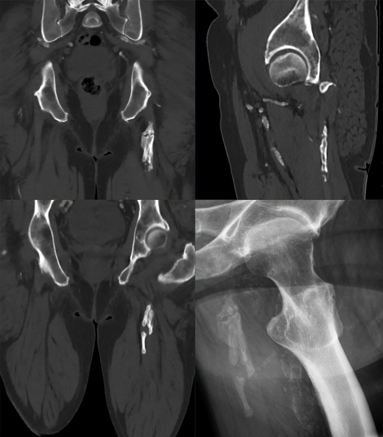
- II.10 Myositis Ossificans (Heterotopic Ossification)
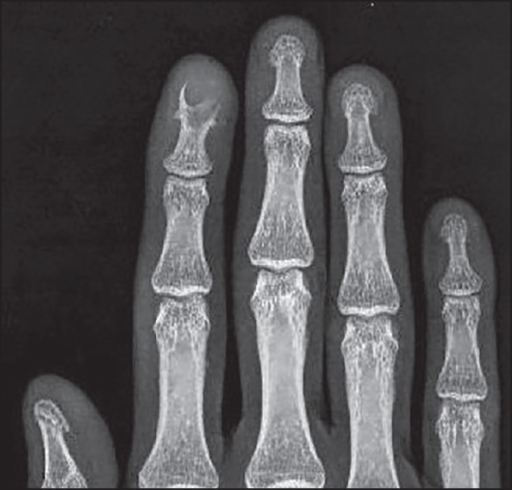
- II.11 Intraosseous Epidermoid Cyst
- II.12 Other “Leave-Me-Alone” Lesions and Important Mimics
- SECTION III --- SYNTHESIS AND EXAM AIDS
- References
- Figure Credits and Licences
Introduction and Scope
“Tumour-like lesions of bone” is the formal heading of an entire category of the World Health Organization (WHO) Histological Classification of Bone Tumours, a classification largely shaped by Fritz Schajowicz, whose monograph devotes a dedicated chapter (Chapter IX) to exactly these entities. The category exists for a single, eminently practical reason. These lesions are not true neoplasms, yet clinically, radiographically, and frequently even histologically they reproduce the appearance of genuine bone tumours. Failure to recognise them leads to two opposite errors, both serious: over-treatment of a trivial, self-healing lesion as if it were a sarcoma, and under-treatment of a sarcoma mistaken for a banal “tumour-like” lesion.
This review covers the classic WHO/Schajowicz list of tumour-like lesions, supplemented by closely related “leave-me-alone” lesions and important mimics that the examiner expects the candidate to place in the differential diagnosis. The principal sources are the standard reference texts of bone pathology and orthopaedic oncology, namely Schajowicz (Tumors and Tumorlike Lesions of Bone, 2nd ed.), Campanacci (Bone and Soft Tissue Tumors), and Dorfman & Czerniak (Bone Tumors). These are integrated with the Bulgarian operative-orthopaedic tradition represented by Boycho Boychev’s Хирургическа ортопедия (Surgical Orthopaedics), from which the indigenous operative techniques and Bulgarian terminology are drawn. Where the molecular genetics and contemporary therapeutics post-date the core reference texts (which were written between 1994 and 2016), the modern consensus is stated explicitly and flagged as such.
SECTION I --- GENERAL CONSIDERATIONS
I.1 Definition and Concept
A tumour-like lesion of bone is a non-neoplastic process (reactive, developmental/dysplastic, metabolic, or of uncertain pathogenesis) that simulates a primary or secondary bone tumour. Schajowicz frames the whole group not by a single sentence but by the recurring qualifier embedded in each WHO definition. The solitary bone cyst is “a common non-neoplastic process of uncertain pathogenesis”; the metaphyseal fibrous defect is “a well-defined non-neoplastic bone lesion”; eosinophilic granuloma is “a non-neoplastic lesion of unknown aetiology”; fibrous dysplasia is “presumably developmental in nature.” The unifying thread is therefore this: lesions that are not tumours but behave radiologically and pathologically as though they were.
Three corollaries follow, and they recur throughout this topic:
- The giant-cell trap. Several of these lesions (solitary bone cyst membrane, brown tumour, giant-cell reparative granuloma, the solid variant of aneurysmal bone cyst) contain osteoclast-type multinucleated giant cells. A pathologist deprived of the clinical and radiological context can readily mislabel any of them a giant-cell tumour. Schajowicz states this explicitly for the solitary bone cyst and repeats it for the brown tumour and reparative granuloma.
- The sarcoma trap. Others (myositis ossificans, the cellular phase of any of these lesions, fibrous dysplasia with a fatigue fracture) display brisk cellularity, mitoses, and immature bone that can be over-read as osteosarcoma. The classic example is the early, cellular centre of myositis ossificans biopsied before it matures.
- The clinico-radiological-pathological triad. The central diagnostic doctrine, stated verbatim by Schajowicz, is that “the histologic aspect does not permit one to establish the anatomoclinical type of any particular lesion … without knowing the complete clinical and radiologic picture.” Histology alone is insufficient. Diagnosis in this field is the deliberate triangulation of clinical data + imaging + pathology, and the biochemistry of calcium metabolism must be added whenever giant cells are seen.
I.2 Classification
The Schajowicz / WHO list (Chapter IX)
Schajowicz’s Chapter IX enumerates ten entities. This list is the most defensible “backbone” answer to the exam question “what are the tumour-like lesions of bone?”, because it is the WHO category:
| № | Schajowicz section title | Synonyms |
|---|---|---|
| 1 | Solitary bone cyst | Simple / unicameral / juvenile bone cyst |
| 2 | Aneurysmal bone cyst | Multilocular haematic bone cyst |
| 3 | Juxta-articular bone cyst | Intraosseous ganglion |
| 4 | Metaphyseal fibrous defect | Non-ossifying fibroma; fibrous cortical defect; histiocytic xanthogranuloma; fibroxanthoma |
| 5 | Eosinophilic granuloma | Histiocytosis X; Langerhans cell histiocytosis |
| 6 | Fibrous dysplasia (and osteofibrous dysplasia) | Fibro-osseous dysplasia |
| 7 | “Myositis ossificans” | Heterotopic ossification |
| 8 | “Brown tumour” | Brown tumour of hyperparathyroidism |
| 9 | Intraosseous epidermoid cyst | Keratin / epidermal inclusion cyst |
| 10 | Giant-cell (reparative) granuloma | Giant-cell reaction |
The quotation marks Schajowicz deliberately places on “myositis ossificans” and “brown tumour” flag both as misnomers: the first is neither an inflammation (-itis) nor confined to muscle, and the second is not a true tumour. To this WHO list it is conventional, and examinable, to add the closely related intraosseous lipoma, the bone island (enostosis) and osteopoikilosis, and massive osteolysis (Gorham-Stout disease), all discussed below.
Modern reclassification (post-dating the core texts)
The molecular era has redrawn some of these boundaries, and a contemporary answer should acknowledge it:
- Aneurysmal bone cyst is no longer regarded as purely reactive. Primary ABC is now a true (benign) neoplasm defined by a USP6 gene rearrangement (most often CDH11-USP6, t(16;17)(q22;p13)); secondary ABC lacks the rearrangement. The same USP6 family of fusions appears in nodular fasciitis and myositis ossificans.[23]
- Langerhans cell histiocytosis is now regarded as a clonal neoplastic proliferation of myeloid dendritic-cell precursors, with the activating BRAF V600E mutation in roughly half of cases (MAP2K1 in many of the rest)[24]. This directly contradicts the older WHO wording “non-neoplastic.”
- Fibrous dysplasia is unified by a post-zygotic, somatic activating GNAS mutation (Gsα, codon R201, chromosome 20q13); the mosaic distribution explains the whole spectrum from a solitary focus to McCune-Albright syndrome.[25]
- Solid ABC, giant-cell reparative granuloma of the appendicular skeleton, and the giant-cell lesion of small bones share the USP6 rearrangement, confirming that they form one biological family. The gnathic (jaw) reparative granuloma, by contrast, is USP6-negative and pathogenetically distinct.[26]
- Non-ossifying fibroma, long taught as a developmental defect, is now regarded by WHO 2020 as a true (benign) neoplasm: activating, mutually exclusive KRAS or FGFR1 mutations driving MAPK signalling are present in over 80 % of sporadic lesions (ICD-O 8830/0).[27]
These updates do not change the clinical management taught in the reference texts, but they sharpen the differential diagnosis (e.g., USP6 FISH helps separate primary ABC and solid ABC from secondary ABC and from telangiectatic osteosarcoma) and they are high-yield “what’s new” points.
I.3 General Diagnostic Approach
- Age, site, and the radiographic pattern do most of the work. Most tumour-like lesions occur in the first two decades, in the metaphysis or the diaphysis, and have a non-aggressive (geographic, type IA Lodwick) margin: a well-defined lytic lesion with a sclerotic rim, intact or merely thinned cortex, and no soft-tissue mass. A short “differential by age and site” is given in Section III.
- Recognise the “leave-me-alone” lesions. A fibrous cortical defect/non-ossifying fibroma, a classic bone island, an asymptomatic intraosseous lipoma of the calcaneus, and a classic vertebra plana in a child can each be diagnosed with confidence on imaging alone and require no biopsy. Biopsy of these lesions risks confusing the pathologist and harming the patient.
- Know the alarm features that mandate biopsy or referral: ill-defined/permeative margins, cortical destruction with a soft-tissue mass, periosteal reaction of aggressive type (sunburst, Codman triangle without fracture), pain disproportionate to the imaging, rapid growth, or an “atypical” site for a putatively benign lesion.
- Always check calcium metabolism when giant cells are present. A lesion that looks like a giant-cell tumour but sits in an unusual site, or is multifocal, demands measurement of serum calcium, phosphate, alkaline phosphatase, and parathyroid hormone before it is called anything. The brown tumour of hyperparathyroidism is histologically indistinguishable from a giant-cell tumour and reparative granuloma.
- Biopsy with the triad in mind. When tissue is needed, fine-needle/trephine aspiration confirms the cystic (fluid) nature of a cyst; an open biopsy must be planned so the tract can be excised at definitive surgery. The pathologist must receive the radiographs and the clinical history, never a jar alone.
Reading the radiograph systematically. A plain film is read for three things, in order: how the lesion is behaving, what it is, and how far it extends. Its behaviour usually matters more than a histological guess. Look at: (i) the margin and zone of transition, graded by Lodwick, where a sharp sclerotic rim (geographic IA) is indolent, a well-defined but rimless edge (IB/IC) less so, and a moth-eaten or permeative pattern signals aggression; (ii) cortical integrity (thinned and expanded versus frankly destroyed); (iii) the periosteal reaction, where a thin solid layer is indolent, whereas lamellated (“onion-skin”), spiculated (“sunburst”) and Codman triangles accompany aggressive growth, though none is specific, since each also occurs with benign periosteum-lifting processes; (iv) the matrix, whether diffuse “ground-glass” (fibrous dysplasia), rings-and-arcs or punctate calcification (cartilage), or dense osteoid; and (v) the site, as most lesions are metaphyseal. The cardinal caveat is that an aggressive-looking lesion need not be malignant: the aneurysmal bone cyst, giant-cell tumour, Langerhans cell histiocytosis and osteomyelitis all mimic malignancy on imaging.
What each modality adds. Radiography is the foundation. CT best shows cortical destruction, matrix mineralisation and fluid-fluid levels (which need roughly 20-30 minutes of stillness to form and are not specific to any lesion). MRI is the most accurate test for marrow and soft-tissue extent, and should be read along the long axis of the bone to capture proximal spread. Scintigraphy screens for multiple lesions and gauges activity, but can be falsely cold (some Langerhans lesions, myeloma). Angiography with embolisation is both diagnostic and therapeutic for vascular lesions such as the aneurysmal bone cyst. Whatever the biopsy shows, the clinical, radiological and pathological data must agree; when they do not, the diagnosis is wrong.
I.4 General Principles of Treatment
The management menu across this group is small and recurs from lesion to lesion:
- Observation for the asymptomatic, radiographically diagnostic, non-fracture-threatening lesion (fibrous cortical defect, small non-ossifying fibroma, bone island, latent simple cyst near skeletal maturity, classic vertebra plana).
- Intralesional injection using corticosteroid (and/or autologous marrow, demineralised bone matrix) for the simple bone cyst and for solitary eosinophilic granuloma; sclerotherapy (doxycycline, polidocanol) for the aneurysmal bone cyst.
- Curettage with or without an adjuvant (phenol, cryotherapy, high-speed burr) and bone grafting, the work-horse operation for symptomatic or fracture-prone cysts, non-ossifying fibromas, reparative granulomas, and intraosseous ganglia.
- En-bloc / marginal resection for expendable bones (rib, fibula, ulna, a metacarpal) or for recurrent/aggressive lesions.
- Treat the cause, not the lesion. This is the cardinal rule for the brown tumour (parathyroidectomy) and, by analogy, the deferral of surgery in osteofibrous dysplasia and the timing of excision in myositis ossificans (only after maturation).
- Selective arterial embolisation, radiotherapy, and systemic agents (denosumab for aggressive ABC; bisphosphonates for fibrous-dysplasia pain; chemotherapy for multisystem LCH) occupy defined niches, detailed under each lesion.
A note that runs through the operative sections: bone grafts placed into an actively expanding lesion (active simple cyst, active fibrous dysplasia, active Gorham osteolysis, an active ABC) tend to be resorbed. Definitive grafting is most durable once the lesion is quiescent, a principle Boychev’s atlas and the Western texts share.
I.5 Surgical Staging, Margins, and Biopsy
Staging benign lesions (Enneking). Benign bone lesions are graded biologically, by the growth rate read off the reactive rim, into three stages, and the stage, more than the histological label, dictates treatment:
- Stage 1 (latent): static or self-healing, with a thick, well-defined sclerotic rim; never becomes malignant. The fibrous cortical defect and non-ossifying fibroma are the prototypes. Treatment: observe, and curette only if necessary.
- Stage 2 (active): grows progressively but stays within natural barriers, with a thin rim. The aneurysmal bone cyst is the prototype. Treatment: intralesional curettage with a high-speed burr, usually curative.
- Stage 3 (aggressive): breaks through the cortex into the soft tissues, with little or no reactive rim. The giant-cell tumour is the prototype. Treatment: extended intralesional curettage with a local adjuvant, or wide resection.
Surgical margins. Four margins are defined by the dissection plane relative to the lesion and its pseudocapsule: intralesional (within the lesion, as in curettage), marginal (through the pseudocapsule, which may leave microscopic disease), wide (the lesion plus a cuff of normal tissue, removed en bloc), and radical (the entire anatomical compartment). Almost every tumour-like lesion is treated adequately by an intralesional procedure; the one entity in this topic that crosses into wide resection is adamantinoma, the malignant end of the osteofibrous-dysplasia spectrum.
Biopsy - principles and hazards. Biopsy is the last diagnostic step, done after staging, because it superimposes real and artefactual changes on the imaging. The rules are well established: a longitudinal incision in line with the future resection, never transverse; through a single compartment and away from the neurovascular bundle; meticulous haemostasis, because a haematoma contaminates the whole field; sampling of the representative periphery or the extraosseous soft-tissue component; and excision of the biopsy tract at the definitive operation. It should be done by the surgeon who will treat the patient, or at the centre where that treatment will happen. Mankin’s surveys put numbers on the cost of ignoring this: roughly 13-18% major diagnostic errors, about 16% complications, and 3-4% unnecessary amputations, several times more frequent when the biopsy was done at the referring rather than the specialist centre, which is why the rule is to refer before biopsy. A lesion that is diagnostic on clinical and radiological grounds, such as the latent “leave-me-alone” lesions, needs no biopsy at all. When tissue is required, image-guided core-needle biopsy (over 90% accurate) is usually the first choice; fine-needle aspiration suits homogeneous tumours but cannot show architecture; open incisional biopsy remains the reference standard when needle sampling is inconclusive.
I.6 Operative Technique for Benign Bone Lesions
Most of the cystic and fibro-osseous lesions in this topic are treated by extended intralesional curettage, a sequence designed to turn a simple curettage, which recurs often, into one that clears the cavity wall:
- A wide cortical window, elliptical, with its long axis along the bone and rounded ends. A rounded oblong window preserves far more bone strength than a sharp-cornered one, and it is the window’s width, not its length, that weakens the bone.
- Thorough hand curettage of the whole cavity, leaving only microscopic disease.
- High-speed power burring of the cavity walls, the step that extends the margin into the reactive shell.
- A local adjuvant to sterilise the residual wall: phenol; liquid nitrogen (cryosurgery), which kills at −21 to −60 °C and leaves a 1-2 cm rim of marrow necrosis over two freeze-thaw cycles, and which cut giant-cell-tumour recurrence from roughly 40-55% after curettage alone to about 3%; argon-beam coagulation; or electrocautery. Polymethylmethacrylate (PMMA) cement adds an exothermic and a structural effect.
- Reconstruction by filling with autograft, allograft, a calcium-phosphate substitute, or PMMA, adding internal fixation where stability demands it. Early cryosurgery series that omitted fixation had 25-32% fracture rates, which structured reconstruction abolished.
The lesion-specific departures from this template are described under each lesion. They include intralesional methylprednisolone or marrow injection and percutaneous decompression for the simple cyst, selective arterial embolisation and sclerotherapy for the aneurysmal bone cyst, steroid injection for eosinophilic granuloma, and deformity-led fixation rather than curettage for fibrous dysplasia. # SECTION II - THE
SECTION II --- INDIVIDUAL LESIONS
II.1 Solitary (Unicameral) Bone Cyst
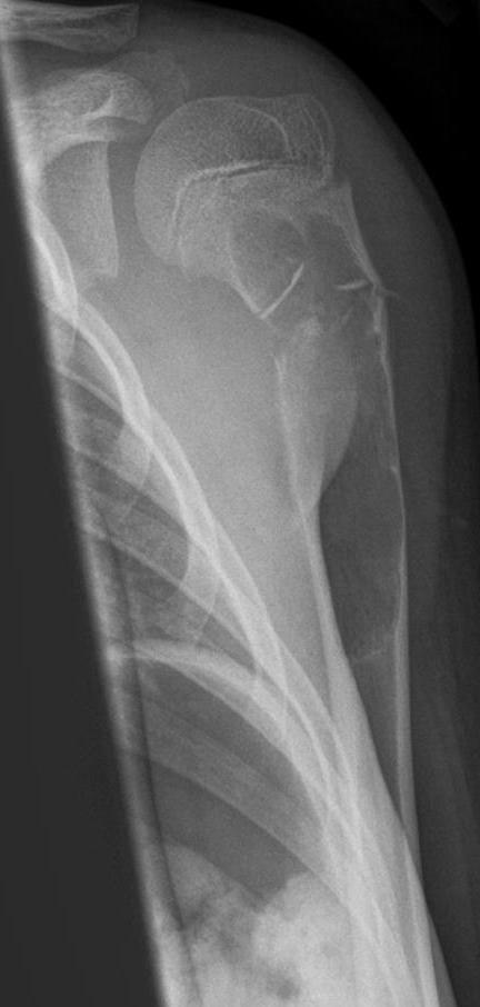
Synonyms / status. Simple, unicameral, or juvenile bone cyst. A non-neoplastic lesion.[28] The term “unicameral” is a mild misnomer because older cysts become multilocular. (A small subset has recently been found to carry EWSR1/FUS-NFATC2 rearrangements, but no defining genetic lesion is established.)
Definition. A solitary, fluid-filled cavity of the metaphysis, lined by a thin fibrous membrane and containing clear or serosanguineous fluid. It begins in childhood adjacent to the growth plate and becomes inactive around skeletal maturity.
Epidemiology. Most present in the first two decades (≈80-90 % under 20 years; symptomatic cysts cluster between 5 and 15 years). There is a male predominance of roughly 2-2.7 : 1 across series (Schajowicz 2.7 : 1; Campanacci 2.3 : 1; Dorfman ≈ 2 : 1). It is among the commonest tumour-like lesions, second in frequency after metaphyseal fibrous defects and exostoses.
Pathogenesis. Unknown. The leading concept is a local haemodynamic disturbance, namely venous outflow obstruction of the fertile metaphysis. Cyst fluid resembles serum and contains bone-resorbing factors (prostaglandins, IL-1, proteolytic enzymes). Campanacci’s manometry distinguished active cysts (intracystic pressure >30 cmH₂O, pulsating) from latent cysts (venous pressure, 6-10 cmH₂O). The cyst does not truly migrate. Rather, the growth plate grows away from it, so that a juxta-physeal “active” cyst becomes a diaphyseal “latent” cyst with time.
Sites. The proximal humerus (≈ 50-60 %, the single commonest site) and proximal femur (≈ 25 %) account for the great majority in children; the ilium, calcaneus, and other long-bone metaphyses follow. The calcaneal cyst of adults is regarded as a separate, atrophic/degenerative entity that does not fracture and does not respond to steroid.
Clinical features. Typically silent until a pathological fracture after trivial trauma; fracture is the presenting event in over 80 % of diagnosed cysts. A humeral cyst may cause 1-4 cm of arm shortening, while pelvic cysts are chance findings.
Imaging. A central, well-marginated, radiolucent metaphyseal lesion that thins and mildly expands but never breaches the cortex and shows no soft-tissue mass; ridges on the inner cortex create a pseudo-trabeculated (“pseudoloculated”) look. There is no periosteal reaction except from a healing fracture. The “fallen fragment / fallen leaf” sign (Reynolds, 1969) is a cortical fragment that drops into the dependent part of the fluid-filled cavity after fracture; it is pathognomonic because it proves the cavity is hollow and fluid-filled. CT/MRI show water-density/T2-bright fluid; classically there are no multiple fluid-fluid levels (those favour ABC). Bone scan shows little uptake. Pathological fracture occurs in roughly two-thirds of symptomatic cysts, the aspirated fluid characteristically shows a raised alkaline phosphatase, and Mirra regards it as the only true primary cyst of bone, defined by its thin lining membrane.[29]
Pathology. A unicameral cavity with clear, straw-coloured or serosanguineous fluid and an eggshell cortex. The lining is a thin grey membrane of loose vascular connective tissue with scattered osteoclast giant cells, haemosiderin, cholesterol clefts, and a characteristic, diagnostically useful finding: cementum-like (“fibrinoid”) calcified material, which historically generated the term “cementoma of long bones.” Two practical points follow. The simple bone cyst is the only true, fluid-filled cyst of bone (every other “cystic” lesion is a solid, tissue-filled tumour), and its curettings are scant, a thin wall only, unlike the voluminous tissue retrieved from a giant-cell tumour or aneurysmal bone cyst.
Differential diagnosis. Chiefly the aneurysmal bone cyst (more expansile, septated, blood-filled with fluid-fluid levels, yields blood not clear fluid at puncture), fibrous dysplasia (ground-glass matrix), intraosseous lipoma (calcaneus, central calcification), and eosinophilic granuloma.
Treatment. What is treated is fracture risk, not the cyst per se.
- Observation for latent cysts near or past puberty, and for unloaded bones (ilium).
- Intralesional methylprednisolone (Scaglietti technique): aspirate, lavage, inject; repeat at ~2-month intervals (typically 2-5 injections); first signs of repair at ~6 months; effective only in active cysts (rarely used after age 15). Adjuncts and alternatives include autologous bone-marrow injection, demineralised bone matrix, and multiple trepanation.
- Curettage and bone grafting, the traditional operation, now reserved for displaced lower-limb fractures, very large cysts, or diagnostic uncertainty.
- Continuous decompression with cannulated screws or a flexible intramedullary nail (modern; particularly for the proximal humerus).
Prognosis and complications. Recurrence after curettage and grafting is reported between ~20 % and ~45 % (Dorfman ~20 %; Schajowicz 30-45 %); steroid injection gives broadly comparable results.[30] The cyst reliably quiesces around puberty. Complications are pathological fracture, residual humeral shortening or femoral deformity, and, after curettage near the physis, growth arrest. Malignant change is exceptional.
Exam pearls. Fallen-fragment sign = simple cyst after fracture. Active vs latent = distance from the physis (Jaffe). Two sites (humerus > femur), two decades, 2 : 1 male. Cementum-like fibrinoid material is the histological clue. The plate moves away; the cyst does not migrate.
II.2 Aneurysmal Bone Cyst
Synonyms / status. Named by Jaffe and Lichtenstein in 1942; Schajowicz preferred “multilocular haematic bone cyst.” Once thought a reactive lesion, primary ABC is now recognised as a true benign neoplasm characterised by a USP6 rearrangement (most often CDH11-USP6). Secondary ABC is a blood-filled cystic change engrafted on a pre-existing lesion (giant-cell tumour, chondroblastoma, osteoblastoma, fibrous dysplasia, non-ossifying fibroma) and lacks the rearrangement.
Definition. An expanding, osteolytic lesion of blood-filled spaces separated by fibrous septa that contain osteoid trabeculae and osteoclast giant cells.
Epidemiology. About 80 % occur before age 20 (peak in the second decade); it is roughly half as frequent as giant-cell tumour and represents ~2.5 % of primary bone tumours. The sex distribution is roughly equal, with a slight female lean in some series.
Pathogenesis. Historically attributed to a local circulatory disturbance with markedly raised venous pressure (Lichtenstein), possibly via an intraosseous arteriovenous shunt, producing a self-perpetuating haemorrhagic process. There is no endothelial lining to the blood spaces, and arguments against a true AV shunt include the absence of endothelium and the raised fibrinolytic activity in cyst fluid. The modern unifying explanation for primary ABC is the USP6 oncogene rearrangement.
Sites. Metaphyses/metadiaphyses of long bones (especially the lower limb), the posterior elements of the spine (the neural arch, spreading into the body, with ABC accounting for a notable share of spinal tumour-like lesions), and the pelvis (about the triradiate cartilage). It is essentially never primary in the epiphysis while the physis is open; eccentric location is typical. Dorfman notes an almost uniform skeletal distribution, unusual among bone lesions.
Clinical features. Pain and swelling, sometimes marked because of the “blow-out” expansion. Pathological fracture is uncommon except as vertebral collapse, which may cause neurological deficit. Growth can be rapid, and symptoms may worsen during pregnancy.
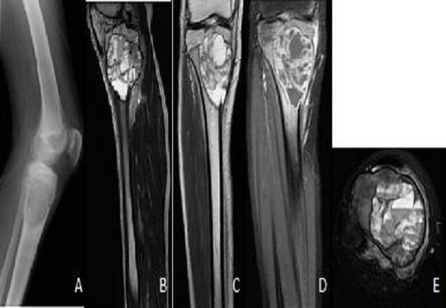
Imaging. An eccentric, expansile, “blown-out / ballooned” lucent lesion with a thin periosteal shell and a “soap-bubble” internal pattern; a Codman-type periosteal reaction may occur at the margin. The “finger-in-the-balloon” sign (Freiberger), a cortical buttress projecting into the expanded lesion, favours ABC over telangiectatic osteosarcoma. CT and MRI show fluid-fluid levels from sedimented blood, and the septa enhance. Critically, fluid-fluid levels are not specific: they also occur in telangiectatic osteosarcoma, giant-cell tumour, chondroblastoma, and a fractured simple cyst. Bone scan shows peripheral uptake with a cold centre (“doughnut” sign). Five radiographic types are described (central; “blow-out”; eccentric; subperiosteal; and subperiosteal with soft-tissue extension).
Pathology. Multilocular, blood-filled “sponge-like” cavities with fibrous septa. Opening the eggshell yields profuse non-pulsatile venous bleeding, and the tissue peels cleanly off the bony wall. Histologically: blood-filled spaces without endothelial lining, septa of fibrohistiocytic tissue with osteoclast giant cells, haemosiderin, and reactive osteoid running parallel to the septum; Campanacci’s “blue reticulated chondroid” (“blue bone”) is characteristic. Mitoses are common but never atypical. Atypical mitoses, anaplasia, necrosis, and tumour osteoid instead indicate telangiectatic osteosarcoma. The solid variant consists largely of giant-cell-reparative-granuloma-like solid tissue and shares the USP6 rearrangement. On unroofing, blood wells up but does not spurt in pulsatile fashion, and the solid curettings are small relative to the cavity. The decisive separation from telangiectatic osteosarcoma is cytologic atypia within the septa, present in the sarcoma and absent here.
Differential diagnosis. The single most important is telangiectatic osteosarcoma; sample the lesion widely and look for atypical cells, tumour osteoid, and necrosis. Also: simple bone cyst (clear vs bloody aspirate), giant-cell tumour (especially around the mature knee, often with secondary ABC), and the brown tumour (check biochemistry). A sacral expansile fluid-filled lesion should suggest ABC superimposed on a giant-cell tumour.
Treatment.
- Intralesional curettage with a high-speed burr and an adjuvant (phenol, cryotherapy, argon-beam) plus bone grafting is the mainstay.
- En-bloc resection for expendable bones gives the lowest recurrence (near 0 %).
- Selective arterial embolisation, often curative for the spine, pelvis, and proximal femur, and used pre-operatively to reduce bleeding.
- Sclerotherapy with doxycycline or polidocanol is an effective minimally invasive option, especially for the spine/pelvis and recurrences.
- Denosumab (anti-RANKL) is used off-label for surgically inaccessible, aggressive (e.g., sacral/spinal) lesions, with caution about rebound hypercalcaemia in children and recurrence on cessation.
- Radiotherapy (20-40 Gy) is reserved for inoperable lesions because of growth-plate damage and post-irradiation sarcoma risk.
Prognosis and complications. Recurrence after curettage ranges widely across series (Campanacci <10 %; Schajowicz ~17 % after curettage, 0 % after resection; Dorfman 20-70 %), almost always within the first two years.[31] Overall outcome is good (~90 % satisfactory). Complications include spinal cord/nerve compression and paraplegia, recurrence, and rare post-irradiation sarcoma.
Exam pearls. Posterior spinal elements; soap-bubble plus fluid-fluid levels (not specific); finger-in-the-balloon sign; no endothelial lining; mitoses present but never atypical; #1 DDx telangiectatic osteosarcoma; primary ABC = USP6 neoplasm.
II.3 Juxta-articular Bone Cyst (Intraosseous Ganglion)
Synonyms / status. The WHO term is juxta-articular bone cyst; “intraosseous ganglion” is the common clinical name. “Synovial cyst of bone” is a misnomer (no synovial lining). A non-neoplastic lesion.
Definition. A benign, often multiloculated subchondral cyst of fibrous tissue with extensive mucoid (myxoid) change, lying in subchondral bone adjacent to a joint but, in most cases, not communicating with it.
Epidemiology. Adults, typically 30-60 years (mean ~40); male predominance (~1.2-1.5 : 1). Often an incidental finding; symptomatic in roughly 60 %.
Pathogenesis. Two types occur. A minority (~16 %) are penetrating, where a soft-tissue ganglion erodes into bone (classic at the medial malleolus or beneath a meniscal cyst). The majority are idiopathic/primary intraosseous, an intramedullary fibroblastic proliferation with mucoid degeneration, probably driven by a local vascular disturbance and minor trauma. Campanacci confirmed the absence of joint communication by intra-operative methylene-blue injection.
The key distinction - ganglion vs subchondral (degenerative) cyst/geode. This is the highest-yield point. The intraosseous ganglion occurs in a younger patient, with a normal joint, no osteoarthritis, no joint communication, and usually a single cyst. The geode is an integral part of osteoarthritis in an older patient, communicates with the joint, often shows “kissing” cysts on both sides, and contains necrotic debris and cartilage. Histologically the two are identical, so the distinction is radiological and clinical.
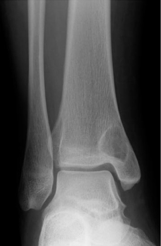
Sites. Subchondral, eccentric, in the epiphysis: medial malleolus (the classic site), carpus (lunate, scaphoid), hip (acetabulum, femoral head), proximal tibia, knee. Bilateral/symmetrical lesions occur. A rare periosteal ganglion sits on the tibial diaphysis far from any joint.
Clinical features. Joint-related pain, sometimes activity-related, over months to years; swelling is rare.
Imaging. A well-defined, oval, eccentric subchondral lucency with a thin sclerotic rim, usually ~1 cm (rarely up to 5 cm), not connected to the joint, with a normal articular surface and joint space. MRI shows fluid signal (low T1, high T2).
Pathology. A uni- or multilocular cavity with a thin fibrous wall and gelatinous mucoid content; the wall is dense collagen with foci of myxoid degeneration and stellate cells, and no synovial or epithelial lining. The mucin dissolves in routine processing unless acetic acid is added to the fixative.
Treatment. Curettage with or without bone grafting; for the penetrating type, excise the soft-tissue ganglion as well. Recurrence is uncommon.
Exam pearls. Medial malleolus and carpal lunate/scaphoid; young patient + no OA + no joint communication + single cyst = ganglion (vs old patient + OA + communicating + kissing cysts = geode); histology identical, so diagnosis is radiological; no synovial lining.
II.4 Fibrous Dysplasia
Synonyms / status. Fibro-osseous dysplasia; named by Lichtenstein (1938) and Lichtenstein & Jaffe (1942). A developmental/dysplastic, non-neoplastic lesion unified by a somatic GNAS mutation.
Definition. A benign, developmental disorder in which medullary bone is replaced by fibro-osseous tissue whose hallmark is the inability to mature beyond woven bone: bland fibroblastic stroma containing trabeculae of immature, non-lamellar bone.
Epidemiology. Monostotic disease outnumbers polyostotic 8-10 : 1. About 70 % present in the first three decades; there is a slight female predominance overall (becoming ~2 : 1 female in polyostotic disease). It is not hereditary. McCune-Albright syndrome is rare (1/100 000-1/1 000 000).
Pathogenesis. A post-zygotic activating mutation of GNAS (the α-subunit of the stimulatory G-protein, Gsα), classically R201 on chromosome 20q13, causes constitutive adenylyl-cyclase activity. Somatic mosaicism explains the spectrum from a single focus to widespread polyostotic disease and the associated endocrinopathies. (Diagnostically important caveat: GNAS mutations also occur in low-grade osteosarcoma, so the molecular test does not reliably separate the two.)
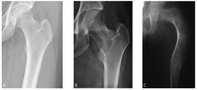
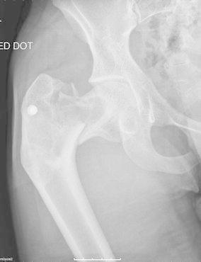
Sites. The proximal femur (the classic site, prone to the “shepherd’s-crook” deformity), tibia, ribs (fibrous dysplasia is the commonest benign expansile rib lesion), and craniofacial bones (maxilla). The lesion is intramedullary, metaphyseal/diaphyseal, and spares the epiphysis while the physis is open. Polyostotic disease tends to be unilateral / one side of the body.
Clinical features. Often an incidental finding; otherwise swelling, deformity, or pathological fracture, with pain from fatigue fractures (classically the medial femoral-neck cortex).
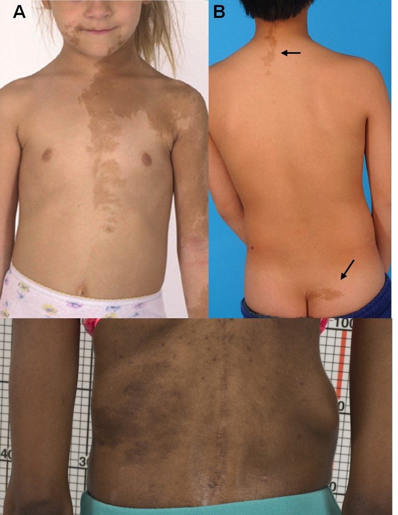
- McCune-Albright syndrome = the triad of polyostotic fibrous dysplasia + café-au-lait macules + endocrinopathy (precocious puberty, most often in girls), with other endocrinopathies (hyperthyroidism, acromegaly/GH excess, Cushing, phosphaturic rickets via FGF23) less commonly.
- Mazabraud syndrome = fibrous dysplasia + intramuscular myxomas (the rarest association).
- Café-au-lait borders: fibrous dysplasia produces irregular, jagged “Coast of Maine” borders that tend to respect the midline, versus the smooth “Coast of California” borders of neurofibromatosis type 1 (a distinction attributed to Albright).
Imaging. The hallmark is a “ground-glass” intramedullary matrix, well-defined, with a sclerotic rind, endosteal scalloping, a smooth thinned cortex, and no periosteal reaction. Deformities (shepherd’s crook of the proximal femur, bowing) and fatigue fractures are common; bone scan is usually “hot,” though Greenspan notes that about 10 % of ground-glass lesions show no increased uptake, and CT attenuation runs a wide 70-400 HU from the microscopic ossification.[32]
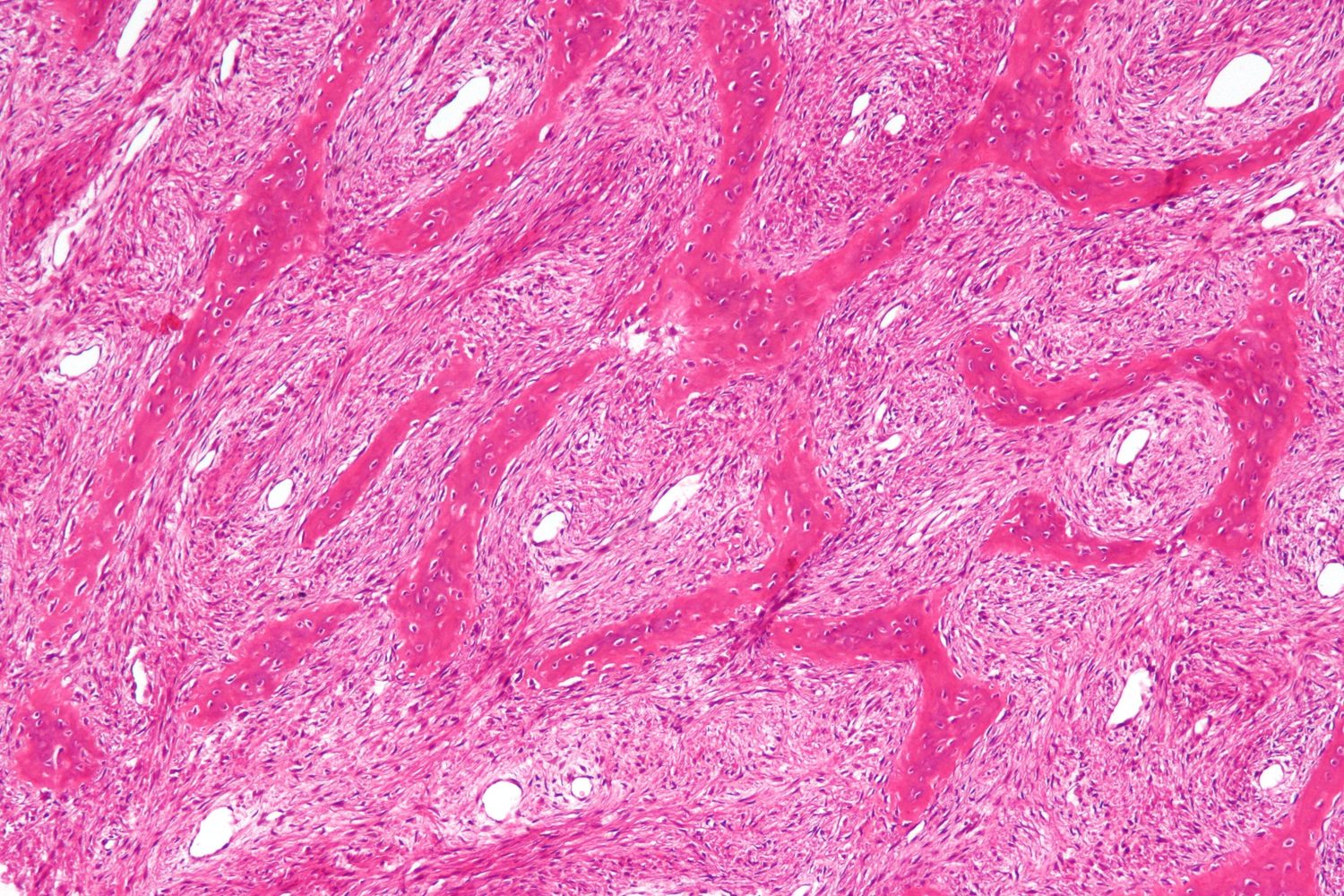
Pathology. Bland spindle-cell stroma in a whorled/storiform pattern with trabeculae of woven (immature) bone shaped like “Chinese characters” that are not rimmed by osteoblasts, the key contrast with osteofibrous dysplasia. Cartilage nodules, cementoid bodies (especially craniofacial), foam cells, giant cells, and secondary aneurysmal-bone-cyst change may occur. A prominent metaplastic cartilage component (fibrocartilaginous dysplasia) can mimic a cartilage neoplasm, and intratrabecular reversal lines can mimic Paget disease.
Differential diagnosis. Low-grade central osteosarcoma (the clinically critical one; look for nuclear atypia and a permeative growth pattern, since GNAS does not discriminate), osteofibrous dysplasia (intracortical, tibia, rimmed trabeculae), Paget disease (older patient, flame-shaped advancing edge, lamellar mosaic bone), enchondroma/Ollier, and non-ossifying fibroma (no metaplastic bone).
Malignant transformation. Rare, under ~1 % (Schajowicz ~0.5 %),[33] higher in polyostotic/McCune-Albright disease and after radiotherapy; usually to osteosarcoma, fibrosarcoma, or MFH, after a mean latency of ~13 years, with a poor prognosis.
Treatment. There is no medical or radiation cure for the dysplasia, and radiotherapy is contraindicated (it raises the malignancy risk). Bisphosphonates reduce fibrous-dysplasia bone pain (modern). Surgery is directed at deformity and fracture: corrective osteotomy with internal fixation (intramedullary devices preferred) for the shepherd’s-crook femur; cortical/fibular strut grafting of the femoral neck. Curettage and grafting usually fail in active/cortical disease (the graft resorbs) and are reserved for the quiescent adult. Pathological fractures heal by callus and are generally treated conservatively or with internal fixation.
Prognosis. Lesions stabilise after puberty; monostotic disease does not become polyostotic. Functional prognosis is poor only in extensive polyostotic disease.
Exam pearls. Ground-glass + Chinese-letter woven bone with NO osteoblastic rimming; shepherd’s crook; Coast-of-Maine café-au-lait; McCune-Albright (polyostotic FD + café-au-lait + precocious puberty), Mazabraud (FD + myxomas); GNAS R201; radiotherapy contraindicated; normal serum calcium, phosphate and PTH separate it from hyperparathyroidism.
II.5 Osteofibrous Dysplasia and the Adamantinoma Spectrum
Synonyms / status. Ossifying fibroma of long bones (Kempson, 1966); osteofibrous dysplasia (Campanacci, 1976); hence the “Kempson-Campanacci lesion.” A benign, often spontaneously regressing fibro-osseous lesion that nonetheless sits at the benign end of a spectrum with adamantinoma. (Schajowicz dissents, regarding it as merely active “intracortical fibrous dysplasia” rather than a separate entity.)[34]
Definition. An intracortical fibro-osseous lesion of the tibia/fibula in young children, resembling fibrous dysplasia but with woven-bone trabeculae rimmed by active osteoblasts and a zonal architecture; most cases contain scattered keratin-positive epithelial cells.
Epidemiology. Rare; male predominance; almost all in the first decade (many under 5 years; some congenital).
Sites. Almost exclusively the diaphysis of the tibia (anterior cortex), occasionally the ipsilateral fibula. Intracortical, in contrast to intramedullary fibrous dysplasia.
Clinical features. Painless anterior or anterolateral bowing/expansion of the tibia. Stress and pathological fractures occur, but, importantly, pseudarthrosis is not a feature (this distinguishes it from congenital pseudarthrosis of the tibia and NF1).
Imaging. Intracortical, anterior-tibial multilocular “bubbly” lucencies with a sclerotic rim and anterior bowing, sometimes veiled by ground-glass. The picture can be radiographically identical to adamantinoma.
Pathology. Less cellular than fibrous dysplasia; woven trabeculae rimmed by plump osteoblasts; a zonal architecture (sparse central woven bone giving way to coarser, partly lamellar bone peripherally); cytokeratin staining reveals scattered single epithelial cells or small nests in most cases (becoming overt epithelial islands in adamantinoma); even keratin-negative lesions show epithelial tonofilaments on electron microscopy, the molecular thread linking osteofibrous dysplasia to differentiated adamantinoma.[35]
The OFD-adamantinoma spectrum. OFD → OFD-like / “differentiated (regressing)” / juvenile intracortical adamantinoma → classic adamantinoma. Two pathogenetic hypotheses are cited. Dorfman’s holds that OFD is a reparative reaction overgrowing a regressing epithelial tumour, analogous to regressing neuroblastoma. Mirra’s holds that OFD harbours epithelial rests that may, years later, give rise to adamantinoma, analogous to notochordal rests becoming chordoma. Shared chromosomal trisomies (7, 8, 12, 21) support a genuine relationship.
Adamantinoma (for contrast). A low-grade malignant, epithelial (cytokeratin- and vimentin-positive; EMA- and factor-VIII-negative) tumour, usually of the tibial diaphysis (80-90 %) in adults aged 20-40, with overt epithelial islands; it metastasises in ~15 % (lung, late) and requires wide resection (intralesional/marginal surgery recurs). Cure approaches ~90 % with adequate surgery.[36]
Treatment of OFD. Defer surgery: OFD recurs if excised before it stabilises and tends to regress spontaneously after ~10-12 years. Observe and brace; correct marked bowing by osteotomy once stable; treat fractures conservatively; biopsy mainly when the course suggests adamantinoma.
Exam pearls. FD = intramedullary, no rimming, any bone; OFD = intracortical, osteoblastic rimming present, tibia/fibula, child <10 y, spontaneous regression; pseudarthrosis NOT a feature; spectrum to adamantinoma (tibia, adult, epithelial, keratin+); defer surgery in OFD.
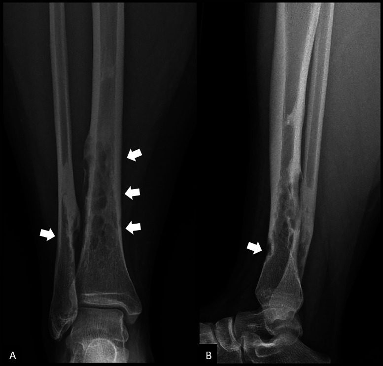
II.6 Metaphyseal Fibrous Defect (Fibrous Cortical Defect / Non-ossifying Fibroma)
Synonyms / status. Fibrous cortical defect (FCD, the small cortical form) and non-ossifying fibroma (NOF, the larger form with medullary extension) are the same fibrohistiocytic process at different sizes; other names are non-osteogenic fibroma, histiocytic xanthogranuloma, fibroxanthoma. WHO’s neutral descriptive term is metaphyseal fibrous defect. It is regarded as a developmental, non-neoplastic defect (though a clonal subset with t(1;4) has been reported).
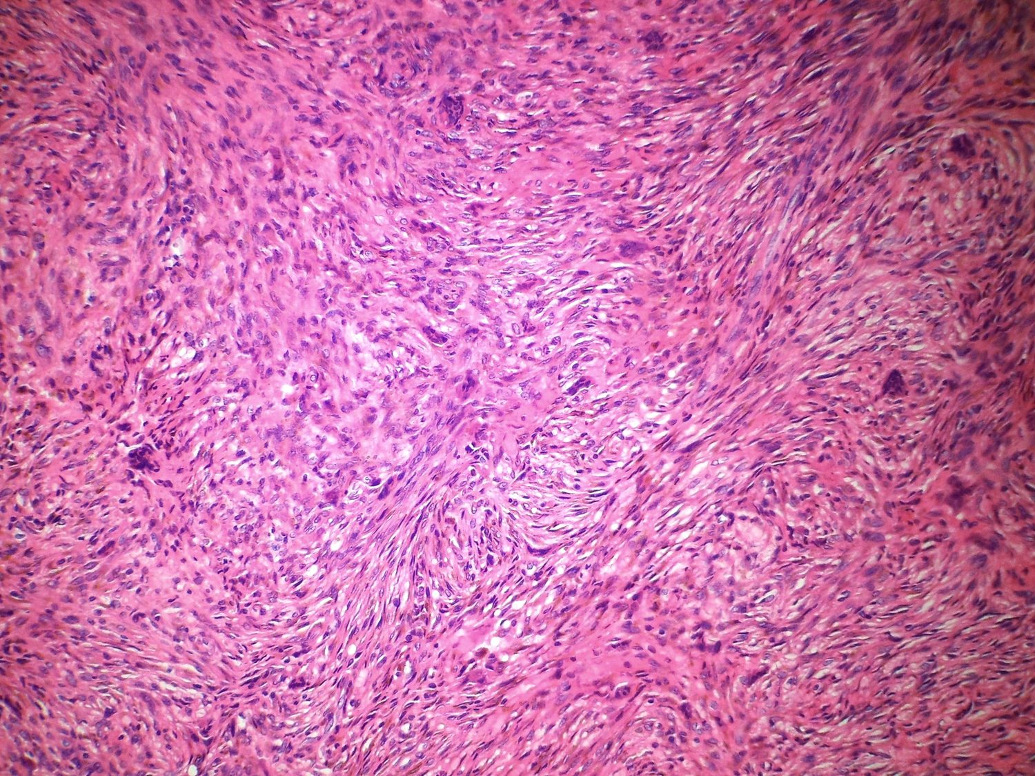
Definition. A well-defined, eccentric, cortically based metaphyseal lesion of storiform fibrous tissue with multinucleated giant cells, foamy (xanthoma) cells, and haemosiderin, histologically identical to benign fibrous histiocytoma, that forms no bone and characteristically resolves spontaneously.
Epidemiology. The commonest tumour-like lesion of bone, and one of the commonest lesions in the skeleton: fibrous cortical defects are present on the radiographs of roughly 30-40 % of children with open physes (Sontag & Pyle found up to 53 % of boys). The peak is in the second decade; there is a male predominance; the lesion is rare in adults because most involute.
Pathogenesis. A developmental defect at the periphery of the physis (subperiosteal/cortical origin), of probable histiocytic lineage (CD68-positive); the lesion enlarges, then heals by sclerosis and ossification at skeletal maturity. WHO 2020 nonetheless now classifies it as a true (benign) neoplasm, with activating KRAS or FGFR1 mutations in over 80 % of sporadic lesions - the developmental and neoplastic views converging on a self-limited, MAPK-driven proliferation.
Sites. Metaphyseal, eccentric, cortically based, long axis parallel to the bone: distal femur > proximal tibia > distal tibia; the fibula is also common. In thin bones (fibula, ulna) it may occupy the whole width. Flat-bone, rib, skull, or pelvic location is atypical and should prompt the label benign fibrous histiocytoma instead.
Clinical features. Usually asymptomatic and incidental. A large NOF can cause pain or a pathological fracture, and a superimposed secondary aneurysmal bone cyst can make a quiet lesion suddenly enlarge.
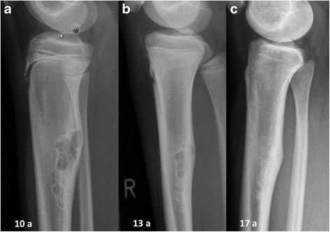
Imaging. An eccentric, cortically based, lytic lesion with a scalloped sclerotic (“soap-bubble”/grape-like) margin, a thinned but intact cortex, and no matrix mineralisation; it migrates away from the physis with growth and heals by sclerosis. The appearance is specific enough to diagnose without biopsy.
Pathology. Cellular storiform spindle cells, osteoclast-type giant cells clustered around haemorrhage, foamy lipid-laden macrophages, and abundant haemosiderin, with no bone formation within the lesion (the feature that separates it from fibrous dysplasia and gave it the name “non-ossifying”).
Differential diagnosis. Fibrous dysplasia (central, with metaplastic bone), giant-cell tumour (epiphyseal, adult), aneurysmal bone cyst (secondary within an NOF), chondromyxoid fibroma (chondroid matrix), and benign fibrous histiocytoma, the histological twin distinguished only by clinical context (older patient and/or unusual site, more often painful and recurrent).
Treatment. Observation for the small, classic, asymptomatic lesion (no biopsy). Curettage and grafting for lesions that are large, painful, develop a secondary ABC, or threaten fracture. The classic fracture threshold: a lesion occupying >50 % of the transverse bone diameter (the Arata/Drennan criterion; >33 mm length is also quoted) is fracture-prone and warrants prophylactic curettage and grafting; the commonest fracture site is the distal tibia.[37]
Jaffe-Campanacci syndrome. Multiple NOFs + café-au-lait macules + extraskeletal anomalies (hypogonadism/cryptorchidism, ocular and cardiovascular malformations) ± mental retardation; increasingly regarded as part of the NF1 spectrum.
Prognosis. Excellent: spontaneous involution and ossification by skeletal maturity; recurrence after curettage is rare; true malignant transformation essentially does not occur (apparent cases were malignant from the outset). Salter’s clinical caution bears repeating: the subperiosteal cortical defect is found in 10-20% of children, and its real danger is over-treatment, or being wrongly blamed for unrelated pain when the true cause lies elsewhere.
Exam pearls. Commonest tumour-like lesion; eccentric metaphyseal lytic lesion with scalloped sclerotic border in a teenager = leave-me-alone, no biopsy; storiform + giant cells + foam cells + haemosiderin with NO bone; >50 % transverse diameter → fracture risk; Jaffe-Campanacci = multiple NOFs + café-au-lait (NF1 spectrum).
 ## II.7 Langerhans Cell Histiocytosis (Eosinophilic Granuloma)
Synonyms / status. Eosinophilic granuloma (the localised bone form), histiocytosis X (Lichtenstein, 1953), Langerhans cell histiocytosis. The older WHO definition calls it non-neoplastic. The modern consensus, codified by WHO 2020, is that LCH is a clonal neoplasm of myeloid dendritic-cell precursors, with BRAF V600E in ~50-60 %.[38]
Definition. A proliferation of Langerhans-type histiocytes admixed with eosinophils, spanning a clinical spectrum from a solitary, self-healing bone lesion to disseminated, potentially fatal disease.
The clinical spectrum (eponyms).
- Solitary eosinophilic granuloma - the commonest (>70-80 %) and most benign form; bone-only; excellent prognosis.
- Hand-Schüller-Christian disease - chronic disseminated form; the classic triad is skull lytic defects + exophthalmos + diabetes insipidus (the complete triad is actually rare).[39] Onset usually at 3-5 years.
- Letterer-Siwe disease - acute disseminated form of infants under ~3 years, with visceral involvement, a seborrhoeic-eczema-like rash, hepatosplenomegaly, and cytopenias; the worst prognosis.
Epidemiology. Children and young adults; peak 5-15 years; male predominance ~2 : 1; rare after 20 (adult lesions favour ribs, mandible, clavicle).
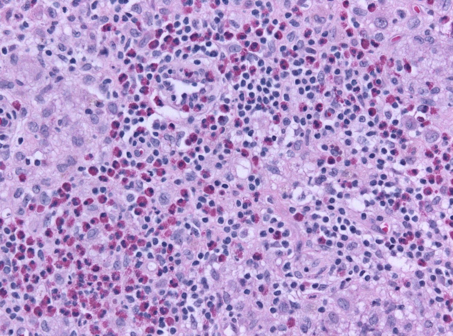
Pathogenesis. Proliferation of Langerhans cells (non-phagocytic dendritic histiocytes). The ultrastructural hallmark is the Birbeck granule (“tennis-racket”/rod shaped), and the immunophenotype is S-100, CD1a, and langerin/CD207 positive. Clonality and BRAF V600E/MAP2K1 mutations underlie the neoplastic reclassification.
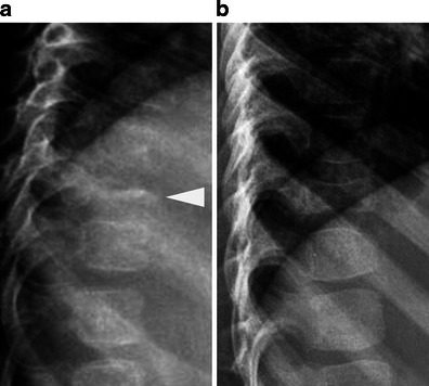
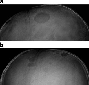
Sites. The skull is the commonest site (~70 % under age 20), then femur, pelvis, ribs, vertebrae, and mandible; it usually spares the hands and feet. Vertebral collapse produces vertebra plana (Calvé disease) with preserved disc spaces.
Clinical features. Pain and local swelling; in the spine, vertebra plana (rarely with neurological signs, since the uniform collapse usually spares alignment); otitis/mastoid involvement and diabetes insipidus in disseminated disease. Laboratory tests are usually normal (occasional mild eosinophilia, raised ESR).
Imaging. A lytic lesion that grows rapidly, then heals: early ill-defined, later with a sclerotic rim. In the skull it is “punched-out” with a bevelled edge / hole-within-a-hole (uneven destruction of the two tables). In long bones it can show an onion-skin periosteal reaction mimicking Ewing sarcoma or osteomyelitis (the “great mimicker”). A useful discriminator from Ewing: in LCH the periosteal lamellae are thicker than the intervening lucent lines. Vertebra plana is characteristic.
Pathology. Sheets of Langerhans cells with grooved, reniform “coffee-bean” nuclei, admixed with eosinophils (which may be lost if acid decalcification is used, a pitfall that makes it resemble osteomyelitis), foamy macrophages, and Touton-type giant cells; Birbeck granules on electron microscopy; S-100 / CD1a / langerin positive. (Charcot-Leyden crystals from eosinophil breakdown are a classic associated finding.) Mitoses may be brisk but do not predict the clinical course. Eosinophil-rich foci (“eosinophilic abscesses”) in a polymorphous inflammatory background are typical, and are the main reason the lesion is mistaken for osteomyelitis.
Differential diagnosis. Osteomyelitis, Ewing sarcoma, lymphoma, and, in long bones, osteosarcoma. LCH is the great mimic of all of them.
Natural history. Solitary lesions are self-limiting and often heal spontaneously. Vertebra plana in a child usually reconstitutes much of its height. If a lesion remains solitary for 12 months, dissemination is very unlikely and cure can be regarded as permanent.
Treatment.
- Solitary EG: intralesional corticosteroid injection (Campanacci’s preference) or curettage (Schajowicz’s preference), often after confirmatory needle biopsy; classic vertebra plana may be managed without biopsy, with bracing.
- Expendable bones: marginal/segmental resection.
- Radiotherapy (low dose) only for surgically inaccessible or neurologically threatening spinal lesions (used sparingly in children).
- Multisystem disease: chemotherapy - historically vinblastine + prednisone (± methotrexate, etoposide); BRAF/MEK inhibitors (vemurafenib, dabrafenib/trametinib) for refractory BRAF-mutated disease (modern).
Prognosis. Excellent for bone-only disease, even when multifocal. Prognosis worsens with younger age at onset (<3 years) and with organ dysfunction; Letterer-Siwe has the poorest survival.
Exam pearls. Spectrum: EG → Hand-Schüller-Christian (skull defects + exophthalmos + diabetes insipidus) → Letterer-Siwe (infant, visceral, worst); skull commonest bone; vertebra plana (Calvé), discs preserved, reconstitutes in children; punched-out bevelled skull lesion; coffee-bean nuclei + eosinophils + Birbeck granules, S-100/CD1a/langerin+; modern = clonal neoplasm, BRAF V600E.
II.8 Brown Tumour of Hyperparathyroidism
Synonyms / status. The focal/tumorous expression of the skeletal disease of hyperparathyroidism (osteitis fibrosa cystica, von Recklinghausen disease of bone); a reactive, non-neoplastic giant-cell lesion, in effect a giant-cell reparative granuloma of known (metabolic) cause. The name comes from the reddish-brown colour of old and recent haemorrhage/haemosiderin.
Definition. A localised, lytic, giant-cell-rich mass arising in a focus of intense PTH-driven osteoclastic resorption, with fibrous/haemorrhagic stroma and reactive bone.
Epidemiology. Overt brown tumours are now rare because hyperparathyroidism is detected early. When they occur they favour females (~3 : 1) and adults after 30 (3rd-4th decades).
Pathogenesis. Parathyroid hormone excess drives osteoclastic resorption, producing hypercalcaemia and hypophosphataemia with compensatory osteoblastic activity (raised alkaline phosphatase). The tumour itself is a hyperplastic, secondary mass of fibroblasts, giant cells, and microhaemorrhage that regresses after the parathyroid adenoma is removed.
- Primary hyperparathyroidism (parathyroid adenoma in ~80-85 %, rarely hyperplasia or carcinoma): ↑Ca, ↓PO₄ - the classic setting for brown tumours.
- Secondary (renal) hyperparathyroidism: hyperphosphataemia with normal/low calcium; brown tumours are uncommon, and the picture is dominated by generalised osteopenia, subperiosteal resorption, and (with combined Ca-PO₄ elevation) soft-tissue tumoral-calcinosis-like deposits.
Sites. Jaw, pelvis, ribs, femur, and the shafts/ends of long and tubular bones; often polyostotic. (Schajowicz emphasises a diaphyseal/shaft predilection; Campanacci ranks the metaepiphysis first, a minor source disagreement.)
Clinical features and laboratory diagnosis. The systemic hypercalcaemic syndrome, “stones, bones, groans, and psychiatric moans” (renal stones, bone pain/fractures, abdominal/peptic symptoms, neuropsychiatric features). The diagnosis is biochemical: ↑ serum (ionised) calcium, ↓ phosphate, ↑ alkaline phosphatase, and, the pathognomonic test, ↑ parathyroid hormone.[40] Caveat: with renal failure the calcium/phosphate changes may be muted, so a normal calcium does not exclude hyperparathyroidism.
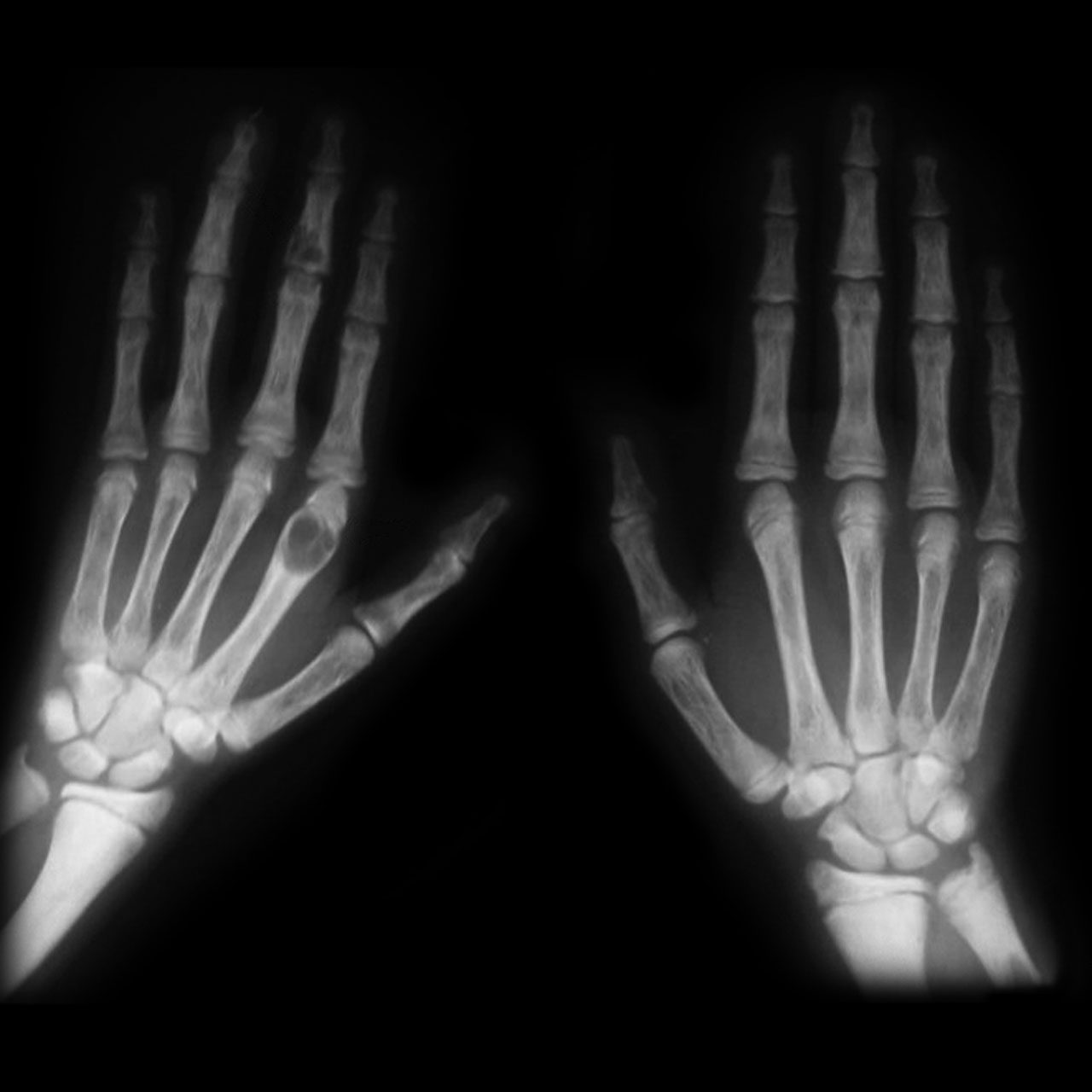
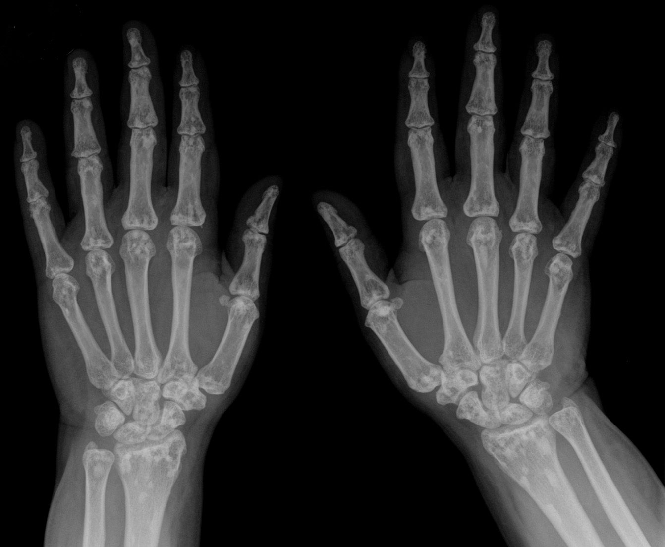
Imaging. Three components: osteopenia, subperiosteal bone resorption (classically the radial border of the phalanges, terminal-tuft resorption producing “drumstick” fingers, the acromial clavicle, “salt-and-pepper” skull), and the brown tumours themselves
- lytic, often trabeculated/expansile, with no prominent periosteal reaction. Loss of the dental lamina dura is characteristic.
Pathology. Marrow replaced by vascular fibrous tissue; small, irregularly distributed osteoclast giant cells clustered around haemorrhage with abundant haemosiderin; reactive osteoid/woven bone with osteoblastic rimming; “dissecting fibro-osteoclasia” within trabeculae. The brown tumour is histologically indistinguishable from a giant-cell tumour and from a reparative granuloma. The diagnosis is made by examining the adjacent bone (microlacunar resorption) and, above all, by the biochemistry: a normal calcium and phosphate exclude a brown tumour and point to a reparative granuloma, while subperiosteal resorption is sought first on the radial border of the middle phalanges of the index and middle fingers.[41]
Treatment. Treat the hyperparathyroidism, not the bone lesion: excise the adenoma (or perform subtotal parathyroidectomy for hyperplasia), and the brown tumours then regress and ossify. Watch for post-operative hypocalcaemia (“hungry bone”). Avoid plaster immobilisation (it worsens regional osteopenia and hypercalcaemia), and defer orthopaedic surgery until after the adenoma is removed where possible.
Prognosis. Skeletal lesions are reversible even when advanced; repair begins at 4-6 months and completes over 1-2 years. Prognosis depends on early diagnosis and on the (less reversible) renal damage.
Exam pearls. A “giant-cell tumour” in an unusual site or that is multifocal → check Ca/PO₄/PTH. ↑Ca, ↓PO₄, ↑PTH, ↑ALP. Subperiosteal resorption of the radial phalanges + salt-and-pepper skull. Treat the gland, not the bone. Earliest radiographic sign = loss of the dental lamina dura; in modern practice most primary hyperparathyroidism is asymptomatic, with overt bone disease in under 2%.
II.9 Giant-Cell (Reparative) Granuloma
Synonyms / status. Giant-cell reaction/lesion of bone; original jaw description by Jaffe (1953); the term “giant-cell reparative granuloma” was proposed by Lorenzo & Dorfman (1980). The word “reparative” is often dropped because trauma is frequently absent. A non-neoplastic/reactive lesion (though appendicular lesions share the USP6 rearrangement of solid ABC and may be clonal).
Definition. A benign giant-cell lesion of fibrous spindle-cell stroma with haemorrhage and unevenly clustered, small giant cells plus reactive bone, distinct from true giant-cell tumour and closely related to the solid variant of aneurysmal bone cyst.
Epidemiology. Younger than giant-cell tumour (peak 2nd decade, most 10-25 years); sex predilection disputed (male in some series, female in others); rare.
Sites. The gnathic bones (mandible, maxilla) are the classic site, then the small tubular bones of the hands and feet; long bones and vertebrae are rare. (Because a true giant-cell tumour essentially never arises in the jaw without Paget disease, a jaw giant-cell lesion is a reparative granuloma or brown tumour by default.)
Clinical features. Pain and swelling; may progress in pregnancy. Mandatory rule-out: measure calcium, phosphate, and PTH in every reparative granuloma, since the brown tumour is histologically identical, and multifocality should raise suspicion of a metabolic or syndromic cause.
Imaging. A lytic, often eccentric, sometimes expansile/trabeculated lesion without a prominent sclerotic rim or periosteal reaction; in small bones it may fill the cross-section. It is frequently mistaken radiographically for an aneurysmal bone cyst.
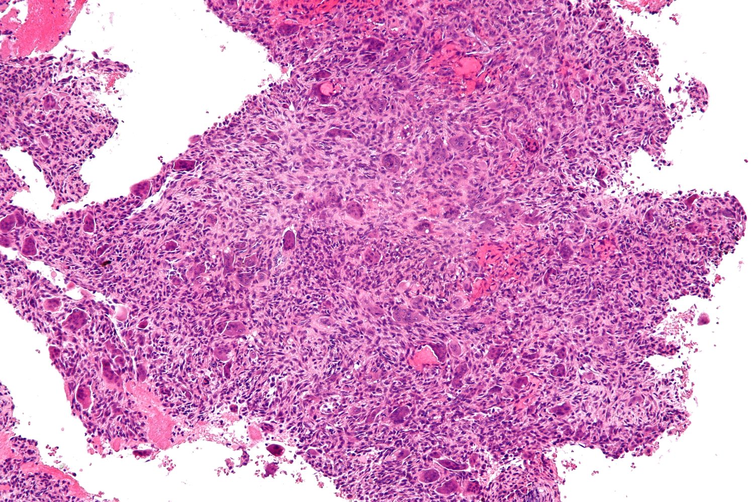
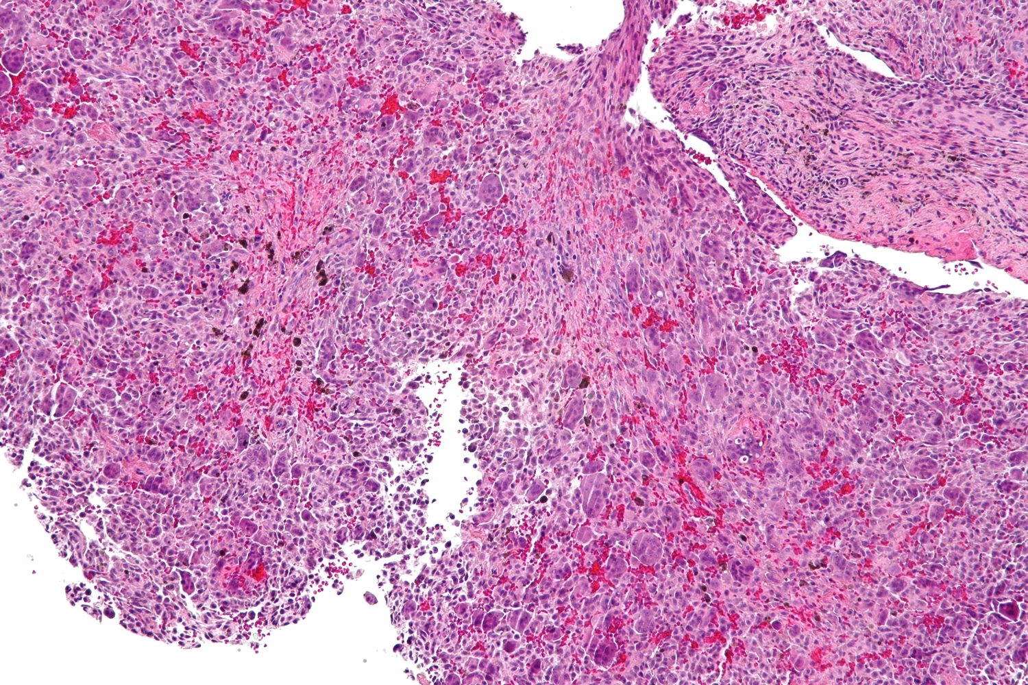
Pathology. Cellular fibrous stroma, areas of haemorrhage, reactive osteoid/bone, and multinucleated giant cells unevenly clustered around haemorrhage, smaller and with fewer nuclei than in giant-cell tumour. About 30 % contain ABC-like cystic foci. Mitoses may be present but are not atypical. The contrast with a true giant-cell tumour lies in the spindled, collagenised stroma and clustered giant cells here, versus the round-to-oval stroma and evenly distributed giant cells of the tumour; and in the solid curettings, which approximate the lesion’s size rather than the blood that fills an aneurysmal bone cyst.
Treatment. Thorough curettage with or without grafting is usually curative for jaw and hand/foot lesions; recurrence is ~30-50 % (usually within ~15 months).[42] Extensive small-bone lesions or recurrences may need marginal en-bloc resection or ray amputation (no recurrence after resection). Radiotherapy is effective but reserved; transient responses to steroid/calcitonin/interferon are reported for maxillary lesions. There is no malignant transformation and no pulmonary spread (unlike giant-cell tumour).
Distinguishing the giant-cell lesions (high-yield). The most useful histological criterion (Dorfman) separating giant-cell tumour from reparative granuloma/brown tumour is the uniform distribution of giant cells and the absence of reactive bone and stromal collagenisation in the unaltered giant-cell tumour, in which the mononuclear stromal-cell nuclei match those of the giant cells. See the comparison table in Section III.3.
Exam pearls. Jaw and small bones of hands/feet; always check Ca/PO₄/PTH (brown tumour is identical); giant cells small and clustered around haemorrhage vs even/large in GCT; solid ABC ≈ extragnathic reparative granuloma (USP6); cherubism = AD bilateral jaw lesions, SH3BP2 gene.
II.10 Myositis Ossificans (Heterotopic Ossification)
Synonyms / status. A misnomer, neither inflammatory nor confined to muscle; better termed heterotopic ossification. The idiopathic, non-traumatic form is the “pseudomalignant osseous tumour of soft tissue” (Fine & Stout). A reactive, self-limiting, non-neoplastic lesion (polyclonal, though a subset shows USP6 rearrangement).
Schajowicz’s classification of heterotopic ossification. (I) Myositis ossificans progressiva (fibrodysplasia ossificans progressiva); (II) myositis ossificans circumscripta - (A) post-traumatic (60-75 %) and (B) without trauma, either associated with systemic disease (paraplegia, head injury, burns, tetanus) or idiopathic.
Definition. A reactive proliferation of fibrous tissue forming large amounts of heterotopic bone (± cartilage) in soft tissue or on the bone surface, undergoing zonal maturation.
Epidemiology. Adolescents and young, athletically active adults; peak in the 2nd-3rd decades; male predominance ~2 : 1.
Pathogenesis. Usually develops at a site of trauma. Neurogenic heterotopic ossification complicates head/spinal-cord injury, burns, and prolonged coma (typically around the hips), and these neurogenic lesions notably show no zonal architecture and no peripheral bone shell. Fibrodysplasia ossificans progressiva is caused by a germline ACVR1 (R206H) mutation.[43]
Sites. Deep soft tissue/muscle of the limbs - classically the quadriceps/thigh, the brachialis at the elbow, and the hip/gluteal region.
Clinical features and time course (high-yield). A painful soft-tissue mass appears a few weeks after the injury. Radiographically: negative in the first ~2 weeks → faint flocculent “dotted-veil” calcification at ~2-4 weeks → peripheral ossification by ~5-6 weeks → full maturation by ~6 months, after which the mass becomes stationary, may regress, or fuses to the cortex (“post-traumatic periosteoma”). Mature lesions are usually under 10 cm.
Imaging and the zonal phenomenon (THE key concept). A well-demarcated mass with peripheral mature ossification and a relatively lucent centre, the reverse of parosteal/extraskeletal osteosarcoma, in which the most mature, dense bone is central/basal and the periphery is the immature, infiltrative zone. On MRI the periphery is a low-signal shell. A radiolucent cleft separating the parosteal lesion from the cortex argues for myositis ossificans (vs the broad cortical fusion of parosteal osteosarcoma).
Pathology and the zonation (and the sarcoma pitfall). The hallmark is zonal maturation: a cellular centre of myofibroblastic granulation-like tissue with mitoses and haemorrhage → osteoid → woven bone → mature lamellar bone with osteoblastic rimming at the periphery. The major pitfall is biopsy of the cellular early centre, which can be over-read as osteosarcoma; but atypical mitoses are absent and the zonation, when present, is diagnostic. Biopsy timing matters.
Key differential - myositis ossificans vs osteosarcoma (“reverse zoning”). This is the single highest-yield point and is tabulated in Section III. In brief: maturation is centrifugal (mature outside) in myositis ossificans and centripetal/central in osteosarcoma; a parosteal lesion fused seamlessly to the posterior distal-femoral cortex should be treated as suspicious.[44]
Treatment. Observation first (some regress). Marginal excision is curative but only after the lesion has fully matured, because early excision recurs. For prophylaxis of heterotopic ossification (after hip arthroplasty/acetabular surgery, and in at-risk neurogenic patients), indomethacin and low-dose single-fraction radiotherapy are standard.
Fibrodysplasia ossificans progressiva (brief). Rare autosomal-dominant disease, onset in the first decade, with congenital great-toe malformation (microdactyly/hallux valgus) and progressive heterotopic ossification (axial → appendicular) causing ankylosis. Ossification develops centrally with no zonation. Biopsy, trauma, and intramuscular injections exacerbate it and must be avoided; death is usually from thoracic restriction.
Exam pearls. Peripheral mature bone + lucent centre = myositis ossificans (zonal); reverse = osteosarcoma. Timeline: dotted veil ~2-4 wk, ossifies ~5-6 wk, mature ~6 months. Don’t biopsy or excise early. Neurogenic HO and FOP lack zonation. FOP = ACVR1, great-toe anomaly. Salter’s two rules: never forcibly stretch or manipulate (it tears more muscle and worsens the process), and never excise early (the cellular phase mimics osteosarcoma and the lesion largely resorbs on its own).
II.11 Intraosseous Epidermoid Cyst
Synonyms / status. Keratin / epidermal inclusion cyst; skull lesions were called “cholesteatoma.” A non-neoplastic lesion in two flavours: an acral (phalangeal) post-traumatic inclusion type and a skull (developmental/congenital) type.
Definition. A bone cyst lined by stratified squamous epithelium and filled with laminated keratin.
Epidemiology. The phalangeal type occurs in adult male manual workers (often with a prior crush/penetrating injury, sometimes at an amputation stump); the skull type presents in children/young adults.
Sites. The distal (terminal) phalanges of the hands (occasionally toes), and the calvaria (frontoparietal, intradiploic). Long-bone involvement is extraordinarily rare.
Clinical features. A slowly enlarging, sometimes painful finger swelling with nail deformity, occasionally a pathological fracture; in the skull, a palpable swelling.
Imaging. A well-demarcated, round, purely lytic defect with a thin sclerotic rim and no internal matrix; cortical disruption is rare.
Pathology. A cavity lined by keratinising stratified squamous epithelium with a granular layer, filled with laminated keratin (“cheesy” content); rupture incites a foreign-body giant-cell reaction with cholesterol clefts.
Differential diagnosis. In the phalanx: enchondroma (the main mimic), glomus tumour, aneurysmal bone cyst; in the skull: eosinophilic granuloma and dermoid cyst (the latter contains dermal adnexa).
Treatment and prognosis. Curettage or excision (amputation only for advanced destructive phalangeal disease). Benign; the acral inclusion type has no reported malignant transformation, whereas developmental skull epidermoids/dermoids may rarely undergo squamous-carcinoma change.
Exam pearls. Lytic distal-phalanx lesion in a male manual worker with prior finger trauma; squamous epithelium + keratin is unique among bone lesions.
II.12 Other “Leave-Me-Alone” Lesions and Important Mimics
These are not all in Schajowicz’s numbered list but are routinely examined alongside it.
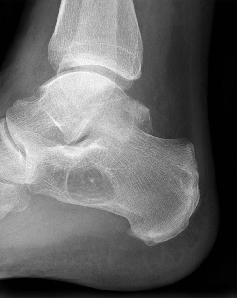
Intraosseous lipoma. A rare benign lesion of mature fat, classically in the calcaneus (also the proximal femur and tibia); usually asymptomatic. Imaging shows a well-defined lytic lesion with a thin sclerotic rim and a central calcified/ossified nidus (fat necrosis), following fat signal on CT/MRI (confirming the diagnosis non-invasively). Milgram’s three stages: (1) viable fat; (2) transitional (partial necrosis + calcification); (3) involuted (necrosis, cyst, calcification). Treatment is observation; curettage only if symptomatic.
Bone island (enostosis) and osteopoikilosis. A bone island is a focus of compact lamellar bone within cancellous bone, a “leave-me-alone” lesion. Imaging: a dense, ovoid sclerotic focus with a “brush border” of radiating thorny spicules, aligned with the trabeculae, and cold-to-mildly-warm on bone scan, the key feature separating it from an osteoblastic metastasis (markedly hot). Osteopoikilosis is an autosomal-dominant sclerosing dysplasia of multiple symmetrical bone islands (with Buschke-Ollendorff syndrome when associated with connective-tissue naevi, LEMD3 mutation); its differential cousins are osteopathia striata and melorheostosis.
Massive osteolysis - Gorham-Stout disease. “Vanishing/phantom/disappearing bone disease” (Gorham & Stout, 1955):[45] a rare, non-neoplastic, self-limited progressive dissolution of bone with proliferation of thin-walled vascular/lymphatic channels replacing bone, then fibrous tissue. It affects children and young adults, crosses joints and bony boundaries, and favours the spine and pelvis (Campanacci’s most frequent sites), the shoulder girdle (scapula, clavicle, humerus), and the maxilla/mandible. Radiographs show concentric resorption with “sucked-candy” tapering, no reactive bone, no periosteal reaction, and no fracture healing. Thoracic disease may cause a chylothorax (a potentially lethal complication); spinal disease may cause paraplegia. There is little osteoclastic activity; osteolysis is driven by the vascular/lymphatic proliferation (modern view: lymphatic-endothelial, D2-40-positive, IL-6/VEGF-mediated). Treatment is unsatisfactory. Radiotherapy may hasten arrest; bisphosphonates, interferon-α, and sirolimus are used (modern); grafts placed during the active phase resorb, so reconstruction is deferred until the disease burns out; chylothorax needs pleurodesis.
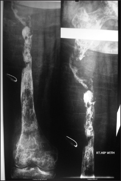 # SECTION III - SYNTHESIS AND EXAM AIDS
SECTION III --- SYNTHESIS AND EXAM AIDS
III.1 Pattern-based Differential Diagnosis
The “FEGNOMASHIC” mnemonic for a lytic/lucent bone lesion shows how the tumour-like lesions sit among the true tumours (entities in bold are tumour-like lesions or relevant mimics covered here):
- F - Fibrous dysplasia / Fibrous cortical defect (NOF)
- E - Enchondroma / Eosinophilic granuloma
- G - Giant-cell tumour
- N - Non-ossifying fibroma (often listed with F)
- O - Osteoblastoma
- M - Metastasis / Myeloma
- A - Aneurysmal bone cyst
- S - Simple (unicameral) bone cyst
- H - Hyperparathyroidism (brown tumour) / Haemangioma
- I - Infection
- C - Chondroblastoma / Chondromyxoid fibroma
Fluid-fluid levels (CT/MRI) are not specific. They turn up in aneurysmal bone cyst, telangiectatic osteosarcoma, giant-cell tumour, chondroblastoma, and even a fractured simple bone cyst.
Two “reverse” rules worth memorising:
- Zonation: peripheral mature bone = myositis ossificans; central mature bone = (parosteal/extraskeletal) osteosarcoma.
- Cyst content at puncture: clear/straw fluid = simple bone cyst; blood = aneurysmal bone cyst.
III.2 Quick Reference - Age, Site, and Signature
| Lesion | Typical age | Classic site | Signature feature |
|---|---|---|---|
| Simple bone cyst | 5-15 y | Proximal humerus, proximal femur | Central; fallen-fragment sign |
| Aneurysmal bone cyst | 2nd decade | Long-bone metaphysis; posterior spine | Blow-out; fluid-fluid levels; USP6 |
| Intraosseous ganglion | 30-60 y | Medial malleolus, carpus | Subchondral; no joint communication |
| Fibrous dysplasia | <30 y | Proximal femur, ribs, craniofacial | Ground-glass; shepherd’s crook; GNAS |
| Osteofibrous dysplasia | <10 y | Anterior tibial cortex | Intracortical; bowing; osteoblastic rimming |
| Non-ossifying fibroma | 5-15 y | Distal femur, proximal tibia | Eccentric, scalloped sclerotic rim |
| Langerhans cell histiocytosis | 5-15 y | Skull, then femur; spine | Punched-out skull; vertebra plana; BRAF |
| Brown tumour | 30-50 y | Jaw, pelvis, long bones | ↑Ca ↓PO₄ ↑PTH; subperiosteal resorption |
| Giant-cell reparative granuloma | 10-25 y | Jaw, hands/feet | Clustered small giant cells; check PTH |
| Myositis ossificans | 15-35 y | Thigh, elbow (brachialis) | Peripheral (zonal) ossification |
| Intraosseous epidermoid | adult | Distal phalanx, skull | Squamous epithelium + keratin |
| Intraosseous lipoma | adult | Calcaneus | Central calcified nidus; fat signal |
III.3 The Giant-Cell Lesions Compared
A recurring exam theme is that several lesions look alike under the microscope. Here are the four that matter, with the features that separate them.
| Feature | Giant-cell tumour | Reparative granuloma | Brown tumour | Solid ABC |
|---|---|---|---|---|
| Nature | Neoplastic, aggressive | Reactive (some clonal) | Reactive (metabolic) | Reactive (≈ extragnathic reparative granuloma) |
| Age / maturity | 20-55 y, skeletally mature | 10-25 y | 30-50 y | young |
| Classic site | Epiphysis of long bone (knee) | Jaw, hands/feet | Jaw, pelvis, long bones; polyostotic | Long bones, axial |
| Jaw? | Essentially never (unless Paget) | Commonest site | Yes | No |
| Giant-cell distribution | Even/uniform, large, many nuclei | Uneven, clustered around haemorrhage; small | Irregular, around haemorrhage; small | Uneven, clustered |
| Reactive bone / collagen | Absent (in unaltered tumour) | Present, often prominent | Present | Present |
| Stromal nuclei | Match the giant-cell nuclei | Bland spindle cells | Differentiated fibroblasts | Fibroblastic |
| Biochemistry | Normal | Normal (must check to exclude HPT) | ↑Ca, ↓PO₄, ↑PTH, ↑ALP | Normal |
| Behaviour | Recurs 25-35 %; rare lung implants | Recurs 30-50 %; no metastases | Regresses after parathyroidectomy | Curettage; may recur |
The single most useful discriminator is Dorfman’s: in the unaltered giant-cell tumour the giant cells are uniformly distributed, reactive bone and stromal collagenisation are absent, and the mononuclear stromal-cell nuclei resemble the giant-cell nuclei. Whenever giant cells turn up in an unusual site or a multifocal lesion, measure calcium, phosphate, and PTH before committing to a diagnosis.
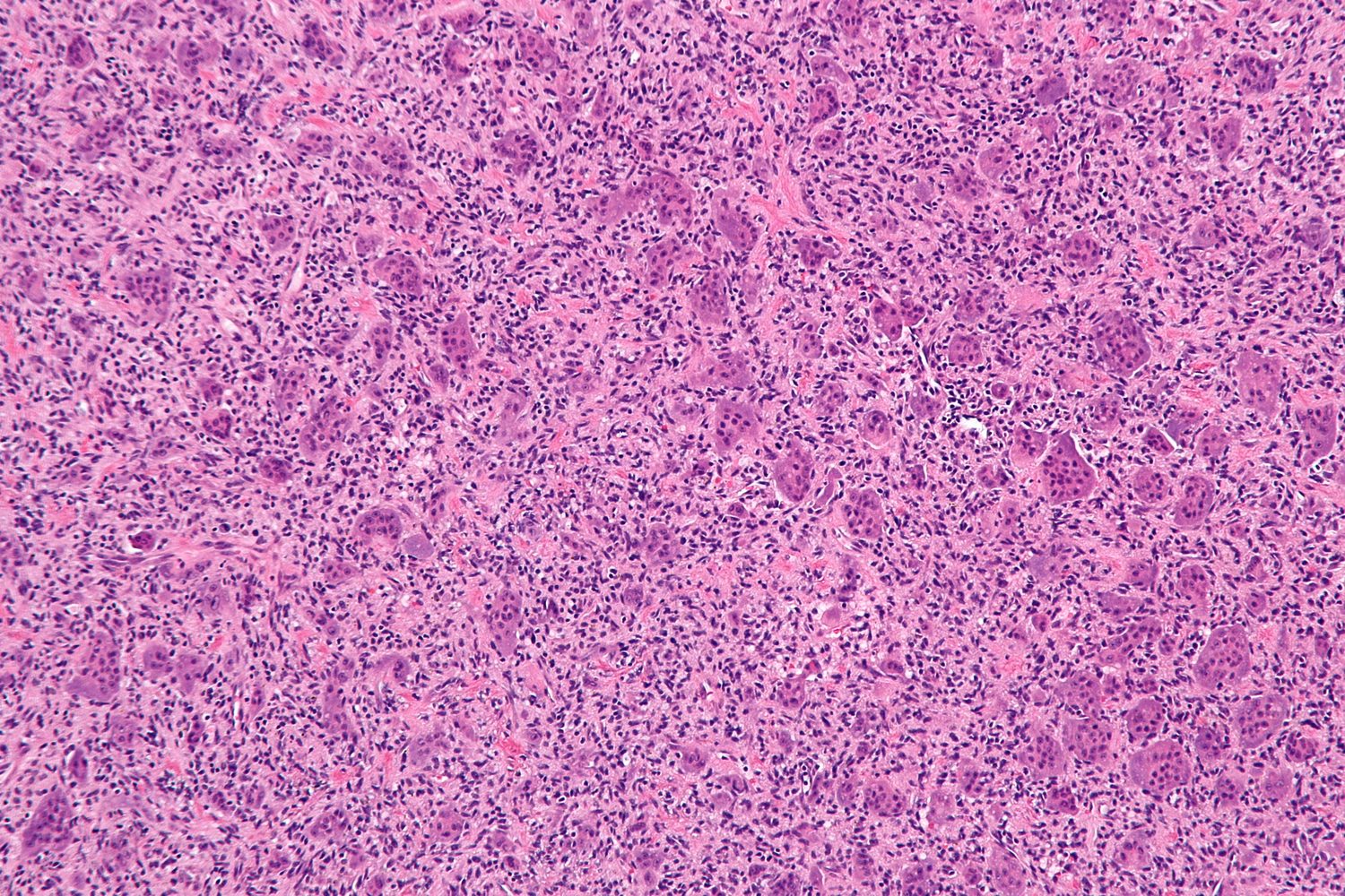
III.4 When to Observe, Biopsy, or Operate
Observe (no biopsy): classic fibrous cortical defect / small NOF; bone island; asymptomatic calcaneal intraosseous lipoma; latent simple bone cyst near skeletal maturity; classic vertebra plana in a child; mature myositis ossificans that is asymptomatic.
Biopsy / confirm tissue: any lesion with alarm features (permeative margin, soft-tissue mass, aggressive periosteal reaction, disproportionate pain, atypical site), and any putatively benign giant-cell lesion before definitive surgery, with the radiographs in the pathologist’s hands.
Operate:
- Curettage ± adjuvant ± graft - symptomatic/fracture-prone simple cyst (or steroid injection), large/painful NOF (>50 % transverse diameter), ABC (with burr/adjuvant), intraosseous ganglion, reparative granuloma, epidermoid cyst.
- Fixation / osteotomy - deformity or fracture of fibrous dysplasia (and the femoral-neck strut graft).
- Resection - expendable-bone ABC/NOF/reparative granuloma; adamantinoma (wide); recurrent disease.
- Treat the cause - parathyroidectomy for the brown tumour; defer surgery in osteofibrous dysplasia and in active Gorham disease.
III.5 The Bulgarian Operative Tradition (Boychev) and Terminology
The Bulgarian operative-orthopaedic school, codified in Boycho Boychev’s Хирургическа ортопедия (Surgical Orthopaedics), contributes two directly relevant techniques and a vocabulary the candidate is expected to use in the Bulgarian examination.
Curettage and filling (“plombage”) of a bone cyst (кюртиране и пломбиране на костна киста). For a solitary cyst (e.g., in the trochanteric region) threatening pathological fracture, Boychev’s described technique runs as follows. Expose the bone, where the cortex is typically ballooned, soft, and crackling on pressure. With a borer/drill (борер) make multiple holes outlining the cyst, join them with a chisel (длето), and remove the resulting bony lid (капак). Take the cyst wall down toward the diaphysis and enter the medullary canal, then curette out the contents (изкюртиране) and scrape the walls to healthy bone (изстъргване до здраво). Pack the cavity densely with cancellous bone grafts (спонгиозни присадъци) from the iliac crest or a bone bank, close in layers, and send the curetted material for histology. The defect heals in 4-6 months under plaster. (A humeral variant is attributed to Ya. Holevich.)
Osteoplastic resection after Boychev (костно-пластична резекция по Бойчев). This is an indigenous limb-salvage technique for large benign (giant-cell tumour, osteochondroma) or slowly-metastasising malignant (fibrosarcoma, chondrosarcoma) tumours about the knee. The tumour-bearing bone end is divided transversely, well clear of the lesion, and removed “ablastically” (no-touch). A local autograft rotated through 180° then bridges the defect, taken from the anterior surface of the tibia when the distal femur is resected, or from the anterior femur when the proximal tibia is resected, and fixed between two long medial and lateral plates whose screws engage both plates, achieving a knee arthrodesis-type reconstruction. Plaster immobilisation continues for more than six months until union. The atlas also catalogues wide resection for tumours (широка резекция при тумори), musculoplastic resection (мускулно-пластична резекция), and distal-femoral resection with allograft (алотрансплантат) replacement.
III.6 High-Yield Revision Summary
- Simple bone cyst - child, proximal humerus, presents with fracture; fallen-fragment sign; steroid injection / curettage; clear fluid.
- Aneurysmal bone cyst - second decade, metaphysis & posterior spine; soap-bubble + fluid-fluid levels (not specific); primary ABC = USP6 neoplasm; #1 DDx telangiectatic osteosarcoma; blood at puncture.
- Intraosseous ganglion - adult, medial malleolus/carpus, subchondral, no joint communication; distinguish from osteoarthritic geode.
- Fibrous dysplasia - ground-glass + Chinese-letter bone, no osteoblastic rimming; shepherd’s crook; GNAS; McCune-Albright; Coast-of-Maine café-au-lait; radiotherapy contraindicated.
- Osteofibrous dysplasia - child, anterior tibial cortex, rimming present, defer surgery; spectrum to adamantinoma.
- Non-ossifying fibroma - commonest tumour-like lesion; eccentric metaphyseal, scalloped sclerotic margin; storiform + giant + foam cells, no bone; observe unless >50 % width.
- Langerhans cell histiocytosis - skull, vertebra plana (Calvé); punched-out bevelled lesion; coffee-bean nuclei + eosinophils + Birbeck granules, S-100/CD1a/langerin; spectrum EG → Hand-Schüller-Christian → Letterer-Siwe; BRAF V600E.
- Brown tumour - ↑Ca, ↓PO₄, ↑PTH; subperiosteal resorption, salt-and-pepper skull; treat the gland.
- Giant-cell reparative granuloma - jaw/hands/feet; clustered small giant cells; check PTH; solid ABC is its appendicular relative.
- Myositis ossificans - post-traumatic thigh/elbow mass; peripheral (zonal) ossification; don’t biopsy/excise early; reverse of osteosarcoma.
- Intraosseous epidermoid - distal phalanx, male manual worker; squamous epithelium + keratin.
- Leave-me-alone & mimics - intraosseous lipoma (calcaneus, fat signal), bone island (cold scan), Gorham-Stout (vanishing bone, sucked-candy, chylothorax).
References
-
Schajowicz F. Tumors and Tumorlike Lesions of Bone: Pathology, Radiology and Treatment. 2nd ed. Berlin: Springer-Verlag, 1994 - Chapter IX, “Tumorlike Lesions.”
-
Campanacci M. Bone and Soft Tissue Tumors: Clinical Features, Imaging, Pathology and Treatment. 2nd ed. Vienna/New York: Springer, 1999 - sections on simple and aneurysmal bone cyst, intraosseous ganglion, massive osteolysis, fibrous and osteofibrous dysplasia, Langerhans cell histiocytosis, brown tumours, and reparative giant-cell granuloma.
-
Dorfman HD, Czerniak B. Dorfman and Czerniak’s Bone Tumors. 2nd ed. Philadelphia: Elsevier, 2016 - Chapters 8 (Fibrous Dysplasia), 9 (Fibrous and Fibrohistiocytic Lesions), 10 (Giant Cell Lesions), 15 (Cystic Lesions), and 23 (Reactive and Metabolic Conditions Simulating Neoplasms of Bone).
-
Бойчев Б. (Boychev B.) Хирургическа ортопедия (Surgical Orthopaedics). Sofia - operative techniques: curettage and plombage of bone cyst; osteoplastic resection after Boychev; wide and musculoplastic resection for tumours.
-
Unni KK, Inwards CY. Dahlin’s Bone Tumors: General Aspects and Data on 10,165 Cases. 6th ed. Philadelphia: Lippincott Williams & Wilkins, 2010 (consulted as a comparative epidemiological reference).
-
Key eponymous and primary sources cited within: Jaffe HL, Lichtenstein L (aneurysmal bone cyst, 1942; fibrous dysplasia, 1938/1942; giant-cell reparative granuloma of the jaw, Jaffe 1953); Lichtenstein L (histiocytosis X, 1953); Kempson RL (ossifying fibroma of long bones, 1966); Campanacci M (osteofibrous dysplasia, 1976); Scaglietti O (intracystic methylprednisolone); Reynolds J (fallen-fragment sign, 1969); Gorham LW, Stout AP (massive osteolysis, 1955); Lorenzo JC, Dorfman HD (giant-cell reparative granuloma, 1980).
-
WHO Classification of Tumours Editorial Board. Soft Tissue and Bone Tumours. WHO Classification of Tumours, 5th ed. Lyon: IARC; 2020.
-
Oliveira AM, Perez-Atayde AR, Inwards CY, et al. USP6 and CDH11 oncogenes identify the neoplastic cell in primary aneurysmal bone cysts and are absent in so-called secondary aneurysmal bone cysts. Am J Pathol. 2004;165(5):1773-1780.
-
Weinstein LS, Shenker A, Gejman PV, Merino MJ, Friedman E, Spiegel AM. Activating mutations of the stimulatory G protein in the McCune-Albright syndrome. N Engl J Med. 1991;325(24):1688-1695.
-
Badalian-Very G, Vergilio JA, Degar BA, et al. Recurrent BRAF mutations in Langerhans cell histiocytosis. Blood. 2010;116(11):1919-1923.
-
Shore EM, Xu M, Feldman GJ, et al. A recurrent mutation in the BMP type I receptor ACVR1 causes inherited and sporadic fibrodysplasia ossificans progressiva. Nat Genet. 2006;38(5):525-527.
-
Gorham LW, Stout AP. Massive osteolysis (acute spontaneous absorption of bone, phantom bone, disappearing bone): its relation to hemangiomatosis. J Bone Joint Surg Am. 1955;37-A(5):985-1004.
-
Enneking WF, Spanier SS, Goodman MA. A system for the surgical staging of musculoskeletal sarcoma. Clin Orthop Relat Res. 1980;(153):106-120.
-
Mankin HJ, Mankin CJ, Simon MA. The hazards of the biopsy, revisited. Members of the Musculoskeletal Tumor Society. J Bone Joint Surg Am. 1996;78(5):656-663.
-
Bickels J, Wittig JC, Malawer MM, et al. Operative Techniques in Orthopaedic Surgical Oncology. 2nd ed. Philadelphia: Wolters Kluwer; 2015.
-
Malawer MM, Sugarbaker PH, eds. Musculoskeletal Cancer Surgery: Treatment of Sarcomas and Allied Diseases. Dordrecht: Kluwer Academic; 2001.
-
Pettersson H, Springfield DS, Enneking WF. Radiologic Management of Musculoskeletal Tumors. Berlin: Springer-Verlag; 1987.
-
Wold LE, Unni KK, Sim FH, Adler C-P, Sundaram M, Inwards CY. Atlas of Orthopedic Pathology. 3rd ed. Philadelphia: Saunders/Elsevier; 2008.
-
Salter RB. Textbook of Disorders and Injuries of the Musculoskeletal System. 3rd ed. Baltimore: Williams & Wilkins; 1999.
-
Greenspan A, Beltran J. Orthopedic Imaging: A Practical Approach. 6th ed. Philadelphia: Wolters Kluwer; 2015.
-
Mirra JM, Picci P, Gold RH. Bone Tumors: Clinical, Radiologic, and Pathologic Correlations. Philadelphia: Lea & Febiger; 1989.
-
Bullough PG. Orthopaedic Pathology. 4th ed. Edinburgh: Mosby; 2004.
-
Oliveira AM, Perez-Atayde AR, Inwards CY, et al. USP6 and CDH11 oncogenes identify the neoplastic cell in primary aneurysmal bone cysts and are absent in so-called secondary aneurysmal bone cysts. Am J Pathol 2004;165(5):1773-80. WHO Classification of Tumours: Soft Tissue and Bone Tumours, 5th ed. (IARC, 2020), pp.447-449, lists aneurysmal bone cyst as a benign neoplasm (ICD-O 9260/0) with a USP6 rearrangement in about 70 % of cases.
-
Badalian-Very G, Vergilio JA, Degar BA, et al. Recurrent BRAF mutations in Langerhans cell histiocytosis. Blood 2010;116(11):1919-23 (BRAF V600E in ~57 %). WHO 2020 (pp.502-504) classifies Langerhans cell histiocytosis as a clonal neoplasm - ICD-O 9751/1, or 9751/3 when disseminated - with MAPK-pathway mutations, chiefly BRAF p.V600E, in over 85 % of cases.
-
Weinstein LS, Shenker A, Gejman PV, et al. Activating mutations of the stimulatory G protein in the McCune-Albright syndrome. N Engl J Med 1991;325(24):1688-95. WHO 2020 (pp.482-484) classifies fibrous dysplasia as a benign fibro-osseous neoplasm (ICD-O 8818/0), with activating GNAS mutations in 50-70 % of cases (p.R201H ≈ 66 %, p.R201C ≈ 31 %).
-
WHO 2020 (pp.447-448) folds the former “giant cell lesion of the small bones” into the solid subtype of aneurysmal bone cyst.
-
WHO 2020, pp.457-458; the germline NF1 mutation underlies the multiple non-ossifying fibromas of neurofibromatosis type 1 and the Jaffe-Campanacci syndrome.
-
WHO 2020 (pp.477-479) retains simple bone cyst as a non-neoplastic cystic lesion of unknown pathogenesis (no ICD-O code; molecular testing “not clinically relevant”), with a local recurrence rate of 10-20 % and spontaneous healing after pathological fracture in about 10 %.
-
Greenspan & Beltran, Orthopedic Imaging, 6th ed., pp.1663-1664; Mirra, Bone Tumors, p.223.
-
Schajowicz p.527; Campanacci p.795 (~30 % in active cysts); Dorfman & Czerniak, Ch. 15 (≈20 % after either curettage or steroid injection).
-
Campanacci p.830; Schajowicz p.545; Dorfman & Czerniak, Ch. 15. The wide spread reflects differences in adjuvant use and case mix across series.
-
Greenspan & Beltran, Orthopedic Imaging, 6th ed., pp.1609, 1619.
-
Dorfman & Czerniak, Ch. 8 (<1 %); Schajowicz p.593 (~0.5 %, citing Schwartz & Alpert).
-
WHO 2020 (pp.470-472) keeps osteofibrous dysplasia a benign fibro-osseous tumour with no assigned ICD-O code and no causative molecular alteration; tellingly, the GNAS mutation of fibrous dysplasia is absent, and germline MET mutations occur in hereditary forms.
-
Bullough, Orthopaedic Pathology, 4th ed., pp.454-455; Greenspan p.1641 (the lamellar-rimmed “dressed trabeculae” of osteofibrous dysplasia versus the naked woven trabeculae of fibrous dysplasia).
-
Campanacci pp.716, 728.
-
Dorfman & Czerniak, Ch. 9 - in a series of NOF pathological fractures, the lesions that fractured exceeded ~50 % of the transverse bone diameter, most often in the distal tibia.
-
WHO 2020, pp.502-504 (ICD-O 9751/1; MAPK mutations in >85 %, chiefly BRAF p.V600E). Mandibular disease causes “floating teeth” from loss of supporting alveolar bone, and the bevelled skull edge reflects uneven destruction of the inner and outer tables - Greenspan pp.1750-1751.
-
Schajowicz p.568 - the complete triad was present in only ~6 of 129 cases in Cheyne’s series.
-
Campanacci p.879 (PTH the most important/pathognomonic test); Schajowicz p.605.
-
Bullough, Orthopaedic Pathology, 4th ed., p.471 (normocalcaemia and normophosphataemia distinguish reparative granuloma from brown tumour); Greenspan p.2147.
-
Schajowicz p.616 (30-50 %); term “giant-cell reparative granuloma” proposed by Lorenzo & Dorfman, 1980; original jaw description Jaffe, 1953.
-
Shore EM, Xu M, Feldman GJ, et al. A recurrent mutation in the BMP type I receptor ACVR1 causes inherited and sporadic fibrodysplasia ossificans progressiva. Nat Genet 2006;38(5):525-7 (617G→A, R206H).
-
Schajowicz p.602 and Dorfman & Czerniak, Ch. 23 - both designate the zonal architecture the single most important feature distinguishing myositis ossificans from surface osteosarcoma.
-
Gorham LW, Stout AP. Massive osteolysis (acute spontaneous absorption of bone, phantom bone, disappearing bone): its relation to hemangiomatosis. J Bone Joint Surg Am 1955;37-A:985-1004; clinical account per Campanacci pp.840-841.
Figure Credits and Licences
All figures are radiographs, clinical photographs, or photomicrographs reproduced from openly-licensed sources; each remains under its original licence. Two items are CC BY-NC-SA (non-commercial) and are marked accordingly.
Radiographs and clinical photographs
- Simple bone cyst, proximal humerus with pathological fracture - Hellerhoff, CC BY-SA 4.0, Wikimedia Commons. https://commons.wikimedia.org/wiki/File:Juvenile_Knochenzyste_Humerus_mit_pathologischer_Fraktur_9W_-_fallen_fragment_CR_ap_-_001.jpg
- Aneurysmal bone cyst, radiograph/MRI - Boubbou M, Atarraf K, Chater L, Afifi A, Tizniti S, CC BY 2.0, Wikimedia Commons. https://commons.wikimedia.org/wiki/File:Cisti_aneurismatica.jpg
- Intraosseous ganglion, distal tibia - Sakamoto A, Oda Y, Iwamoto Y, CC BY 3.0, Wikimedia Commons. https://commons.wikimedia.org/wiki/File:Ganglio_intraosseo.png
- Fibrous dysplasia, ground-glass femoral neck - “Basmajoor”, CC BY-SA 4.0, Wikimedia Commons. https://commons.wikimedia.org/wiki/File:R%C3%B6ntgenfoto_Fibreuze_Dysplasie.png
- Fibrous dysplasia, shepherd’s-crook femur - Al-Mouazzen L et al., CC BY, via PMC. https://pmc.ncbi.nlm.nih.gov/articles/PMC3800513/
- Café-au-lait macules (McCune-Albright) - Boyce AM, Collins MT (NIH), CC BY 2.0, Wikimedia Commons. https://commons.wikimedia.org/wiki/File:Mccune-albrightsyndrome1.jpg
- Osteofibrous dysplasia, distal tibia/fibula - Chabot C, Janssens de Varebeke S, CC BY 4.0, via PMC. https://pmc.ncbi.nlm.nih.gov/articles/PMC12396180/
- Non-ossifying fibroma, natural history - Herget GW et al., CC BY 4.0, Wikimedia Commons. https://commons.wikimedia.org/wiki/File:Fibroma_non_ossificante-RX.jpg
- Vertebra plana and punched-out skull (Langerhans cell histiocytosis) - Khung S, Budzik J-F, Amzallag-Bellenger E, et al., Insights into Imaging 2013;4(5):569-579, CC BY, via PMC. https://pmc.ncbi.nlm.nih.gov/articles/PMC3781243/
- Brown tumours of the hand - Frank Gaillard, CC BY-SA 3.0, Wikimedia Commons. https://commons.wikimedia.org/wiki/File:Brown_tumours_of_the_hands.jpg
- Intraosseous lipoma, calcaneus - Hellerhoff, CC BY-SA 4.0, Wikimedia Commons. https://commons.wikimedia.org/wiki/File:Typisches_Lipom_des_Calcaneus_52M_-_CR_seitlich_-_001.jpg
- Bone islands (osteopoikilosis) - Hellerhoff, CC BY-SA 3.0, Wikimedia Commons. https://commons.wikimedia.org/wiki/File:Osteopoikilie_Haende.jpg
- Gorham-Stout disease, femur - Baba AN et al., CC BY-NC-SA 3.0 (non-commercial), via PMC. https://pmc.ncbi.nlm.nih.gov/articles/PMC3227364/
- Intraosseous epidermoid cyst, distal phalanx - Prasad K et al., CC BY-NC-SA 3.0 (non-commercial), via PMC. https://pmc.ncbi.nlm.nih.gov/articles/PMC4564511/
- Heterotopic ossification, hamstring musculature - Hellerhoff, CC BY-SA 4.0, Wikimedia Commons. https://commons.wikimedia.org/wiki/File:Heterotope_Ossifikationen_im_Verlauf_der_ischiokruralen_Muskulatur_80M_-_CT_und_CR_-_001.jpg


Photomicrographs (histology)
- Fibrous dysplasia (“Chinese-letter” woven bone) - Nephron, CC BY-SA 3.0, Wikimedia Commons. https://commons.wikimedia.org/wiki/File:Fibrous_dysplasia_-_intermed_mag.jpg
- Non-ossifying fibroma (storiform stroma, giant cells) - Sarahkayb, CC BY-SA 4.0, Wikimedia Commons. https://commons.wikimedia.org/wiki/File:Bone_Metaphyseal_Fibrous_Defect_Medium_Power.JPG
- Aneurysmal bone cyst - Nephron, CC BY-SA 3.0, Wikimedia Commons. https://commons.wikimedia.org/wiki/File:Aneurysmal_bone_cyst_-_intermed_mag.jpg
- Giant-cell tumour of bone - Nephron, CC BY-SA 3.0, Wikimedia Commons. https://commons.wikimedia.org/wiki/File:Giant_cell_tumour_of_bone_-_intermed_mag.jpg
- Giant-cell (reparative) granuloma - Nephron, CC BY-SA 3.0, Wikimedia Commons. https://commons.wikimedia.org/wiki/File:Central_giant_cell_granuloma_-_intermed_mag.jpg
- Langerhans cell histiocytosis (eosinophils + Langerhans cells) -


