Contents
- Introduction and scope
- Part I - Definitions and nomenclature
- Part II - Classification and genetics
- Part III - Diagnostic approach
- Part IV - Cross-cutting orthopaedic and medical management
- Part V - Achondroplasia and the FGFR3 group
- Part VI - Pseudoachondroplasia and multiple epiphyseal dysplasia (the COMP group)
- Part VII - Spondyloepiphyseal dysplasias and the type II collagen group
- Part VIII - Diastrophic dysplasia
- Part IX - Metaphyseal chondrodysplasias
- Part X - The mucopolysaccharidoses
- Part XI - Osteogenesis imperfecta
- Part XII - Osteopetrosis and the dense-bone group
- Part XIII - Cleidocranial dysplasia
- Bulgarian terminology (Боев / Boychev tradition) - glossary
- Image attributions
Introduction and scope
The systemic skeletal dysplasias (osteochondrodysplasias) are a large family of rare, genetically determined disorders of the growth and development of cartilage and bone. Individually each is uncommon, but together they affect roughly 1 in 5,000 births and they dominate the differential diagnosis of disproportionate short stature.[1] More than 450 distinct disorders are now recognised. No examiner expects all of them. What is expected is a clear grasp of how the dysplasias are named and classified, how a child with one is diagnosed, the orthopaedic problems that recur across the whole group, and the handful of prototypic entities: achondroplasia, the spondyloepiphyseal dysplasias, diastrophic dysplasia, the mucopolysaccharidoses, osteogenesis imperfecta, osteopetrosis and cleidocranial dysplasia.
Two ideas organise the topic. First, the dysplasias share a small set of orthopaedic problems (upper-cervical instability, thoracolumbar kyphosis and spinal stenosis, scoliosis, lower-limb malalignment and early arthritis, and short stature), so once the specific diagnosis is made the management runs along these common lines. Second, classification has shifted from morphology to molecular pathway: the 2019 nosology groups disorders by the gene and biological mechanism (FGFR3, type II collagen, COMP, the sulfation transporter, the collagen-I/osteogenesis-imperfecta group, the lysosomal/storage group, and so on), which both explains the radiographic pattern and guides genetic counselling.
Part I - Definitions and nomenclature
An osteochondrodysplasia (skeletal dysplasia) is a generalised, intrinsic abnormality of the growth and development of cartilage and/or bone: a developmental disorder of the skeleton treated as an organ.[2] It must be distinguished from two neighbouring terms:
- A dysostosis is a malformation of individual bones, singly or in combination, a defect of a part, usually of a single embryological field (for example Klippel-Feil or cleidocranial involvement of specific bones). The dysostoses form a separate category in the nosology and “do not present as true chondrodysplasias with widespread epiphyseal or metaphyseal changes.”[3]
- A dystrophy / disruption denotes a metabolic or destructive process rather than an intrinsic developmental one.
Short stature is a height more than 2 standard deviations below the mean for age; a “dwarfing condition” is disproportionate short stature, described first as short-trunk versus short-limb.[4] Short-limb shortening is localised by segment: rhizomelic (the root segment, femur and humerus; the pattern of achondroplasia), mesomelic (the middle segment, forearm and leg; e.g. dyschondrosteosis), acromelic (the distal segment, hands and feet), and micromelic (the whole limb). Disproportion is quantified by the upper-to-lower segment (US:LS) ratio, normally about 1.6 at birth and falling to about 0.93 by adulthood as the limbs grow, and by the arm span.[5]
Dysplasias are also localised by the growth region primarily affected (epiphyseal, physeal, metaphyseal, diaphyseal and spondylo- (vertebral)) and by their combinations: spondyloepiphyseal (SED), spondylometaphyseal (SMD) and spondyloepimetaphyseal (SEMD).[6] Many entities carry descriptive or eponymous names: diastrophic (“twisted”), metatropic (“changing form”), chondrodysplasia punctata (“stippled”), and the eponyms Kniest, Morquio, McKusick, Schmid, Jansen.
Part II - Classification and genetics
The 2019 ISDS nosology
The reference classification is the Nosology and Classification of Genetic Skeletal Disorders, maintained by the International Skeletal Dysplasia Society; the 2019 (tenth) revision contains 461 disorders in 42 groups, with a causative gene identified in about 92% (437 genes).[7] Its organising principle is hybrid: some groups are defined by the causal gene, some by a shared radiographic feature, and some by clinical course or skeletal region. This reflects that one gene can cause many phenotypes (FGFR3, COL2A1, COMP each span lethal dysplasia to mild short stature) and one phenotype can arise from many genes (multiple epiphyseal dysplasia, osteogenesis imperfecta).[8] It descended from the 1970 International Nomenclature of Constitutional Diseases of Bone (the morphological era, of which Rubin’s classification was part) and has progressively become molecular.
The orthopaedically important molecular families make a useful framework:[9]
- FGFR3 group: achondroplasia, hypochondroplasia, thanatophoric dysplasia (a constitutively active receptor that down-regulates endochondral growth).
- Type II collagen group (COL2A1): SED congenita, Kniest, Stickler type 1, achondrogenesis 2.
- Type XI collagen group: Stickler types 2-3, OSMED.
- Sulfation group (SLC26A2 / DTDST): diastrophic dysplasia, achondrogenesis 1B, recessive multiple epiphyseal dysplasia.
- COMP / cartilage-matrix group: pseudoachondroplasia and multiple epiphyseal dysplasia.
- Metaphyseal group: Schmid (COL10A1), Jansen (PTHR1), cartilage-hair hypoplasia (RMRP).
- TRPV4 group: metatropic dysplasia, Kozlowski SMD.
- Osteogenesis imperfecta / decreased-bone-density group (COL1A1, COL1A2 + recessive genes).
- Dense-bone (osteopetrosis/sclerosing) group.
- Lysosomal storage / dysostosis multiplex group (the mucopolysaccharidoses).
- Cleidocranial group (RUNX2).
Lovell’s complementary pathogenetic schema classifies by mechanism: defects of structural macromolecules (collagens, COMP), of metabolic pathways and transport (the sulfate transporter; the lysosomal enzymes), of hormones, receptors and signal transduction (FGFR3, the PTH/PTHrP receptor), and of transcription factors (RUNX2 in cleidocranial dysplasia, where a well-formed cartilage model fails to ossify).[10]
Part III - Diagnostic approach
Diagnosis integrates clinical assessment, a radiographic skeletal survey and molecular testing.[11]
- History and examination record the birth length (short in achondroplasia and SED, but normal at birth in pseudoachondroplasia and the storage disorders, a key discriminator), head circumference (large in achondroplasia), respiratory difficulty in infancy (small thorax or foramen-magnum stenosis), and syndromic clues (retinal detachment in Kniest/SED; immune deficiency in cartilage-hair hypoplasia; cardiac disease in chondroectodermal dysplasia). Parental heights and family history are taken.
- Anthropometry establishes whether the short stature is proportionate or disproportionate, short-trunk or short-limb, and (if short-limb) rhizomelic, mesomelic or acromelic, using the US:LS ratio and arm span.
- A neurological examination is mandatory, because upper-cervical cord compromise is common.
- The radiographic skeletal survey (lateral skull and cervical spine, AP whole spine, pelvis, an arm, a hand and the legs) is the cornerstone. Whether the epiphyses, metaphyses or spine are the primary site narrows the differential, and certain signs are nearly pathognomonic: caudal narrowing of the lumbar interpedicular distance in achondroplasia, a double-layered patella in (recessive) multiple epiphyseal dysplasia, and iliac horns in nail-patella syndrome.[12]
- Laboratory and genetic testing covers calcium, phosphate and alkaline phosphatase (to exclude rickets and hypophosphatasia), a urinary screen for storage products, and increasingly molecular mutation analysis with a geneticist, who often becomes the child’s principal physician.[13]
- Prenatally, the femur length best distinguishes the common conditions: under 40% of the mean suggests achondrogenesis, 40-60% thanatophoric dysplasia or osteogenesis imperfecta type II, and over 80% achondroplasia or OI type III. The lethal dysplasias (combined incidence ~15 per 100,000) include thanatophoric dysplasia, achondrogenesis, atelosteogenesis, short-rib-polydactyly syndrome and OI type II.[14]
Part IV - Cross-cutting orthopaedic and medical management
The problems that recur across the dysplasias, organised by anatomical region, make up the high-yield examinable core.
Cervical spine instability and the foramen magnum
Two distinct cervical problems must be separated. Atlantoaxial (C1-C2) instability from odontoid hypoplasia is the hallmark of the short-trunk dysplasias, classically SED congenita and tarda, Morquio (MPS IV), Kniest and metatropic dysplasia (and Larsen syndrome), and it also affects pseudoachondroplasia (a short-limb dysplasia, with cervical instability in about half of patients). It is screened with flexion-extension radiographs (and MRI), and the first symptoms are reduced endurance and abnormal respiratory control rather than overt myelopathy. The thresholds for occipitocervical or C1-C2 fusion vary by disorder but cluster around an atlanto-dens interval greater than about 8 mm, a space-available-for-cord under about 13-14 mm, or any cord signal change or neurological sign.[15] By contrast, achondroplasia does not have increased atlantoaxial instability; its danger is foramen-magnum stenosis, which compresses the cervicomedullary junction in infancy and causes hypotonia, central and obstructive sleep apnoea, and a 2-5% risk of sudden infant death. The American Academy of Pediatrics recommends screening every achondroplastic infant with polysomnography and MRI/CT, and a symptomatic infant is treated by foramen-magnum decompression (with C1 laminectomy and dural expansion).[16]
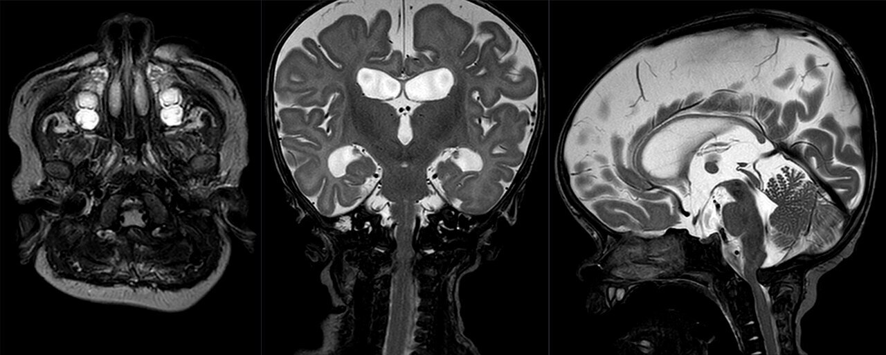
Foramen-magnum stenosis in achondroplasia. Infant craniocervical MRI (axial, coronal and sagittal T2): the sagittal view shows the narrowed foramen magnum and cervicomedullary junction, with ventriculomegaly. Hellerhoff, Wikimedia Commons (CC BY-SA 4.0).
Foramen-magnum stenosis in achondroplasia. Infant craniocervical MRI (axial, coronal and sagittal T2): the sagittal view shows the narrowed foramen magnum and cervicomedullary junction, with ventriculomegaly. Hellerhoff, Wikimedia Commons (CC BY-SA 4.0).
Thoracolumbar kyphosis and spinal stenosis
In achondroplasia, a thoracolumbar kyphosis (gibbus) appears in infancy from trunk hypotonia and a large head but usually resolves once the child begins to walk; the Pauli protocol of counselling (no unsupported sitting, firm-backed seating) and selective bracing is used, and surgery (anterior arthrodesis ± posterior instrumented fusion) is reserved for a kyphosis of 50° or more persisting at age 5, observing two rules: no hardware in the canal and no overcorrection of the tethered cord.[17] The other great achondroplasia problem is symptomatic spinal canal stenosis in the second to third decade, the consequence of short pedicles and the caudally narrowing interpedicular distance. Neurogenic claudication relieved by squatting is the clue, and wide multilevel decompression (with fusion in the skeletally immature) is needed in roughly 30% of patients.[18]
Scoliosis, lower-limb realignment and the hip
Scoliosis is frequent and often sharply angular in SED, diastrophic, metatropic and Kniest dysplasias. Bracing is largely ineffective, and a progressive curve is treated by instrumented fusion (growing constructs in the very young), with a high pseudarthrosis rate in some entities favouring combined anterior-posterior surgery.[19] Lower-limb malalignment takes several forms: genu varum in achondroplasia and the metaphyseal dysplasias, genu valgum in chondroectodermal dysplasia, MED and the mucopolysaccharidoses, and “windswept” deformity in pseudoachondroplasia. It is corrected by guided growth (hemiepiphysiodesis) in the growing child and osteotomy near maturity, with arthrography to localise the deformity. Coxa vara (a neck-shaft angle under about 100°, classically in SED and cleidocranial dysplasia) is corrected by valgus osteotomy, and premature hip arthritis is common in SED, pseudoachondroplasia, MED and diastrophic dysplasia, eventually requiring custom or constrained total joint arthroplasty.[20]
Short stature and limb lengthening
Limb lengthening is an option mainly for entities without a high degenerative-arthritis risk (achondroplasia, hypochondroplasia) rather than those with epiphyseal disease. It can add substantial height but is lengthy and carries a complication rate around 40%, and humeral lengthening (for reach and self-care) is the most appreciated and least risky.[21]
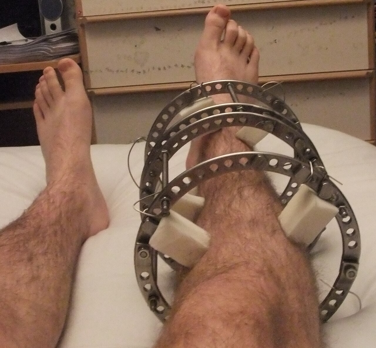
Limb lengthening. An Ilizarov ring external fixator on the leg - the apparatus used for gradual lengthening and deformity correction in short stature. Pagemaker787, Wikimedia Commons (CC BY-SA 4.0).
Anaesthetic and medical considerations
Anaesthesia is hazardous because of cervical instability or stenosis, a difficult airway (midface hypoplasia, limited neck motion, laryngotracheomalacia) and restrictive lung disease from a small thorax, so cervical stability must be confirmed before any general anaesthetic.[23] Care is multidisciplinary (genetics, ENT for the near-universal otitis media of achondroplasia, ophthalmology for the retinal detachment of Kniest/SED, cardiology, dentistry, dietetics and patient-support groups), and caesarean delivery is often required in affected mothers because of a narrow pelvis.
Part V - Achondroplasia and the FGFR3 group
Achondroplasia
Achondroplasia is the commonest skeletal dysplasia (~1 in 25,000) and the prototype rhizomelic dwarfism. It is caused by a gain-of-function mutation of FGFR3 (almost always the G380R substitution in the transmembrane domain), is autosomal dominant, and arises as a new mutation in roughly 80% of cases, with a paternal-age effect; the constitutively active receptor suppresses endochondral ossification while sparing membranous and articular cartilage.[24] The clinical picture is rhizomelic disproportionate short stature (adult height about 131 cm in men, 124 cm in women) with a large head, frontal bossing, midface hypoplasia, a trident hand, thoracolumbar gibbus in infancy, exaggerated lumbar lordosis and genu varum; intelligence is normal. The radiographic hallmarks are caudal narrowing of the lumbar interpedicular distance, short pedicles, square iliac wings with a small sciatic notch (“champagne-glass” pelvis), an inverted-V (chevron) distal femoral physis and short, flared-metaphysis tubular bones. Because the epiphyses are normal, degenerative arthritis is rare.[25]
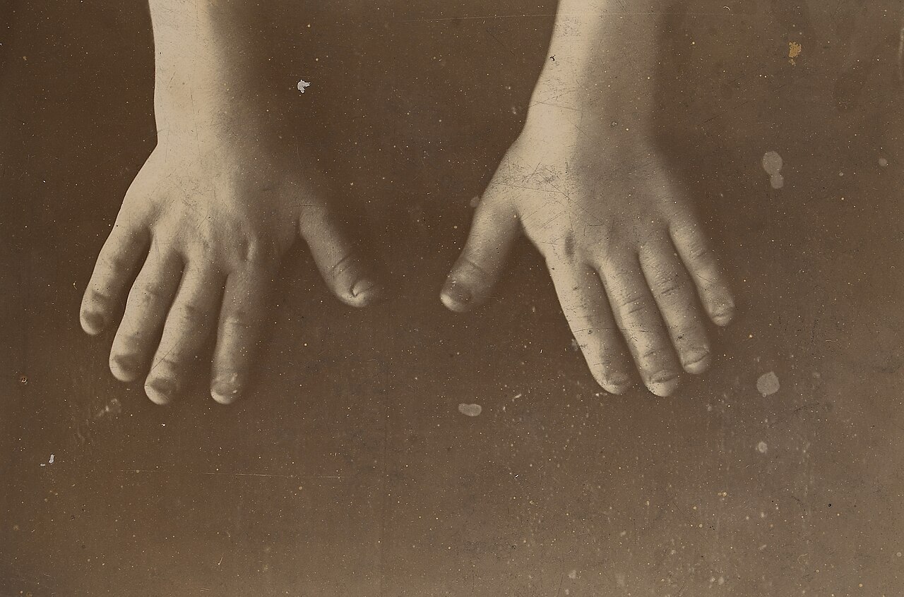
Trident hand in achondroplasia. The short, stubby digits with a persistent gap between the middle and ring fingers. Wellcome Collection (CC BY 4.0).
Trident hand in achondroplasia. The short, stubby digits with a persistent gap between the middle and ring fingers. Wellcome Collection (CC BY 4.0).
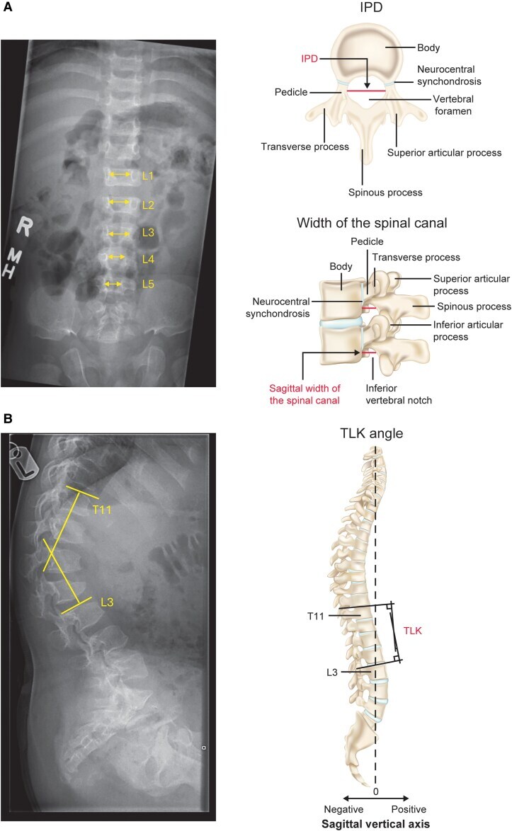
Radiographic signs of achondroplasia. (A) AP lumbar spine - the interpedicular distance fails to widen caudally (measured L1-L5); (B) lateral spine - thoracolumbar kyphosis (T11-L3). The right-hand panels are explanatory anatomy. Hoover-Fong J et al., J Endocr Soc 2026;10(3):bvag008, Fig. 1 (CC BY 4.0).
Radiographic signs of achondroplasia. (A) AP lumbar spine - the interpedicular distance fails to widen caudally (measured L1-L5); (B) lateral spine - thoracolumbar kyphosis (T11-L3). The right-hand panels are explanatory anatomy. Hoover-Fong J et al., J Endocr Soc 2026;10(3):bvag008, Fig. 1 (CC BY 4.0).
The management is the cross-cutting approach above applied to one disorder: foramen-magnum decompression for the symptomatic infant, thoracolumbar kyphosis control, lumbar decompression for adult stenosis, osteotomy/guided growth for genu varum (fibular shortening is not indicated), the limb-lengthening debate, and vosoritide as a growth-promoting option.[26]
Hypochondroplasia and thanatophoric dysplasia
Hypochondroplasia is a milder, allelic FGFR3 disorder (commonly the N540K mutation), often diagnosed only at school age, with a relatively long trunk, short limbs, lumbar lordosis and bowing, and, unlike achondroplasia, little risk of foramen-magnum or spinal stenosis.[27] Thanatophoric dysplasia, also FGFR3, is the commonest lethal skeletal dysplasia: type 1 has curved (“telephone-receiver”) femora, type 2 has straight femora with a cloverleaf skull, and death is in early infancy from respiratory insufficiency.[28]
Part VI - Pseudoachondroplasia and multiple epiphyseal dysplasia (the COMP group)
Pseudoachondroplasia is an autosomal-dominant disorder of COMP (cartilage oligomeric matrix protein) in which the mutant protein cannot be secreted and accumulates in the chondrocyte’s rough endoplasmic reticulum. The diagnostic point is that the child is normal at birth and has a normal face and skull (distinguishing it from achondroplasia), then develops short-limbed dwarfism with a waddling gait from about 18 months; there is generalised ligamentous laxity, lower-limb malalignment (often “windswept”), early osteoarthritis of the hips and knees, and odontoid hypoplasia with atlantoaxial instability in a minority.[29] Treatment is realignment osteotomy (avoiding overcorrection), cervical fusion for instability, and early (often constrained) joint arthroplasty.
Multiple epiphyseal dysplasia (MED, Fairbank/Ribbing) is genetically heterogeneous (dominant COMP, COL9 and MATN3; recessive SLC26A2/DTDST). It presents after age 2 with joint pain, a waddling gait and mild short stature with a normal face and a normal (or near-normal) spine, and its hallmark is small, fragmented, irregular epiphyses with early osteoarthritis. The classic differential is bilateral Legg-Calvé-Perthes disease: MED is symmetrical and synchronous with primary acetabular changes and abnormal knees and ankles, whereas Perthes is usually asymmetrical with a normal acetabulum. The double-layered patella points to recessive MED.[30] Management is guided growth or osteotomy for malalignment and early joint replacement.
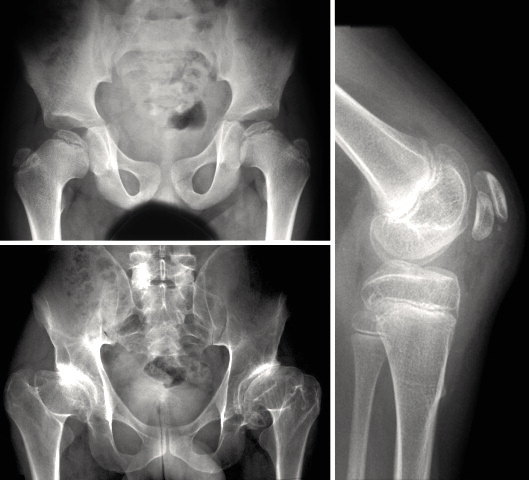
Multiple epiphyseal dysplasia. Small, irregular, fragmented epiphyses at the hips (AP pelvis) and knee. Bellllacullllen, Wikimedia Commons (CC BY-SA 3.0).
Multiple epiphyseal dysplasia. Small, irregular, fragmented epiphyses at the hips (AP pelvis) and knee. Bellllacullllen, Wikimedia Commons (CC BY-SA 3.0).
Part VII - Spondyloepiphyseal dysplasias and the type II collagen group
Spondyloepiphyseal dysplasia congenita (SEDC) is an autosomal-dominant disorder of type II collagen (COL2A1) producing short-trunk dwarfism with a short neck, a barrel chest with pectus carinatum, normal-sized hands and feet, coxa vara, genu valgum and progressive myopia with retinal detachment.[31] The orthopaedically critical feature is odontoid hypoplasia with atlantoaxial instability and cord compression, which must be screened for routinely and fused when unstable or symptomatic. The spine also shows platyspondyly with posterior wedging and a sharply angular scoliosis, and the coxa vara is corrected by valgus osteotomy when the neck-shaft angle falls below 100°.[32]
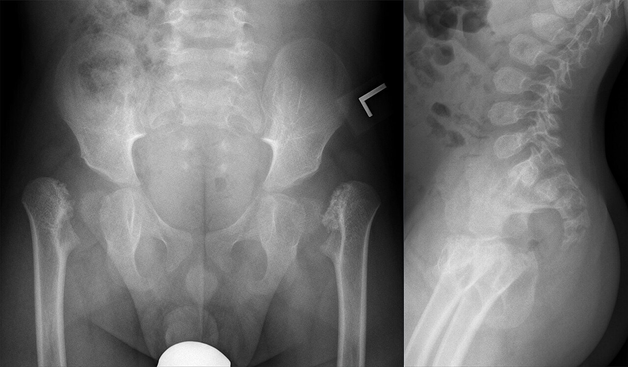
Spondyloepiphyseal dysplasia congenita. Infant AP pelvis (delayed femoral-head ossification, dysplastic epiphyses) and lateral spine (ovoid, flattened vertebrae - platyspondyly). Hellerhoff, Wikimedia Commons (CC BY-SA 4.0).
Spondyloepiphyseal dysplasia congenita. Infant AP pelvis (delayed femoral-head ossification, dysplastic epiphyses) and lateral spine (ovoid, flattened vertebrae - platyspondyly). Hellerhoff, Wikimedia Commons (CC BY-SA 4.0).
SED tarda is a later-onset, milder form (most commonly X-linked, from the SEDL/sedlin gene), often first mistaken for bilateral Perthes, with an arm span exceeding height; it too can have odontoid problems.[33]
Several related type II/XI collagen disorders are worth recognising. Kniest dysplasia (COL2A1) gives stiff knobby joints, cleft palate, myopia, “dumbbell” femora and atlantoaxial instability. Stickler syndrome (COL2A1/COL11) is the arthro-ophthalmopathy of myopia with retinal detachment, cleft palate/Pierre Robin and deafness, but no dwarfism. Metatropic dysplasia (TRPV4) is the “changing form” with a coccygeal tail and cervical instability.[34]
Part VIII - Diastrophic dysplasia
Diastrophic dysplasia is an autosomal-recessive disorder of the sulfate transporter SLC26A2 (DTDST), common in Finland through a founder effect, that produces undersulfated cartilage proteoglycan. Its clinical signature is highly recognisable: short-limbed dwarfism with multiple joint contractures, a “hitchhiker thumb” (a proximally placed, abducted thumb), rigid cast-resistant equinovarus clubfeet, and a “cauliflower ear” (an acute cystic swelling of the pinna in the first weeks of life that hardens into deformity), often with cleft palate.[35] The spine shows a cervical kyphosis present at birth (which may resolve but occasionally progresses to myelopathy) and a progressive scoliosis; atlantoaxial instability, by contrast, is almost never seen. Management is demanding: observation or fusion of the cervical kyphosis, bracing and fusion of scoliosis, soft-tissue release of the recurrent clubfeet (with talectomy as salvage), and joint-preserving surgery then arthroplasty for the contracted, prematurely arthritic hips and knees.[36]
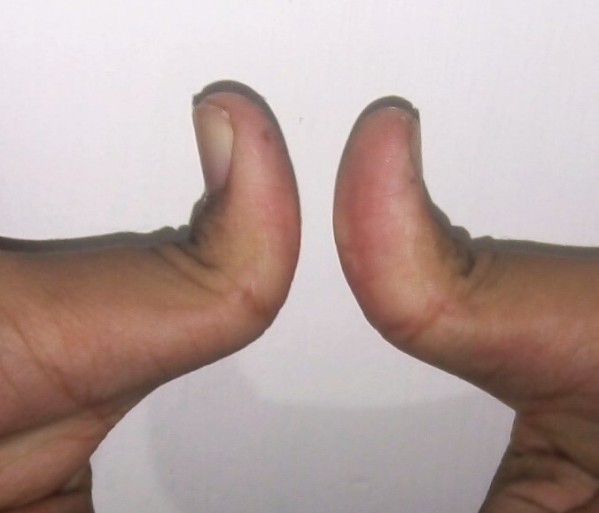
The “hitchhiker thumb” sign. Marked proximal placement and abduction of the thumbs - the characteristic hand sign of diastrophic dysplasia (illustrated here as the posture/sign). Rollcloud, Wikimedia Commons (CC0).
The “hitchhiker thumb” sign. Marked proximal placement and abduction of the thumbs - the characteristic hand sign of diastrophic dysplasia (illustrated here as the posture/sign). Rollcloud, Wikimedia Commons (CC0).
Part IX - Metaphyseal chondrodysplasias
The metaphyseal chondrodysplasias affect the metaphyses while sparing the epiphyses, and their main differential is rickets, from which they are distinguished by normal calcium, phosphate and alkaline phosphatase, so vitamin D is contraindicated.[37] The three named types are:
- Schmid type (COL10A1, autosomal dominant): the commonest and mildest, presenting in the second year with genu varum, a waddling gait and coxa vara that mimic rickets; osteotomy is deferred until after physeal closure.[38]
- Jansen type (an activating mutation of the PTH/PTHrP receptor, PTHR1): severe, with grossly expanded fragmented metaphyses and asymptomatic hypercalcaemia.[39]
- Cartilage-hair hypoplasia / McKusick type (RMRP, autosomal recessive; first described in the Amish): short-limbed dwarfism with fine sparse hair, a variable immunodeficiency, anaemia and an increased risk of lymphoma and skin cancer; live vaccines are avoided, and bone-marrow transplantation corrects the immune defect but not the bone disease.[40]
Part X - The mucopolysaccharidoses
The mucopolysaccharidoses (MPS) are lysosomal storage disorders in which incompletely degraded glycosaminoglycans accumulate, producing a stereotyped skeletal picture, dysostosis multiplex: a macrocephalic skull with a J-shaped sella, oar-shaped (paddle) ribs, anterior-inferior vertebral beaking with a thoracolumbar gibbus, flared iliac wings with coxa valga, and proximally pointed metacarpals.[41] The pattern is the same across types, while the enzyme, inheritance and systemic features differ:
| Type | Eponym | Enzyme / gene | Notes |
|---|---|---|---|
| MPS I | Hurler / Scheie | α-L-iduronidase (IDUA) | corneal clouding; Hurler severe with intellectual disability, Scheie mild with carpal tunnel syndrome |
| MPS II | Hunter | iduronate-2-sulfatase (IDS) | X-linked; clear corneae |
| MPS III | Sanfilippo | four enzymes (heparan-sulfate degradation) | severe behavioural/cognitive decline, mild somatic disease |
| MPS IV | Morquio | GALNS (IVA) / GLB1 (IVB) | keratan sulfate in urine; normal intelligence; the orthopaedically critical MPS |
| MPS VI | Maroteaux-Lamy | arylsulfatase B (ARSB) | normal intelligence; short stature, stiff joints |
| MPS VII | Sly | β-glucuronidase (GUSB) | wide phenotypic spectrum |
[42] Intelligence is preserved in Morquio and Maroteaux-Lamy and impaired in the severe Hurler, Hunter and Sanfilippo forms.
The orthopaedic problems are those of dysostosis multiplex: atlantoaxial instability from odontoid hypoplasia (most prominent in Morquio) requiring cervical fusion; thoracolumbar gibbus; genu valgum; hip dysplasia with coxa valga; and the carpal tunnel syndrome and trigger digits classic in Hurler and Hunter.[43] Morquio stands out as a short-trunk dwarfism with platyspondyly, odontoid hypoplasia, coxa valga (in contrast to the coxa vara of SED) and genu valgum, where the first symptom of cord compression is reduced endurance and anaesthesia is hazardous. Disease-modifying treatment, namely enzyme replacement therapy (elosulfase for Morquio is documented in the sources; laronidase, idursulfase and galsulfase for the other types are standard practice) and haematopoietic stem-cell transplantation for Hurler, improves the systemic disease but does not reverse the established skeletal deformity, which still requires orthopaedic correction.[44]
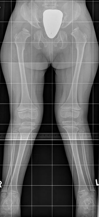
Genu valgum in Morquio disease (MPS IV). Standing lower-limb radiograph showing knock-knees with metaphyseal and epiphyseal dysplasia. Kinderradiologie Olgahospital, Klinikum Stuttgart, Wikimedia Commons (CC BY-SA 4.0).
Genu valgum in Morquio disease (MPS IV). Standing lower-limb radiograph showing knock-knees with metaphyseal and epiphyseal dysplasia. Kinderradiologie Olgahospital, Klinikum Stuttgart, Wikimedia Commons (CC BY-SA 4.0).
Part XI - Osteogenesis imperfecta
Osteogenesis imperfecta (OI) is the prototype disorder of bone fragility. About 85-90% of cases are autosomal-dominant defects of type I collagen (COL1A1 or COL1A2), with the remaining ~10% caused by a growing list of mostly recessive collagen-processing genes (CRTAP, P3H1/LEPRE1, PPIB, SERPINF1, FKBP10, and others) that now number the disorder up to type 17 or beyond.[45] The clinical core, present in every patient, is bone fragility with recurrent fractures and generalised osteoporosis, accompanied in varying degree by blue sclerae, dentinogenesis imperfecta, conductive or mixed hearing loss, ligamentous laxity, wormian skull bones, short stature, basilar invagination, scoliosis and “popcorn” epiphyses.[46]
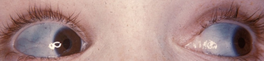
Blue sclerae in osteogenesis imperfecta. The thin sclera transmits the underlying choroid, giving the characteristic blue hue (eye-only crop). Fred & van Dijk, Images of Memorable Cases (CC BY-SA 3.0, cropped).
Blue sclerae in osteogenesis imperfecta. The thin sclera transmits the underlying choroid, giving the characteristic blue hue (eye-only crop). Fred & van Dijk, Images of Memorable Cases (CC BY-SA 3.0, cropped).
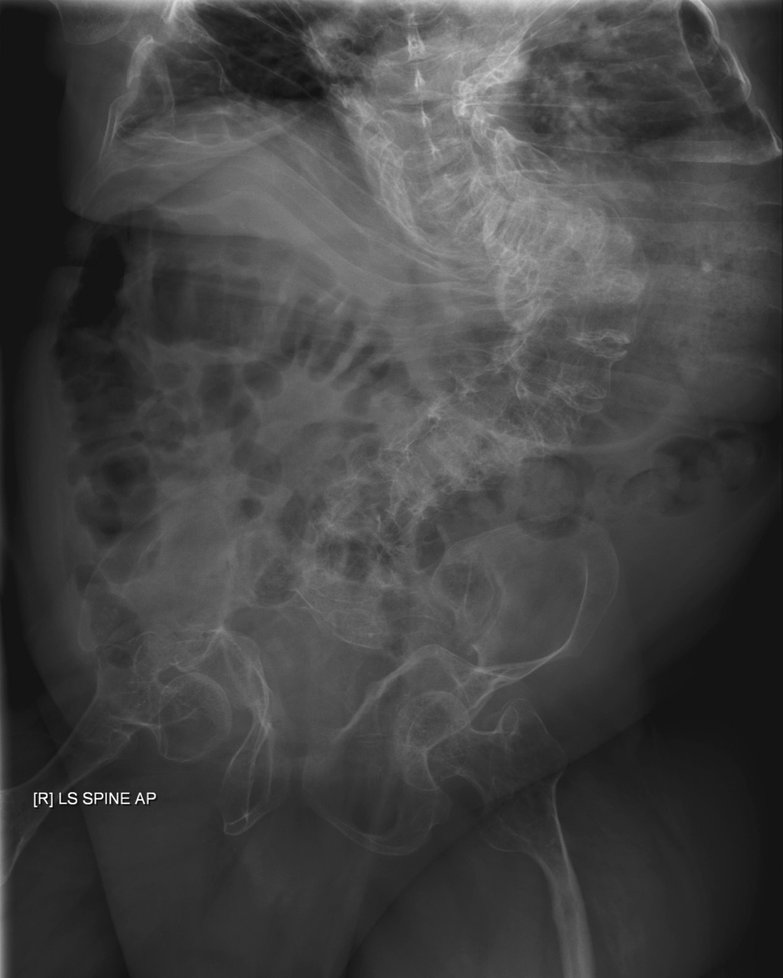
Bone fragility in osteogenesis imperfecta. Adult radiograph showing gracile (thin) bones with severe scoliosis and thoracic deformity. Fredrick Brennan, Wikimedia Commons (public domain).
Bone fragility in osteogenesis imperfecta. Adult radiograph showing gracile (thin) bones with severe scoliosis and thoracic deformity. Fredrick Brennan, Wikimedia Commons (public domain).
The Sillence classification remains the clinical framework:[47]
- Type I: mildest, non-deforming, autosomal dominant, blue sclerae, often normal height.
- Type II: perinatally lethal (crumpled bones, beaded ribs); about 90% die within weeks.
- Type III: the severe, progressively deforming survivable form, with marked limb deformity, kyphoscoliosis, short stature and popcorn epiphyses.
- Type IV: moderate, with white or grey sclerae and frequent dentinogenesis imperfecta.
- Type V: autosomal dominant (IFITM5), distinguished by hyperplastic fracture callus, calcification of the interosseous membranes (limiting forearm rotation, with radial-head dislocation) and a mesh-like bone histology.
A perennial examination point is the differential with non-accidental injury: blue sclerae, wormian bones, a positive family history, and fractures after the child is mobile favour OI, whereas metaphyseal corner fractures, retinal haemorrhage, skin marks and fractures before independent mobility favour abuse. The two can also coexist.[48]
Management combines medical and surgical measures. Cyclical bisphosphonates (pamidronate, zoledronate) reduce bone pain and increase bone density and vertebral size (with debated effects on fracture rate). Deformity is corrected by realignment with intramedullary fixation: the Sofield-Millar “shish-kebab” multiple osteotomy, and telescoping/elongating rods (Bailey-Dubow, Fassier-Duval) that grow with the child and substantially outperform static implants. Scoliosis bracing is ineffective, so a significant curve (a thoracic curve over 60° impairs pulmonary function) is treated by instrumented fusion; basilar invagination and the anaesthetic risks (difficult positioning, possible malignant hyperthermia, platelet-related bleeding) must be anticipated.[49]
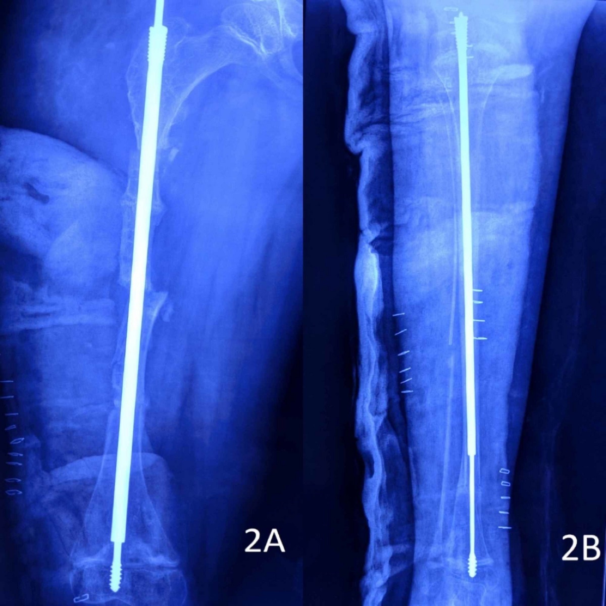
Intramedullary rodding in osteogenesis imperfecta. Post-operative radiographs of a long bone with a telescoping (Fassier-Duval-type) intramedullary rod after realignment. Behera et al., Cureus 2020, via Wikimedia Commons (CC BY 4.0).
Intramedullary rodding in osteogenesis imperfecta. Post-operative radiographs of a long bone with a telescoping (Fassier-Duval-type) intramedullary rod after realignment. Behera et al., Cureus 2020, via Wikimedia Commons (CC BY 4.0).
Part XII - Osteopetrosis and the dense-bone group
Osteopetrosis is a group of disorders of defective bone resorption from absent or dysfunctional osteoclasts, producing dense but brittle bone with defective remodelling (Erlenmeyer-flask metaphyses) and the radiographic signs of “bone-in-bone” vertebrae, sandwich/rugger-jersey vertebrae and a dense skull base.[50]
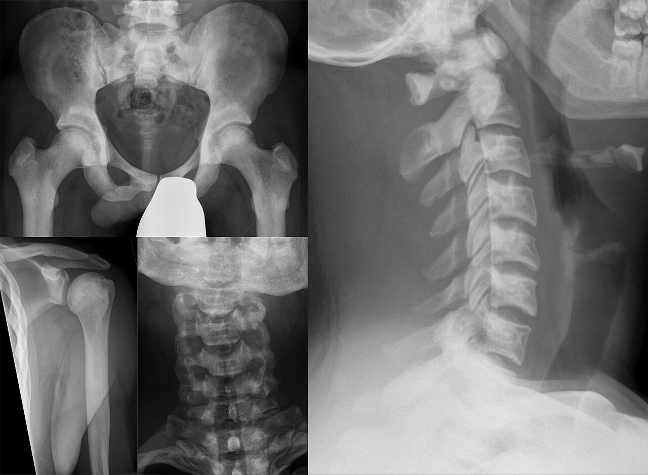
Osteopetrosis. Diffusely dense (“marble”) bones of the pelvis, shoulder and cervical spine, with the “bone-in-bone” vertebral appearance. Hellerhoff, Wikimedia Commons (CC BY-SA 4.0).
Osteopetrosis. Diffusely dense (“marble”) bones of the pelvis, shoulder and cervical spine, with the “bone-in-bone” vertebral appearance. Hellerhoff, Wikimedia Commons (CC BY-SA 4.0).
Three forms are distinguished:
- Malignant infantile (autosomal recessive), genes TCIRG1, CLCN7, OSTM1, with marrow failure (pancytopenia), hepatosplenomegaly from extramedullary haematopoiesis, optic atrophy and other cranial-nerve compressions, and death in the first decade unless treated; haematopoietic stem-cell transplantation is the definitive treatment.[51]
- Carbonic anhydrase II (CA2) deficiency (autosomal recessive): the triad of osteopetrosis, renal tubular acidosis and cerebral calcification, treated with alkali, potassium and (for the bone disease) marrow transplantation; notably there is no marrow failure.[52]
- Benign adult autosomal dominant (Albers-Schönberg, “marble bone disease,” type II; CLCN7), with fractures, mandibular osteomyelitis, cranial-nerve palsies and rugger-jersey vertebrae; the orthopaedic challenges are delayed union, nonunion and difficult surgery in dense bone.[53]
Among the related sclerosing dysplasias, pycnodysostosis (cathepsin K, CTSK; autosomal recessive) combines dense bone with acro-osteolysis of the distal phalanges, an obtuse mandibular angle and persistent open fontanelles (the dysplasia attributed to Toulouse-Lautrec), while Camurati-Engelmann disease, craniometaphyseal dysplasia, osteopoikilosis and melorheostosis complete the group.[54]
Part XIII - Cleidocranial dysplasia
Cleidocranial dysplasia is an autosomal-dominant disorder of the osteoblast transcription factor RUNX2 (CBFA1), in which a well-formed cartilage model fails to ossify normally.[55] The diagnostic features are absent or hypoplastic clavicles (allowing the shoulders to be drawn together in front of the chest, with complete absence in only about 10%), a large brachycephalic skull with delayed closure of the fontanelles and sutures and wormian bones, dental anomalies with supernumerary and retained teeth, mild short stature, delayed ossification of the pubis, coxa vara and an increased incidence of scoliosis (often a double thoracic curve).[56] The clavicles need no treatment; coxa vara is corrected by valgus osteotomy when the neck-shaft angle is below 100° with a Trendelenburg gait, a progressive scoliosis prompts MRI (for the associated syringomyelia), and caesarean delivery is often required because of cephalopelvic disproportion.[57]
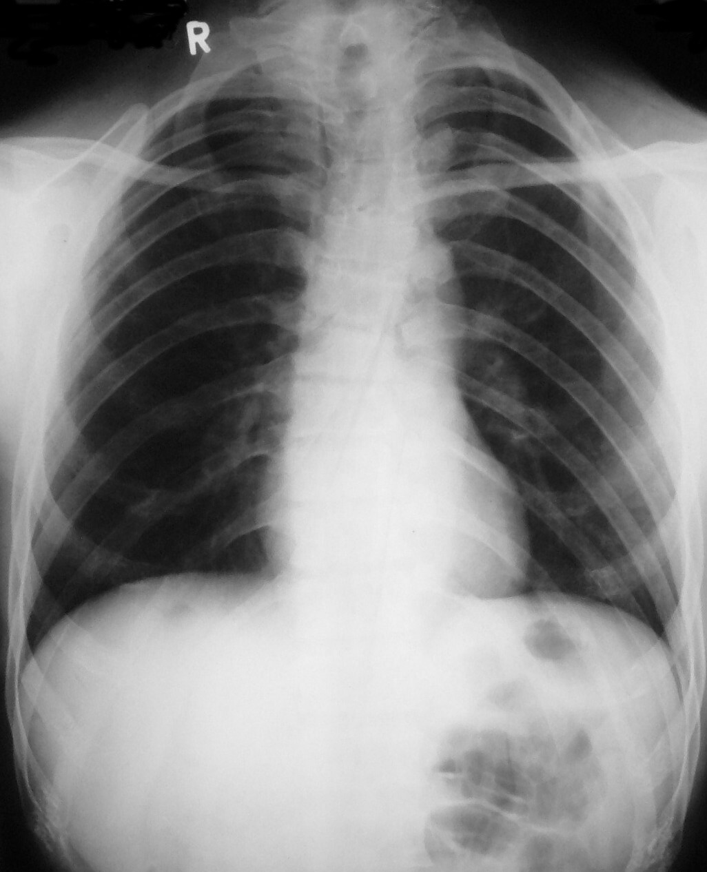
Cleidocranial dysplasia. Chest radiograph showing absent/hypoplastic clavicles and a bell-shaped thorax. Garg & Agrawal, Cases Journal 2008;1:377, via Wikimedia Commons (CC BY 2.0).
Cleidocranial dysplasia. Chest radiograph showing absent/hypoplastic clavicles and a bell-shaped thorax. Garg & Agrawal, Cases Journal 2008;1:377, via Wikimedia Commons (CC BY 2.0).
Bulgarian terminology (Боев / Boychev tradition) - glossary
The following Bulgarian equivalents bridge the international literature with the terminology of the Bulgarian school of orthopaedics (in the tradition of Бойчо Бойчев / Boycho Boychev) used in the state examination.
| English term | Bulgarian term (Cyrillic) | Transliteration |
|---|---|---|
| Skeletal dysplasia | Скелетна дисплазия | Skeletna displaziya |
| Osteochondrodysplasia | Остеохондродисплазия | Osteohondrodisplaziya |
| Dysostosis | Дизостоза | Dizostoza |
| Disproportionate short stature / dwarfism | Диспропорционален нисък ръст (нанизъм) | Disproportsionalen nisak rast (nanizam) |
| Rhizomelic | Ризомелично скъсяване | Rizomelichno skasyavane |
| Achondroplasia | Ахондроплазия | Ahondroplaziya |
| Hypochondroplasia | Хипохондроплазия | Hipohondroplaziya |
| Pseudoachondroplasia | Псевдоахондроплазия | Psevdoahondroplaziya |
| Multiple epiphyseal dysplasia | Множествена епифизарна дисплазия | Mnozhestvena epifizarna displaziya |
| Spondyloepiphyseal dysplasia | Спондилоепифизарна дисплазия | Spondiloepifizarna displaziya |
| Diastrophic dysplasia | Диастрофична дисплазия | Diastrofichna displaziya |
| Metaphyseal chondrodysplasia | Метафизарна хондродисплазия | Metafizarna hondrodisplaziya |
| Mucopolysaccharidosis | Мукополизахаридоза | Mukopolizaharidoza |
| Osteogenesis imperfecta | Незавършена костна остеогенеза (остеогенезис имперфекта) | Nezavarshena kostna osteogeneza |
| Osteopetrosis (marble bone disease) | Остеопетроза (мраморна болест) | Osteopetroza (mramorna bolest) |
| Cleidocranial dysplasia | Клейдокраниална дисплазия | Kleidokranialna displaziya |
| Foramen magnum | Голям тилен отвор (форамен магнум) | Golyam tilen otvor (foramen magnum) |
| Odontoid hypoplasia | Хипоплазия на одонтоида (зъбовидния израстък) | Hipoplaziya na odontoida |
| Atlantoaxial instability | Атлантоаксиална нестабилност | Atlantoaksialna nestabilnost |
| Spinal canal stenosis | Стеноза на гръбначния канал | Stenoza na grabnachniya kanal |
| Kyphosis / gibbus | Кифоза (гибус) | Kifoza (gibus) |
| Coxa vara | Кокса вара | Koksa vara |
| Genu varum | Варусно коляно (геню варум) | Varusno kolyano (genu varum) |
| Intramedullary rod | Интрамедуларен пирон | Intramedularen piron |
| Bisphosphonate | Бифосфонат | Bifosfonat |
| Blue sclerae | Сини склери | Sini skleri |
| Limb lengthening | Удължаване на крайник | Udalzhavane na kraynik |
Image attributions
(Figure attributions and licences are listed in the figure MANIFEST and inserted with each image. All images are openly licensed [CC0 / CC BY / CC BY-SA] or used under their stated terms; any non-commercial [NC] item is flagged as such and must not be used in a commercial product.)
References
-
Lovell & Winter, Pediatric Orthopaedics, ch. 7 The Skeletal Dysplasias (Sponseller & Ain), p. 196. The aggregate ~1/5,000 figure is standard background; the extracts give mainly entity-specific incidences.
-
Lovell, p. 196; ISDS Nosology and Classification of Genetic Skeletal Disorders, 2019 revision, p. 25.
-
Nosology 2019, p. 25.
-
Lovell, pp. 196, 198.
-
Lovell, p. 199.
-
Lovell, p. 197; Nosology 2019, pp. 6-7.
-
Nosology 2019, pp. 0-1, 24.
-
Nosology 2019, pp. 1, 24.
-
Nosology 2019, pp. 2-16; Lovell, pp. 196-197.
-
Lovell, pp. 196-197.
-
Lovell, pp. 197-199.
-
Lovell, p. 199.
-
Lovell, p. 199.
-
Lovell, pp. 197, 229-230.
-
Lovell, pp. 198, 214-219.
-
Lovell, pp. 200-202; Spranger atlas, achondroplasia section, pp. 31-32.
-
Lovell, pp. 200, 203-204.
-
Lovell, pp. 204-205; Spranger, p. 32.
-
Lovell, pp. 207-216.
-
Lovell, pp. 202-203, 208-222.
-
Lovell, pp. 198, 202-203; Campbell’s, pp. 1463-1467.
-
Spranger, p. 32 (notes “FGFR3 signal-transduction strategies in clinical testing”); vosoritide is the realisation of that strategy and is standard current teaching, not named verbatim in the extracts.
-
Lovell, p. 198.
-
Spranger, p. 31; Lovell, pp. 199-200.
-
Spranger, p. 31; Lovell, pp. 201-202.
-
Lovell, pp. 200-205; Spranger, p. 32.
-
Spranger, p. 38; Lovell, p. 205.
-
Spranger, p. 27; Lovell, p. 229.
-
Spranger, pp. 49-50; Lovell, pp. 218-219.
-
Spranger, pp. 53-55; Lovell, pp. 219-220.
-
Spranger, pp. 98-99; Lovell, p. 214.
-
Lovell, pp. 214-215.
-
Lovell, pp. 216-217.
-
Spranger, pp. 111, 123-124; Lovell, pp. 205-207, 213-214.
-
Spranger, p. 276; Lovell, pp. 209-211.
-
Lovell, pp. 211-212.
-
Spranger, p. 59; Lovell, pp. 222-223.
-
Spranger, p. 59; Lovell, p. 223.
-
Spranger, p. 80.
-
Spranger, pp. 63-64; Lovell, p. 222.
-
Spranger atlas, MPS section, pp. 134, 140-142.
-
Spranger, pp. 135-136, 143.
-
Spranger, pp. 143-144, 136, 155.
-
Spranger, pp. 143, 123, 150; the specific non-Morquio ERT agent names and the “skeleton not reversed” point are standard teaching beyond these extracts.
-
Spranger, OI section, pp. 523-525.
-
Campbell’s, p. 1459; Spranger, pp. 527, 543.
-
Spranger, p. 524; Campbell’s, p. 1459.
-
Spranger, pp. 527-528.
-
Campbell’s, pp. 1459-1462; Spranger, p. 538.
-
Spranger, OI/dense-bone section, pp. 601-602.
-
Spranger, pp. 606-607.
-
Spranger, pp. 617-618.
-
Spranger, pp. 613-614.
-
Spranger, pp. 627-628, 637-639.
-
Lovell, pp. 197, 226.
-
Lovell, pp. 226-227.
-
Lovell, pp. 226-227, 231.