Contents
- Introduction and scope
- Part I - Hallux valgus: definition and pathoanatomy
- Part II - Hallux valgus: aetiology and assessment
- Part III - Hallux valgus: the radiographic angles and classification
- Part IV - Hallux valgus: conservative care and the surgical menu
- Part V - Hallux valgus surgery: complications
- Part VI - Hallux rigidus
- Part VII - Lesser-toe deformities
- Part VIII - Metatarsalgia and the plantar keratoses
- Part IX - Morton’s (interdigital) neuroma
- Part X - Freiberg infraction, the bunionette and sesamoid disorders
- Bulgarian terminology (Боев / Boychev tradition) - glossary
- Image attributions
- References
Introduction and scope
The static deformities of the forefoot are among the commonest reasons an adult comes to the orthopaedic clinic, the typical patient being a woman in well-fitting shoes she can no longer tolerate. They comprise hallux valgus, hallux rigidus, the lesser-toe deformities, and the metatarsalgia group (plantar keratoses, Morton’s neuroma, Freiberg infraction, the bunionette and sesamoid disorders). The examiner expects a confident command of hallux valgus (its pathoanatomy, the radiographic angles that drive treatment, and the menu of osteotomies and fusions matched to the deformity), of hallux rigidus (the Coughlin-Shurnas grade and the cheilectomy-versus-fusion decision), of the lesser-toe deformities (which joint each involves, flexible versus fixed, the plantar plate and the crossover toe), and of the metatarsalgia group (the discrete-versus-diffuse plantar keratosis, the interdigital neuroma, and Freiberg’s disease).
Two ideas run through the topic. First, match the operation to the deformity and the patient, not to the X-ray: in hallux valgus the choice of osteotomy is dictated by the intermetatarsal angle, the distal articular angle and joint congruity, and surgery is offered for pain, not appearance. Second, the forefoot is a balanced load-sharing system. Shorten or elevate one metatarsal and the load, with the pain, simply transfers to the next, so “transfer metatarsalgia” is the shadow that follows almost every forefoot operation.
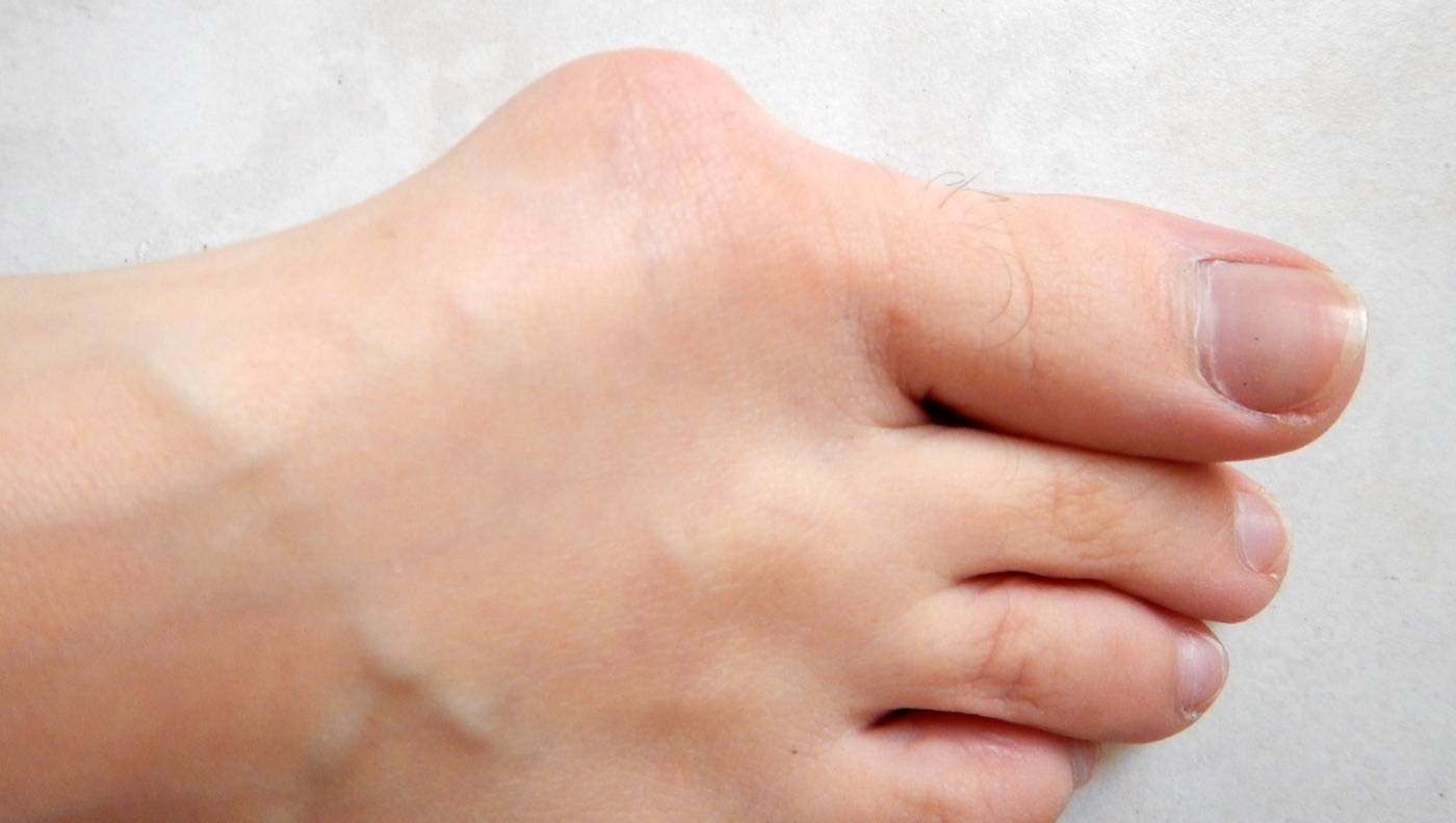
Hallux valgus (bunion). Clinical photograph of a left foot: a prominent medial eminence at the first metatarsophalangeal joint with lateral deviation of the great toe overriding the second. Lamiot, Wikimedia Commons, CC BY-SA 4.0.
Hallux valgus (bunion). Clinical photograph of a left foot: a prominent medial eminence at the first metatarsophalangeal joint with lateral deviation of the great toe overriding the second. Lamiot, Wikimedia Commons, CC BY-SA 4.0.
Part I - Hallux valgus: definition and pathoanatomy
Hallux valgus is a static subluxation of the first metatarsophalangeal (MTP) joint, with lateral deviation of the great toe and medial deviation of the first metatarsal (metatarsus primus varus).[1] The word bunion (from the Latin bunio, a turnip) is non-specific and has been applied to several conditions, so it is best avoided as a diagnosis.[2]
The deformity is driven by a self-reinforcing imbalance of forces. No muscle inserts on the metatarsal head, so its position is governed by the proximal phalanx. Once the phalanx deviates laterally, the tendons that crossed the joint come to lie lateral to its axis and become deforming forces.[3] The lateral structures (adductor hallucis, lateral capsule) contract while the medial capsule attenuates; the metatarsal head is pushed medially off the sesamoid sling (which is anchored to the phalanx, not the head, so it stays put), the crista erodes, the extensor hallucis longus bowstrings laterally, and the hallux pronates.[4] The medial eminence is not a bony overgrowth but simply the metatarsal head exposed by lateral migration of the phalanx (mean width ~4.4-4.6 mm).[5] A rounded metatarsal head is intrinsically unstable and is found in 71-91% of hallux valgus feet, whereas a flat or chevron-shaped head resists progression.[6]
Part II - Hallux valgus: aetiology and assessment
The cause is multifactorial, with extrinsic and intrinsic contributions:
- Footwear is the principal extrinsic factor; hallux valgus is almost confined to shod populations (33% of shod versus 2% of unshod feet in the classic Sim-Fook & Hodgson study).[7]
- Heredity is strong: a family history is present in 58-88% of patients (84% in Coughlin’s series), most consistent with autosomal-dominant inheritance with incomplete penetrance.[8]
- Female sex predominates overwhelmingly in surgical series (over 90%).[9]
- Pes planus / hyperpronation, metatarsus primus varus and metatarsus adductus are associated, the last two especially in the juvenile form.[10]
- First-ray (first tarsometatarsal) hypermobility is debated. By the Klaue measurement, normal sagittal mobility is ~5 mm and hallux valgus feet ≥9 mm, but Coughlin’s work suggests the hypermobility is largely an effect of the deformity (it normalises after a realignment that spares the tarsometatarsal joint) and is truly pathological in only ~10%.[11]
The deformity is bilateral in about 84% and usually becomes symptomatic from the third decade onward.[12] The chief complaint is pain over the medial eminence in 70-75%, often with transfer metatarsalgia under the second metatarsal (about half of moderate-severe cases).[13] Assessment records the first-MTP range (normal ~87° total), the first-ray mobility, the presence of pes planus and a gastrocnemius contracture, the neurovascular status, and any associated lesser-toe deformity or interdigital neuroma.[14]
Part III - Hallux valgus: the radiographic angles and classification
Weight-bearing radiographs define the deformity and dictate the operation. The key angles and their normal values are:
- Hallux valgus angle (HVA), first-metatarsal versus proximal-phalanx axis: normal < 15°.
- First-second intermetatarsal angle (IMA): normal < 9°.
- Distal metatarsal articular angle (DMAA / PASA), the lateral tilt of the articular surface on the metatarsal head: normal ≤ ~6°. A value over ~10-15° is associated with a congruent deformity and contraindicates a purely intra-articular soft-tissue realignment.
- Hallux valgus interphalangeus (HVI), the phalangeal contribution, abnormal when > 10°.[15]
By the Coughlin/Mann severity grading: mild (HVA < 20°, IMA ≤ 11°), moderate (HVA 20-40°, IMA 11-16°) and severe (HVA > 40°, IMA > 16°).[16] Joint congruity matters just as much. A congruent joint (articular surfaces parallel, no lateral phalangeal shift) is stable and does not progress, its valgus arising from a high DMAA; an incongruent (subluxated) joint is progressive (Piggott found only 9% congruent).[17] The sesamoid position is graded on the AP/axial view, although the apparent lateral displacement is largely an artefact of first-ray pronation.[18]
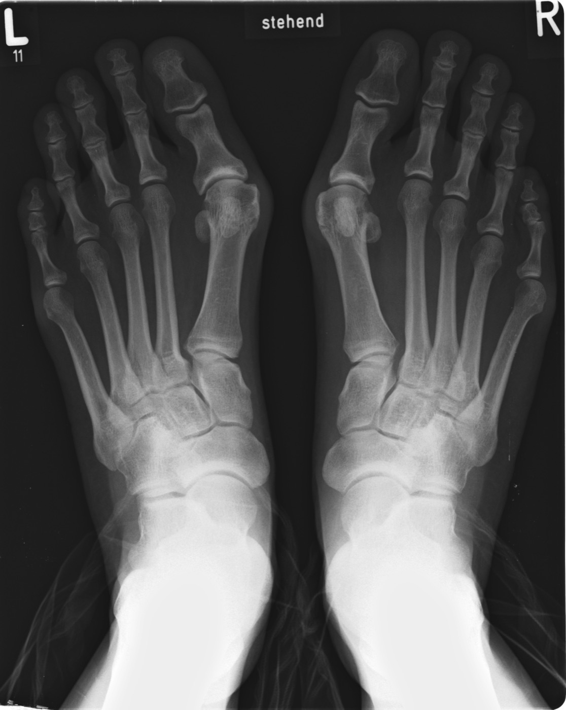
Bilateral hallux valgus. Standing AP radiograph of both forefeet: medial eminence, lateral hallux deviation, increased intermetatarsal angle and lateral sesamoid subluxation. Michael Nebel, Wikimedia Commons, CC BY-SA 2.0 DE.
Bilateral hallux valgus. Standing AP radiograph of both forefeet: medial eminence, lateral hallux deviation, increased intermetatarsal angle and lateral sesamoid subluxation. Michael Nebel, Wikimedia Commons, CC BY-SA 2.0 DE.
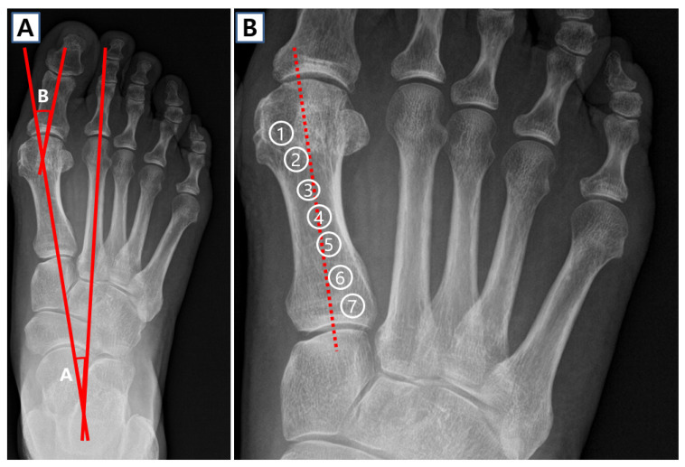
Radiographic parameters of hallux valgus. (A) Hallux valgus angle (HVA) and intermetatarsal angle (IMA) constructed on an AP radiograph; (B) the seven-grade tibial-sesamoid position classification. Cho et al., J Clin Med 2025;14:5072, Fig. 1 (CC BY 4.0).
Radiographic parameters of hallux valgus. (A) Hallux valgus angle (HVA) and intermetatarsal angle (IMA) constructed on an AP radiograph; (B) the seven-grade tibial-sesamoid position classification. Cho et al., J Clin Med 2025;14:5072, Fig. 1 (CC BY 4.0).
Part IV - Hallux valgus: conservative care and the surgical menu
The first principle is that surgery is for pain, not for cosmesis. Conservative care relieves symptoms (a wide, soft, low-heeled shoe with a roomy toe box, plus pads, spacers or night splints), though orthoses do not prevent progression.[19]
The operation is matched to the deformity by the IMA, the DMAA and joint congruity:
- Distal soft-tissue procedure (modified McBride): lateral release (adductor, lateral capsule, transverse metatarsal ligament) plus medial plication, for an incongruent joint with HVA < 30° and IMA < 11°. The fibular sesamoid is preserved (its excision causes hallux varus), and because the metatarsus primus varus is the limiting factor, an osteotomy is added in roughly 95% of cases.[20]
- Distal metatarsal osteotomy (chevron / Austin), for mild-to-moderate deformity (HVA < 30° or IMA < 13°), translating the head laterally about 5-6 mm. A congruent joint is acceptable if the DMAA ≤ 15° (otherwise a biplanar/medial closing-wedge variant corrects the DMAA). The feared complication is avascular necrosis of the head, which the sources agree follows excessive dorsolateral neck stripping.[21]
- Scarf (midshaft Z) osteotomy, for moderate-to-severe deformity with IMA 14-20°; technically demanding, with “troughing” and hallux varus among its pitfalls.[22]
- Proximal metatarsal osteotomy (crescentic, proximal chevron,
Ludloff, Mau) with distal soft-tissue release, for
moderate-to-severe incongruent deformity with **HVA > 30° and IMA
13°**.[23]
- Lapidus procedure (first tarsometatarsal arthrodesis), for a large IMA (≥ 16°), documented first-ray hypermobility, recurrent deformity or arthritis of that joint; nonunion is the main concern (most series 5-24%).[24]
- Akin osteotomy (medial closing wedge of the proximal phalanx), for hallux valgus interphalangeus, as an adjunct; it does not reduce the IMA and is inadequate as an isolated procedure.[25]
- First-MTP arthrodesis, for severe deformity (HVA often > 50°), rheumatoid disease, spasticity, advanced arthritis or salvage; fused in about 15° valgus and 15-20° of dorsiflexion relative to the shaft (roughly 10-15° to the floor), with the IMA correcting automatically.[26]
- Keller resection arthroplasty, for the elderly, low-demand patient with arthritis; it corrects only about half the deformity and risks a cock-up toe and transfer metatarsalgia.[27]
Juvenile hallux valgus is distinguished by a high DMAA and an open physis (not a contraindication to osteotomy), and it recurs more often.[28]
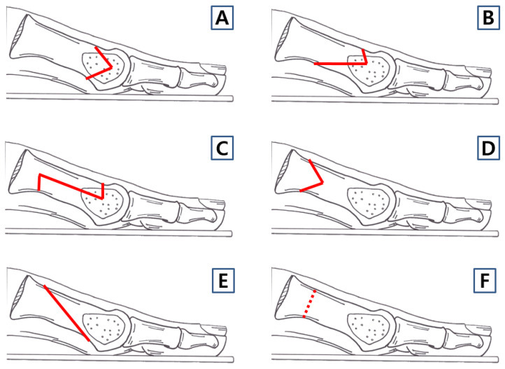
First-metatarsal osteotomy techniques (schematic). Labelled lateral diagrams of the principal first-metatarsal osteotomies used for hallux valgus. Cho et al., J Clin Med 2025;14:5072, Fig. 3 (CC BY 4.0).
First-metatarsal osteotomy techniques (schematic). Labelled lateral diagrams of the principal first-metatarsal osteotomies used for hallux valgus. Cho et al., J Clin Med 2025;14:5072, Fig. 3 (CC BY 4.0).
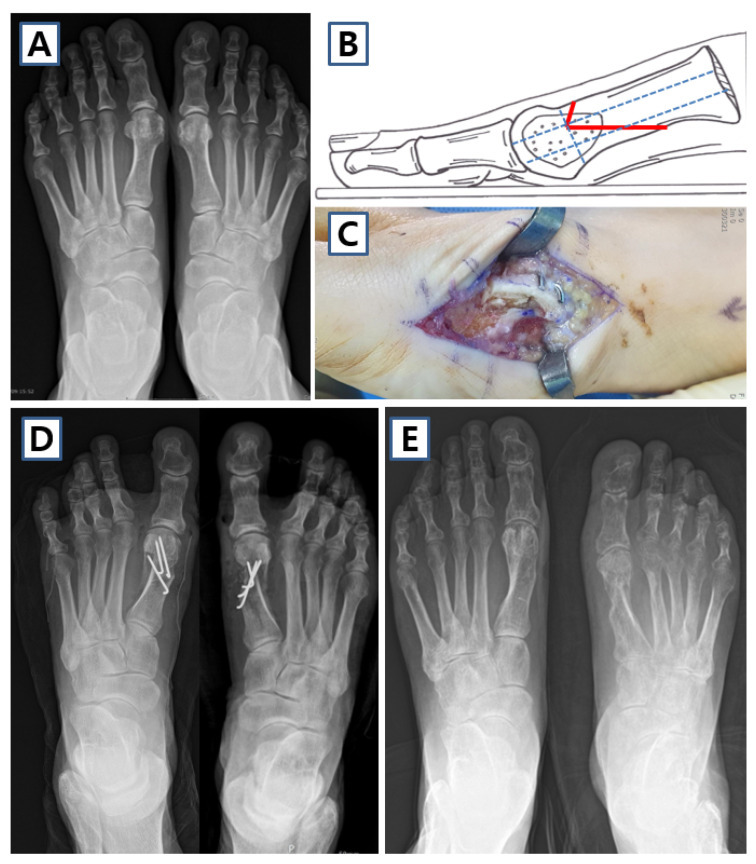
Distal chevron osteotomy for hallux valgus. Pre-operative deformity (A), the osteotomy (B-C) and the corrected, screw-fixed post-operative alignment (D-E). Cho et al., J Clin Med 2025;14:5072, Fig. 4 (CC BY 4.0).
Distal chevron osteotomy for hallux valgus. Pre-operative deformity (A), the osteotomy (B-C) and the corrected, screw-fixed post-operative alignment (D-E). Cho et al., J Clin Med 2025;14:5072, Fig. 4 (CC BY 4.0).
Part V - Hallux valgus surgery: complications
The characteristic complications mirror the principles:
- Recurrence, usually from choosing the wrong procedure, failing to correct the IMA, or missing a congruent joint with a high DMAA.[29]
- Hallux varus (overcorrection), classically after fibular sesamoidectomy with over-aggressive medial-eminence resection or capsular plication. A fixed varus is treated by a split extensor-hallucis-longus transfer, often with interphalangeal fusion.[30]
- Avascular necrosis of the metatarsal head, from excessive periosteal stripping at the dorsolateral neck; salvaged by arthrodesis.[31]
- Malunion: shortening or dorsiflexion (elevatus) of the metatarsal, which transfers load and produces transfer metatarsalgia.[32]
- First-MTP stiffness and persistent pain in about 10%.[33]
Part VI - Hallux rigidus
Hallux rigidus is degenerative arthritis of the first MTP joint, with a dorsal osteophyte and progressive loss of dorsiflexion. It is the second commonest disorder of the great-toe joint after hallux valgus, affecting roughly 1 in 40 people over 50, usually bilaterally and with a strong family history.[34] The most commonly cited cause is trauma (an osteochondral injury or repetitive microtrauma). A flat or chevron-shaped joint surface predisposes, whereas first-ray mobility, metatarsal length and metatarsus primus elevatus are not proven causes; elevatus is a secondary finding (up to 5 mm of elevation is normal).[35]
The patient has dorsal pain and a hard dorsal prominence, with pain at the extremes of motion. The grind (axial-load) test reproducing pain at the midrange of motion signals advanced cartilage loss and a poor prognosis for joint-sparing surgery.[36] Radiographs show a dorsal osteophyte (the “dripping candle wax” appearance on the lateral) and a flattened metatarsal head.[37] The Coughlin-Shurnas classification grades the disease 0-4 on dorsiflexion range, radiographic change and pain pattern. Grades 3 and 4 can look identical radiographically and are distinguished clinically by pain at the midrange of motion in grade 4.[38]
Treatment is matched to the grade. Grade 0 has conservative care (a stiff-soled or rocker shoe, a Morton extension, judicious injection). For grades 1-2, and for grade 3 if more than half the articular cartilage remains, the operation is a cheilectomy (excision of the dorsal 20-30% of the metatarsal head, aiming for about 60° of dorsiflexion), with a ~92% long-term success; a dorsal closing-wedge phalangeal osteotomy (Moberg) repositions the available motion. First-MTP arthrodesis is the gold standard for grade 4 (and grade 3 with less than half the cartilage), with interposition or resection arthroplasty as alternatives.[39]
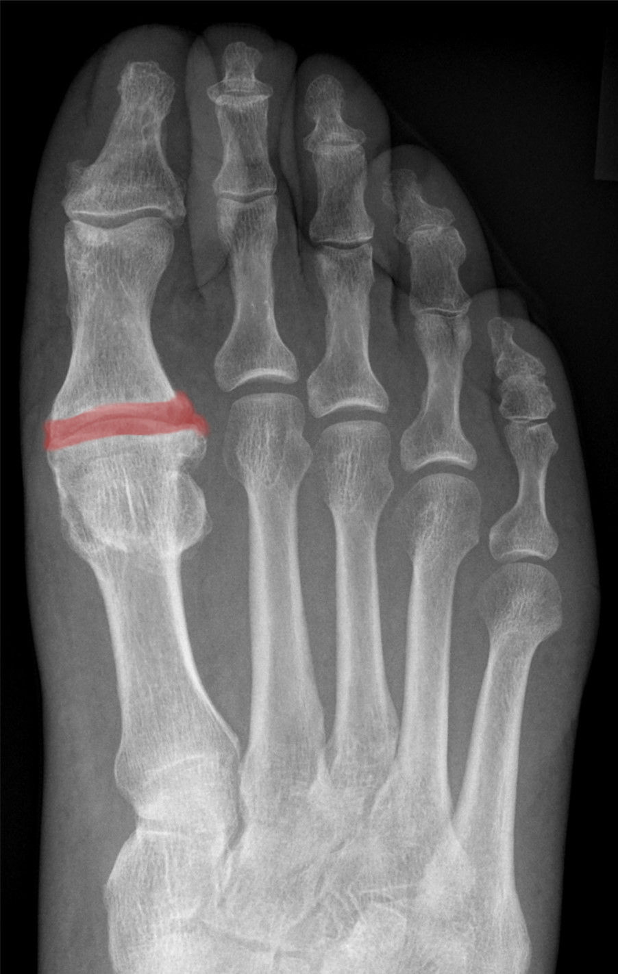
Hallux rigidus. AP radiograph with the first-MTP joint space highlighted, showing narrowing in a 66-year-old man without hallux valgus. Hellerhoff, Wikimedia Commons, CC BY-SA 4.0.
Hallux rigidus. AP radiograph with the first-MTP joint space highlighted, showing narrowing in a 66-year-old man without hallux valgus. Hellerhoff, Wikimedia Commons, CC BY-SA 4.0.
Part VII - Lesser-toe deformities
The lesser-toe deformities are defined by the joint each involves: a mallet toe is flexion at the distal interphalangeal (DIP) joint; a hammer toe is flexion at the proximal interphalangeal (PIP) joint (a complex hammer toe adds MTP hyperextension); a claw toe is PIP flexion plus MTP extension, usually affecting all the lesser toes and often neuromuscular; and a curly toe is a paediatric flexion-and-rotation deformity.[40] They are 4-5 times commoner in women, increasing with age, and the second toe is most often affected.[41]
The deformities arise from an imbalance between the extrinsic and intrinsic muscles, with the plantar plate, the principal stabiliser of the MTP joint, attenuating under chronic hyperextension.[42] The single most useful sign of MTP instability is the vertical drawer (Thompson-Hamilton) test: with the toe dorsiflexed about 25°, dorsal pressure subluxes an unstable joint.[43] The key distinction for treatment is flexible versus fixed.
- A flexible hammer toe is corrected by a flexor-to-extensor (Girdlestone-Taylor) tendon transfer, which converts the long flexor into a substitute for the lost intrinsic action.[44]
- A fixed hammer toe needs a PIP resection arthroplasty or arthrodesis.[45]
- A flexible mallet (or curly) toe is treated by a simple flexor tenotomy, a fixed mallet toe by DIP condylectomy.[46]
- A claw toe combines an MTP soft-tissue release with correction of the toe (and of any associated cavus foot).[47]
- A Weil shortening osteotomy of the metatarsal decompresses and realigns an unstable or subluxed MTP joint, but it characteristically causes a “floating toe” because it shifts the intrinsic tendons dorsal to the joint axis.[48]
The crossover (second) toe is the dorsomedial deviation of the second toe from plantar-plate and lateral-collateral failure. Coughlin and Nery grade the instability 0-IV (and the plantar-plate tear anatomically 0-IV), treating it by soft-tissue release, a Weil osteotomy, direct plantar-plate repair and, for a dislocated joint, a flexor-to-extensor transfer.[49]
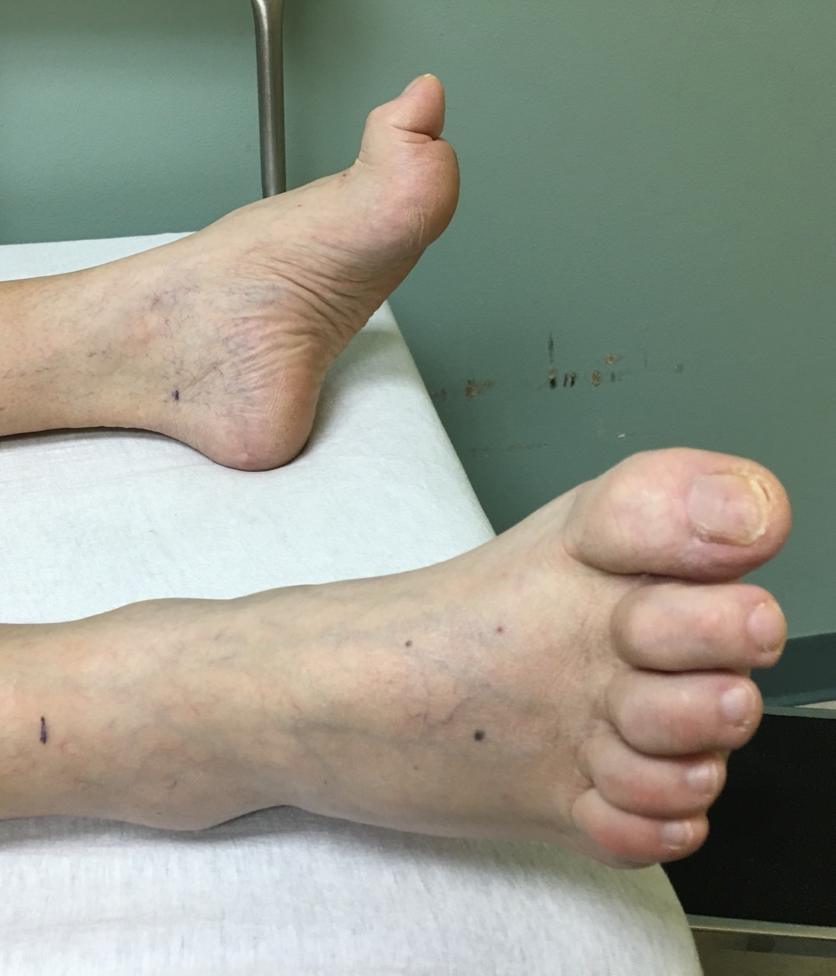
Hammer/claw toes. Clinical photograph showing flexion deformity of the lesser toes with dorsal prominence at the proximal interphalangeal joints. Wwcmdmsha (neurosigns.org), Wikimedia Commons, CC BY-SA 4.0.
Hammer/claw toes. Clinical photograph showing flexion deformity of the lesser toes with dorsal prominence at the proximal interphalangeal joints. Wwcmdmsha (neurosigns.org), Wikimedia Commons, CC BY-SA 4.0.
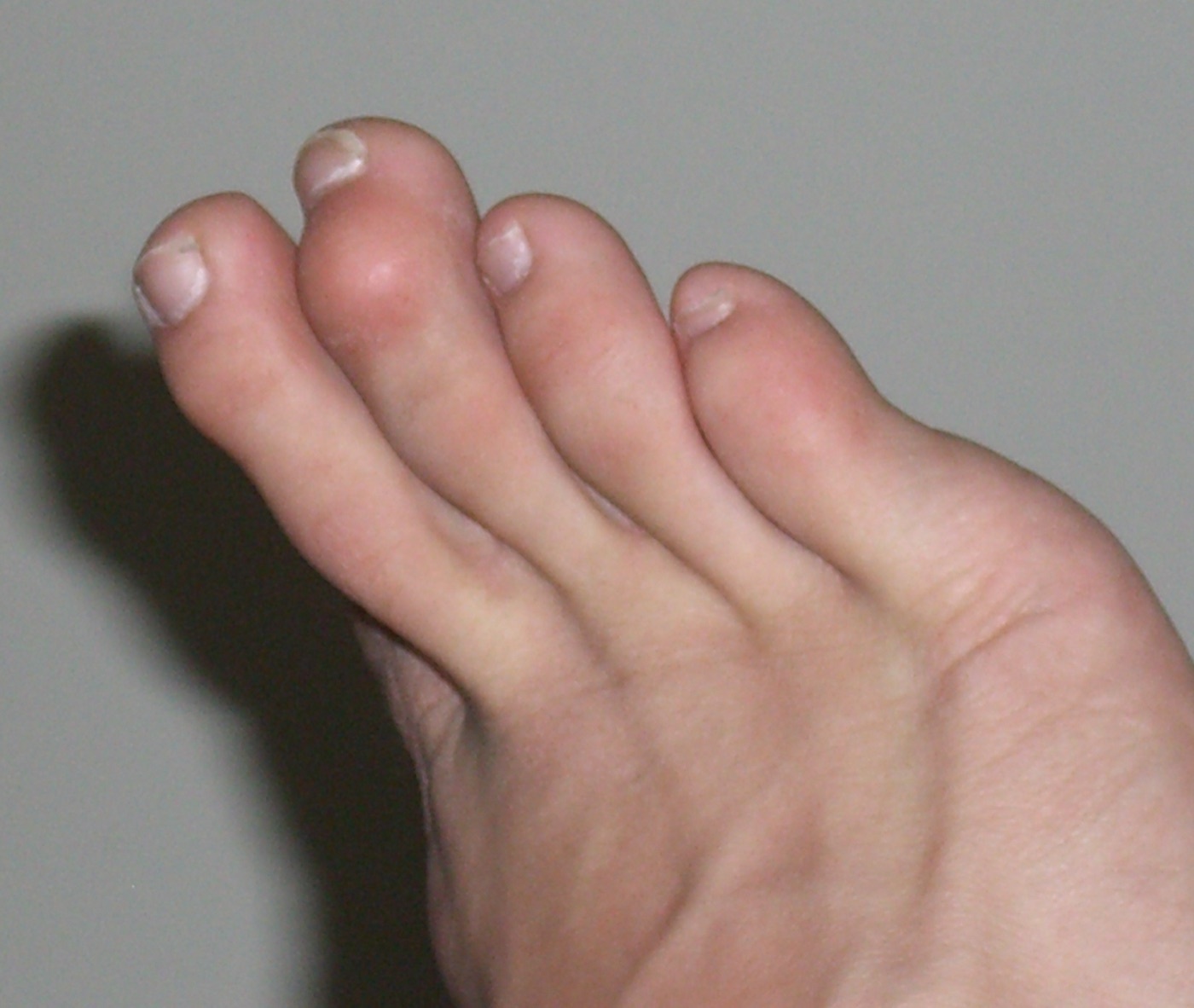
Mallet toe. Distal interphalangeal flexion of a lesser toe - useful for contrasting the joint level with the hammer/claw deformity above. Bprender22, Wikimedia Commons, public domain.
Mallet toe. Distal interphalangeal flexion of a lesser toe - useful for contrasting the joint level with the hammer/claw deformity above. Bprender22, Wikimedia Commons, public domain.
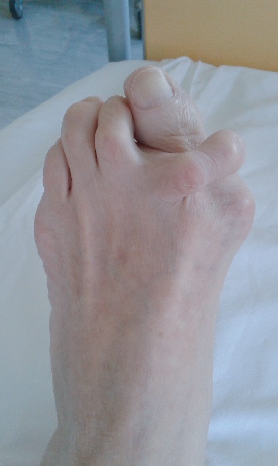
Crossover second toe. The second toe deviates dorsomedially and crosses over the great toe, here with an associated hallux valgus. Case Rep Orthop 2020;2020:3901458, Fig. 1 (CC BY 4.0).
Crossover second toe. The second toe deviates dorsomedially and crosses over the great toe, here with an associated hallux valgus. Case Rep Orthop 2020;2020:3901458, Fig. 1 (CC BY 4.0).
Part VIII - Metatarsalgia and the plantar keratoses
Metatarsalgia is pain under the lesser metatarsal heads, and the central clinical task is to find its source before operating. A plantar keratosis is divided into two types whose treatment is opposite:
- a discrete (localised) intractable plantar keratosis (IPK), with a central keratotic core, lies under a prominent fibular condyle of a metatarsal head (or under the tibial sesamoid) and is treated by an elevating procedure (a DuVries plantar condylectomy);
- a diffuse keratosis, without a core, lies under the second metatarsal head and reflects an overloaded or long metatarsal, and is treated by a shortening procedure (a Weil osteotomy).[50]
A keratosis must be distinguished from a plantar wart (verruca): the skin lines run unchanged across a callus but diverge around a wart, and a wart bleeds when pared (it has end-arteries) whereas a keratosis does not.[51] Forefoot overload has several causes, among them a prominent condyle, a long lesser metatarsal, first-ray insufficiency (a short first metatarsal - the Morton foot - or a hypermobile first ray, or one unloaded by hallux valgus), a cavus or equinus foot, and MTP subluxation.[52]
The most important surgical lesson is transfer metatarsalgia: no osteotomy is precise enough to guarantee the load will not move to the next metatarsal, and an iatrogenic transfer lesion after first-ray surgery should be treated by correcting the first ray (a plantarflexion osteotomy or fusion), not by shortening the second metatarsal.[53]
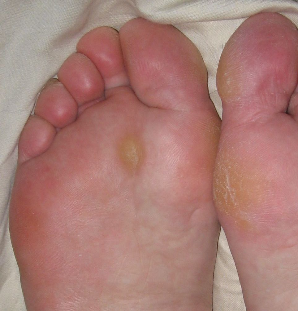
Plantar keratosis. A discrete callus over the central sub-metatarsal region of the sole. Jmarchn, Wikimedia Commons, CC BY-SA 3.0.
Plantar keratosis. A discrete callus over the central sub-metatarsal region of the sole. Jmarchn, Wikimedia Commons, CC BY-SA 3.0.
Part IX - Morton’s (interdigital) neuroma
Morton’s neuroma is not a true neoplasm but a perineural fibrosis (an entrapment neuralgia) of a common digital nerve, occurring distal to the transverse intermetatarsal ligament.[54] It arises most often in the third web space, then the second (a first- or fourth-space neuroma scarcely exists), is far commoner in women (roughly 8-10 times, with reported ratios from about 4:1 upward), and is bilateral in about 15%.[55] The leading mechanism is entrapment of the nerve beneath the transverse intermetatarsal ligament as the metatarsal heads are loaded.[56]
The patient describes plantar burning pain radiating into the toes of the interspace, relieved by removing the shoe and rubbing the foot. The cardinal sign is Mulder’s click, a palpable, often audible clunk on mediolateral compression, diagnostic only when it reproduces the patient’s pain.[57] The diagnosis is clinical, supported where needed by ultrasound or MRI and by a diagnostic local-anaesthetic injection; the differential includes MTP synovitis, a stress fracture and Freiberg’s disease.[58] Conservative care is tried first (a wide shoe, a metatarsal pad, a corticosteroid injection that helps about a third). Persistent symptoms are treated by excision of the common digital nerve, through a dorsal approach, cutting the nerve well proximal to the metatarsal head so the stump retracts away from the weight-bearing surface. About 80% are satisfied, and most recurrences are due to inadequate resection or a stump neuroma.[59]
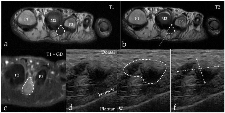
Morton’s (interdigital) neuroma. Second-web-space neuroma on MRI (T1, T2 and contrast-enhanced, top) and ultrasound (bottom), outlined and measured. Diagnostics 2025;15:1339, Fig. 4 (CC BY 4.0).
Morton’s (interdigital) neuroma. Second-web-space neuroma on MRI (T1, T2 and contrast-enhanced, top) and ultrasound (bottom), outlined and measured. Diagnostics 2025;15:1339, Fig. 4 (CC BY 4.0).
Part X - Freiberg infraction, the bunionette and sesamoid disorders
Freiberg infraction
Freiberg’s infraction is osteonecrosis of a lesser metatarsal head, most often the second (the third in about a quarter), presenting in adolescent girls (about 5:1, one of the few osteochondroses with a female predominance), and attributed to repetitive microtrauma of the longer second metatarsal.[60] The Smillie staging (I-V) runs from an epiphyseal fissure, through flattening and central collapse, to loose-body formation and end-stage arthritis.[61] Treatment is by stage: rest or a stiff shoe early; joint débridement with removal of the dorsal 20-30% of the head for established disease; and, for collapse, the Gauthier dorsal closing-wedge osteotomy, which rotates the intact plantar cartilage up into the joint. Head resection and shortening are avoided.[62]
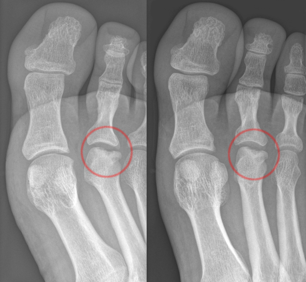
Freiberg infraction. AP radiograph with the second metatarsal head circled, showing flattening and sclerosis (osteonecrosis). Hellerhoff, Wikimedia Commons, CC BY-SA 3.0.
Freiberg infraction. AP radiograph with the second metatarsal head circled, showing flattening and sclerosis (osteonecrosis). Hellerhoff, Wikimedia Commons, CC BY-SA 3.0.
Bunionette (tailor’s bunion)
A bunionette is a symptomatic prominence of the fifth metatarsal head. It is classically grouped into three patterns (an enlarged head, lateral bowing of the metatarsal, and an increased fourth-fifth intermetatarsal angle). Treatment is by a wide shoe, a contouring lateral condylectomy, or an osteotomy chosen by the deformity: a distal chevron (head shifted medially ~4-5 mm) for a curved metatarsal with a slightly widened angle, or a proximal oblique diaphyseal osteotomy for a wider angle or lateral bowing. Simple ostectomy alone tends to remove too much head and destabilise the joint.[63]
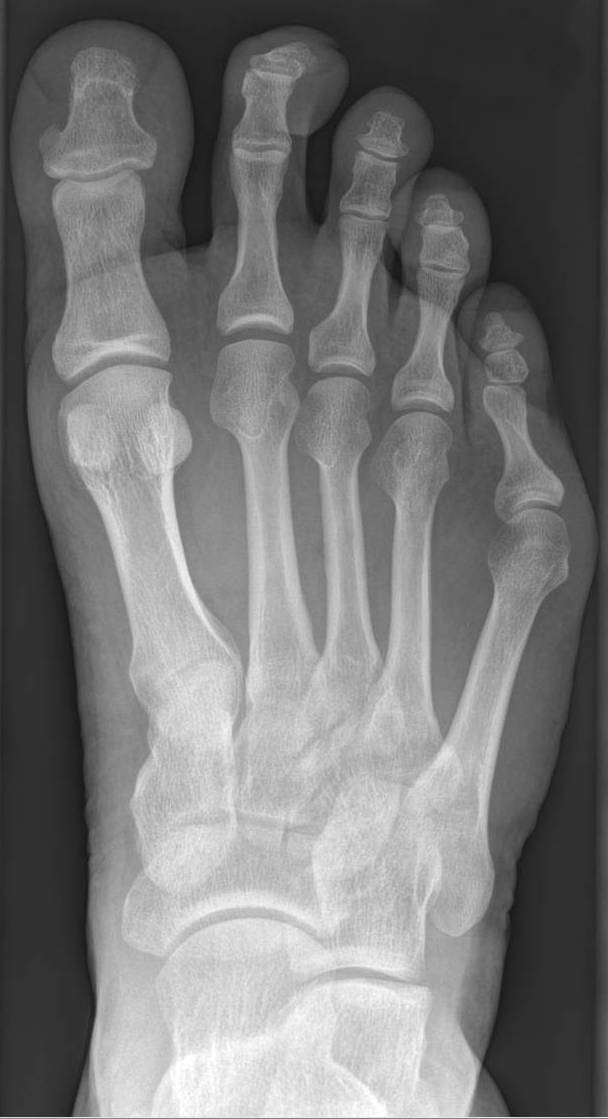
Bunionette (tailor’s bunion). AP radiograph showing a laterally prominent fifth metatarsal head with an increased fourth-fifth intermetatarsal angle. Hellerhoff, Wikimedia Commons, CC BY-SA 4.0.
Bunionette (tailor’s bunion). AP radiograph showing a laterally prominent fifth metatarsal head with an increased fourth-fifth intermetatarsal angle. Hellerhoff, Wikimedia Commons, CC BY-SA 4.0.
Sesamoid disorders
The hallux carries two sesamoids within the flexor hallucis brevis, anchored distally to the proximal phalanx by the plantar plate.[64] The common disorders are sesamoiditis, osteonecrosis, and the distinction of a stress fracture from a bipartite sesamoid, the rule being that the fragments of a fracture sum to the size of a normal sesamoid, whereas a bipartite sesamoid sums to something larger (and the tibial sesamoid is the one more often bipartite).[65] Excision has predictable consequences to remember: tibial (medial) sesamoidectomy lets the hallux drift into valgus, and fibular (lateral) sesamoidectomy into varus, so the flexor brevis and the abductor or adductor must be repaired, and both sesamoids should never be removed together (removing one already shifts load onto the other).[66] A sesamoidectomy is avoided in a cavus foot, where the load merely shifts. Turf toe is an acute hyperextension injury of the hallux plantar-plate/sesamoid complex.[67]
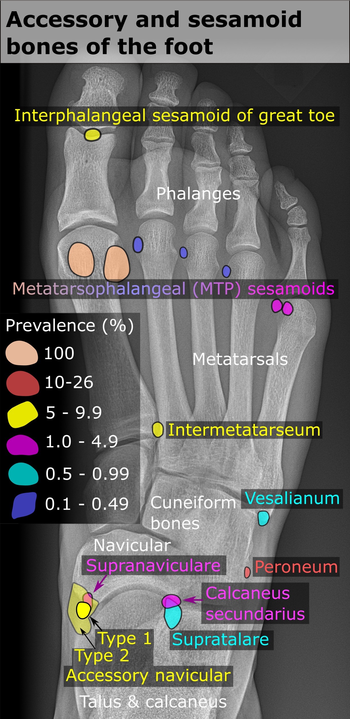
Sesamoid and accessory bones of the foot. Labelled dorsoplantar radiograph showing the hallux metatarsophalangeal sesamoids and the accessory ossicles. Mikael Häggström, Wikimedia Commons, CC0.
Sesamoid and accessory bones of the foot. Labelled dorsoplantar radiograph showing the hallux metatarsophalangeal sesamoids and the accessory ossicles. Mikael Häggström, Wikimedia Commons, CC0.
To tie the topic together: in hallux valgus, read the HVA, the IMA, the DMAA and the joint congruity, then match the operation (soft-tissue release and a distal osteotomy for the smaller deformity, a proximal osteotomy or Lapidus for the larger, an arthrodesis for the severe or arthritic) and operate for pain, not appearance. Grade hallux rigidus by Coughlin-Shurnas and choose cheilectomy while cartilage remains, fusion when it does not. Define each lesser-toe deformity by its joint and its flexibility, and remember the plantar plate behind the crossover toe. Separate the discrete keratosis (elevate) from the diffuse one (shorten), recognise the third-web-space neuroma by Mulder’s click, and watch always for the transfer lesion that follows a disturbed metatarsal cascade.
Bulgarian terminology (Боев / Boychev tradition) - glossary
The following Bulgarian equivalents connect the international literature to the terminology of the Bulgarian school of orthopaedics (in the tradition of Бойчо Бойчев / Boycho Boychev) used in the state examination.
| English term | Bulgarian term (Cyrillic) | Transliteration |
|---|---|---|
| Hallux valgus (bunion) | Халукс валгус (булка/бунион) | Haluks valgus (bunion) |
| Medial eminence | Медиална костна проминенция | Medialna kostna prominentsiya |
| Metatarsus primus varus | Metatarsus primus varus (варус на I метатарзал) | Metatarsus primus varus |
| Hallux valgus angle | Ъгъл на халукс валгус | Agal na haluks valgus |
| Intermetatarsal angle | Интерметатарзален ъгъл | Intermetatarzalen agal |
| Sesamoid | Сезамовидна кост | Sezamovidna kost |
| Hallux rigidus | Халукс ригидус | Haluks rigidus |
| Cheilectomy | Хейлектомия | Heylektomiya |
| Dorsal osteophyte | Дорзален остеофит | Dorzalen osteofit |
| Arthrodesis (fusion) | Артродеза | Artrodeza |
| Osteotomy | Остеотомия | Osteotomiya |
| Hammer toe | Чукообразен пръст | Chukoobrazen prast |
| Claw toe | Ноктовиден (нокътообразен) пръст | Noktoviden prast |
| Mallet toe | Чукче-пръст (дистална флексия) | Chukche-prast |
| Crossover toe | Кръстосан (преметнат) пръст | Krastosan prast |
| Plantar plate | Плантарна плочка | Plantarna plochka |
| Metatarsalgia | Метатарзалгия | Metatarzalgiya |
| Plantar keratosis / callus | Плантарна кератоза / калус | Plantarna keratoza / kalus |
| Morton’s (interdigital) neuroma | Мортонов (интердигитален) невром | Mortonov (interdigitalen) nevrom |
| Mulder’s click | Знак (щракане) на Мълдър | Znak (shtrakane) na Maldar |
| Freiberg infraction | Болест на Фрайберг | Bolest na Frayberg |
| Bunionette (tailor’s bunion) | Шивашки бунион (булка на V метатарзал) | Shivashki bunion |
| Transfer metatarsalgia | Трансферна метатарзалгия | Transferna metatarzalgiya |
Image attributions
(Figure attributions and licences are listed in the figure MANIFEST and inserted with each image. All images are openly licensed [CC0 / Public Domain / CC BY / CC BY-SA] or used under their stated terms; any non-commercial [NC] item is flagged as such and must not be used in a commercial product.)
References
-
Mann’s Surgery of the Foot and Ankle (9th ed.), p. 158.
-
Mann’s, p. 157.
-
Mann’s, p. 158, p. 161.
-
Mann’s, p. 159, p. 161, p. 164-165.
-
Mann’s, p. 165, p. 183.
-
Mann’s, p. 161, p. 183, p. 186.
-
Mann’s, p. 169.
-
Mann’s, p. 170.
-
Mann’s, p. 167-168.
-
Mann’s, p. 171, p. 183, p. 193.
-
Mann’s, p. 173, p. 176-177.
-
Mann’s, p. 166.
-
Mann’s, p. 161, p. 195.
-
Mann’s, p. 198-199.
-
Mann’s, p. 177, p. 181, p. 186, p. 189.
-
Mann’s, p. 177-179.
-
Mann’s, p. 180, p. 184.
-
Mann’s, p. 172-173, p. 195.
-
Mann’s, p. 197, p. 199.
-
Mann’s, p. 205-206, p. 209.
-
Mann’s, p. 220-221, p. 230-231; Myerson, Reconstructive Foot and Ankle Surgery, p. 9, p. 45.
-
Mann’s, p. 238, p. 241-242.
-
Mann’s, p. 242-243, p. 251.
-
Mann’s, p. 261, p. 266.
-
Mann’s, p. 214-216, p. 218.
-
Mann’s, p. 268, p. 272, p. 274.
-
Mann’s, p. 276-278.
-
Mann’s, p. 195, p. 214.
-
Mann’s, p. 297-302.
-
Mann’s, p. 300-303, p. 306-308.
-
Mann’s, p. 289-291.
-
Mann’s, p. 284-288.
-
Mann’s, p. 295, p. 307.
-
Mann’s, p. 933, p. 935.
-
Mann’s, p. 937-939.
-
Mann’s, p. 940, p. 946.
-
Mann’s, p. 940.
-
Mann’s, p. 941-944.
-
Mann’s, p. 943, p. 947-948, p. 951; Myerson, p. 79-80, p. 89.
-
Mann’s, p. 324-326, p. 363.
-
Mann’s, p. 326, p. 329.
-
Mann’s, p. 332-333.
-
Mann’s, p. 375.
-
Mann’s, p. 353-356.
-
Mann’s, p. 340-343.
-
Mann’s, p. 359-360.
-
Mann’s, p. 363-364.
-
Mann’s, p. 332, p. 400; Myerson, p. 120.
-
Mann’s, p. 368-371, p. 376, p. 380-382.
-
Mann’s, p. 432-435.
-
Mann’s, p. 430.
-
Mann’s, p. 428, p. 434-435.
-
Myerson, p. 127-128, p. 130.
-
Mann’s, p. 622.
-
Mann’s, p. 624, p. 626.
-
Mann’s, p. 624.
-
Mann’s, p. 626-627.
-
Mann’s, p. 628-629.
-
Mann’s, p. 630-634, p. 638.
-
Mann’s, p. 451-452.
-
Mann’s, p. 452.
-
Mann’s, p. 453-454.
-
Myerson, p. 139.
-
Mann’s, p. 159.
-
Myerson, p. 83-84.
-
Myerson, p. 91.
-
Myerson, p. 84, p. 96.