Contents
- Introduction and Scope
- SECTION I - THE ACUTELY IRRITABLE HIP IN THE CHILD
- SECTION II - TRANSIENT SYNOVITIS OF THE HIP
- SECTION IV - SYNTHESIS AND EXAM AIDS
- References
- Figure Credits and Licences
Introduction and Scope
Topic 5 of the syllabus pairs two conditions that present alike, a young child with a painful, irritable hip and a limp, yet sit at opposite ends of the urgency scale. Transient synovitis is the commonest and most benign cause: a self-limiting post-viral synovitis that resolves within a week or two. Infantile (neonatal) septic arthritis of the hip, the Bulgarian exam’s кърмачески остеоартрит, is a surgical emergency. It destroys the joint within days and can leave a child with a permanently maimed hip. The whole topic therefore turns on a single clinical question: is this hip septic, or merely synovitic? This review covers the shared diagnostic approach first, then each condition in turn.
The sources are the paediatric-orthopaedic references in the library: the dedicated Pediatric and Adolescent Hip (Alshryda et al.), Lovell & Winter’s Pediatric Orthopaedics, Salter, Staheli, Campbell’s Operative Orthopaedics, and Graf’s Hip Sonography. Antibiotic regimens are given per current Sanford/IDSA practice and flagged as such, and the Bulgarian terminology is drawn from Boychev.
SECTION I - THE ACUTELY IRRITABLE HIP IN THE CHILD
I.1 The Clinical Problem
A child who refuses to walk, limps, or holds the hip still presents a recurring emergency-department dilemma. The hip is splinted by reflex in flexion, abduction and external rotation, the position of greatest capsular volume, which minimises the pain of any effusion. This posture is identical whether the effusion is septic or sterile.[1] Hip pain is frequently referred to the knee or anterior thigh, so a child with “knee pain” and a normal knee must have the hip examined. The task is not to diagnose transient synovitis, which is a label of exclusion, but to exclude septic arthritis, because a missed septic hip is catastrophic while a treated transient synovitis is trivial.[2]
I.2 The Diagnostic Work-up
- Inflammatory markers. CRP is the most useful single test. It rises within 4-6 h, has a short half-life, and serves both as the best early marker and the best monitor of response. ESR rises and falls more slowly; the white-cell count is the least sensitive, raised in only a minority even of true infection. Normal markers make sepsis unlikely but do not exclude it, particularly in the culture-negative Kingella picture and the early neonate.[3]
- Ultrasound is the key bedside test of the irritable hip. It sensitively detects an effusion (the capsule lifted off the femoral neck) without radiation or sedation, and guides aspiration. Its limitation is that an effusion is non-specific: it occurs in transient synovitis just as in sepsis, so ultrasound confirms fluid but not its nature.[4]
- Joint aspiration is the definitive test and is mandatory whenever septic arthritis is genuinely suspected. A synovial white-cell count above ~50,000/µL with a high (>75-90 %) neutrophil fraction, a positive Gram stain, or a positive culture indicate sepsis (counts of 10,000-75,000 are a grey zone, and the threshold is neither perfectly sensitive nor specific). A non-specific effusion on ultrasound does not settle the question: when in doubt, aspirate.[5]
- The radiograph is usually normal early, since bone changes need ~7-14 days, and serves mainly to exclude fracture, tumour, Perthes and slipped epiphysis; it may show soft-tissue swelling or slight medial joint-space widening.
- MRI is the most comprehensive test, showing effusion together with osteomyelitis, pyomyositis and abscess, but it must not delay drainage when the clinical picture and ultrasound already indicate sepsis.[6]
The Kocher criteria were derived to separate septic arthritis from transient synovitis of the child’s hip, using four independent predictors: (1) a history of fever >38.5 °C, (2) inability to bear weight, (3) ESR ≥40 mm/h, and (4) a white-cell count >12,000/µL. In the original series the predicted probability of septic arthritis rose with the number of predictors present (about 3 %, 40 %, 93 % and 99.6 % for one to four). These figures must be used with caution. Later validations gave substantially lower probabilities; an external series found only ~59 % with all four. The criteria are specific but not highly sensitive, and they cannot distinguish septic arthritis from osteomyelitis or pyomyositis. Adding CRP improves prediction (a five-variable model reaches ~98 % with all five; a two-variable model of CRP >20 mg/L plus inability to bear weight gives ~74 %, and neither factor gives <1 %). The criteria guide judgement; they do not replace it.[7]
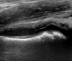
Figure 1. Hip ultrasound - fluid distending the anterior joint recess over the femoral neck (effusion). The key bedside test of the irritable hip; the effusion itself is non-specific (it occurs in both transient synovitis and sepsis). Ruiz Santiago et al., Radiol Res Pract 2016, CC BY 4.0, via Wikimedia Commons.
Figure 1. Hip ultrasound - fluid distending the anterior joint recess over the femoral neck (effusion). The key bedside test of the irritable hip; the effusion itself is non-specific (it occurs in both transient synovitis and sepsis). Ruiz Santiago et al., Radiol Res Pract 2016, CC BY 4.0, via Wikimedia Commons.
I.3 Differential Diagnosis of the Limping Child
Several conditions should be run through before settling on transient synovitis in an acutely irritable or limping child’s hip. The list includes septic arthritis (the must-not-miss), osteomyelitis (especially of the intracapsular proximal femur) and pyomyositis, Legg-Calvé-Perthes disease, slipped capital femoral epiphysis (in the older or adolescent child), juvenile idiopathic arthritis, post-streptococcal reactive arthritis / acute rheumatic fever and Lyme arthritis, trauma/non-accidental injury, and malignancy. Leukaemia matters most here, with its diffuse bone pain, anaemia, cytopenias and metaphyseal lucent bands, followed by the bone tumours. Age narrows the field: a septic hip and transient synovitis dominate the toddler and young child, Perthes the 4-8-year-old, and slipped epiphysis the overweight adolescent.[8]
SECTION II - TRANSIENT SYNOVITIS OF THE HIP
Synonyms / definition. Irritable hip, toxic synovitis, “observation hip.” A benign, self-limiting, non-specific (sterile) synovitis of the hip and the commonest cause of acute hip pain and limp in young children. It is a diagnosis of exclusion, reached only once septic arthritis has been ruled out.[9]
Epidemiology. Typically affects children aged about 3-8 years (peak ~5-6), with a male predominance, and is almost always unilateral. (These demographic specifics are standard teaching rather than stated in the cited chapters, which frame transient synovitis chiefly as the benign comparator in the septic-arthritis work-up.)[10]
Aetiology / pathogenesis. Uncertain, though a post-viral / post-infectious mechanism is favoured. A recent upper-respiratory illness is common, and a recent viral prodrome points away from bacterial sepsis and toward transient synovitis or post-streptococcal reactive arthritis. The pathology is a non-specific synovitis with a sterile effusion.[11]
Clinical features. An acute limp or reluctance to bear weight (though the child can often still walk), hip or referred knee/thigh pain, and mild restriction of motion, especially internal rotation and abduction. The child generally looks well, with low-grade or no fever. Compared with a septic hip, the pain is milder, the range of motion less restricted, and the course waxes and wanes rather than progressing relentlessly.[12]
Diagnosis. A diagnosis of exclusion reached when septic arthritis has been ruled out: the child is well, markers are normal or only mildly raised, ultrasound shows a (non-specific) effusion, and the radiograph is normal (it may show soft-tissue fullness or slight medial joint-space widening, the Waldenström sign of standard teaching). The Kocher criteria estimate the probability of sepsis; when that probability is appreciable or the picture is at all atypical, aspirate the hip.[13]
Treatment. Supportive, meaning rest, analgesia/NSAIDs, and observation, with symptoms settling within about 1-2 weeks. Clinical (± radiographic) follow-up confirms resolution and avoids missing early Perthes.[14]
Prognosis. Benign and self-limiting; recurrence is occasional. There is a small, well-recognised association with later Legg-Calvé-Perthes disease (reported in a few per cent), which is the reason for follow-up.[15]
Exam pearls. Commonest cause of the irritable hip in a young child; well child, can often weight-bear, mild ROM loss, low/no fever, recent viral illness; diagnosis of exclusion; normal radiograph + non-specific US effusion; Kocher criteria; resolves in 1-2 weeks; follow up for Perthes; if in doubt, aspirate. # SECTION III - INFANTILE / NEONATAL SEPTIC ARTHRITIS OF THE HIP
Definition and why the infant hip is special. Pyogenic infection of the hip joint in the neonate or infant is a surgical emergency. The infant hip is uniquely vulnerable for three anatomical reasons.[16]
- The proximal femoral metaphysis is intracapsular, so metaphyseal osteomyelitis that breaks through the cortex discharges directly into the joint. The hip is one of the few joints where the metaphysis lies inside the capsule, along with the proximal humerus, proximal radius and distal lateral tibia.
- In infants (classically under ~12-18 months), transphyseal vessels cross the not-yet-formed growth plate, letting infection pass into the epiphysis and joint and damage the physis. The newest text argues that this route is overstated: the physis usually acts as a barrier, and transphyseal spread is seen mainly in overwhelming sepsis, so it should be presented as the classic teaching with that caveat.
- The femoral head’s blood supply (the medial circumflex / retinacular vessels) runs intracapsularly. A tense pyogenic effusion therefore raises intracapsular pressure and, with septic thrombosis, can strangle that supply and cause avascular necrosis. This is why the hip is drained, not merely aspirated, and never splinted in extension.
Epidemiology and route. Septic arthritis is about twice as common as osteomyelitis in childhood, and the hip is the most commonly involved joint in infants. Spread is haematogenous (neonatal sepsis, umbilical or femoral venous catheters) or contiguous from proximal-femoral osteomyelitis. Neonatal infection is often multifocal.[17]
Causative organisms by age. Staphylococcus aureus is the commonest at every age (cover MRSA empirically where prevalent). In the neonate, add group B Streptococcus and Gram-negative enterics, together with gonococcus acquired through the birth canal. Under about 4 years, Kingella kingae is now a leading cause. It is a fastidious Gram-negative that is culture-negative on routine media (it needs blood-culture broth or PCR), causes mild disease, and is β-lactam-sensitive with few sequelae. H. influenzae type b is now rare after vaccination, and Salmonella features in sickle-cell disease.[18]
Clinical features. Pseudoparalysis, in which the infant stops moving the limb, is the most reliable sign. The hip is held flexed, abducted and externally rotated, and is painful on movement and on nappy changes. In a neonate the systemic picture is non-specific (poor feeding, lethargy, irritability, instability) and fever is unreliable, so a high index of suspicion is essential. Coexisting proximal-femoral osteomyelitis or pyomyositis is common.[19]
Diagnosis. Aspiration of the hip is mandatory and definitive (synovial white-cell count, Gram stain, culture) and should be performed promptly. In the hip it is usually done under anaesthesia, with arthrographic or ultrasound confirmation of needle position. Ultrasound is the key bedside test for the effusion and may show a tense effusion lifting or even dislocating the femoral head. The radiograph is often normal early; later signs are joint-space widening, lateral displacement and frank subluxation/dislocation of the femoral head, then destruction. Joint-space widening, however, is a late sign, and its absence does not exclude sepsis. Send blood cultures and CRP, which should fall daily on effective treatment; failure to normalise predicts sequelae. In a sick neonate, antibiotics need not be withheld before aspiration, as the yield is preserved.[20]
Treatment - a surgical emergency. The mainstay is urgent open arthrotomy with irrigation and drainage of the hip (the anterior Smith-Petersen approach is standard), together with empirical-then-targeted intravenous antibiotics. The hip and the neonate are the classic indications for open drainage rather than aspiration alone, because of the avascular-necrosis risk and the depth of the joint. Outcomes are good when drainage is within about five days of onset, and failure to improve by 72-96 h should prompt re-exploration for a missed focus. Empirical antibiotics are chosen by age, covering S. aureus/MRSA always. In the neonate, current Sanford/IDSA practice favours cefotaxime (avoiding ceftriaxone in neonates) plus vancomycin to cover group B Streptococcus, Gram-negatives and MRSA, with a β-lactam (not vancomycin/clindamycin) for Kingella. Treatment is intravenous initially and switched to oral as CRP falls, for roughly 2-3 weeks (longer if there is concomitant osteomyelitis). The hip is positioned comfortably and never splinted in extension; a spica may be used in a functional position.[21]
Complications and sequelae. The hip joint can be wrecked. The recognised sequelae are avascular necrosis of the femoral head (up to complete resorption), physeal/growth-plate damage (limb-length discrepancy, coxa vara, coxa breva, coxa magna), chondrolysis (cartilage destruction begins within ~8 h of inoculation and continues even after the bacteria are cleared), pathological subluxation/dislocation, and fibrous or bony ankylosis. The destroyed infant hip is the classic “Tom Smith arthritis.” The late sequelae are graded, and the Choi classification of septic-arthritis sequelae of the hip is the usual reference. Systemic complications include septic/toxic shock and venous thromboembolism.[22]
Prognosis. Outcome is determined by age (worse under 6 months/neonate), delay (>4 days), hip involvement, and organism virulence (MRSA/S. pyogenes worse; Kingella mild). Prompt drainage and antibiotics usually give rapid recovery with few sequelae. Delay or a destroyed head requires later reconstruction, ranging from femoral-head reshaping to arthroplasty for the collapsed or incongruent hip.[23]
Exam pearls. Neonatal septic hip = emergency: ultrasound, aspirate, urgent open arthrotomy + IV antibiotics. The hip is opened (not just aspirated) because the intracapsular femoral-head vessels are throttled by pus → AVN. Pseudoparalysis in a septic neonate = septic hip until proven otherwise. Organisms: S. aureus always; neonate GBS + Gram-negatives; <4 y Kingella. Cartilage dies within ~8 h. Joint-space widening/lateral displacement is a late sign. Destroyed hip = Tom Smith arthritis; sequelae graded by Choi.
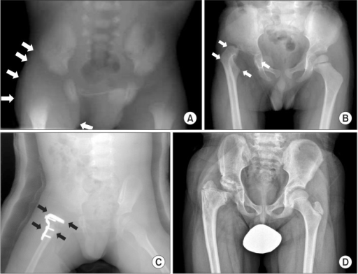
Figure 2. Infantile septic arthritis of the right hip and its sequelae: (A) neonatal peri-hip soft-tissue swelling; (B) pathological dislocation of the femoral head at 3 years; (C) reconstruction (varus-derotation osteotomy with arthroplasty); (D) follow-up at 10 years. Choi et al., Clin Orthop Surg 2015, CC BY-NC 3.0 (non-commercial), via PMC.
Figure 2. Infantile septic arthritis of the right hip and its sequelae: (A) neonatal peri-hip soft-tissue swelling; (B) pathological dislocation of the femoral head at 3 years; (C) reconstruction (varus-derotation osteotomy with arthroplasty); (D) follow-up at 10 years. Choi et al., Clin Orthop Surg 2015, CC BY-NC 3.0 (non-commercial), via PMC.
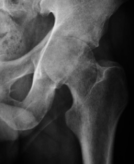
Figure 3. Septic arthritis - a destructive monoarticular process with peri-articular osteopenia (an adult hip, shown to illustrate the radiographic pattern). Ruiz Santiago et al., Radiol Res Pract 2016, CC BY 4.0, via Wikimedia Commons.
Figure 3. Septic arthritis - a destructive monoarticular process with peri-articular osteopenia (an adult hip, shown to illustrate the radiographic pattern). Ruiz Santiago et al., Radiol Res Pract 2016, CC BY 4.0, via Wikimedia Commons.
SECTION IV - SYNTHESIS AND EXAM AIDS
IV.1 Transient Synovitis vs Septic Arthritis
| Feature | Transient synovitis | Septic arthritis |
|---|---|---|
| General appearance | Well child | Often ill / toxic (subtle in neonate) |
| Fever | Low-grade or absent | Higher (>38.5 °C in Kocher) |
| Weight-bearing | Often able | Typically refuses; infant pseudoparalysis |
| Pain / range of motion | Milder, less restricted | Severe pain, markedly restricted |
| Course | Waxes and wanes, then resolves | Relentlessly progressive |
| CRP / ESR / WBC | Normal or mildly raised | Elevated |
| Ultrasound effusion | Present but small, non-specific | Larger effusion more likely |
| Aspirate | Sterile, <50,000 cells | >50,000/µL, >75-90 % neutrophils, +Gram/culture |
| Treatment | Rest, NSAIDs, observe | Urgent drainage + IV antibiotics |
IV.2 The Kocher Criteria
Fever >38.5 °C · non-weight-bearing · ESR ≥40 mm/h · WBC >12,000/µL. The original probabilities of septic arthritis were ~3 % (1), 40 % (2), 93 % (3), 99.6 % (4), but later validations are lower (about 59 % with all four in one external series). The criteria are specific rather than sensitive, and they do not separate septic arthritis from osteomyelitis or pyomyositis. CRP adds discriminatory power.
IV.3 Organisms by Age and Empirical Antibiotics
| Age | Likely organisms | Empirical cover (Sanford/IDSA) |
|---|---|---|
| Neonate (<1 mo) | S. aureus, group B Streptococcus, Gram-negative enterics, gonococcus | cefotaxime + vancomycin (avoid ceftriaxone in neonates) |
| <4 years | S. aureus, Kingella kingae, S. pyogenes, S. pneumoniae | anti-staphylococcal agent + a cephalosporin (covers Kingella) |
| Older child | S. aureus (± MRSA), S. pyogenes | anti-staphylococcal; vancomycin if MRSA risk |
| Sickle-cell disease | Salmonella, S. aureus | add Gram-negative cover |
IV.4 Differential of the Limping Child (by Age)
- 0-3 years: septic arthritis/osteomyelitis, transient synovitis, non-accidental injury, developmental dysplasia.
- 4-8 years: transient synovitis, septic arthritis, Perthes disease, juvenile idiopathic arthritis.
- 9-15 years: slipped capital femoral epiphysis, septic arthritis, JIA.
- Any age: malignancy (leukaemia), reactive/Lyme arthritis, trauma.
IV.5 Bulgarian Terminology and the Boychev Note
The Bulgarian syllabus term кърмачески остеоартрит denotes septic arthritis of the hip in infancy. Boychev’s operative atlas describes drainage arthrotomy (дренажна артротомия) of an acutely infected joint: the joint is opened, sometimes in several places, and drained with tubes under antibiotic cover. This is the operative principle applied to the septic infant hip.
| Bulgarian | English |
|---|---|
| кърмачески остеоартрит | infantile septic arthritis (of the hip) |
| транзиторен синовит | transient synovitis |
| гноен (септичен) артрит | pyogenic (septic) arthritis |
| куцане; накуцване | limp / limping |
| ставен излив | joint effusion |
| ставна пункция | joint aspiration |
| дренажна артротомия | drainage arthrotomy |
| аваскуларна некроза | avascular necrosis |
| псевдопарализа | pseudoparalysis |
IV.6 High-Yield Revision
- The central question is always “septic or not.” Transient synovitis is a diagnosis of exclusion; the septic hip is an emergency.
- Kocher criteria (fever >38.5 °C, non-weight-bearing, ESR ≥40, WBC >12,000) estimate the probability of sepsis; CRP refines it; aspiration settles it.
- Transient synovitis: well child, recent viral illness, mild signs, normal film with a non-specific US effusion, resolves in 1-2 weeks, follow up for Perthes.
- Infantile septic hip: pseudoparalysis, and the intracapsular metaphysis and femoral-head vessels make it a drain-don’t-just-aspirate emergency; S. aureus/GBS/Gram-negatives/Kingella; complications AVN, growth arrest, dislocation, Tom Smith arthritis.
References
- Alshryda S, Howard JJ, Huntley JS, Schoenecker JG, eds. The Pediatric and Adolescent Hip: Essentials and Evidence. Cham: Springer; 2019.
- Flynn JM, Weinstein SL, et al., eds. Lovell and Winter’s Pediatric Orthopaedics. 8th ed. Philadelphia: Wolters Kluwer; 2020 (musculoskeletal infection).
- Salter RB. Textbook of Disorders and Injuries of the Musculoskeletal System. 3rd ed. Baltimore: Williams & Wilkins; 1999.
- Staheli LT. Fundamentals of Pediatric Orthopedics. 5th ed. Philadelphia: Wolters Kluwer; 2016.
- Graf R. Hip Sonography: Diagnosis and Management of Infant Hip Dysplasia. 2nd ed. Berlin: Springer; 2006.
- Azar FM, Beaty JH, eds. Campbell’s Operative Orthopaedics. 14th ed. Philadelphia: Elsevier; 2020 (infectious arthritis).
- Kocher MS, Zurakowski D, Kasser JR. Differentiating between septic arthritis and transient synovitis of the hip in children: an evidence-based clinical prediction algorithm. J Bone Joint Surg Am. 1999;81(12):1662-1670.
- Choi IH, Bowen JR, Gillespie R. Late sequelae of septic arthritis of the hip in infancy and childhood. J Pediatr Orthop. 1990;10(3):365-372.
- Бойчев Б. (Boychev B.) Хирургическа ортопедия (Surgical Orthopaedics). Sofia.
Note on currency: antibiotic regimens follow current Sanford/IDSA practice (notably cefotaxime rather than ceftriaxone in neonates), and the Kocher probabilities are reported with their later, lower revalidation figures. Demographic details of transient synovitis and the “Tom Smith”/Choi sequelae nomenclature are standard paediatric-orthopaedic teaching where not stated verbatim in the cited chapters.
Figure Credits and Licences
- Fig 1 Hip ultrasound, effusion (transient synovitis) - Ruiz Santiago F et al., Radiology Research and Practice 2016;2016:6369237, CC BY 4.0, via Wikimedia Commons. https://commons.wikimedia.org/wiki/File:Ultrasonography_of_joint_effusion_in_transient_synovitis_of_the_hip.jpg
- Fig 2 Infantile septic hip and sequelae - Choi IH et al., Clinics in Orthopedic Surgery 2015, CC BY-NC 3.0 (non-commercial use only), via PMC. https://pmc.ncbi.nlm.nih.gov/articles/PMC4329523/
- Fig 3 Septic arthritis of the hip, radiographic pattern (adult) - Ruiz Santiago F et al., Radiology Research and Practice 2016;2016:6369237, CC BY 4.0, via Wikimedia Commons. https://commons.wikimedia.org/wiki/File:X-ray_of_septic_arthritis_of_the_hip.jpg


-
Lovell & Winter’s Pediatric Orthopaedics, p.1222; The Pediatric and Adolescent Hip (Alshryda), p.284.
-
Lovell p.1261-1262; Campbell’s Operative Orthopaedics, Ch.22, p.958.
-
Lovell p.1220-1231; Alshryda p.291-292; Staheli, Fundamentals of Pediatric Orthopedics, p.384-385.
-
Alshryda p.284-285; Staheli p.383; Graf, Hip Sonography, p.42; Lovell p.1255.
-
Alshryda p.285, p.289-290; Lovell p.1257-1259; Campbell’s p.958.
-
Campbell’s p.958-959; Alshryda p.285-286, p.288; Lovell p.1245-1254.
-
Kocher MS, Zurakowski D, Kasser JR. Differentiating between septic arthritis and transient synovitis of the hip in children. J Bone Joint Surg Am 1999;81(12):1662-70; Lovell p.1262-1263 (original, prospective-validation and Luhmann figures); Campbell’s p.958 (Singhal CRP model).
-
Alshryda p.277-288; Lovell p.1195, p.1263-1264; Salter p.245-264.
-
Alshryda p.287 (“a post-infectious arthritis”); Salter p.246 (non-specific inflammatory disorder); Lovell p.1262. “Commonest cause of childhood hip pain/limp” is standard teaching.
-
Standard paediatric-orthopaedic teaching (age 3-8, male predominance, unilateral); the cited sources discuss transient synovitis within the septic-arthritis differential rather than as a stand-alone entity.
-
Alshryda p.284; Lovell p.1220-1221, p.1262.
-
Lovell p.1261-1262; Alshryda p.287.
-
Alshryda p.285, p.288, p.291; Lovell p.1262-1263; Campbell’s p.958-959. The Waldenström sign eponym is standard radiological teaching.
-
Supportive management and the ~1-2-week course are standard teaching; the principle of NSAIDs/observation for a self-limited inflammatory hip and of serial follow-up (symptoms improve, sepsis worsens) is supported by Salter p.262 and Lovell p.1221.
-
Benign self-limited course and the small Perthes association are standard teaching; the cited sources confirm the benign, cyclic nature (Lovell p.1262; Salter p.262) but do not quantify the Perthes link.
-
Alshryda p.275-278 (intracapsular metaphysis; femoral-head supply; AVN; and the dispute over transphyseal vessels, p.276-277); Lovell p.1206, p.1218; Staheli p.370, p.373.
-
Alshryda p.283; Lovell p.1205-1206; Staheli p.371, p.373.
-
Lovell p.1204-1205, p.1220, p.1265; Alshryda p.283, p.294; Staheli p.376-379.
-
Staheli p.379-380; Lovell p.1220, p.1237; Alshryda p.275, p.284.
-
Alshryda p.285, p.289-290; Lovell p.1224-1238, p.1257-1259; Staheli p.381-385; Graf p.42; Lovell p.1227 (CRP and outcome).
-
Alshryda p.294-299; Lovell p.1237, p.1265-1269; Staheli p.388; antibiotic specifics per current Sanford/IDSA practice (cefotaxime preferred over ceftriaxone in neonates).
-
Alshryda p.278-279, p.296-297; Lovell p.1218-1220, p.1233, p.1238. The Choi classification (Choi IH et al., J Pediatr Orthop 1990;10:365) is cited for sequelae grading; “Tom Smith arthritis” is the standard eponym for the destroyed infant hip.
-
Staheli p.379; Lovell p.1261-1262; Alshryda p.283, p.297, p.314.