Contents
- Introduction and scope
- Part I - Definition and the three-dimensional concept
- Part II - Classification by age of onset
- Part III - Epidemiology
- Part IV - Etiology and pathogenesis
- Part V - Natural history
- Part VI - Clinical and radiographic evaluation
- Part VII - Assessment of skeletal maturity
- Part VIII - Classification systems for treatment
- Part IX - Non-operative management
- Part X - Operative treatment
- Part XI - Complications of surgery
- Part XII - Outcomes
- Part XIII - Adult and untreated late-onset scoliosis
- Bulgarian terminology (Боев / Boychev tradition) - glossary
- Image attributions
Introduction and scope
“Scoliotic disease” in the examination syllabus means idiopathic scoliosis, the structural three-dimensional spinal deformity of unknown cause that accounts for roughly 80% of all scoliosis.[1] This summary follows the disease in the order an examiner expects: what scoliosis is and why it is a three-dimensional deformity; how it is classified by age of onset; its epidemiology, etiology and natural history; the clinical and radiographic work-up, including the assessment of skeletal maturity; the two classification systems that drive surgery (King and Lenke); and finally non-operative and operative management, their complications and outcomes, and the distinct problem of adult scoliosis.
Three ideas anchor the whole topic. The first is that idiopathic scoliosis is a diagnosis of exclusion: atypical features such as a left thoracic curve, pain, a neurological sign or a very young child must trigger a search for a secondary cause, usually with MRI. The second is that growth drives the curve. The risk of progression depends on how much growth remains, so judging skeletal maturity is the central clinical skill, and the timing of every treatment decision flows from it. The third is that the goal of treatment is to control progression and produce a balanced spine in all three planes: bracing prevents progression without correcting, while surgery aims for a balanced, level fusion that spares as many lumbar motion segments as possible.
Part I - Definition and the three-dimensional concept
Scoliosis is a lateral curvature of the spine with a Cobb angle greater than 10° measured on a standing radiograph, accompanied by vertebral rotation. The Scoliosis Research Society sets the diagnostic threshold at 10° or more; a curve below 10° is not scoliosis but “spinal asymmetry.”[2] “Idiopathic” simply means without an identified cause.
Although it is detected as a side-to-side curve, scoliosis is a three-dimensional deformity with a coupled component in each plane:[3]
- Coronal plane: the lateral deviation that gives the condition its name.
- Axial (transverse) plane: vertebral rotation, greatest at the apex. Through the attached ribs this rotation produces the posterior rib prominence (rib hump) that makes early clinical detection possible, along with a corresponding anterior chest-wall asymmetry.
- Sagittal plane: characteristically a loss of normal thoracic kyphosis (thoracic hypokyphosis, even segmental lordosis). The Somerville-Dickson concept holds that an early evolution to thoracic lordosis lets the column buckle and rotate. Conversely, increased kyphosis or a left thoracic curve should raise suspicion of a non-idiopathic cause such as a syrinx or Chiari malformation.
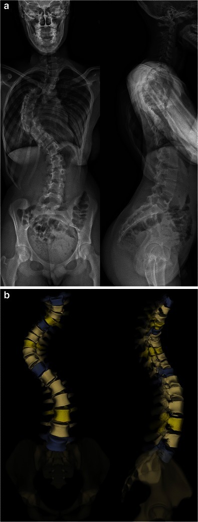
Adolescent idiopathic scoliosis in three planes. Standing antero-posterior and lateral radiographs (A) of a right thoracic / left lumbar curve, with the biplanar (EOS) three-dimensional reconstruction (B) showing the coupled lateral deviation, axial rotation and sagittal change. Amzallag-Bellenger E et al., Insights Imaging 2014;5:571-583 (CC BY 4.0).
Adolescent idiopathic scoliosis in three planes. Standing antero-posterior and lateral radiographs (A) of a right thoracic / left lumbar curve, with the biplanar (EOS) three-dimensional reconstruction (B) showing the coupled lateral deviation, axial rotation and sagittal change. Amzallag-Bellenger E et al., Insights Imaging 2014;5:571-583 (CC BY 4.0).
A curve is described as structural when it lacks normal flexibility and fails to correct on supine side-bending films, and non-structural (functional) when it corrects or over-corrects. The classic example is the compensatory lumbar curve of a leg-length discrepancy, which disappears when the legs are levelled.[4] The vocabulary used to describe a curve (apical, end and neutral vertebrae; major and minor curves; primary and compensatory curves) is defined in the radiographic section below and underpins both classification systems.
Part II - Classification by age of onset
Idiopathic scoliosis is traditionally divided by the age at which it is first recognised, because age at onset is one of the strongest determinants of natural history: the younger the child, the more growth remains and the greater the potential for progression.[5] The exact cut-offs differ slightly between sources, a common examination pitfall:
| Group | Typical age range | Sex tendency | Curve-side tendency |
|---|---|---|---|
| Infantile | birth-3 years | boys (≈3:2) | left-sided; thoracic in ~75-90% |
| Juvenile | 4-9/10 years | girls (rising with age) | right thoracic |
| Adolescent | ≥10 years to maturity | girls (rising with curve size) | right thoracic |
[6]
Adolescent idiopathic scoliosis (AIS) is by far the most common form (around 89% of cases in a large Boston series), developing in previously healthy children during the pubertal growth spurt.[7] Juvenile idiopathic scoliosis (8-21% of cases) is a transitional group; once a juvenile curve reaches 30° it is almost always progressive, and thoracic juvenile curves come to arthrodesis in over 90% of cases despite bracing.[8] Infantile idiopathic scoliosis (<1% of cases in North America, historically commoner in Europe) is unusual in being male-predominant and left-sided, and in that a large fraction resolve spontaneously (see natural history below).[9]
A modern overlay on this scheme is the concept of early-onset scoliosis (EOS): any scoliosis, of any cause, developing before the age of 10.[10] The age of 10 is chosen because it changes the treatment strategy. Under 10 the priority is “growth-friendly” treatment, whereas at 10 and older definitive fusion becomes the principal surgical option. EOS matters so much because of the lung: the alveoli multiply from about 20 million at birth to 250 million by age 4, and most alveolar development is complete by age 8, so a curve that distorts the thorax during this window produces restrictive lung disease, pulmonary hypertension and, ultimately, cor pulmonale. This is Campbell’s thoracic insufficiency syndrome, the inability of the thorax to support normal respiration and lung growth.[11]
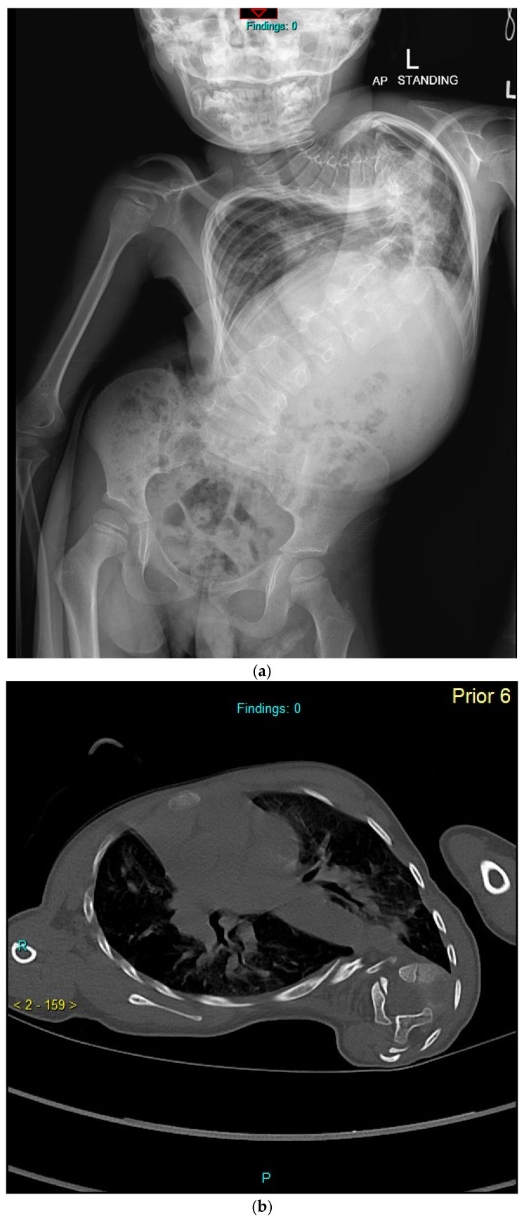
Infantile idiopathic scoliosis. Standing antero-posterior radiograph of a young child with a severe early-onset curve (A) and an axial chest CT (B) showing the marked apical vertebral rotation and chest-wall deformity that threaten lung development. Balasubramanian SG et al., J Clin Med 2026;15(2):754 (CC BY 4.0).
Infantile idiopathic scoliosis. Standing antero-posterior radiograph of a young child with a severe early-onset curve (A) and an axial chest CT (B) showing the marked apical vertebral rotation and chest-wall deformity that threaten lung development. Balasubramanian SG et al., J Clin Med 2026;15(2):754 (CC BY 4.0).
Part III - Epidemiology
Idiopathic scoliosis is common in its mild forms and rare in its severe ones. The prevalence of curves greater than 10° is about 2-3% of adolescents, but the prevalence falls steeply as the curve magnitude rises: curves over 20° are present in roughly 0.3-0.5%, and curves over 30-40° in only about 0.1-0.2%.[12]
The most testable epidemiological fact is the female predominance that increases with curve magnitude. Among small curves the sex ratio is roughly equal (≈1:1); among curves large enough to need treatment it rises to about 1 boy to 7-10 girls, and girls also tend to have larger curves.[13] Familial clustering is well documented: scoliosis is roughly three times more frequent when a parent is affected and about seven times more frequent when a sibling is affected, with scoliosis found in around 11% of first-degree relatives.[14]
Part IV - Etiology and pathogenesis
The cause of idiopathic scoliosis is unknown and almost certainly multifactorial. It is best understood not as one disease but as a final common pathway in which the vulnerable, rapidly growing adolescent spine decompensates into a self-perpetuating rotational deformity.[15] Examiners ask for “the theories,” so the major lines of evidence are worth knowing:
- Genetic. The condition is strongly heritable (twin concordance is higher in monozygotic than dizygotic pairs, though the reported figures vary widely between studies), does not follow simple Mendelian inheritance, and is associated with validated risk genes. LBX1 is the best-replicated, alongside GPR126, PAX1, BNC2 and others, while severity associates with estrogen-receptor and other genes. A DNA-based prognostic test (ScoliScore) was marketed but proved non-replicable outside Caucasian populations.[16]
- Neuromuscular / central nervous system. Idiopathic scoliosis is uniquely human. Abnormalities of proprioception, postural balance, somatosensory evoked potentials, and brainstem/vestibular and cerebellar morphology have all been described, and a syrinx or Chiari malformation is found in a meaningful minority of young patients presenting as “idiopathic.”[17]
- Melatonin and neuroendocrine. Pinealectomy reliably produces scoliosis in chickens and bipedal rats, but human melatonin data are inconsistent. A melatonin-signalling defect in osteoblasts (rather than simple deficiency) has been proposed.[18]
- Connective tissue and the intervertebral disc. Scoliosis accompanies Marfan and Ehlers-Danlos syndromes. In true 3-D analysis the early Cobb increase begins at the disc (disc wedging precedes vertebral wedging), part of Stokes’ mechanically-driven “vicious cycle.”[19]
- Bone, growth and relative anterior spinal overgrowth. Affected adolescents tend to be taller and thinner with a higher peak height velocity. Relative overgrowth of the anterior column produces thoracic lordosis and rotational buckling, and low bone mineral density is an independent predictor of progression. The Hueter-Volkmann principle, reduced growth on the compressed concave side, perpetuates the wedging once a curve begins.[20]
- Biomechanical, leptin/autonomic and neurotransmitter theories complete the picture, all consistent with a multifactorial “perfect storm.”[21]
Part V - Natural history
The natural history justifies every treatment threshold, which makes it the heart of the topic.
Predicting progression during growth
Four factors predict that a curve will progress: a larger curve at presentation, more growth remaining, a thoracic (vs lumbar) apex, and double (vs single) curves. Progression is fastest during the peak height velocity of the growth spurt.[22] The classic quantitative guide is the Lonstein and Carlson probability of progression, which combines curve magnitude and Risser sign. In a skeletally immature child (Risser 0-1) a 5-19° curve progresses about 22% of the time, a 20-29° curve about 68%, and a 30-59° curve about 90%; by maturity (Risser ≥3) the risk falls below 10%.[23]
The infantile curve: resolution versus progression
A large fraction of infantile idiopathic curves resolve spontaneously (reported widely, up to ~90% in some British series), while a smaller progressive group can become severe.[24] The prognostic tool is Mehta’s rib-vertebra angle difference (RVAD), the difference between the rib-vertebra angles on the concave and convex sides at the apical vertebra. An RVAD below 20° predicts resolution (≈83-90%); an RVAD of 20° or more predicts progression. Mehta’s rib phase complements it. In phase 1 the rib head does not overlap the apical vertebral body (measure the RVAD), whereas in phase 2 the rib head overlaps the body, and a phase-2 rib is itself a near-certain sign of progression.[25] Any infantile or juvenile curve of 20° or more (or one with an absent abdominal reflex) warrants whole-neuraxis MRI, since intraspinal anomalies are found in up to 20%.[26]
The untreated adolescent curve into adulthood
The definitive long-term data come from the Iowa series (Weinstein and Ponseti), which followed untreated late-onset idiopathic scoliosis for over 40-50 years and corrected the over-stated mortality claims of older, mixed-aetiology studies.[27] The key points:
- Curves continue to progress after maturity in about two-thirds of patients (68%). Thoracic curves of 50-75° at maturity progress most (≈1°/year); thoracic curves under 30° generally do not progress, and lumbar curves over 30° tend to progress.[28]
- Back pain is modestly more common than in controls (about 60-65% vs 35%) but is usually not disabling. Most idiopathic scoliosis is painless, so significant pain should always prompt the question “is this truly idiopathic?”[29]
- Pulmonary compromise and cor pulmonale occur essentially only with very large early-onset thoracic curves (vital capacity falls as a thoracic curve exceeds ~70°, and the risk of right-heart failure is confined to curves beyond ~90-100°).[30]
- Mortality is not increased in studies restricted to adolescent idiopathic scoliosis. The old “twice expected mortality” figures came from cohorts that mixed in congenital and paralytic curves.[31]
Part VI - Clinical and radiographic evaluation
History and physical examination
Idiopathic scoliosis is usually silent and painless, detected at a school or sports screening or noticed as asymmetry by a parent.[32] Examination from behind, with the patient gowned to expose the back from the gluteal cleft up, looks for asymmetry of the shoulders, scapulae and waist, a trunk shift, and a paravertebral prominence. Trunk balance is assessed with a plumb-line dropped from C7 to the gluteal cleft (a patient may have a level head yet a shifted trunk). The legs are checked for length discrepancy, the skin for café-au-lait spots (neurofibromatosis) and midline dysraphic stigmata, and the joints for laxity (Marfan, Ehlers-Danlos).[33]
A focused neurological examination is mandatory, including the superficial abdominal reflexes, since a persistently asymmetric or absent abdominal reflex is a classic clue to an intraspinal lesion such as a syrinx. The findings that should prompt MRI of the neuraxis are worth memorising: a left thoracic curve, significant pain, any neurological abnormality, true hyperkyphosis, a unilateral cavus foot, rapid progression, and onset before age 10. In juvenile and infantile curves a neuraxis anomaly is found in roughly one in five, so MRI is routine in that group.[34]
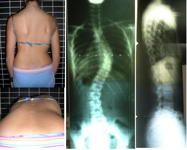
Clinical detection of scoliosis. A standing posterior photograph and the Adams forward-bend view (left) reveal the trunk asymmetry and rib hump, confirmed on antero-posterior and lateral radiographs (right). Rigo M, Negrini S, Weiss HR et al. (SOSORT), via Wikimedia Commons (CC BY 2.0).
Clinical detection of scoliosis. A standing posterior photograph and the Adams forward-bend view (left) reveal the trunk asymmetry and rib hump, confirmed on antero-posterior and lateral radiographs (right). Rigo M, Negrini S, Weiss HR et al. (SOSORT), via Wikimedia Commons (CC BY 2.0).
Screening - the Adams test and the scoliometer
The screening manoeuvre is the Adams forward-bend test: the patient bends forward at the waist with knees straight and palms together, and the examiner looks along the back for a rib hump. A scoliometer (inclinometer) laid across the prominence quantifies the angle of trunk rotation (ATR). The accepted referral rule is that an ATR of 5° may be dismissed and an ATR of 7° or more is referred for a standing PA radiograph (an ATR of about 5-7° corresponds loosely to a Cobb angle of ~15-20°).[35] School screening (forward-bend plus scoliometer) is supported by the orthopaedic societies for girls aged 10-12 and boys 13-14, but it remains controversial because of over-referral and low predictive value. The US Preventive Services Task Force currently rates the evidence as insufficient rather than recommending for or against it.[36]
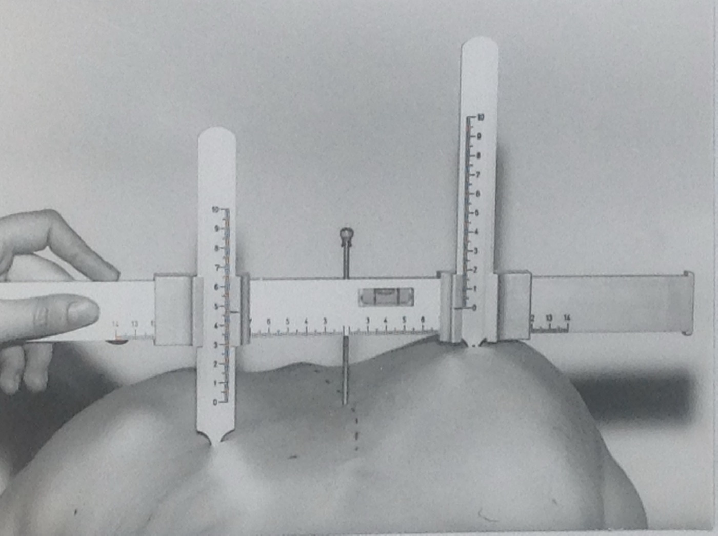
Scoliometer (inclinometer). The device is laid across the apex of the rib prominence with the patient bent forward to read the angle of trunk rotation; an angle of 7° or more prompts a screening radiograph. Hans-Günther Götze, via Wikimedia Commons (CC BY-SA 4.0).
Scoliometer (inclinometer). The device is laid across the apex of the rib prominence with the patient bent forward to read the angle of trunk rotation; an angle of 7° or more prompts a screening radiograph. Hans-Günther Götze, via Wikimedia Commons (CC BY-SA 4.0).
Radiographic measurement
The standard study is a standing full-length postero-anterior and lateral radiograph on a long (14×36-inch) cassette. The films are taken PA rather than AP to cut radiation to the breast and thyroid several-fold, and they must be standing, because gravity increases the measured curve, so a first standing film after earlier supine films can give a false impression of progression.[37]
The curve is measured by the Cobb method (Cobb, 1948): lines are drawn along the endplate of the upper end vertebra (the most tilted vertebra at the top of the curve) and the lower end vertebra, perpendiculars are erected, and the angle of intersection is the Cobb angle. Because the inter- and intra-observer error is about ±5°, a change of at least 5° (6° by the criterion most surgeons accept) is required to call true progression.[38]
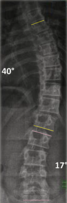
The Cobb method. Lines are drawn along the endplates of the upper and lower end vertebrae of each curve and perpendiculars erected; the angle of intersection is the Cobb angle (here 40° and 17°). Radiograph: Weiss HR, Seibel S, Kleban A; annotation by Mikael Häggström MD, via Wikimedia Commons (CC BY 4.0).
The Cobb method. Lines are drawn along the endplates of the upper and lower end vertebrae of each curve and perpendiculars erected; the angle of intersection is the Cobb angle (here 40° and 17°). Radiograph: Weiss HR, Seibel S, Kleban A; annotation by Mikael Häggström MD, via Wikimedia Commons (CC BY 4.0).
Other measurements include:
- The apical vertebra (most rotated and most laterally deviated) and the neutral vertebra (no rotation; symmetric pedicles).
- Vertebral rotation, graded by the Nash-Moe pedicle method (0-IV) or the Perdriolle torsionmeter, both imprecise and now largely superseded by CT or low-dose 3-D imaging.[39]
- Structural versus non-structural status on supine side-bending (flexibility) films (and, for large stiff curves, fulcrum-bending or traction films), which is essential for surgical level selection.[40]
- Coronal balance (the C7 plumb line relative to the central sacral vertical line; balanced if within 2 cm) and sagittal balance (positive if the C7 plumb falls more than 2 cm anterior to the sacrum).[41]
Advanced imaging has well-defined roles: MRI for the indications listed above; CT for bony detail and navigation; and EOS low-dose biplanar slot-scanning, which delivers a fraction of the radiation of conventional radiography (a “micro-dose” protocol cuts the dose roughly 45-fold) and allows 3-D reconstruction while preserving the upright, gravity-loaded posture.[42]
Part VII - Assessment of skeletal maturity
Because growth drives the curve, judging how much growth remains is the central skill that times every treatment decision. Progression risk peaks at the peak height velocity (PHV) of the growth spurt, about 8-10 cm/year with half of it in the trunk, which occurs before menarche and before the Risser sign appears.[43] The tools, in order of usefulness:
- Risser sign: ossification of the iliac crest apophysis, progressing lateral-to-medial over 18-24 months, graded 0-5 (in the American system the grade reflects the extent of excursion across the single apophysis, whereas the European system subdivides the apophysis and records grades with a “+” notation, so be explicit which is meant). Risser 4 marks the cessation of spinal growth and Risser 5 the end of height gain. Its weakness is that the apophysis appears after the most rapid growth, so Risser 0 spans the dangerous early spurt and is no more accurate than chronological age at predicting progression.[44]
- Triradiate cartilage: its closure occurs during Risser 0, around the time of peak spinal growth. An open triradiate cartilage marks a very immature, high-risk patient and is the key flag for crankshaft risk after isolated posterior fusion.[45]
- Sanders skeletal-maturity stage: an 8-stage hand-and-wrist (digital epiphyseal) system that correlates best with the curve-acceleration/peak-growth window, including within the Risser-0 phase where Risser is blind. Many now regard it as superior to Risser for timing.[46]
- Tanner staging and menarche: menarche follows peak height velocity by about a year and so signals the decelerating, lower-risk phase of growth.[47]
No single index is perfect, so the prudent approach is to pool all available maturity data when counselling a family.[48]
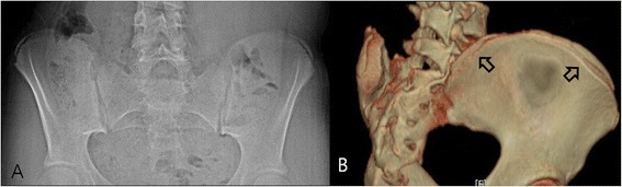
Risser sign. Antero-posterior pelvic radiograph (A) and three-dimensional CT (B) of the iliac crest apophysis, which ossifies from lateral to medial over 18-24 months and is graded 0-5 as an index of skeletal maturity. Yang JH et al., J Orthop Surg Res 2014;9:101 (CC BY 4.0).
Risser sign. Antero-posterior pelvic radiograph (A) and three-dimensional CT (B) of the iliac crest apophysis, which ossifies from lateral to medial over 18-24 months and is graded 0-5 as an index of skeletal maturity. Yang JH et al., J Orthop Surg Res 2014;9:101 (CC BY 4.0).
Part VIII - Classification systems for treatment
Two coronal-based classifications guide surgical decision-making, and both ultimately answer the same question: which curves are structural and must be fused, and which are compensatory and can be spared.
King-Moe classification (1983)
The King classification was derived from 405-455 thoracic curves treated with Harrington instrumentation, and it describes five thoracic curve types to help decide whether to fuse the thoracic curve alone or both the thoracic and lumbar curves:[49]
- King I: a true double (S-shaped) curve in which both thoracic and lumbar curves cross the midline and the lumbar curve is larger and/or less flexible.
- King II: the “false double major”, where the thoracic curve is larger and both cross the midline but the lumbar curve is more flexible. This is the classic candidate for selective thoracic fusion.
- King III: a thoracic curve in which the lumbar curve does not cross the midline.
- King IV: a long thoracic curve in which L4 tilts into the curve.
- King V: a double thoracic curve with a structural upper curve and a positive T1 tilt.
King introduced the practical concepts of the stable vertebra (the most proximal vertebra distal to the curve that is bisected by the central sacral vertical line) and curve flexibility. Its limitations are important: it covers only thoracic curves, ignores the sagittal plane, has only moderate inter-observer reliability, and was designed for distraction instrumentation.[50]
Lenke classification (2001)
The Lenke classification is the modern standard. It is a triad of curve type, lumbar modifier and sagittal modifier, built after determining which of the three curve regions (proximal thoracic, main thoracic, thoracolumbar/lumbar) are structural.[51]
A region is structural if it does not bend out below 25° on side-bending, or if it has junctional kyphosis of +20° or more (proximal thoracic T2-T5, or thoracolumbar T10-L2).[52] The major curve is the one with the largest Cobb angle and is always fused.
- Curve type 1-6: 1 main thoracic; 2 double thoracic; 3 double major; 4 triple major; 5 thoracolumbar/lumbar; 6 thoracolumbar/lumbar-main thoracic.
- Lumbar modifier A / B / C: the relationship of the central sacral vertical line to the apical lumbar vertebra, with A between the pedicles, B touching the apical body, and C completely medial to the apical body.
- Sagittal thoracic modifier − / N / +: the T5-T12 kyphosis, with − less than +10°, N +10° to +40°, and + more than +40°.
The power of Lenke is that it functions as a template for fusion: fuse every structural curve (the major and any structural minors) and spare the non-structural ones. It is reproducible, with about 85% intra-observer reliability and 84-90% agreement between surgeons. Approximate equivalences to the older system are King II ≈ Lenke 1B/1C, King V ≈ Lenke 2, King I ≈ Lenke 3.[53]
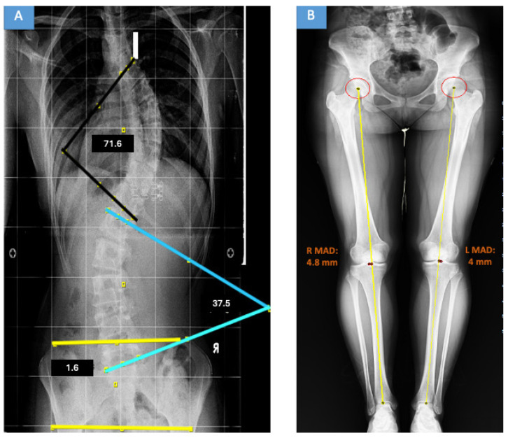
Lenke classification on a full-spine radiograph. Standing antero-posterior film of a Lenke 3C double-major curve with the Cobb angles drawn (main thoracic 71.6°, thoracolumbar/lumbar 37.5°). Aydın AS et al., Medicina (Kaunas) 2026;62(5):978, panel A (CC BY 4.0).
Lenke classification on a full-spine radiograph. Standing antero-posterior film of a Lenke 3C double-major curve with the Cobb angles drawn (main thoracic 71.6°, thoracolumbar/lumbar 37.5°). Aydın AS et al., Medicina (Kaunas) 2026;62(5):978, panel A (CC BY 4.0).
Part IX - Non-operative management
Observation
Curves below about 20° are observed with periodic examination and films; curves of 20-25° in a growing child are followed every 4-6 months during rapid growth; and curves over 30° at maturity are checked occasionally for slow late progression.[54]
Bracing
The cardinal principle is that the goal of bracing is to prevent progression, not to correct the curve. The in-brace correction is generally lost after weaning, and families must be told so.[55] The Scoliosis Research Society indications are a skeletally immature patient (Risser 0-2, premenarchal or within a year of menarche), aged 10 or more, with a primary curve of 25-40°.[56] The brace works by applying corrective transverse, bending and rotational forces during growth (Hueter-Volkmann growth modulation).
Brace types:[57]
- Milwaukee (CTLSO): the historical standard, a pelvic girdle with a superstructure, now reserved for high thoracic curves (apex above T7) and rarely prescribed because it is conspicuous.
- Boston / underarm TLSO: the most widely used brace worldwide, for curves with an apex below T7.
- Charleston and Providence: night-time bending braces for single thoracolumbar/lumbar curves.
- Rigo-Chêneau: a curve-specific, derotational TLSO that preserves the sagittal profile and complements scoliosis-specific exercise.
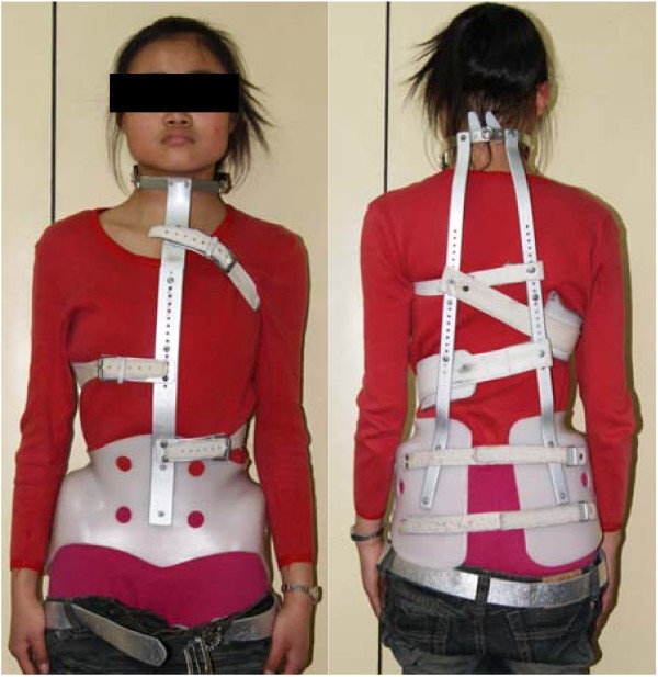
Milwaukee brace (CTLSO). The historical cervico-thoraco-lumbo-sacral orthosis, with a pelvic girdle and a superstructure carrying occipital and throat pads, now reserved for high thoracic curves. Jun Jiang et al., via Wikimedia Commons (CC BY 2.0; the patient’s face is redacted in the original).
Milwaukee brace (CTLSO). The historical cervico-thoraco-lumbo-sacral orthosis, with a pelvic girdle and a superstructure carrying occipital and throat pads, now reserved for high thoracic curves. Jun Jiang et al., via Wikimedia Commons (CC BY 2.0; the patient’s face is redacted in the original).
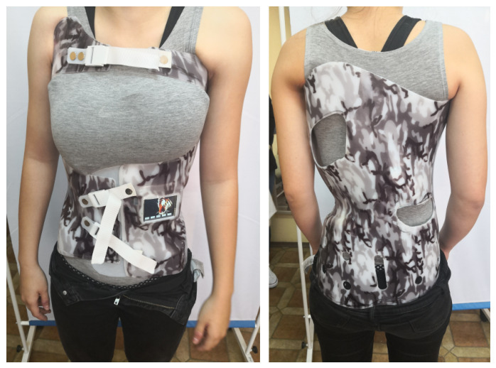
Boston-type underarm TLSO. The thoraco-lumbo-sacral orthosis is the most widely used brace for adolescent idiopathic scoliosis, applied to curves with an apex below T7. Tymińska-Wójcik P et al., Sensors (Basel) 2026;26(10):3169 (CC BY 4.0).
Boston-type underarm TLSO. The thoraco-lumbo-sacral orthosis is the most widely used brace for adolescent idiopathic scoliosis, applied to curves with an apex below T7. Tymińska-Wójcik P et al., Sensors (Basel) 2026;26(10):3169 (CC BY 4.0).
The landmark evidence is the BrAIST trial (Weinstein et al., 2013), a randomised and preference-cohort study of immature patients with 20-40° curves, whose primary outcome was prevention of progression to the surgical threshold of 50°. Bracing succeeded in about 72-75% of patients versus 42-48% with observation, the trial was stopped early for demonstrated benefit, and the effect was strongly dose-dependent. The critical wear threshold was about 13 hours per day, with a number-needed-to-treat of roughly 3 to prevent one surgery.[58]
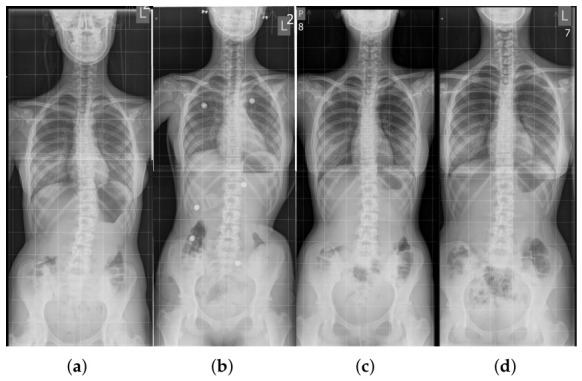
In-brace correction. Serial full-spine radiographs of an adolescent idiopathic curve - before bracing (a), in the brace with embedded sensors (b), at follow-up (c) and out of the brace (d) - illustrating that the brace holds the curve corrected during growth. Tymińska-Wójcik P et al., Sensors (Basel) 2026;26(10):3169 (CC BY 4.0).
In-brace correction. Serial full-spine radiographs of an adolescent idiopathic curve - before bracing (a), in the brace with embedded sensors (b), at follow-up (c) and out of the brace (d) - illustrating that the brace holds the curve corrected during growth. Tymińska-Wójcik P et al., Sensors (Basel) 2026;26(10):3169 (CC BY 4.0).
Physiotherapeutic scoliosis-specific exercises (PSSE: Schroth, SEAS) teach three-plane autocorrection and have level-1 support as a first step for small curves not yet in the bracing range, and as an adjunct to bracing.[59] By contrast, the evidence does not support generic exercise, chiropractic manipulation, or electrical stimulation as a cure; electrical stimulation performs no better than the natural history.[60]
Serial casting for infantile and early-onset scoliosis
For progressive infantile idiopathic curves, serial elongation-derotation-flexion (EDF / Mehta) casting can use the child’s growth as a corrective force to cure the resolvable curve or, in larger or syndromic curves, to delay definitive surgery while preserving thoracic and lung growth. Casts are changed under anaesthesia every 8-16 weeks. The crucial technical principle is that the cast must never push the ribs toward the spine, which would narrow the chest and produce iatrogenic thoracic insufficiency. The best response is predicted by younger age at the start (under about 2 years), a smaller curve, and an idiopathic rather than syndromic diagnosis.[61]
Part X - Operative treatment
Indications and goals
Surgery is considered for progressive curves and for curves of about 45-50° or more in a growing child, and for thoracic curves over 50° at maturity (which continue to progress).[62] The goals are to arrest progression, achieve a fusion that is balanced in both the coronal and sagittal planes, correct the rotation and rib hump, and preserve as many distal lumbar motion segments as possible while avoiding complications.[63]
Evolution of instrumentation
The history of instrumentation is a recurring examination theme:[64]
- Harrington rod (1962): concave distraction, the first major advance, but it flattened the lumbar lordosis (“flat-back”), gave no rotational control, and required postoperative casting.
- Luque (sublaminar wires): segmental fixation that obviated casting but carried a higher early neurological risk from passing wires under the laminae.
- Cotrel-Dubousset (CD): dual rods with hooks and the rod-derotation manoeuvre, introducing three-dimensional thinking (though the manoeuvre was later shown to act mainly by lateral translation rather than true derotation).
- Modern all-pedicle-screw constructs: segmental three-column fixation giving the most powerful coronal and axial control and allowing direct vertebral derotation, with coronal correction around 64-70%.
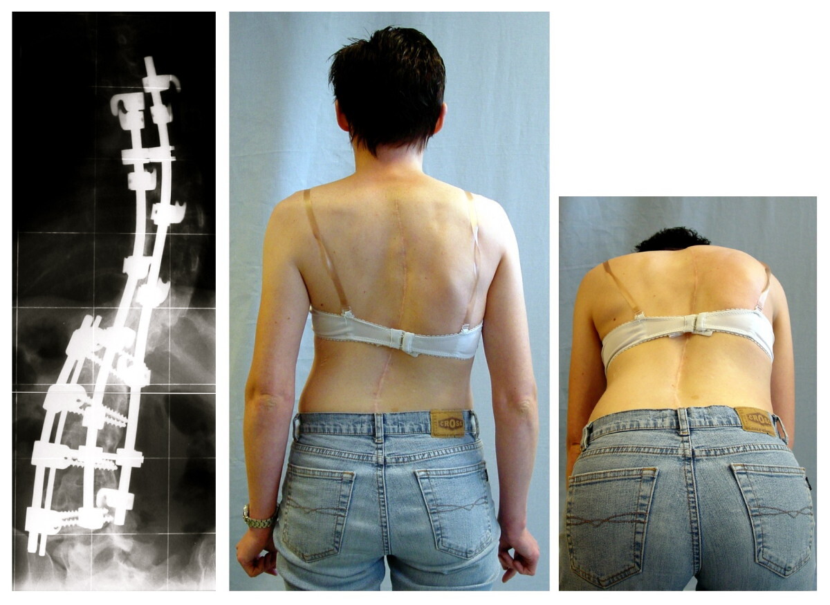
Posterior instrumented spinal fusion. Postoperative radiograph showing a dual-rod, segmental construct correcting an adolescent curve, with the clinical result and the healed midline incision. Weiss HR, Goodall D, Scoliosis 2008;3:9, via Wikimedia Commons (CC BY 2.0).
Posterior instrumented spinal fusion. Postoperative radiograph showing a dual-rod, segmental construct correcting an adolescent curve, with the clinical result and the healed midline incision. Weiss HR, Goodall D, Scoliosis 2008;3:9, via Wikimedia Commons (CC BY 2.0).
Approaches and level selection
Posterior spinal fusion is the workhorse. Anterior fusion (open or thoracoscopic) has a niche in single thoracolumbar/lumbar (Lenke 5) curves, saving distal levels and, by removing the anterior growth plates, avoiding crankshaft in the very immature, at the cost of measurable pulmonary-function loss.[65] Fusion levels are chosen using the stable, neutral and end vertebrae and related tools (the last-touched and sagittal-stable vertebrae), and stopping short of these causes “adding-on.”[66]
The most nuanced concept is selective thoracic fusion: in a structural main-thoracic curve with a flexible, non-structural compensatory lumbar curve (King II / Lenke 1C), the lumbar curve is left unfused to preserve lumbar motion, and it spontaneously balances out. The key technical caution is to avoid over-correcting the thoracic curve, which throws the patient into coronal decompensation; most immediate post-operative trunk shift resolves over 6-12 months as the lumbar curve compensates.[67] Thoracoplasty (convex rib resection) for the rib hump is now rarely needed because direct vertebral derotation addresses the axial deformity.[68]
Growth-friendly and fusionless surgery
For juvenile and early-onset idiopathic scoliosis the aim is to delay definitive fusion while controlling the curve and allowing the trunk and lungs to grow: traditional growing rods (lengthened every 6-12 months), VEPTR, magnetically controlled growing rods (MCGR) that avoid repeated open lengthenings, and Shilla growth-guidance.[69] The modern fusionless option for the skeletally immature adolescent is vertebral body tethering (VBT), a flexible cord under tension across convex anterior screws that exploits the Hueter-Volkmann principle to let the concave side catch up while preserving spinal motion. The ideal candidate has substantial growth remaining (open triradiate), and the main risk is over-correction.[70]
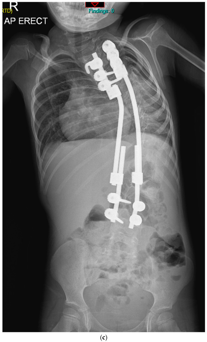
Growth-friendly surgery for early-onset scoliosis. Erect antero-posterior radiograph of a young child with a traditional growing-rod construct, which controls the curve while allowing the trunk and lungs to keep growing before any definitive fusion. Balasubramanian SG et al., J Clin Med 2026;15(2):754 (CC BY 4.0).
Growth-friendly surgery for early-onset scoliosis. Erect antero-posterior radiograph of a young child with a traditional growing-rod construct, which controls the curve while allowing the trunk and lungs to keep growing before any definitive fusion. Balasubramanian SG et al., J Clin Med 2026;15(2):754 (CC BY 4.0).
Part XI - Complications of surgery
- Neurological injury is the most feared complication, with an incidence below 1% at high-volume centres. It is guarded against by intra-operative neuromonitoring combining somatosensory evoked potentials (SSEP) and the more sensitive transcranial motor evoked potentials (MEP), backed up by the Stagnara wake-up test when signals change. Raising the mean arterial pressure alone restores a fifth of monitoring losses, and persistent changes mandate reversing the corrective manoeuvres.[71]
- Blood loss is reduced with tranexamic acid, cell salvage and controlled hypotension.[72]
- Infection complicates about 1.4-1.6% of AIS fusions (far less than neuromuscular cases), with obesity the major risk factor. An early deep infection is treated by debridement with implant retention, whereas a late, indolent infection (often Cutibacterium acnes) usually requires implant removal.[73]
- The crankshaft phenomenon is continued anterior column growth tethered by an isolated posterior fusion in a very immature child (open triradiate cartilage), producing progressive rotational deformity. It is defined radiographically by an increase in Cobb angle, rotation and RVAD, and is avoided by anterior fusion or growth-friendly surgery.[74]
- Proximal junctional kyphosis (PJK), defined as more than 10° of kyphosis between the upper instrumented vertebra and the two vertebrae above it, occurs in 9-27%, rarely symptomatic in adolescents but a real problem in adults. Adding-on / distal decompensation is the analogous distal problem, related to skeletal immaturity and level selection.[75]
- Superior mesenteric artery (SMA) syndrome (duodenal compression after curve correction or traction, presenting with bilious vomiting in a thin patient) and flat-back syndrome (the iatrogenic loss of lumbar lordosis of the Harrington era) round out the classic list.[76]
Part XII - Outcomes
Modern instrumented fusion maintains correction well: at 15 years or more, thoracic curves lose only a few degrees and the sagittal contours are preserved, with a very low pseudarthrosis rate.[77] Health-related quality of life is measured with the scoliosis-specific SRS-22 questionnaire, and notably the radiographic parameters correlate poorly with patient-reported outcomes (coronal balance is the one that tracks satisfaction).[78] Most patients miss only a few weeks of school and return to non-contact sport by about six months.[79] The chief long-term caveat is that fusion extending to L4 or below is associated with more lumbar disc degeneration, which is the rationale for sparing the distal lumbar segments wherever possible.[80] No randomised trial has ever compared surgery with non-operative care for surgical-range AIS.[81]
Part XIII - Adult and untreated late-onset scoliosis
Scoliosis in the adult is a different problem. Where the child is treated to prevent progression and its cosmetic and pulmonary sequelae, the adult is treated for pain, neurogenic symptoms from stenosis, and sagittal imbalance, and global sagittal alignment, more than the size or location of the curve, determines disability.[82] Surgery combines decompression, fusion and realignment (using osteotomies such as the pedicle subtraction osteotomy and pelvic fixation for long fusions to the sacrum), and it carries a substantially higher complication rate than adolescent surgery. This is one argument for treating a progressive curve while the patient is still young, when the operation is shorter, blood loss is less, fewer levels are needed, and the sacrum can usually be spared.[83] In older adults the SRS-Schwab classification, with its spinopelvic modifiers (pelvic incidence-lumbar lordosis mismatch, pelvic tilt and sagittal vertical axis), guides realignment.[84]
Bulgarian terminology (Боев / Boychev tradition) - glossary
The following Bulgarian equivalents bridge the international literature with the terminology of the Bulgarian school of orthopaedics (in the tradition of Бойчо Бойчев / Boycho Boychev) used in the state examination.
| English term | Bulgarian term (Cyrillic) | Transliteration |
|---|---|---|
| Idiopathic scoliosis | Идиопатична сколиоза | Idiopatichna skolioza |
| Scoliosis | Сколиоза | Skolioza |
| Scoliotic disease | Сколиотична болест | Skoliotichna bolest |
| Spine / vertebral column | Гръбначен стълб | Grabnachen stalb |
| Vertebra | Прешлен | Preshlen |
| Lateral curvature | Странично изкривяване | Stranichno izkrivyavane |
| Vertebral rotation | Ротация на прешлените | Rotatsiya na preshlenite |
| Rib hump / gibbus | Ребрена гърбица | Rebrena garbitsa |
| Cobb angle | Ъгъл по Коб | Agal po Kob |
| Curve / spinal curve | Кривина | Krivina |
| Thoracic | Гръден (торакален) | Graden (torakalen) |
| Lumbar | Поясен (лумбален) | Poyasen (lumbalen) |
| Structural curve | Структурна кривина | Strukturna krivina |
| Non-structural curve | Неструктурна (функционална) кривина | Nestrukturna (funktsionalna) krivina |
| Skeletal maturity | Костна зрялост | Kostna zryalost |
| Risser sign | Признак на Рисер | Priznak na Riser |
| Growth spurt | Пубертетен растежен скок | Pubertetna rastezhen skok |
| Forward-bend test (Adams) | Тест с навеждане напред (Адамс) | Test s navezhdane napred (Adams) |
| Scoliometer | Сколиометър | Skoliometar |
| Brace / orthosis | Корсет (ортеза) | Korset (orteza) |
| Milwaukee brace | Корсет тип Милуоки | Korset tip Miluoki |
| Spinal fusion / arthrodesis | Спинална фузия (артродеза) | Spinalna fuziya (artrodeza) |
| Instrumentation | Инструментация | Instrumentatsiya |
| Pedicle screw | Транспедикуларен винт | Transpedikularen vint |
| Curve progression | Прогресия на кривината | Progresiya na krivinata |
| Trunk asymmetry | Асиметрия на трупа | Asimetriya na trupa |
| Menarche | Менархе (първа менструация) | Menarhe (parva menstruatsiya) |
Image attributions
(Figure attributions and licences are listed in the figure MANIFEST and inserted with each image. All images are openly licensed [CC0 / CC BY / CC BY-SA] or used under their stated terms; any non-commercial [NC] item is flagged as such and must not be used in a commercial product.)
References
-
Lovell & Winter, Pediatric Orthopaedics, ch. 17 Idiopathic Scoliosis, p. 653; Rothman-Simeone, The Spine, idiopathic-scoliosis chapter, p. 408. Idiopathic scoliosis is a diagnosis of exclusion: congenital, neuromuscular, syndromic, tumoral, infective and intraspinal causes must first be ruled out.
-
Harms Study Group, Idiopathic Scoliosis: The Harms Study Group Treatment Guide, 2nd ed., pp. 28, 54; Lovell, p. 664; Rothman, p. 408.
-
Harms, p. 36; Lovell, pp. 648-649.
-
Moe’s Textbook of Scoliosis and Other Spinal Deformities, 3rd ed., pp. 58-59.
-
Lovell, pp. 652-653.
-
Moe’s, pp. 56-58 (0-3 / 3-10 / >10); Lovell, p. 652 (0-3 / 4-10 / 11-17); Rothman, p. 408 (to 2 yr 11 mo / 3-9 yr 11 mo / 10-17 yr 11 mo). Infantile sex ratio and left-sidedness: Growing Spine, p. 171; Lovell, p. 664.
-
Lovell, p. 664; Harms, p. 36.
-
Lovell, p. 665; Growing Spine, p. 172.
-
Growing Spine, p. 171; Lovell, p. 664.
-
Lovell, p. 664; Growing Spine, pp. 169-170.
-
Growing Spine, pp. 94, 171-172.
-
Lovell, pp. 664-665; Rothman, p. 408; Harms, pp. 54-55.
-
Moe’s, p. 105 (5-10° ≈ equal; 11-20° 1.6:1; >20° 6.4:1 girls:boys); Lovell, p. 665 (≈1:8 among those requiring treatment); Harms, p. 55.
-
Lovell, pp. 649, 654; Rothman, p. 409.
-
Harms, p. 50; Lovell, p. 652.
-
Harms, p. 44; Rothman, pp. 408-409; Growing Spine, pp. 49-50.
-
Harms, pp. 36, 49-50; Lovell, p. 653.
-
Harms, pp. 43, 46; Lovell, pp. 651-652.
-
Harms, pp. 43, 46.
-
Lovell, pp. 648-651; Harms, pp. 46-47.
-
Harms, pp. 36-39, 49, 53, 55.
-
Lovell, pp. 665-666.
-
Lovell, pp. 666-667.
-
Lovell, p. 664; Growing Spine, pp. 171-172.
-
Lovell, pp. 664-665; Growing Spine, p. 175; Rothman, p. 409.
-
Growing Spine, pp. 169, 176.
-
Harms, p. 54; Rothman, p. 1289.
-
Lovell, p. 666; Rothman, pp. 408-409.
-
Lovell, p. 667; Rothman, p. 1286.
-
Lovell, pp. 666-667; Moe’s, pp. 108-109.
-
Lovell, p. 666; Moe’s, pp. 106-107.
-
Harms, p. 60.
-
Moe’s, pp. 63-64, 68; Harms, pp. 60-61; Lovell, pp. 656-657.
-
Lovell, pp. 655-656, 663; Harms, pp. 61-62, 68; Moe’s, p. 68.
-
Harms, p. 63; Lovell, pp. 656, 658, 666.
-
Harms, pp. 75-76; Lovell, p. 666.
-
Harms, pp. 63, 67; Lovell, pp. 657-658.
-
Harms, p. 29; Moe’s, pp. 85-88; Lovell, pp. 658-659 (most surgeons accept a 6° difference as the criterion for progression).
-
Moe’s, pp. 88-90; Lovell, p. 660.
-
Moe’s, p. 59; Lovell, p. 658; Harms, p. 63.
-
Harms, p. 68.
-
Harms, pp. 65-68; Lovell, p. 658.
-
Lovell, pp. 665-666.
-
Lovell, pp. 658, 661; Moe’s, pp. 92-93; Harms, p. 70.
-
Lovell, pp. 661-663; Harms, p. 70.
-
Harms, pp. 56, 70-71; Lovell, p. 661.
-
Moe’s, p. 63; Lovell, p. 666.
-
Harms, p. 71.
-
Moe’s, pp. 257-258; Lovell, p. 687; Rothman, p. 1287.
-
Rothman, p. 1287; Lovell, p. 687.
-
Lovell, pp. 687-688; Harms, pp. 139, 144.
-
Harms, pp. 128, 144-148; Rothman, p. 1288.
-
Harms, pp. 139-140, 150; Lovell, p. 688.
-
Lovell, p. 667; Harms, p. 76.
-
Harms, pp. 75-76; Lovell, p. 668; Moe’s, p. 112.
-
Harms, p. 75; Lovell, pp. 667-668.
-
Harms, pp. 76-77; Lovell, pp. 668-669; Moe’s, pp. 112-116.
-
Harms, pp. 78-79, 84.
-
Harms, pp. 80-81.
-
Harms, pp. 77-78; Moe’s, pp. 116-117; Lovell, p. 670.
-
Growing Spine, pp. 535-540.
-
Rothman, pp. 410-412; Harms, pp. 78, 99.
-
Harms, pp. 127, 139-140; Lovell, p. 707.
-
Harms, pp. 99-102; Lovell, p. 701; Moe’s, p. 261.
-
Harms, pp. 140, 165, 258-259; Lovell, pp. 701-702; Rothman, p. 413.
-
Harms, pp. 139-143; Moe’s, p. 257.
-
Harms, pp. 127-135; Lovell, p. 688.
-
Harms, pp. 156, 164.
-
Lovell, pp. 679-680; Rothman, p. 412.
-
Harms, p. 102; Lovell, pp. 678, 707; Rothman, p. 412.
-
Harms, p. 278; Lovell, p. 704; Rothman, p. 1300.
-
Harms, pp. 276-277; Lovell, p. 704.
-
Harms, pp. 268-274.
-
Harms, p. 283; Lovell, p. 679.
-
Harms, pp. 104, 282; Lovell, p. 706.
-
Harms, pp. 280, 302.
-
Harms, pp. 253-254, 277, 309.
-
Harms, pp. 140, 256.
-
Harms, p. 292.
-
Harms, pp. 127, 260, 265.
-
Harms, pp. 262-265.
-
Rothman, pp. 1286-1287, 1301; Harms, pp. 298, 300.
-
Harms, pp. 298-303; Rothman, pp. 1297-1301.
-
Rothman, pp. 1288-1289; Harms, p. 298.