Contents
- Introduction and scope
- Part I - Definition and terminology
- Part II - Epidemiology
- Part III - Etiology and pathogenesis
- Part IV - Classification
- Part V - Clinical features
- Part VI - Imaging
- Part VII - Treatment
- Part VIII - Complications and sequelae
- Bulgarian terminology (Боев / Boychev tradition) - glossary
- Image attributions
Introduction and scope
Slipped capital femoral epiphysis (SCFE) is the displacement of the proximal femoral epiphysis relative to the metaphysis through the physis. It is the commonest hip disorder of adolescence, and a diagnosis missed at the cost of a hip.[1] The examiner expects the at-risk demographic and the endocrine screen for the atypical patient, the two classifications that matter (the temporal scheme and, above all, the Loder stable/unstable functional classification that drives prognosis, with the Southwick/Wilson severity grades), the radiographic signs (Klein’s line / Trethowan sign, the metaphyseal blanch sign of Steel), and the treatment logic built around in-situ fixation and the avascular-necrosis risk.
Two ideas run through the topic. First, the head stays in the acetabulum while the neck displaces anteriorly and rotates externally, so the epiphysis comes to lie posteromedially. The name is a misnomer, and this geometry explains both the clinical signs (out-toeing, obligate external rotation) and where the fixation screw must go. Second, stability, not slip magnitude, is the dominant determinant of outcome: a stable slip has a very low risk of avascular necrosis, whereas an unstable slip carries a risk of roughly a third to a half, which is why forceful reduction is avoided.
Part I - Definition and terminology
In SCFE the femoral head (capital epiphysis) remains within the acetabulum while the femoral neck and shaft displace anteriorly and rotate externally, so the head appears posteromedial relative to the neck.[2] It is the neck that moves, not the head, so the name is a misnomer (the older synonym is “adolescent coxa vara”). Histologically it is a Salter-Harris type I failure through the physis (chiefly its proliferative and hypertrophic zones), the hypertrophic zone being abnormally widened (from the normal 15-30% of physeal width up to about 80%).[3] The condition spans a spectrum, from a symptomatic “pre-slip” with physeal widening but no displacement to an acute severe displacement.
Part II - Epidemiology
SCFE presents around puberty (the adolescent growth spurt), with a mean age of about 13-13.5 years in boys and 11-12 years in girls, a male predominance (roughly 1.5-2:1), and a left-sided predilection.[4] The dominant risk factor is obesity (around 50-80% of patients are above the 80th-90th weight percentile). It is bilateral in a substantial proportion: about 20% at presentation, rising to perhaps 40-60% over time (Campbell quotes 25-40%), and most sequential (“metachronous”) slips occur within 18 months of the first.[5] Racial variation is marked, with the highest rates in Polynesian, then African-American and Hispanic children. An onset before age 10 or after about 16, or in an underweight child, is atypical and should prompt a search for an underlying disorder.
Part III - Etiology and pathogenesis
The final common pathway is that shear force across the physis exceeds its strength, whether a physiologic load on a weakened physis or an excessive load on a normal one.[6] The contributing factors are mechanical and endocrine:
- Mechanical: obesity; the pubertal growth spurt; relative femoral retroversion; an increasingly vertical (oblique) physis with a decreasing neck-shaft angle; thinning of the perichondrial ring and loss of the protective epiphyseal tubercle at puberty; and acetabular over-coverage. The shear across the head during gait can reach several times body weight.[7]
- Endocrine / metabolic: endocrinopathy underlies about 5-8% of cases and raises the risk roughly six-fold. The disorders to know are hypothyroidism, growth-hormone deficiency or therapy, hypogonadism and panhypopituitarism, plus renal osteodystrophy (renal rickets) and prior radiation.[8]
The practical rule (Loder and Greenfield) is the age-weight test for the “atypical” slip: a patient outside the ages of 10-16, or below the 50th weight percentile, is likely to have an underlying endocrine or metabolic cause and should be screened (thyroid function, plus growth-hormone evaluation for short stature). Routine screening of the typical obese adolescent is not warranted.[9]
Part IV - Classification
Three classifications are used together.
- Temporal (symptom-duration): acute (symptoms under 3 weeks), chronic (over 3 weeks, the commonest, 80-90%), and acute-on-chronic. This scheme has little prognostic value on its own.[10]
- Loder (functional / stability), the most prognostic. A stable slip is one in which the child can bear weight, with or without crutches; an unstable slip is one in which the child cannot bear weight even with crutches. This single distinction drives outcome: in Loder’s original series avascular necrosis occurred in 47% of unstable slips and 0% of stable ones, and pooled data give roughly 1.5% in stable versus about 33% in unstable slips.[11] One caveat is that clinical weight-bearing is only a surrogate, and Ziebarth found it correlates poorly with the physeal stability seen at open surgery.
- Severity (radiographic). The Southwick lateral head-shaft (epiphyseal-shaft) angle grades the slip mild (<30°), moderate (30-50°) and severe (>50°) (Campbell uses a 30°/60° division), and the Wilson percentage displacement grades it mild (<33%), moderate (33-50%) and severe (>50%).[12]
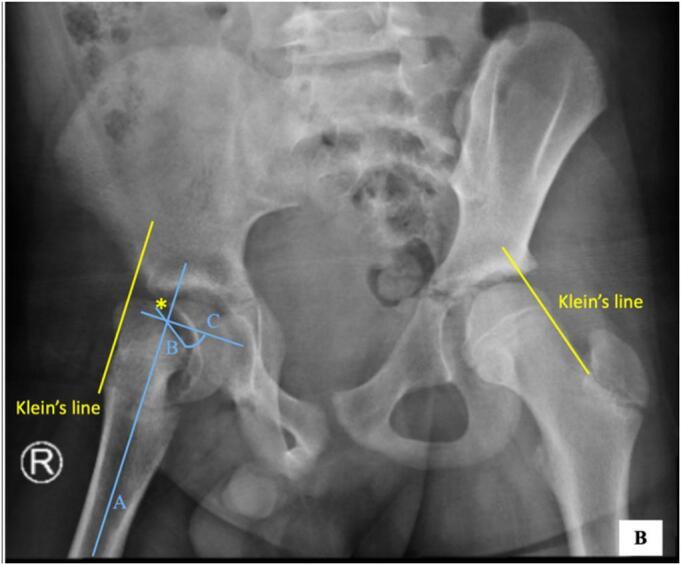
Severe (unstable) SCFE. Antero-posterior pelvis of a severe right-sided slip with marked posteroinferior displacement; Klein’s line is drawn on both sides (positive Trethowan on the right) and the slip angle is constructed. Kurniawan et al., Int J Surg Case Rep 2024;123:110264, Fig. 2 (CC BY 4.0).
Severe (unstable) SCFE. Antero-posterior pelvis of a severe right-sided slip with marked posteroinferior displacement; Klein’s line is drawn on both sides (positive Trethowan on the right) and the slip angle is constructed. Kurniawan et al., Int J Surg Case Rep 2024;123:110264, Fig. 2 (CC BY 4.0).
Part V - Clinical features
The classic presentation is a limp with pain in the hip, groin or medial thigh, or in the knee.[13] Referred knee or distal-thigh pain is the great pitfall. It is the only complaint in a quarter to nearly half of patients, and the resulting failure to examine the hip leads to delay and missed diagnoses, so the hip must be examined in every child presenting with knee or thigh pain. The affected limb is held short and externally rotated with an out-toeing gait; the hallmark sign is obligatory external rotation of the hip as it is flexed (Drehmann’s sign), with loss of internal rotation, abduction and flexion.[14] An unstable slip presents like a fracture, with sudden severe pain and inability to bear weight, often after minor trauma. A child in whom SCFE is suspected should be kept non-weight-bearing until it is excluded.
Part VI - Imaging
The standard study is an antero-posterior pelvis with a frog-leg lateral of both hips, the lateral being more sensitive for a mild slip. In a suspected unstable slip the frog-leg position is avoided (it can displace the slip); a cross-table (true) lateral is used instead.[15] The radiographic signs are:
- Klein’s line and the Trethowan sign: a line drawn along the superior border of the femoral neck normally intersects part of the lateral epiphysis; in SCFE it passes above the epiphysis (the positive Trethowan sign). The modified Klein line, a difference of 2 mm or more in the epiphysis lateral to the line compared with the other side, raises the sensitivity from about 40% to 80%.[16]
- The metaphyseal blanch sign of Steel: a crescentic area of increased density at the metaphysis from the head lying behind the neck.[17]
- Physeal widening and irregularity (the earliest, pre-slip sign), reduced epiphyseal height, and an increased teardrop-to-metaphysis distance.
The slip is measured with the Southwick angle. CT confirms physeal closure or screw position, and MRI is the best modality for the very early (pre-)slip and for detecting avascular necrosis.[18]
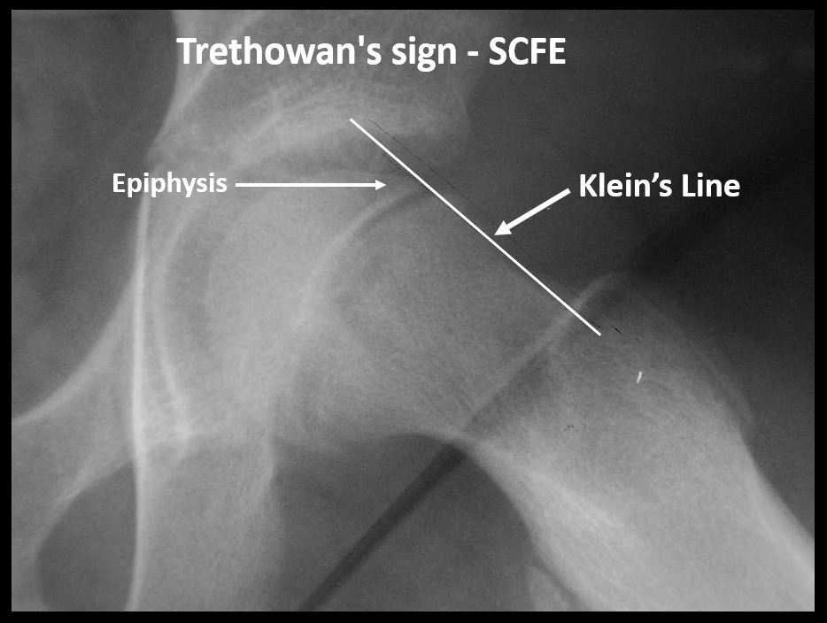
Klein’s line and the Trethowan sign. A line along the superior femoral neck normally clips the lateral epiphysis; here it passes above the slipped epiphysis (positive Trethowan sign). Dr. Vijaya Chandar, via Wikimedia Commons (CC0).
Klein’s line and the Trethowan sign. A line along the superior femoral neck normally clips the lateral epiphysis; here it passes above the slipped epiphysis (positive Trethowan sign). Dr. Vijaya Chandar, via Wikimedia Commons (CC0).
Part VII - Treatment
The goal is to stabilise the hip in situ, catching the slip early so it does not progress and complications are avoided. A chronic or stable slip is not forcibly reduced, because manipulation greatly increases the risk of avascular necrosis. The hip spica and bone-peg epiphysiodesis of the past are obsolete, and treatment is universally operative.[19]
Stable SCFE - in-situ fixation
The gold standard for a mild or moderate stable slip is in-situ fixation with a single percutaneous cannulated screw across the physis, which has the lowest rate of avascular necrosis; a second screw adds little mechanically while increasing the risk of joint penetration.[20] The screw is placed “centre-centre”: in the centre of the epiphysis on both views and perpendicular to the physis. Because the neck has displaced anteriorly, the screw is started on the anterior femoral neck (more proximal and posterior the more severe the slip), with several threads crossing into the epiphysis and the tip kept clear of the subchondral bone. Joint penetration must be excluded with a live-fluoroscopy “approach-withdraw” check, since persistent penetration causes chondrolysis.[21]
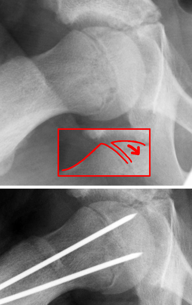
In-situ fixation. A slipped epiphysis (top, arrow) stabilised in situ with implants crossing the physis (bottom), without reduction. Dr. Jochen Lengerke, via Wikimedia Commons (public domain).
In-situ fixation. A slipped epiphysis (top, arrow) stabilised in situ with implants crossing the physis (bottom), without reduction. Dr. Jochen Lengerke, via Wikimedia Commons (public domain).
Unstable SCFE
The same principle applies, but the avascular-necrosis risk is high. Forceful manipulation is contraindicated (only the gentle, incidental reduction that occurs with positioning is accepted), and one or two screws are used, a second placed inferiorly, avoiding the superolateral quadrant and its vessels, sometimes with capsular decompression of the tamponading haematoma.[22] Timing is debated, but the evidence favours stabilising within 24 hours (or, if that is not possible, delaying about a week to let the inflammation settle). The modified Dunn (subcapital realignment) osteotomy is performed through a surgical hip dislocation with an extended retinacular soft-tissue flap that protects the medial femoral circumflex artery, and it allows anatomical reduction of a severe or unstable slip. In expert hands it can give a very low avascular-necrosis rate, but pooled series outside those centres report rates of around 18% or more, so it is reserved for experienced hip surgeons.[23]
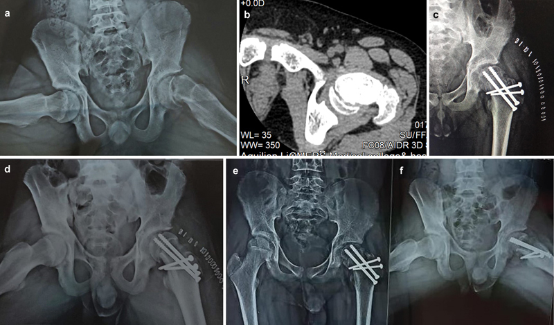
Modified Dunn realignment. A severe slip (a, with axial CT b and frog-lateral c) corrected by subcapital realignment through a surgical hip dislocation, fixed with cannulated screws (d-f). Bhuyan BK, Rev Bras Ortop 2024;59(6):e913-e921, Fig. 2 (CC BY 4.0).
Modified Dunn realignment. A severe slip (a, with axial CT b and frog-lateral c) corrected by subcapital realignment through a surgical hip dislocation, fixed with cannulated screws (d-f). Bhuyan BK, Rev Bras Ortop 2024;59(6):e913-e921, Fig. 2 (CC BY 4.0).
Severe chronic slip and prophylactic fixation
A severe chronic slip is fixed in situ and, if it leaves a disabling deformity, addressed later by a proximal femoral realignment osteotomy. The more proximal (subcapital/Dunn) the level, the better the correction but the higher the avascular-necrosis risk, so the intertrochanteric Imhäuser and base-of-neck osteotomies are favoured for safety, with osteochondroplasty of the residual cam bump.[24] Prophylactic fixation of the contralateral hip is debated. It is not routine, but it is recommended for a young child (open triradiate cartilage or a low modified Oxford bone-age score), for an endocrinopathy or renal disease, and where follow-up is unreliable.[25]
Part VIII - Complications and sequelae
- Avascular necrosis (osteonecrosis) is the dominant and most feared complication, strongly tied to the unstable slip (about 33-47%, versus near-zero in stable slips) and to forceful reduction. The blood supply at risk is the medial femoral circumflex artery and its ascending (retinacular) branches, which enter posterosuperiorly, hence the rule to keep hardware out of the superoposterior head.[26]
- Chondrolysis: acute cartilage necrosis with joint-space narrowing (under 3 mm) and stiffness; historically attributed to unrecognised pin penetration but now thought to have an immune component, since it occurs in untreated and spica-treated hips too.[27]
- Femoroacetabular (cam) impingement and premature osteoarthritis, the long-term residual-deformity sequel. The slip almost inevitably leaves a cam deformity (loss of head-neck offset, a raised alpha angle), producing impingement in a third or more of young adults and a recognised pathway to early hip arthritis; SCFE underlies a measurable share of “idiopathic” osteoarthritis.[28]
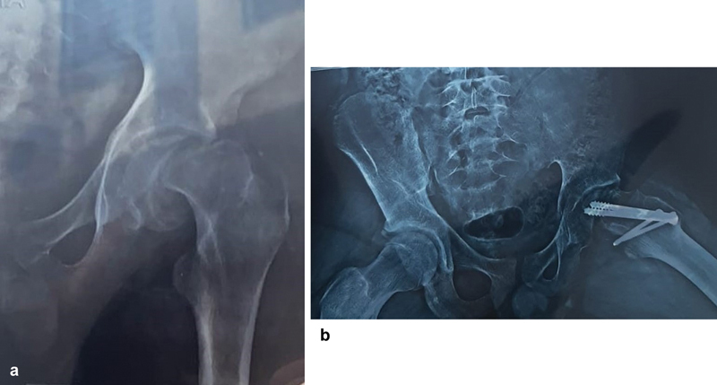
Avascular necrosis after SCFE. A severe slip (a) and the follow-up film after fixation (b) showing osteonecrosis of the femoral head - the dominant complication, far commoner after unstable slips. Bhuyan BK, Rev Bras Ortop 2024;59(6):e913-e921, Fig. 3 (CC BY 4.0).
Avascular necrosis after SCFE. A severe slip (a) and the follow-up film after fixation (b) showing osteonecrosis of the femoral head - the dominant complication, far commoner after unstable slips. Bhuyan BK, Rev Bras Ortop 2024;59(6):e913-e921, Fig. 3 (CC BY 4.0).
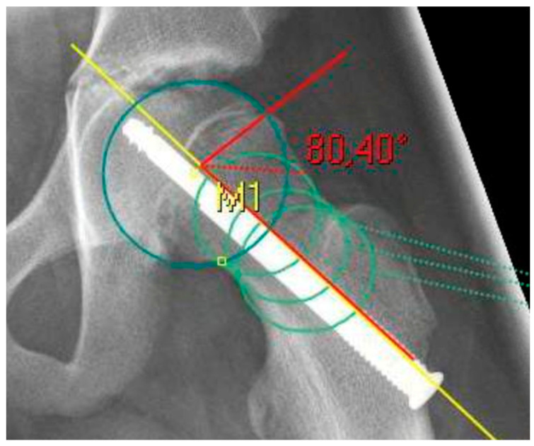
Residual cam deformity (femoroacetabular impingement). After a healed slip, the loss of head-neck offset gives a raised alpha angle (here 80.4°) - the cam morphology that drives impingement and early osteoarthritis. Wirries et al., Children (Basel) 2021;8(11):992, Fig. 1 (CC BY 4.0).
Residual cam deformity (femoroacetabular impingement). After a healed slip, the loss of head-neck offset gives a raised alpha angle (here 80.4°) - the cam morphology that drives impingement and early osteoarthritis. Wirries et al., Children (Basel) 2021;8(11):992, Fig. 1 (CC BY 4.0).
To tie the topic together: fix the stable slip in situ with a single central screw, handle the unstable slip urgently and gently (never forcibly), screen the atypical patient for endocrine disease, and counsel that avascular necrosis, which is governed by stability, and later cam impingement are the outcomes to fear.
Bulgarian terminology (Боев / Boychev tradition) - glossary
The following Bulgarian equivalents bridge the international literature with the terminology of the Bulgarian school of orthopaedics (in the tradition of Бойчо Бойчев / Boycho Boychev) used in the state examination.
| English term | Bulgarian term (Cyrillic) | Transliteration |
|---|---|---|
| Slipped capital femoral epiphysis | Хлъзгане на проксималната бедрена епифиза | Hlazgane na proksimalnata bedrena epifiza |
| Adolescent epiphysiolysis | Адолесцентна епифизиолиза | Adolestsentna epifizioliza |
| Femoral head (capital epiphysis) | Бедрена глава (капитална епифиза) | Bedrena glava (kapitalna epifiza) |
| Femoral neck | Бедрена шийка | Bedrena shiyka |
| Growth plate (physis) | Растежна зона (физа) | Rastezhna zona (fiza) |
| Stable slip | Стабилно хлъзгане | Stabilno hlazgane |
| Unstable slip | Нестабилно хлъзгане | Nestabilno hlazgane |
| Klein’s line | Линия на Клайн | Liniya na Klayn |
| Slip angle | Ъгъл на хлъзгане | Agal na hlazgane |
| In-situ fixation | Фиксация in situ (на място) | Fiksatsiya in situ (na myasto) |
| Cannulated screw | Канюлиран винт | Kanyuliran vint |
| Pinning | Пиниране (фиксация с винт) | Piniране (fiksatsiya s vint) |
| Osteotomy | Остеотомия | Osteotomiya |
| Avascular necrosis | Аваскуларна некроза | Avaskularna nekroza |
| Chondrolysis | Хондролиза | Hondroliza |
| Femoroacetabular impingement | Фемороацетабуларен импинджмънт | Femoroatsetabularen impindzhmant |
| Obesity | Затлъстяване | Zatlastyavane |
| External rotation | Външна ротация | Vanshna rotatsiya |
| Out-toeing | Походка с навънобърнати ходила | Pohodka s navanobarnati hodila |
| Hypothyroidism | Хипотиреоидизъм | Hipotireoidizam |
Image attributions
(Figure attributions and licences are listed in the figure MANIFEST and inserted with each image. All images are openly licensed [CC0 / CC BY / CC BY-SA] or used under their stated terms; any non-commercial [NC] item is flagged as such and must not be used in a commercial product.)
References
-
The Pediatric and Adolescent Hip, ch. 8 SCFE, p. 207; Lovell & Winter, Pediatric Orthopaedics, SCFE chapter, p. 1252.
-
Lovell, p. 1252; Pediatric and Adolescent Hip, p. 207; Campbell’s, p. 1768.
-
Lovell, p. 1259; Pediatric and Adolescent Hip, p. 209.
-
Lovell, p. 1252; Pediatric and Adolescent Hip, p. 211; Campbell’s, p. 1768.
-
Lovell, pp. 1252-1253, 1260-1261; Pediatric and Adolescent Hip, pp. 211, 223; Campbell’s, pp. 1768, 1772.
-
Lovell, p. 1253; Pediatric and Adolescent Hip, p. 208.
-
Pediatric and Adolescent Hip, p. 209; Lovell, pp. 1254, 1258; Campbell’s, p. 1768.
-
Lovell, pp. 1253-1254.
-
Pediatric and Adolescent Hip, pp. 213, 247; Lovell, p. 1253; Campbell’s, p. 1772.
-
Lovell, p. 1255; Pediatric and Adolescent Hip, p. 214; Campbell’s, pp. 1768-1769.
-
Pediatric and Adolescent Hip, pp. 214, 246; Lovell, p. 1255; Campbell’s, p. 1769.
-
Pediatric and Adolescent Hip, p. 215; Lovell, p. 1256; Campbell’s, p. 1769.
-
Pediatric and Adolescent Hip, p. 212; Lovell, p. 1255; Campbell’s, p. 1768.
-
Lovell, pp. 1255-1256; Pediatric and Adolescent Hip, p. 212.
-
Pediatric and Adolescent Hip, p. 215; Lovell, p. 1256.
-
Pediatric and Adolescent Hip, p. 215; Lovell, p. 1256; Campbell’s, p. 1768.
-
Pediatric and Adolescent Hip, pp. 215-216; Campbell’s, p. 1768.
-
Pediatric and Adolescent Hip, pp. 216-217; Lovell, pp. 1256-1257.
-
Pediatric and Adolescent Hip, pp. 217-219; Lovell, pp. 1261-1262.
-
Pediatric and Adolescent Hip, p. 219; Lovell, p. 1268; Campbell’s, p. 1769.
-
Lovell, pp. 1266-1268; Pediatric and Adolescent Hip, pp. 225-227.
-
Pediatric and Adolescent Hip, pp. 221-227; Campbell’s, pp. 1769-1773.
-
Pediatric and Adolescent Hip, pp. 229-235; Campbell’s, pp. 1773-1776.
-
Pediatric and Adolescent Hip, pp. 220-221, 236; Campbell’s, pp. 1772-1779.
-
Pediatric and Adolescent Hip, pp. 223-224; Lovell, pp. 1260-1261; Campbell’s, p. 1772.
-
Pediatric and Adolescent Hip, pp. 238-240; Lovell, pp. 1255, 1259-1260; Campbell’s, p. 1779.
-
Pediatric and Adolescent Hip, p. 245; Lovell, p. 1262; Campbell’s, p. 1780.
-
Pediatric and Adolescent Hip, p. 245; Lovell, pp. 1258, 1260-1261; Campbell’s, pp. 1780-1783.