Contents
- Introduction and Scope
- SECTION I - GENERAL CONSIDERATIONS
- I.1 The Spectrum of Bone and Joint Infection
- I.2 Routes of Infection and Pathogenesis
- I.3 Diagnostic Approach
- I.4 Principles of Treatment
- II.1 Acute Haematogenous Osteomyelitis
- II.2 Subacute Osteomyelitis and Brodie Abscess
- II.3 Chronic Osteomyelitis
- II.4 Special Forms
- II.5 Septic (Pyogenic) Arthritis
- SECTION III - OSTEOARTICULAR TUBERCULOSIS
- SECTION IV - SYNTHESIS AND EXAM AIDS
- References
- Figure Credits and Licences
Introduction and Scope
This review covers Topic 4 of the orthopaedics syllabus: the pyogenic (suppurative) infections of bone and joint (acute and chronic osteomyelitis and septic arthritis) and osteoarticular tuberculosis, of which spinal TB (Pott disease) is the most important. Two organising contrasts run through the whole topic. The first is pyogenic versus tuberculous. Pyogenic infection is acute, “hot,” enzyme-driven, and destroys cartilage within days; tuberculosis is chronic, “cold,” and indolent, sparing the joint space for months. The second is acute versus chronic. Once bone is dead and a biofilm has formed, antibiotics alone almost never cure, and surgery becomes essential.
Sources are the standard references in the library: Zimmerli (Bone and Joint Infections) for the modern microbiology and management of pyogenic infection, Campbell’s Operative Orthopaedics for operative principles, Tuli (Tuberculosis of the Skeletal System) as the dedicated text for osteoarticular TB, the paediatric texts Lovell & Winter and Salter (childhood osteomyelitis and septic arthritis), Greenspan for imaging, the Sanford Guide for antimicrobials, and the Bulgarian operative atlas of Boychev. Antibiotic and anti-tuberculous regimens that have moved on from the older textbooks are stated according to current practice and the IDSA and WHO guidelines, and flagged as such.
SECTION I - GENERAL CONSIDERATIONS
I.1 The Spectrum of Bone and Joint Infection
- Osteomyelitis - infection of bone (marrow, cortex, periosteum). Classified by route (Waldvogel: haematogenous; contiguous-focus/exogenous; secondary to vascular insufficiency/neuropathy) and, for surgical purposes, by Cierny-Mader anatomical + host staging. By tempo it is acute, subacute (Brodie abscess), or chronic. The dividing line is histological (the presence of dead bone) rather than a fixed time.[1]
- Septic (pyogenic) arthritis - infection within a synovial joint; a surgical emergency because cartilage is destroyed within days.
- Osteoarticular tuberculosis - a chronic granulomatous infection by Mycobacterium tuberculosis, reaching bone or joint haematogenously from a visceral focus; the spine is the commonest site.
I.2 Routes of Infection and Pathogenesis
- Haematogenous spread is the commonest route in children. In bone it seeds the metaphysis of rapidly growing long bones, because the metaphyseal capillary loops turn sharply into wide, slow-flowing venous sinusoids where bacteria are trapped, and the region is relatively poor in phagocytes.[2] In a joint, the vascular synovium, which lacks a limiting basement membrane, is seeded directly.
- Contiguous spread carries metaphyseal osteomyelitis into a joint where the metaphysis is intracapsular (hip, shoulder, ankle/elbow); in infants under about a year, transphyseal vessels let metaphyseal infection cross the open physis into the epiphysis and joint, threatening the growth plate.[3]
- Direct inoculation - open fracture, surgery, penetrating wound, joint injection.
- The cascade of acute osteomyelitis: nidus → inflammatory exudate → rising intramedullary pressure → pus tracks through the cortex to raise a subperiosteal abscess → periosteal stripping devascularises cortex → sequestrum and chronic infection.
I.3 Diagnostic Approach
- Inflammatory markers. ESR and CRP are sensitive but non-specific; CRP is the marker of choice for monitoring because it normalises far sooner than ESR. The white-cell count is the least sensitive. In children, a normal CRP and ESR make pyogenic infection unlikely (but do not exclude subacute or Kingella disease).[4]
- Microbiology is the goal. Blood cultures are positive in roughly half of cases. Bone biopsy/aspirate with culture (and histology) is the gold standard; ideally it is obtained within a 1-2-week antibiotic-free window, as prior antibiotics sharply reduce yield. For a joint, aspiration is mandatory.
- Imaging sequence. The plain radiograph lags 10-12 days behind the infection (early films are normal or show only soft-tissue swelling); it is still the first study. MRI is the most sensitive early test (marrow oedema: low T1, high T2/STIR) and best shows abscesses and soft-tissue extent. The three-phase bone scan is positive within 24-72 h and useful for multifocal or occult disease; labelled-WBC or PET studies help when prior surgery/fracture confounds. CT best shows cortical bone and sequestra.[5]
- The pathognomonic radiographic signs of chronic osteomyelitis (sequestrum, involucrum, cloaca) and the “probe-to-bone” test in the diabetic foot are diagnostic shortcuts.
I.4 Principles of Treatment
- Antibiotics: empirical cover (always including Staphylococcus aureus), then targeted to culture; route and duration individualised. The historical default was 4-6 weeks, much of it intravenous. Modern evidence (the OVIVA trial) shows oral is non-inferior to intravenous when a highly bioavailable agent and an intact gut allow, and for native vertebral osteomyelitis 6 weeks equals 12 weeks. Rifampicin (always in combination) is added for staphylococcal biofilm.[6]
- Surgery is required for an abscess, for failure of antibiotics, and for all chronic / dead-bone / implant-associated disease, because biofilm on dead bone or metal resists both host defences and antibiotics. The principles are drainage, radical debridement/sequestrectomy to bleeding “paprika-sign” bone, dead-space management (antibiotic beads/cement, Papineau open grafting, the Masquelet induced-membrane technique, muscle/free flaps), skeletal stabilisation, and soft-tissue coverage. The implant is treated: removed if stability allows, or retained under suppression until union and then removed.[7]
- Tuberculosis inverts the emphasis: anti-tuberculous chemotherapy is the mainstay, with rest and selective, monitored surgery.
II.1 Acute Haematogenous Osteomyelitis
Definition / epidemiology. Acute infection of the metaphysis of a growing long bone, predominantly of children (bimodal: under 2 years and 8-12 years; about half of childhood cases are under 5), with a male predominance. After physeal closure it becomes uncommon, and in adults it shifts to the vertebral column. Long-bone haematogenous osteomyelitis is rare in adults.[8]
Causative organisms - by age and host (high-yield).
- Staphylococcus aureus is the commonest organism at every age. MRSA and PVL-producing strains cause more destruction (deep-vein thrombosis, septic emboli, pathological fracture).
- Neonates (≤2 months): S. aureus, group B Streptococcus, and Gram-negative enterics (E. coli, Klebsiella).
- Under ~4 years: Kingella kingae - now often the commonest in this band; a fastidious Gram-negative that is culture-negative on routine media (needs blood-culture broth/PCR) and presents mildly, often afebrile with normal markers.
- Sickle-cell disease: Salmonella (and S. aureus), classically diaphyseal.
- Intravenous drug use / plantar puncture wound: Pseudomonas aeruginosa.
- Low-virulence/implant/post-spinal-surgery infection: coagulase-negative staphylococci and Cutibacterium acnes.[9]
Clinical features. Pain, local tenderness, refusal to use the limb; fever is variable (up to 40 % of children are afebrile). In neonates and infants the picture is non-specific (poor feeding, irritability, pseudoparalysis) and often multifocal. Because of transphyseal vessels and intracapsular metaphyses, it is frequently complicated by septic arthritis and growth-plate injury.[10]
Treatment. Prompt empirical antibiotics (covering S. aureus; add Gram-negative cover in neonates, Kingella/Hib cover under 4 years; vancomycin where MRSA prevalence is high). Surgery is indicated for a subperiosteal/intraosseous abscess or for failure to improve within 24-48 h of appropriate antibiotics, with drainage by cortical drilling/windowing. Modern paediatric regimens favour a short intravenous course (a few days) then oral switch once the child is improving and CRP is falling.[11]
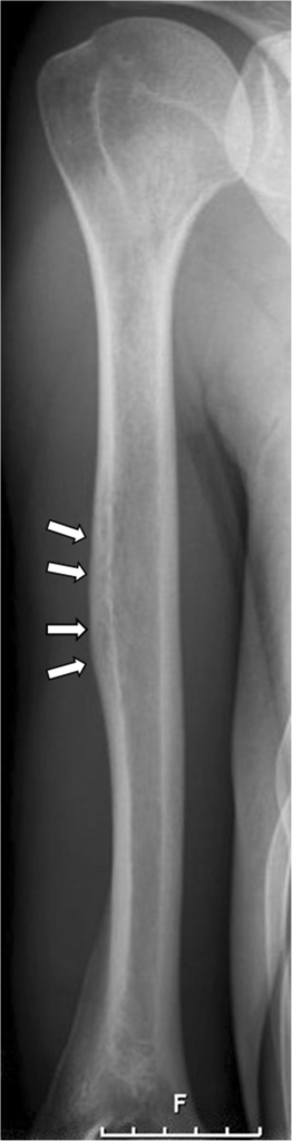
Figure 1. Acute osteomyelitis of the humeral diaphysis (radiograph). Prodinger et al., BMC Infect Dis 2013, CC BY 2.0, via PMC.
Figure 1. Acute osteomyelitis of the humeral diaphysis (radiograph). Prodinger et al., BMC Infect Dis 2013, CC BY 2.0, via PMC.
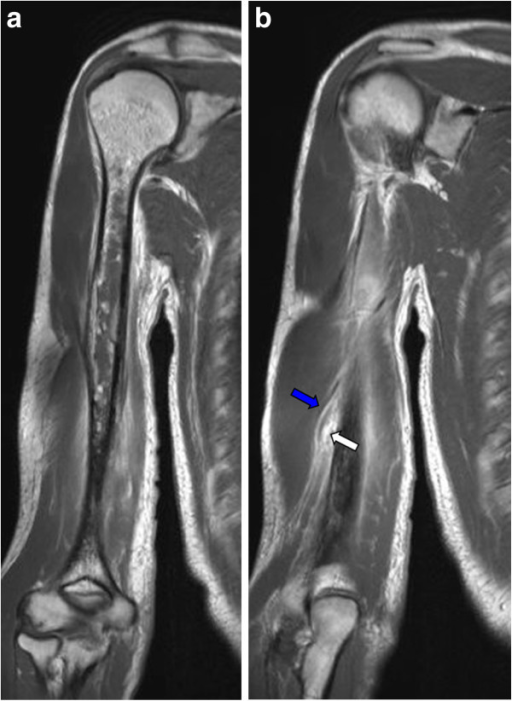
Figure 2. The same humerus on contrast MRI - marrow change is evident far earlier than on the radiograph. Prodinger et al., 2013, CC BY 2.0, via PMC.
Figure 2. The same humerus on contrast MRI - marrow change is evident far earlier than on the radiograph. Prodinger et al., 2013, CC BY 2.0, via PMC.
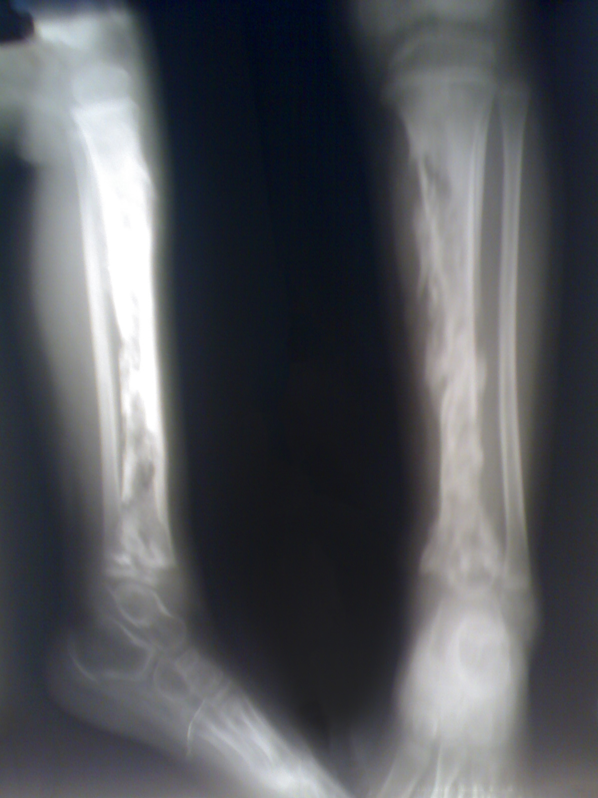
Figure 3. Acute osteomyelitis of the tibia in a child, with intraosseous abscess. sarindam7, CC BY-SA 4.0, Wikimedia Commons.
Figure 3. Acute osteomyelitis of the tibia in a child, with intraosseous abscess. sarindam7, CC BY-SA 4.0, Wikimedia Commons.
II.2 Subacute Osteomyelitis and Brodie Abscess
A more indolent infection (low-virulence organism and/or partly treated), with mild or no fever, often a normal white count, an ESR raised in only about half, and frequently negative cultures. The Brodie abscess is its localised form: a chronic intraosseous abscess in the metaphysis of a long bone in a child or young adult, seen radiographically as a well-defined lytic cavity with a sclerotic rim, which can mimic osteoid osteoma, chondroblastoma or Ewing sarcoma. S. aureus and S. epidermidis predominate.[12]
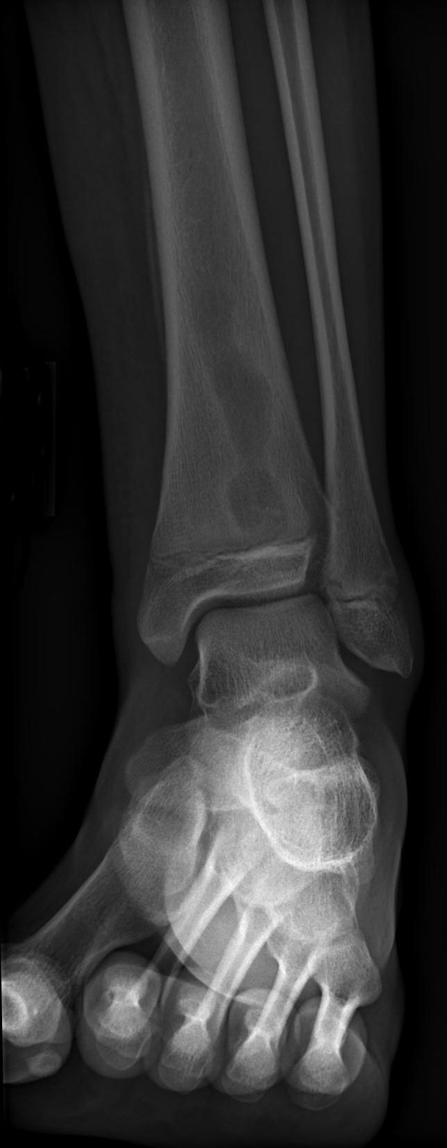
Figure 4. Brodie abscess - well-defined lytic lesion with a sclerotic rim, distal tibia. Jto410, CC BY-SA 3.0, Wikimedia Commons.
Figure 4. Brodie abscess - well-defined lytic lesion with a sclerotic rim, distal tibia. Jto410, CC BY-SA 3.0, Wikimedia Commons.
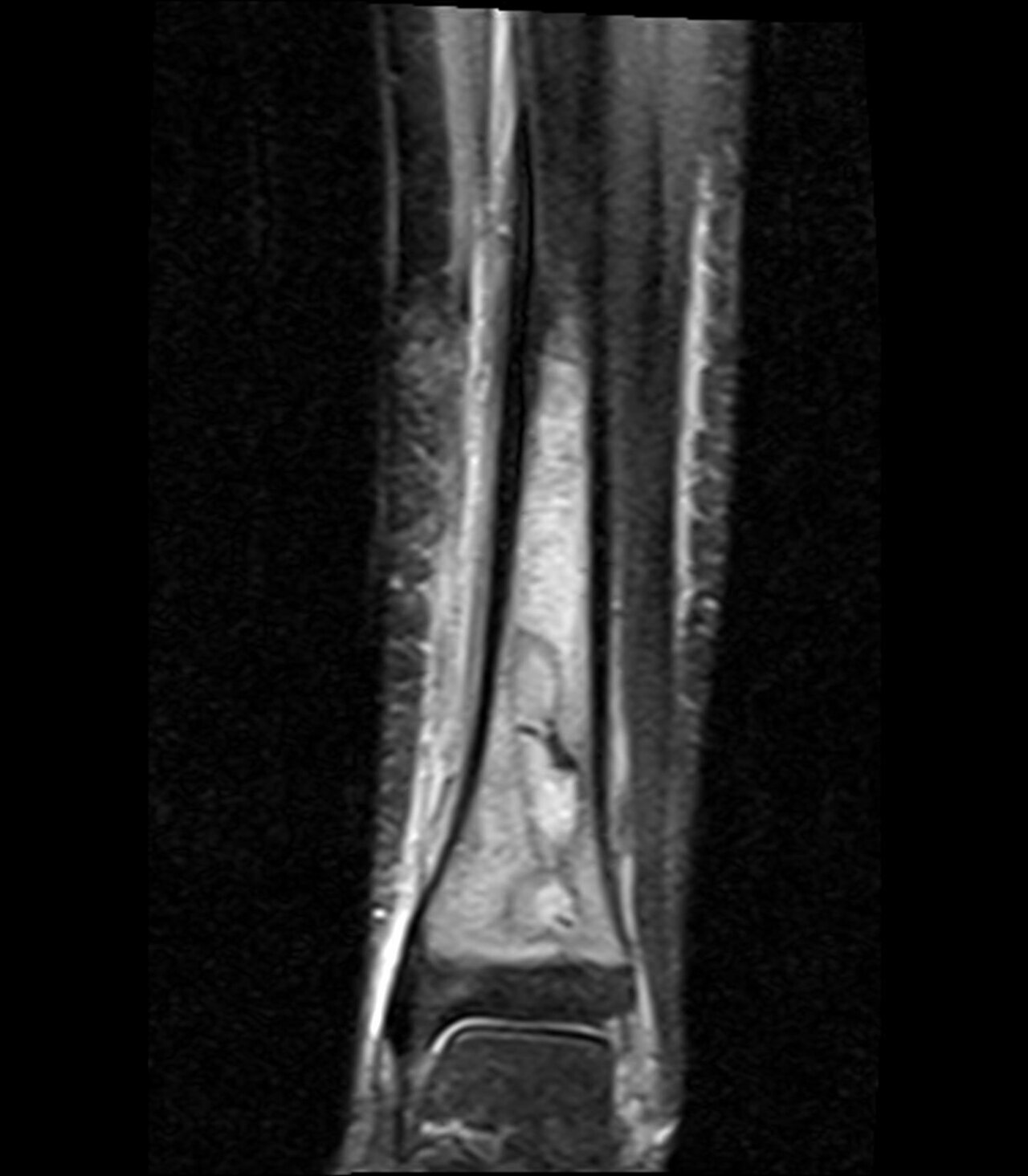
Figure 5. Brodie abscess - fluid-filled cavity on coronal STIR MRI. Jto410, CC BY-SA 3.0, Wikimedia Commons.
Figure 5. Brodie abscess - fluid-filled cavity on coronal STIR MRI. Jto410, CC BY-SA 3.0, Wikimedia Commons.
II.3 Chronic Osteomyelitis
Defining pathology - the classic quartet. Chronic osteomyelitis is infected dead bone within a compromised soft-tissue envelope. The terms to know: a sequestrum is a fragment of necrotic bone colonised by biofilm; an involucrum is the sheath of periosteal new bone that forms around it; a cloaca is the opening through which pus (and sometimes the sequestrum) drains; a sinus tract runs to the skin. Sequestrum, involucrum and cloaca together are radiologically pathognomonic. Biofilm, intracellular persistence, and small-colony variants explain antibiotic failure and recurrence decades later. Sinus-tract swabs do not reflect the deep organism; only bone culture does.[13]
Cierny-Mader staging combines an anatomical type (I medullary, II superficial, III localised full-thickness sequestrum excisable without instability, IV diffuse with instability) and a physiological host class (A normal, B compromised - systemic [Bs] and/or local [Bl], C treatment worse than disease), giving 12 stages. These guide whether treatment is simple or complex, curative or palliative, limb-sparing or ablative.[14]
Treatment is surgical: radical (oncological) debridement and sequestrectomy to bleeding bone, dead-space management (antibiotic beads/cement, Papineau grafting, the Masquelet induced-membrane technique, bone transport, muscle/free flaps), stabilisation and coverage, with antibiotics adjunctive. Malignant (squamous-cell) transformation - a Marjolin ulcer - should be suspected in a long-standing draining sinus that changes (new pain, foul discharge) and biopsied.[15]
Variants of chronic osteomyelitis:
- Sclerosing osteomyelitis of Garré - dense bony sclerosis with thickening but no abscess or sequestrum, in children/young adults, cultures usually negative; treated by fenestration ± antibiotics.
- Chronic recurrent multifocal osteomyelitis (CRMO) / SAPHO - an autoinflammatory, sterile (non-infectious) disease of children with relapsing multifocal bone pain (clavicle, metaphyses, spine), associated with palmoplantar pustulosis; cultures are negative - treat with NSAIDs (± TNF-α inhibitors/bisphosphonates), not antibiotics.[16]
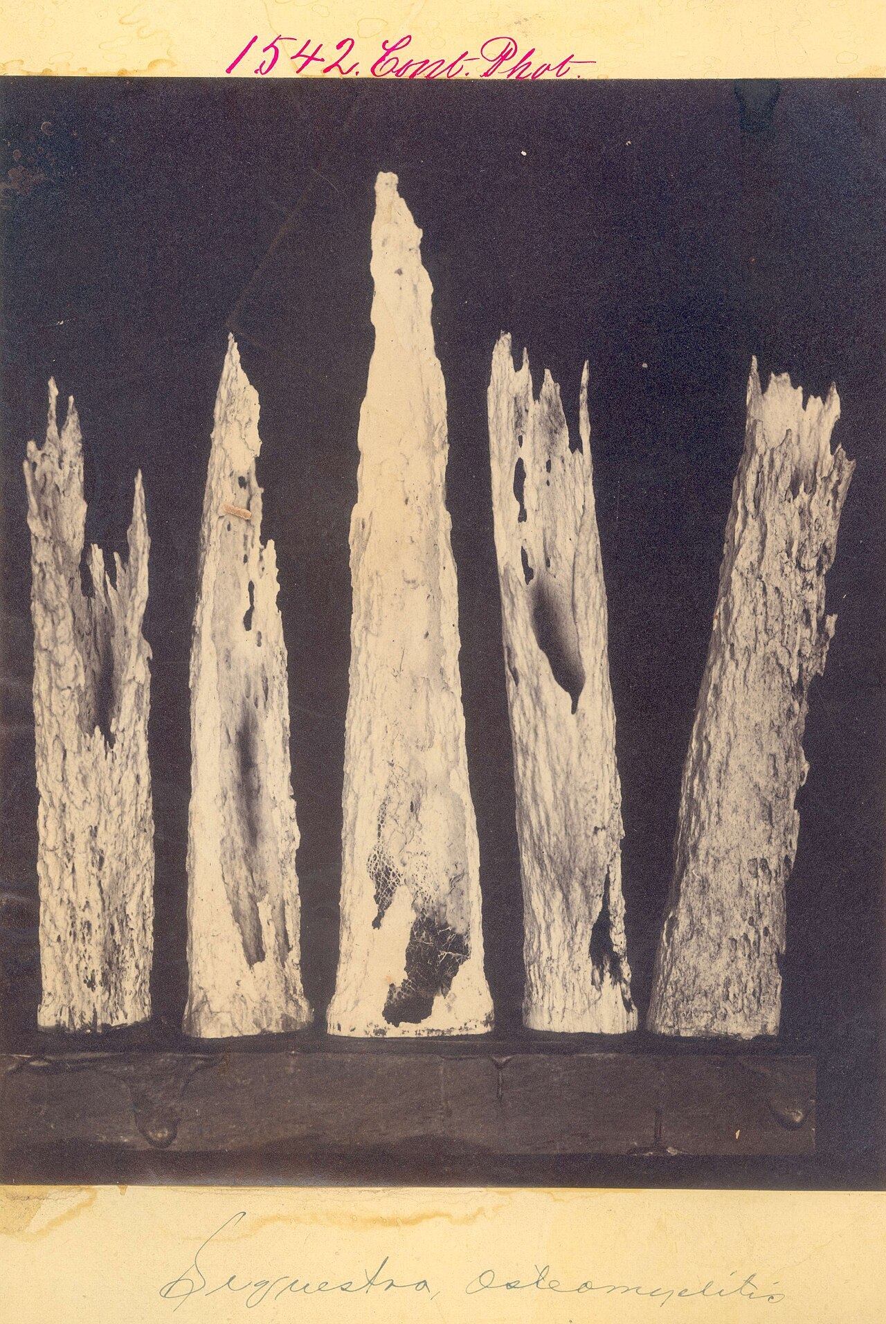
Figure 6. Chronic osteomyelitis - bony sequestra (necrotic, biofilm-colonised fragments). Otis Historical Archives, NMHM, CC BY 2.0, via Wikimedia Commons.
Figure 6. Chronic osteomyelitis - bony sequestra (necrotic, biofilm-colonised fragments). Otis Historical Archives, NMHM, CC BY 2.0, via Wikimedia Commons.
II.4 Special Forms
- Diabetic-foot osteomyelitis - osteomyelitis on a background of neuropathy and ischaemia, usually polymicrobial, spreading contiguously from a foot ulcer. The probe-to-bone test supports the diagnosis, and indium-WBC scanning helps separate it from neuropathic (Charcot) arthropathy. It is one of the few settings where antibiotic-only cure is realistic in selected cases.[17]
- Fracture-related / implant-associated infection (FRI) - defined by a biofilm on the implant; always needs surgery, and the implant is removed once the fracture has united (or earlier, with exchange).
- Vertebral osteomyelitis - the adult haematogenous form; S. aureus in ~50 %, with back pain and a raised ESR/CRP. MRI is the key test; it is managed mostly without surgery, with 6 weeks of antibiotics (equal to 12), reserving surgery for neurological deficit, instability, undrainable abscess or intractable pain.[18]
II.5 Septic (Pyogenic) Arthritis
Why it is an emergency. Within hours of infection, proteolytic enzymes from leucocytes, synovium and bacteria degrade articular cartilage (glycosaminoglycan loss is measurable by ~8 hours and cartilage destruction is well established by 4-6 days), and the process continues even after the bacteria are killed. Joint destruction is therefore prevented only by urgent drainage, not antibiotics alone.[19]
Epidemiology and joints. Most common in children (over 95 % haematogenous); in childhood it is about twice as common as osteomyelitis. The knee is the commonest joint in adults, whereas the hip predominates in infants and young children.[20]
Organisms mirror osteomyelitis (S. aureus overall; GBS/Gram-negatives in neonates; Kingella under 4 years*;* Salmonella* in sickle cell), with one important addition. In sexually active adults, Neisseria gonorrhoeae causes a disseminated syndrome of migratory polyarthralgia, tenosynovitis and dermatitis, often culture-negative from the joint and responsive to antibiotics without drainage. (Gonococcal arthritis in a child beyond the neonatal period is evidence of sexual abuse.)[21]
Clinical features. A hot, swollen, exquisitely painful joint with severe pain on any passive movement; the septic hip is held in flexion, abduction and external rotation (the position of maximum capsular volume), and an infant shows pseudoparalysis. Neonatal presentation is subtle and demands a full sepsis work-up.[22]
Diagnosis. Joint aspiration before antibiotics is mandatory. A synovial white-cell count above ~50,000/µL with >90 % neutrophils suggests sepsis (though counts overlap with crystal arthritis and JIA, and are lower with gonococcus/Kingella), assessed alongside Gram stain, culture and crystal examination. Add blood cultures and CRP/ESR; ultrasound detects a hip effusion and guides aspiration. The Kocher criteria distinguish a septic hip from transient synovitis in a child by four predictors - fever >38.5 °C, inability to bear weight, ESR ≥40 mm/h, white count >12,000/µL - giving a probability of septic arthritis of roughly 3 %, 40 %, 93 % and 99.6 % for 1-4 predictors (later validations give lower figures, so the criteria guide rather than replace clinical judgement).[23]
Treatment. Urgent joint drainage (repeated aspiration or arthroscopic lavage for accessible joints; open arthrotomy is mandatory for the hip and shoulder and in neonates, because pus under pressure in the hip threatens the femoral head’s single intra-articular blood supply with avascular necrosis) plus empirical-then-targeted intravenous antibiotics, with joint rest followed by early mobilisation. Adjunctive short-course dexamethasone improved outcomes in paediatric trials, though its routine use remains debated.[24]
Complications. Chondrolysis and secondary osteoarthritis, fibrous/bony ankylosis, and, in children, growth-plate damage, avascular necrosis of the femoral head, and joint dislocation/instability. These are worst with delay beyond 4 days, hip/shoulder involvement, neonatal age, and virulent (PVL-MRSA) organisms.[25]
Exam pearls (pyogenic). S. aureus at every age; neonate adds GBS/Gram-negatives, under-4 adds Kingella (culture-negative), sickle-cell Salmonella, IVDU/puncture Pseudomonas. CRP best for monitoring; radiograph lags 10-12 days, MRI most sensitive early, bone biopsy/aspiration the gold standard. Chronic osteomyelitis = dead bone + biofilm → surgery mandatory (sequestrum/involucrum/cloaca; Cierny-Mader). CRMO/SAPHO is sterile → NSAIDs not antibiotics. Septic arthritis is an emergency - aspirate (WBC >50,000, >90 % PMN), drain urgently (open arthrotomy for hip/neonate), Kocher criteria for the child’s hip.
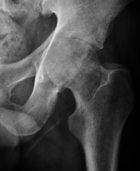
Figure 7. Septic arthritis of the hip with joint destruction. Ruiz Santiago et al., CC BY 4.0, Wikimedia Commons.
Figure 7. Septic arthritis of the hip with joint destruction. Ruiz Santiago et al., CC BY 4.0, Wikimedia Commons.
SECTION III - OSTEOARTICULAR TUBERCULOSIS
III.1 General Principles
Epidemiology. Skeletal tuberculosis occurs in about 1-3 % of all TB (and ~10-15 % of extrapulmonary TB). The spine is the commonest site (~50 %), followed by the hip and knee. In the endemic world it affects the first three decades of life; elsewhere it increasingly affects the elderly and immunocompromised (HIV, anti-TNF therapy, transplantation).[26]
Pathogenesis. Mycobacterium tuberculosis reaches bone or joint haematogenously from a primary pulmonary, lymph-node or visceral focus, usually 2-3 years after the primary infection; in the spine it travels via Batson’s paravertebral venous plexus. The disease is paucibacillary (few organisms, hence the low culture yield). Its hallmark is the caseating granuloma: epithelioid cells and Langhans giant cells with a lymphocyte cuff around central caseation necrosis. Liquefaction produces the “cold abscess”, a collection without the heat or erythema of pyogenic pus, which tracks along fascial planes. In a joint, the tuberculous pannus destroys cartilage from the periphery first, sparing the central weight-bearing surface and the joint space for months. This is the opposite of pyogenic arthritis.[27]
Diagnosis. Diagnosis demands a high index of suspicion in chronic, insidious mono-arthritis. The Mantoux test, a raised ESR, and chest imaging support it, but confirmation rests on biopsy with histology (caseating granuloma), acid-fast (Ziehl-Neelsen) staining and culture. Culture is the gold standard, though it is slow and positive in only a minority because the disease is paucibacillary. GeneXpert MTB/RIF (nucleic-acid amplification) is the modern rapid test, detecting M. tuberculosis and rifampicin resistance within hours on tissue, pus or synovial fluid; a negative result does not exclude TB.[28]
Anti-tuberculous chemotherapy. The first-line drugs are isoniazid (H), rifampicin (R), pyrazinamide (Z) and ethambutol (E), given as a two-phase regimen: an intensive phase of 2 months HRZE followed by a continuation phase of HR. Current WHO/standard practice treats drug-susceptible skeletal and spinal TB with a 6-month regimen (2HRZE/4HR), extended to 9-12 months in selected cases. The older literature (Tuli) used much longer 9-18-month courses, now regarded as historical. Isoniazid must be in every regimen. The drug-toxicity flags are worth memorising: ethambutol → optic neuritis; pyrazinamide → hepatotoxicity and hyperuricaemia; rifampicin → orange body fluids, hepatotoxicity and enzyme induction; streptomycin → eighth-nerve and renal toxicity. GeneXpert-detected rifampicin resistance triggers a WHO MDR-TB (bedaquiline-based) regimen.[29]
III.2 Spinal Tuberculosis (Pott Disease)
Sites and pattern. This is the commonest skeletal TB, with a thoracolumbar predilection, usually involving 2-3 contiguous vertebrae. The paradiscal lesion is commonest: destruction of adjacent vertebral end-plates with disc-space narrowing as the earliest sign. The avascular disc is not primarily infected; it loses its nutrition as the bone is destroyed. Other patterns are central (concentric collapse mimicking vertebra plana or tumour), anterior/subperiosteal (abscess stripping the anterior longitudinal ligament and scalloping several bodies), and posterior (neural arch).[30]
Deformity and abscess. Anterior vertebral collapse produces an angular kyphosis (gibbus): a knuckle with 1-2 bodies, a true gibbus with ≥3. It is worst in the thoracic spine and in children, where anterior growth-plate loss combines with continued posterior growth. The cold abscess tracks characteristically. Cervical disease drains to a retropharyngeal/prevertebral collection, while dorsolumbar disease drains down the psoas sheath to the groin and medial thigh (a psoas abscess gives an apparent hip-flexion deformity without rotational limitation).[31]
Pott paraplegia (neurological complications). This occurs in 10-30 %, highest with lower-thoracic disease; the classic distinction is:
- Early-onset paraplegia - during active disease, from inflammatory oedema, granulation tissue, a (cold) abscess or caseum pressing on the cord; potentially reversible, good prognosis.
- Late-onset paraplegia - years later, from healed disease, due to mechanical causes (a bony ridge/internal gibbus, sequestra, canal stenosis, peridural fibrosis, severe deformity); worse prognosis, and the mechanical cause must be removed surgically.
Cord oedema/myelitis is reversible, whereas myelomalacia, syrinx and cord atrophy are irreversible. Tuli’s neurological staging (I negligible → IV severe, with flexor spasms/flaccid paralysis/sphincter involvement) grades severity, and higher stages recover less.[32]
Imaging. The radiograph (disc narrowing, vertebral destruction, gibbus, paravertebral shadow) lags behind the disease. MRI is the best single test, showing pre-destructive marrow/disc oedema, paravertebral and epidural abscess, and cord status, while CT shows bony detail and posterior elements. Because imaging lags behind biological healing, apparent radiological deterioration in the first months despite clinical improvement should not cause alarm.[33]
Management - the “middle-path regimen” (Tuli). This means neither indiscriminate radical surgery nor pure neglect, but chemotherapy + rest/bracing + monitored, selective surgery: anti-tuberculous drugs, rest on a hard bed (traction for cervical lesions), serial ESR/imaging, gradual mobilisation in a spinal brace, and aspiration of abscesses (most paravertebral abscesses resolve on drugs and are not routinely drained). Surgery is indicated for a neurological deficit not improving (or worsening) on a fair drug trial, instability, a large/threatening abscess, doubtful diagnosis, recurrence, or prevention/correction of severe kyphosis. The classic operation is anterior radical debridement with an anterior strut bone graft (Hodgson & Stock’s “Hong Kong operation”), increasingly combined with posterior instrumentation. Laminectomy is contraindicated in ordinary paradiscal disease because it removes intact posterior support and worsens kyphosis; it is reserved for posterior-element disease or an intraspinal granuloma.[34]
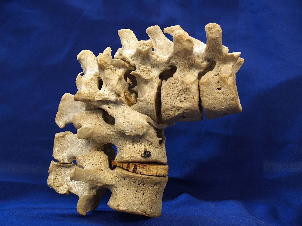
Figure 8. Pott disease - angular kyphosis (gibbus) from tuberculous vertebral collapse (dry specimen). Otis Historical Archives, NMHM, CC BY 2.0, via Wikimedia Commons.
Figure 8. Pott disease - angular kyphosis (gibbus) from tuberculous vertebral collapse (dry specimen). Otis Historical Archives, NMHM, CC BY 2.0, via Wikimedia Commons.
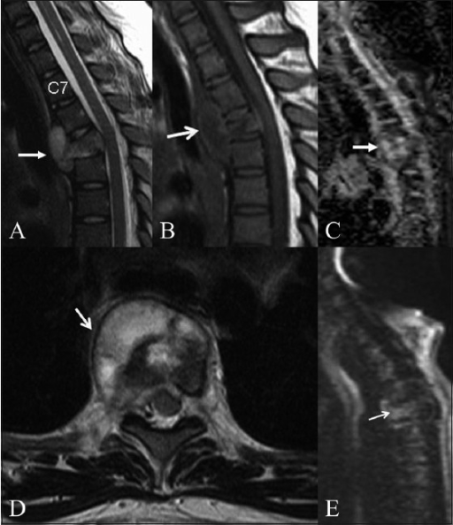
Figure 9. Spinal tuberculosis - vertebral destruction with paravertebral/epidural extension (MRI). Palle et al., Indian J Radiol Imaging 2010, CC BY, via PMC.
Figure 9. Spinal tuberculosis - vertebral destruction with paravertebral/epidural extension (MRI). Palle et al., Indian J Radiol Imaging 2010, CC BY, via PMC.
III.3 Tuberculosis of the Peripheral Joints
General pattern. Onset is synovial or osseous; tuberculous pannus then erodes the joint from the margins/non-contact areas first, so the joint space is preserved late. The radiographic signature is the Phemister triad: (1) juxta-articular osteoporosis, (2) peripheral (marginal) erosions, (3) gradual, late joint-space narrowing. Disease is monoarticular in ~90 %, and a cold abscess or sinus may form. This indolent, peripheral, space-sparing pattern contrasts sharply with the acute, central, rapidly space-narrowing destruction of pyogenic arthritis.[35]
Tuberculosis of the hip. Disease progresses through stages, from synovitis to advanced arthritis with subluxation. The classic deformity progression is characteristic: early synovitis holds the hip in flexion-abduction-external rotation (apparent lengthening), then adductor/flexor spasm pulls it into flexion-adduction-internal rotation with apparent then true shortening; advanced destruction of the acetabular roof and head gives the “wandering (migrating) acetabulum” with a broken Shenton line. Treatment depends on stage: chemotherapy with traction (which relieves spasm and prevents migration) and, for the healed destroyed joint, arthrodesis, or, in selected long-quiescent cases under drug cover, total hip replacement (with a 10-30 % reactivation risk).[36]
Tuberculosis of the knee. This presents as boggy synovial “white swelling” (tumor albus); advanced disease gives the **triple deformity
- flexion, posterior subluxation and external rotation** of the tibia. Treatment is chemotherapy with traction (double traction for the triple deformity); synovectomy/joint debridement for early disease; arthrodesis when more than half the cartilage is destroyed; and arthroplasty only in selected quiescent cases.[37]
Tuberculous osteomyelitis and spina ventosa. Tuberculous osteomyelitis of long bones is rare and treated like chronic pyogenic osteomyelitis (with drug cover). Spina ventosa is TB dactylitis of the short tubular bones in children: fusiform “ballooning” expansion of a metacarpal/metatarsal/phalanx by intramedullary granulation.[38]
Exam pearls (TB). Spine = commonest skeletal site (~50 %); skeletal TB ~1-3 % of all TB. Caseating granuloma + Langhans giant cells; cold abscess (no heat); paucibacillary → biopsy/culture + GeneXpert. Anti-TB = 2HRZE/4HR (WHO 6-month default; bone TB extendable). Pott: paradiscal lesion, disc spared early, gibbus, psoas cold abscess; early-onset paraplegia (active/inflammatory) good, late-onset (healed/mechanical) poor; middle-path regimen, Hong Kong operation, no laminectomy. Peripheral TB: Phemister triad, joint space preserved late (vs pyogenic), monoarticular; hip “wandering acetabulum,” knee “white swelling”+triple deformity; spina ventosa in children.
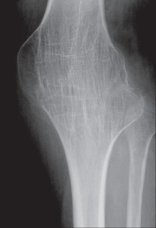
Figure 10. Tuberculous arthritis - bony ankylosis of the knee (a TB sequela). De Backer et al., Indian J Radiol Imaging 2009, CC BY, via PMC.
Figure 10. Tuberculous arthritis - bony ankylosis of the knee (a TB sequela). De Backer et al., Indian J Radiol Imaging 2009, CC BY, via PMC.
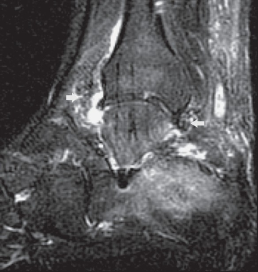
Figure 11. Tuberculous arthritis of the ankle - effusion and synovitis (fat-saturated T2 MRI). De Backer et al., 2009, CC BY, via PMC.
Figure 11. Tuberculous arthritis of the ankle - effusion and synovitis (fat-saturated T2 MRI). De Backer et al., 2009, CC BY, via PMC.
SECTION IV - SYNTHESIS AND EXAM AIDS
IV.1 Pyogenic vs Tuberculous Arthritis
| Feature | Pyogenic (septic) arthritis | Tuberculous arthritis |
|---|---|---|
| Onset / course | Acute, hot, febrile, days | Insidious, “cold,” chronic, months |
| Cartilage destruction | Central / diffuse, early (enzymatic) | Peripheral / marginal first |
| Joint space | Narrows early/rapidly | Preserved long, narrows late |
| Juxta-articular osteoporosis | Less prominent early | Marked (part of Phemister triad) |
| Abscess | Hot, acute pus | Cold abscess, sinus |
| Synovial WBC | >50,000/µL, >90 % PMN | Lower, lymphocyte-rich |
| Organism | S. aureus etc. | M. tuberculosis (paucibacillary) |
| Mainstay of treatment | Urgent drainage + antibiotics | Chemotherapy (+ rest, selective surgery) |
IV.2 Causative Organisms by Age / Host
| Setting | Likely organism(s) |
|---|---|
| All ages (commonest) | Staphylococcus aureus |
| Neonate | S. aureus, group B Streptococcus, Gram-negative enterics |
| Under ~4 years | Kingella kingae (culture-negative), S. aureus, S. pneumoniae |
| Sickle-cell disease | Salmonella (and S. aureus) - diaphyseal |
| IV drug use / foot puncture | Pseudomonas aeruginosa |
| Sexually active adult (joint) | Neisseria gonorrhoeae |
| Implant / post-spinal-surgery | Coagulase-negative staph, Cutibacterium acnes |
IV.3 Imaging Quick Reference
- Radiograph lags 10-12 days; first study; shows late destruction, sequestrum/involucrum/cloaca, gibbus.
- MRI is the most sensitive early study (marrow oedema) and is best for abscess, soft-tissue and cord involvement, and spinal TB.
- Three-phase bone scan turns positive in 24-72 h; in septic arthritis the uptake appears on both sides of the joint.
- CT shows cortical bone, sequestra and the posterior spinal elements, and guides biopsy.
- Ultrasound detects joint effusion (hip) and guides aspiration.
IV.4 Antimicrobial Regimens at a Glance (current practice)
- Acute osteomyelitis/septic arthritis: empirical anti-staphylococcal cover (vancomycin if MRSA risk), tailored by age; switch intravenous→oral on clinical improvement and falling CRP; add rifampicin for staphylococcal biofilm. Vertebral osteomyelitis: 6 weeks = 12 weeks.
- Tuberculosis: 2 months HRZE then 4 months HR (WHO 6-month default for drug-susceptible bone/spinal TB; extend to 9-12 months in selected cases); MDR-TB → WHO bedaquiline-based regimen.
IV.5 The Bulgarian Operative Tradition (Boychev) and Terminology
Boychev’s atlas places bone- and joint-infection surgery within its general operative chapters, and the Bulgarian school contributed several techniques relevant here.
- Drainage arthrotomy (дренажна артротомия) is used for a severely infected joint. The joint is opened, sometimes in several places, and drained with rubber tubes or glove strips under antibiotic cover.
- Joint resection (резекция на става) for joint infection: a single-pole resection drains and excises a destroyed pole, whereas a two-pole resection (intra- or extra-articular) excises both articular ends. In tuberculous osteoarthritis the synovium is excised as well (an incorporated synovectomy). The extra-articular resection does not open the joint and is therefore favoured for infection.
- Arthrodesis (артродеза) is a curative indication for osteoarticular tuberculosis and pyogenic osteoarthritis. Intra-articular fusion down to healthy cancellous bone eliminates the diseased joint, and nearly all such fusions are reinforced with iliac cancellous autografts. Filling a residual osteomyelitic cavity or Brodie abscess with cancellous bone chips is the one explicitly infective bone-grafting application.
- Distinctly Bulgarian contributions: the lengthening arthrodesis (Konforti and L. Boychev 1959 for knee/ankle, Holevich 1963 for hip), which fuses a destroyed joint while correcting limb shortening; the Holevich-Macholov compression hip arthrodesis (1966); and fascial / Vitallium-cup arthroplasty for the tuberculous hip and knee (Balchev, Hadzhistamov).
Glossary (Bulgarian → English).
| Bulgarian | English |
|---|---|
| остеомиелит; остеомиелитна кухина | osteomyelitis; osteomyelitic cavity |
| секвестр; секвестректомия | sequestrum; sequestrectomy |
| гноен артрит / остеоартрит; емпием | pyogenic arthritis / osteoarthritis; empyema |
| абсцес на Броди; фистула | Brodie abscess; sinus / fistula |
| костно-ставна туберкулоза | osteoarticular tuberculosis |
| студен абсцес; гибус | cold abscess; gibbus |
| дренажна артротомия; дренаж | drainage arthrotomy; drainage |
| резекция на става (едно-/двуполюсна) | joint resection (single-/two-pole) |
| синовектомия | synovectomy |
| артродеза; удължаваща артродеза | arthrodesis; lengthening arthrodesis |
| спонгиозни (костни) автоприсадъци | cancellous bone autografts |
| остеосинтеза; гипсова превръзка | osteosynthesis; plaster cast |
IV.6 High-Yield Revision Summary
- Acute haematogenous osteomyelitis - child, metaphysis, S. aureus; (neonate +GBS/GN, <4 y +Kingella, sickle Salmonella, IVDU Pseudomonas); radiograph lags, MRI early; antibiotics ± drainage of abscess.
- Brodie abscess - subacute, lytic lesion + sclerotic rim, often culture-negative.
- Chronic osteomyelitis - dead bone + biofilm (sequestrum/involucrum/cloaca); Cierny-Mader; surgery mandatory; Marjolin in a chronic sinus.
- CRMO/SAPHO - sterile, autoinflammatory → NSAIDs, not antibiotics. Garré - sclerotic, no sequestrum.
- Septic arthritis - emergency; aspirate (>50,000 WBC, >90 % PMN); urgent drainage (open arthrotomy for hip/neonate); Kocher criteria for the child’s hip; complications AVN/growth arrest.
- Skeletal TB - spine commonest; caseating granuloma + Langhans cells; cold abscess; biopsy/culture + GeneXpert; 2HRZE/4HR.
- Pott disease - paradiscal lesion, disc spared early, gibbus, psoas abscess; early-onset paraplegia good / late-onset poor; middle-path regimen, Hong Kong operation, no laminectomy.
- Peripheral TB arthritis - Phemister triad, joint space preserved late; hip “wandering acetabulum”; knee “white swelling” + triple deformity; spina ventosa in children.
References
- Zimmerli W (ed). Bone and Joint Infections: From Microbiology to Diagnostics and Treatment. 2nd ed. Hoboken: Wiley-Blackwell; 2021.
- Azar FM, Beaty JH, eds. Campbell’s Operative Orthopaedics. 14th ed. Philadelphia: Elsevier; 2020 (Part VII, Infections - Chs 20-23).
- Tuli SM. Tuberculosis of the Skeletal System: Bones, Joints, Spine and Bursal Sheaths. 4th ed. New Delhi: Jaypee; 2010.
- Flynn JM, Weinstein SL, et al., eds. Lovell and Winter’s Pediatric Orthopaedics. 8th ed. Philadelphia: Wolters Kluwer; 2020 (musculoskeletal infection).
- Salter RB. Textbook of Disorders and Injuries of the Musculoskeletal System. 3rd ed. Baltimore: Williams & Wilkins; 1999.
- Greenspan A, Beltran J. Orthopedic Imaging: A Practical Approach. 6th ed. Philadelphia: Wolters Kluwer; 2015.
- Gilbert DN, Chambers HF, et al. The Sanford Guide to Antimicrobial Therapy 2022. Antimicrobial Therapy, Inc.
- Berbari EF, Kanj SS, Kowalski TJ, et al. IDSA Clinical Practice Guideline for the Diagnosis and Treatment of Native Vertebral Osteomyelitis in Adults. Clin Infect Dis. 2015;61(6):e26-e46.
- World Health Organization. WHO Consolidated Guidelines on Tuberculosis: Treatment - Drug-Susceptible Tuberculosis Treatment. Geneva: WHO (current edition).
- Li HK, Rombach I, Zambellas R, et al. (OVIVA). Oral versus intravenous antibiotics for bone and joint infection. N Engl J Med. 2019;380(5):425-436.
- Bernard L, Dinh A, Ghout I, et al. Antibiotic treatment for 6 weeks versus 12 weeks in patients with pyogenic vertebral osteomyelitis: a randomised controlled trial. Lancet. 2015;385(9971):875-882.
- Бойчев Б. (Boychev B.) Хирургическа ортопедия (Surgical Orthopaedics). Sofia.
Note on currency: antibiotic and anti-tuberculous regimens (intravenous-to-oral switch and OVIVA; 6 = 12 weeks for vertebral osteomyelitis; the WHO 2HRZE/4HR regimen; GeneXpert MTB/RIF) reflect current IDSA/WHO practice and post-date parts of the older textbooks; where Tuli’s drug durations (9-18 months) conflict with the WHO 6-month regimen, current practice is followed.
Figure Credits and Licences
All figures are radiographs, CT/MR images, or anatomical specimens reproduced from openly-licensed sources; each remains under its original licence, attributed below.
- Fig 1 & 2 Acute osteomyelitis, humerus (radiograph + MRI) - Prodinger PM et al., BMC Infect Dis 2013;13:266, CC BY 2.0, via PMC. https://www.ncbi.nlm.nih.gov/pmc/articles/PMC3679722/
- Fig 3 Osteomyelitis of the tibia in a child - sarindam7, CC BY-SA 4.0, Wikimedia Commons. https://commons.wikimedia.org/wiki/File:Osteomyelitis_of_Tibia_in_Child.jpg
- Fig 4 & 5 Brodie abscess (radiograph + STIR MRI) - Jto410, CC BY-SA 3.0, Wikimedia Commons. https://commons.wikimedia.org/wiki/File:BrodieAbscessRadiograph.jpg
- Fig 6 Osteomyelitic sequestra (specimen) - Otis Historical Archives, NMHM, CC BY 2.0, via Wikimedia Commons. https://commons.wikimedia.org/wiki/File:4821323599_676b57f7b5_bSEQUESTRA_OSTEOMYELITIS.jpg
- Fig 7 Septic arthritis of the hip - Ruiz Santiago F et al., CC BY 4.0, Wikimedia Commons. https://commons.wikimedia.org/wiki/File:X-ray_of_septic_arthritis_of_the_hip.jpg
- Fig 8 Tuberculous kyphosis (gibbus) specimen - Otis Historical Archives, NMHM, CC BY 2.0, via Wikimedia Commons. https://commons.wikimedia.org/wiki/File:Spine_-_Kyphosis_from_Tuberculosis.jpg
- Fig 9 Spinal tuberculosis (MRI) - Palle L et al., Indian J Radiol Imaging 2010;20:279, CC BY, via PMC. https://www.ncbi.nlm.nih.gov/pmc/articles/PMC3056625/
- Fig 10 & 11 Tuberculous arthritis, knee + ankle - De Backer AI et al., Indian J Radiol Imaging 2009;19:176, CC BY, via PMC. https://www.ncbi.nlm.nih.gov/pmc/articles/PMC2766888/


-
Zimmerli, Bone and Joint Infections, 2nd ed., pp.287-291; Campbell’s Operative Orthopaedics, Ch.21 p.926.
-
Greenspan, Orthopedic Imaging, 6th ed., p.1991; Campbell’s p.927; Zimmerli p.296.
-
Zimmerli pp.145, 296; Lovell & Winter’s Pediatric Orthopaedics, p.1206.
-
Zimmerli pp.297-299, 318-319; Campbell’s p.929; Lovell p.1220.
-
Campbell’s pp.929-930; Greenspan pp.1992-1994; Zimmerli pp.298, 321.
-
Zimmerli pp.323-326, 352-353 (OVIVA - Li et al., N Engl J Med 2019; vertebral 6=12 weeks - Bernard et al., Lancet 2015); regimens per Sanford Guide / IDSA current practice.
-
Campbell’s pp.935-941; Zimmerli pp.290-291, 347-352. # SECTION II - PYOGENIC INFECTIONS
-
Campbell’s pp.926-927; Zimmerli pp.295, 312.
-
Campbell’s pp.927-928; Zimmerli pp.295-302.
-
Campbell’s p.928; Zimmerli pp.296-297.
-
Campbell’s pp.930-931; Zimmerli pp.304-305; regimens per Sanford/IDSA current practice.
-
Campbell’s pp.931-932; Greenspan pp.2001-2004.
-
Greenspan p.2002; Zimmerli pp.347-350; Campbell’s pp.932-935.
-
Campbell’s pp.933-934; Zimmerli pp.291-292.
-
Campbell’s pp.935-941, 933; Zimmerli pp.351-352.
-
Campbell’s pp.941-943; Zimmerli pp.290-291.
-
Zimmerli pp.290, 349, 351; Campbell’s p.934.
-
Zimmerli pp.312-326 (6=12 weeks - Bernard et al., Lancet 2015).
-
Lovell pp.1219-1220; Campbell’s p.959; Zimmerli pp.139, 150.
-
Zimmerli pp.139, 146, 162; Lovell pp.1203, 1206.
-
Zimmerli pp.140-143; Campbell’s p.960.
-
Lovell p.1222; Zimmerli pp.146-147.
-
Zimmerli pp.147-148; Lovell pp.1259-1263 (Kocher criteria and the lower revalidation/Luhmann probabilities); Campbell’s p.958.
-
Zimmerli pp.151-154; Campbell’s pp.960-961; antibiotic regimens per Sanford/IDSA current practice.
-
Zimmerli pp.151, 155; Campbell’s p.959.
-
Tuli, Tuberculosis of the Skeletal System, pp.23-25, 212; Campbell’s pp.985-986.
-
Tuli pp.28-32, 29 (peripheral cartilage destruction); Campbell’s p.986.
-
Tuli pp.40-43, 53-56 (NAAT/PCR; GeneXpert per current WHO/standard practice); Campbell’s p.986.
-
Tuli pp.64-83 (drugs; Tuli’s longer durations flagged); the 6-month 2HRZE/4HR default and MDR regimens per WHO/standard practice; Campbell’s p.990 endorses the shorter 6-9-month rifampicin-based course.
-
Tuli pp.219-232 (types; disc relatively spared/indolent vs pyogenic).
-
Tuli pp.214-215, 225-228.
-
Tuli pp.258-267 (Griffiths-Seddon-Roaf early vs late; Tuli staging Table 22.3).
-
Tuli pp.219-235.
-
Tuli pp.285-290, 337-341 (middle-path regimen; surgical indications; Hong Kong operation; laminectomy contraindicated).
-
Tuli pp.130, 91, 133; Campbell’s p.986 (relative joint-space sparing). The Phemister triad is the standard term for these findings.
-
Tuli pp.88-125 (stages, deformity, wandering acetabulum, salvage and reactivation risk).
-
Tuli pp.130-147 (white swelling, triple deformity, treatment by stage).
-
Campbell’s pp.997-998; Tuli pp.155-158.