Contents
- Scope and orientation
- Part I - Proximal Femoral Focal Deficiency (Congenital Femoral Deficiency)
- Part II - Fibular hemimelia (longitudinal fibular deficiency)
- Part III - Tibial hemimelia (longitudinal tibial deficiency)
- Part IV - Congenital pseudarthrosis of the tibia (CPT)
- Part V - Congenital angular deformities (bowing) of the tibia
- Bulgarian terminology glossary
- Figure credits and licences
Scope and orientation
This topic gathers the major congenital longitudinal and dysplastic deficiencies of the lower limb that an examiner expects to be discussed together, because their differential diagnosis and decision-making overlap heavily:
- Proximal femoral focal deficiency (PFFD) / congenital femoral deficiency (CFD), the femoral segment;
- Fibular hemimelia (longitudinal fibular deficiency), the commonest long-bone deficiency;
- Tibial hemimelia (longitudinal tibial deficiency), the rare, often inherited one;
- Congenital pseudarthrosis of the tibia (CPT) and the congenital angular deformities (bowing) of the tibia, which sit at the interface of “anomaly” and “deformity.”
Three threads run through the whole topic and are worth fixing in mind before the detail:
- These deficiencies travel together. A femoral deficiency is accompanied by a fibular deficiency in the large majority of cases, and the foot and ankle problems of the fibula frequently dictate what can be done for the femur. Examine the whole limb.
- The governing surgical question is almost always the same: what is the predicted limb-length discrepancy at skeletal maturity, and is the proximal joint (hip or knee) stable enough to reconstruct? The answer sorts each patient into “reconstruct and lengthen” versus “ablate/realign and fit a prosthesis.”
- For the bowed infantile tibia, the direction of the bow is the diagnosis. Anterolateral is sinister, posteromedial is benign, anteromedial points to the fibula. Memorising this single rule prevents the classic catastrophe: osteotomising a bowed tibia that was actually pre-pseudarthrotic.
A Bulgarian terminology glossary and a viva self-test appendix follow the clinical sections, and full image credits are listed at the end.
Part I - Proximal Femoral Focal Deficiency (Congenital Femoral Deficiency)
I.1 Definition and terminology
Proximal femoral focal deficiency (PFFD) is the historical, radiograph-based name for the severe end of a single congenital spectrum. The contemporary umbrella term is congenital femoral deficiency (CFD), which unites what older texts split into two diagnoses, the mild “congenital short femur” and the severe “PFFD”, once it was recognised that these are points on one continuum of the same developmental failure.[1]
The spectrum runs from a mildly hypoplastic short femur (with mild coxa vara and an anterolateral femoral bow), through severe coxa vara with a proximal femoral pseudarthrosis, to complete absence of the proximal femur, and at the extreme of the entire femur.[2]
CFD is a deficiency of a whole developmental field, not of one bone. Although the femoral abnormality dominates the radiograph, the acetabulum, hip musculature, vasculature, knee cruciate ligaments, tibia, fibula and foot are all involved to varying degrees.[3] The distal limb is frequently the part that determines management, most obviously through the very common coexisting fibular deficiency.
I.2 Epidemiology and associations
- Incidence ≈ 1 in 50,000 live births. The classic figure of 1/52,029 (Edinburgh Register) rests on a single case and is acknowledged within the source literature itself as probably inaccurate; the practical round number is ~1/50,000.[4]
- Bilateral in ~15% of cases; bilateral involvement is uncommon and tends to cluster with recognised genetic syndromes (e.g. Pierre-Robin).[5]
- Association with fibular hemimelia: 70-80%. This is the single most important association. The deficient, valgus, ball-and-socket ankle of fibular deficiency is “almost part of” CFD and is the feature that most often contraindicates a Van Nes rotationplasty. (One section of the same text gives “up to 70%”; cite the 70-80% range.)[6]
- ~50% have anomalies of the other limbs.[7]
- Cruciate-ligament deficiency is near-universal. On MRI (Manner et al., 34 knees) the ACL was abnormal in 100% (85% absent, 15% hypoplastic); the PCL was absent in 24% and hypoplastic in 21%. An arthroscopic series found the ACL deficient in 71% and the PCL in 48%, with an anterior drawer in every patient. The CFD knee is always unstable in the antero-posterior plane.[8]
- Coxa vara is the most constant proximal deformity; the typical pattern is proximal coxa vara with distal femoral valgus. Associated findings include a dysplastic/retroverted acetabulum and a hypoplastic or dislocated patella.[9]
I.3 Embryology and aetiology
CFD is sporadic and, in the unilateral form, essentially not inherited: in Paley’s series of more than 1,000 unilateral cases, only one patient had a parent with unilateral CFD.[10] The current model is a somatic insult to the proximal femoral anlage in the developing limb bud; a heritable (germ-line) mechanism is invoked only for some multi-limb or syndromic cases.[11]
A teratogenic mechanism is supported by the thalidomide experience: thalidomide reliably produces femoral deficiency, and the CFD phenotype resembles its effects, with the timing of a transient exposure determining severity.[12] In non-pseudarthrosis cases, MRI confirms that a cartilaginous anlage still bridges the head and shaft even when nothing is ossified on plain film. This is the basis of the “delayed ossification overstages the deformity” pitfall discussed below.[13] Heritable exceptions that do show inheritance include femoral hypoplasia-unusual facies syndrome (autosomal dominant) and Goltz syndrome (X-linked).[14]
I.4 Classification
Several systems coexist. The examiner-critical distinction is that Aitken, Pappas and Hamanishi are radiographic/morphological, whereas Gillespie-Torode and Paley are treatment-oriented: they sort patients into “can reconstruct” versus “cannot.”
Aitken (1959) - classes A / B / C / D
The most widely used scheme, based on the femoral head, the acetabulum and the femoral segment, with progressively severe dysplasia from A to D. It does not include the mild congenital-short-femur group.[15]
| Class | Femoral head | Acetabulum | Femoral segment / head-shaft relationship |
|---|---|---|---|
| A | Present | Adequate | Very short femur; cartilaginous neck with a subtrochanteric/neck pseudarthrosis that eventually forms a bony connection in many (but not all) patients |
| B | Present | Adequate (may be faulty) | No persisting bony or cartilaginous connection between head and shaft - they do not move as a unit; shaft often capped by an ossified tuft |
| C | Absent, or only a small ossific nidus | Severely dysplastic | Short shaft with a proximal tuft; no true neck/trochanter |
| D | Absent | Absent (flat lateral pelvic wall) | Deformed short shaft, no tuft |
Practical reconstruct-ability: A and B are reconstructable; C and D are managed prosthetically.[16]
Gillespie & Torode (1983) - the “can / cannot reconstruct” split
Defined by hip and knee function rather than by a planned operation:[17]
- Group I, congenital short femur: good hip and knee function; coxa vara, lateral bow, valgus knee; femur ~40-60% of the contralateral side; mobile joints, no fixed contractures → a candidate for lengthening.
- Group II, true/classic PFFD: hip and knee inadequate for painless weight-bearing; femur <40% of normal; deficient or absent head/neck; fixed contractures → amputation and/or rotationplasty.
Gillespie later revised this to three groups (A = short femur, lengthenable; B = unstable hip, prosthetic; C = only a small distal tuft, prosthetic).[18]
Paley (1998) - types 1-4, reconstruction-oriented
Designed to be age-independent (the type does not change as the proximal femur ossifies) and to assign each subtype its own surgical prescription, it underpins the SUPERhip/SUPERknee algorithm. It is based on femoral integrity, hip stability and the level/mobility of the knee.[19]
- Type 1, intact femur, mobile hip and knee. 1a normal proximal ossification (subtypes for genu valgum, retroversion, coxa vara); 1b delayed proximal ossification.
- Type 2, mobile pseudarthrosis (greater-trochanteric apophysis present): 2a head mobile in acetabulum; 2b head partially fused to acetabulum; 2c head + acetabulum fused or absent.
- Type 3, diaphyseal deficiency (greater-trochanteric apophysis absent): 3a knee motion ≥45°; 3b knee motion <45°; 3c complete distal femoral deficiency.
- Type 4, distal femoral deficiency with a normal proximal end.
Treatment maps directly onto type: 1a → equalisation only; 1b and 2 → complex reconstruction (SUPERhip/SUPERknee); 3 → prosthetic management or rotationplasty.[20]
Pappas, Hamanishi, Kalamchi (named briefly)
- Pappas (1983): nine classes by femoral deficiency and associated deformity, severity decreasing as the class number rises; the first scheme to give midshaft/distal deficiencies their own class, but it omits joint stability and so cannot guide treatment.[21]
- Hamanishi (1980): five groups scoring whole-limb involvement, devised partly to separate thalidomide-related from idiopathic CFD; flawed by age-variable cut-offs.[22]
- Kalamchi (1985): types I-V relating femoral deficiency to limb function (IIIA ossifies into a varus bony bridge, IIIB ends in pseudarthrosis).[23]
I.5 Clinical features
The posture of the limb is effectively pathognomonic:
- A short, bulbous proximal thigh held in flexion, abduction and external rotation, tapering rapidly toward the knee.[24]
- The foot of the affected limb lies at the level of the contralateral knee in severe cases; in a milder congenital short femur the foot reaches the mid-tibial level of the normal side.[25]
- Hip: fixed flexion and abduction contracture, an external-rotation deformity, limited abduction when coxa vara is present, and telescoping/pistoning on examination (a sign that the head and shaft are not in continuity, i.e. true PFFD).[26]
- Knee: flexion contracture, a laterally displaced or dislocated patella, and invariably AP instability from cruciate deficiency. Paradoxically the laxity is more obvious in milder shortening, because severe contractures mask it.[27]
- A large projected limb-length discrepancy dominates the long-term problem.[28]
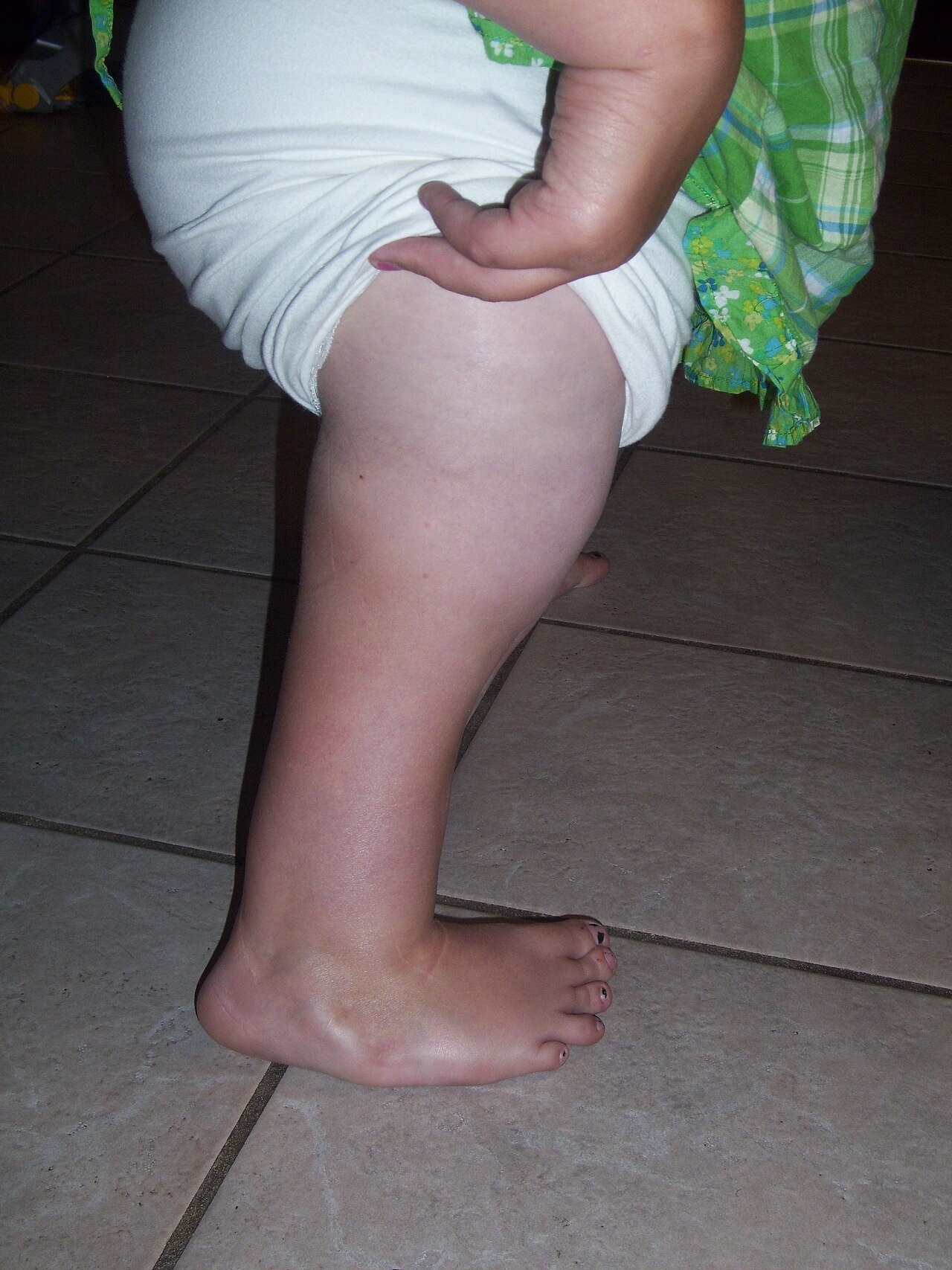
Clinical appearance of congenital femoral deficiency: a markedly short lower limb. In the severe form the thigh segment is short and bulbous and the foot of the affected side sits at the level of the opposite knee. (Wikimedia Commons, “Doughty74”, CC BY-SA 3.0.)
Clinical appearance of congenital femoral deficiency: a markedly short lower limb. In the severe form the thigh segment is short and bulbous and the foot of the affected side sits at the level of the opposite knee. (Wikimedia Commons, “Doughty74”, CC BY-SA 3.0.)
I.6 Imaging
- Serial plain radiographs: full-length standing AP of both legs (patellae forward), long-leg laterals in maximum extension (to capture the knee flexion contracture and true length), and a supine AP pelvis for the centre-edge angle and neck-shaft angle.[29]
- Pitfall: delayed ossification overstages the deformity. The proximal femur may be entirely cartilaginous yet unossified, making the radiographic class look worse than the true cartilaginous anatomy. A “bulbous” proximal fragment predicts that the head/neck will later ossify and may be managed as a short femur. Paley’s classification and the use of MRI were introduced because plain films at birth poorly predict final morphology.[30]
- Arthrography / MRI define the cartilaginous head and neck, whether the head is joined to the shaft, whether it is fused to the acetabulum, and the cruciate status, and they markedly improve correct categorisation over radiographs alone.[31]
- MR angiography before rotationplasty: vascular anomalies are common (a smaller/shorter femoral artery; ~10% have a dominant persistent ischiadic artery with a diminutive femoral artery).[32]
- LLD prediction, the Paley Multiplier method: because the short/long femur ratio stays roughly constant through growth, the discrepancy at maturity can be predicted from early radiographs and used to plan the number and timing of lengthenings and of contralateral epiphysiodesis.[33]
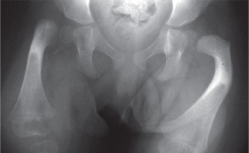
Antero-posterior radiograph of both hips in an infant with congenital femoral deficiency: the proximal femur on the affected side is short and deficient compared with the normal contralateral hip. (Simpson-White et al., Acta Orthop 2013;84:323, Fig. 1; CC BY.)
Antero-posterior radiograph of both hips in an infant with congenital femoral deficiency: the proximal femur on the affected side is short and deficient compared with the normal contralateral hip. (Simpson-White et al., Acta Orthop 2013;84:323, Fig. 1; CC BY.)
I.7 Treatment framework
The whole strategy hinges on the predicted limb-length discrepancy at maturity and whether the hip (and knee) can be reconstructed. Most decisions can wait until 2.5-3 years of age, with an extension prosthesis (“prosthosis”) bridging the interval; bilateral CFD is managed predominantly non-operatively.[34] There are three pathways: (1) reconstruction with lengthening, (2) rotationplasty, (3) knee fusion + foot ablation with a prosthesis.
A. Small/moderate discrepancy + a reconstructable hip → lengthening
The conventional threshold is to lengthen when the predicted discrepancy is <20 cm, the hip is (or can be made) stable, and the knee, ankle and foot are functional.[35] This means multiple staged lengthenings, usually combined with contralateral epiphysiodesis and a shoe lift. Paley’s algorithm writes a “life plan” to maturity and then proceeds in order:[36]
- Preparatory hip/knee surgery (~age 2-3): correct coxa vara (neck-shaft angle <110°), retroversion and the flexion/abduction contractures; perform a pelvic osteotomy if the acetabulum is deficient; release the fascia lata, which tethers the knee, before any lengthening. BMP may be added to promote femoral-neck ossification, and lengthening is deferred until the neck ossifies.
- Serial femoral (± tibial) lengthenings, a safe 5-8 cm per stage, performed over a pre-placed rod, which sharply reduces regenerate-fracture rates. The “Rule of 4”: first lengthening at about age 4, then every ~4 years, finishing by ~14.
- Contralateral epiphysiodesis as an equalisation adjuvant for up to ~5 cm, sparing one lengthening; ipsilateral guided growth (8-plate) corrects knee valgus.
The named reconstructions are the SUPERhip (a three-dimensional proximal femoral osteotomy with selective soft-tissue releases and an abductor slide, to untether the proximal femur and prevent recurrent coxa vara) and the SUPERknee (cruciate reconstruction, patellar realignment and capsulotomy for the unstable, contracted CFD knee).[37]
B. Very large discrepancy / a non-reconstructable hip → prosthetic management
When the predicted discrepancy exceeds ~20 cm, or lengthening is not chosen, the plan is prosthetic and surgery is used to make the residual limb a better lever.[38] The components:
- Foot ablation, Syme (ankle disarticulation) versus Boyd. Both create a residuum that can be fitted. Boyd (which retains the calcaneus) gives a more durable, end-bearing residuum but leaves a longer limb with less room for a prosthetic foot; Syme allows more choice of prosthetic foot but risks heel-pad migration and skin problems. The choice is largely surgeon-dependent.[39]
- Knee fusion (arthrodesis): converts the femur and tibia into a single long lever that sits easily in the prosthesis. At least one (usually both) knee physis is excised so the residuum is not too long, since an above-knee prosthetic knee needs ~7-7.5 cm of space.[40]
- Van Nes rotationplasty: the limb is rotated 180° so the foot
points backwards and the ankle functions as a knee, driving a
transtibial (below-knee-type) prosthesis. The aim is to place the
ankle/hindfoot at the level of the contralateral knee at
maturity.[41]
- Functional rationale: a more energy-efficient gait and better function than Syme + knee fusion, because an active ankle drives the prosthetic knee. It functions better than an above-knee prosthesis but not as well as a true below-knee amputee, since the deficient hip remains the limiting factor.[42]
- Indications: an unreconstructable proximal deficiency (Gillespie II, Aitken C/D, Paley 3) with a near-normal, stable, strong ankle and foot.
- Contraindications: an unstable or deficient ankle, typically the valgus, ball-and-socket ankle of the associated fibular deficiency, and severe equinus/valgus of the foot.[43]
- Main complication: loss of the rotation with growth (derotation), addressed by technique modifications (e.g. the Brown variant).[44]
Role of contralateral epiphysiodesis
Across the lengthening pathway, a well-timed contralateral (distal femoral/proximal tibial) epiphysiodesis equalises up to ~5 cm and can remove one whole lengthening from the life plan; its timing is set by the multiplier method.[45]
Part II - Fibular hemimelia (longitudinal fibular deficiency)
II.1 Definition and significance
Fibular hemimelia is partial or complete absence of the fibula and is the most common congenital long-bone deficiency.[46] Incidence is roughly 7.4-20 per million live births; the cause is unknown and most cases are sporadic, arising from a disruption of embryonic limb development.[47] The presentation spans a wide spectrum, from a mildly short fibula with a near-normal limb to complete fibular absence with a severely deformed, shortened leg.[48] The apparent incidence rises sharply if PFFD cases are counted, since up to ~80% of PFFD patients carry a coexisting fibular deficiency.[49]
II.2 Classification
Achterman-Kalamchi (1979) is the most commonly used scheme:[50]
- Type I, incomplete (partial) deficiency: IA the proximal fibular epiphysis lies distal to the proximal tibial physis and the distal physis proximal to the talar dome (mild); IB 30-50% fibular shortening, the fibula not articulating with the talus.
- Type II, complete fibular absence, with or without a tiny distal remnant.
Birch (1998, modified 2011) is a functional scheme built around one question, is the foot functional? (functional = ≥3 rays giving a stable plantigrade platform):[51]
- Type 1, functional foot, graded by % limb-length inequality (1A <6% → orthosis/epiphysiodesis; 1B 6-10%; 1C 11-30%; 1D >30% → multiple lengthenings or amputation).
- Type 2, non-functional foot (<3 rays) → amputation; subdivided by upper-limb function (2A functional upper limbs → early foot amputation; 2B non-functional upper limbs → amputation contraindicated, because the foot is used as a hand).
The earlier Coventry-Johnson scheme and Stanitski’s morphological scheme (which records the ball-and-socket ankle and tarsal coalition) are also cited, and Paley’s ankle-based classification (types 1-4) drives the modern “Super-ankle” reconstruction.[52]
II.3 Associated anomalies
- Femoral shortening / PFFD: the reported frequency varies widely (Amstutz 15%; Bohne & Root ~65%; Kalamchi 70% of type I and 50% of type II; Rodriguez-Ramirez 72%). Cite a range and note the disagreement.[53]
- A hypoplastic lateral femoral condyle (the commonest associated anomaly in one series, 93%) → genu valgum.[54]
- A ball-and-socket ankle (~80%) with characteristic hindfoot valgus.[55]
- Tarsal coalition (~51% radiographically; commoner still on anatomical study, where it is often cartilaginous and radiographically occult early) and an equinovalgus foot with absent lateral (postaxial) rays (~44%).[56]
- An anteromedial tibial bow with an overlying skin dimple.[57]
- Cruciate (ACL ± PCL) deficiency (the ACL is absent in ~95%) with AP knee instability and a small patella, but no varus/valgus laxity.[58]
II.4 Clinical features and imaging
The clinical limb shows a rigid equinovalgus foot with missing lateral rays, a short leg (± short thigh), a valgus knee, an anterior/anteromedial tibial bow with a dimple, and AP knee instability; milder cases present only as a slightly short limb.[59] Imaging is a full-length standing AP (with a lift under the short leg) plus a lateral, showing the short fibula, the ball-and-socket ankle, a triangular distal tibial epiphysis and hypoplastic tibial spines. MRI before a Super-ankle procedure clarifies the unossified hindfoot. The ultimate discrepancy is predicted with the Multiplier method to frame the reconstruction-versus-amputation discussion.[60]
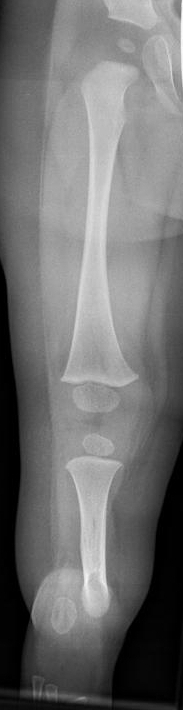
Antero-posterior radiograph of the lower leg in an 18-month-old child with fibular hemimelia: the tibia is present while the fibula is short and hypoplastic (Achterman-Kalamchi type II pattern). (Kinderradiologie Olgahospital, Klinikum Stuttgart, Wikimedia Commons, CC BY-SA 4.0.)
Antero-posterior radiograph of the lower leg in an 18-month-old child with fibular hemimelia: the tibia is present while the fibula is short and hypoplastic (Achterman-Kalamchi type II pattern). (Kinderradiologie Olgahospital, Klinikum Stuttgart, Wikimedia Commons, CC BY-SA 4.0.)
II.5 Treatment
The decision is driven by foot function and the projected discrepancy:[61]
- Functional plantigrade foot + manageable discrepancy → reconstruction/lengthening (± ankle realignment): observation and a shoe lift for projected discrepancies under ~2 cm; epiphysiodesis or staged lengthening for larger ones. The ankle valgus and the cruciate-related knee instability are addressed before lengthening, to prevent subluxation.
- Non-functional foot or a very large discrepancy → Syme or Boyd amputation + prosthetic fitting.
- The historical foot-ray rule (Birch): ≥3 rays = a potentially functional, salvageable foot; <3 rays = non-functional → amputation (unless the upper limbs are non-functional, type 2B).
The genuine controversy is the functional foot with a 10-30% discrepancy, where reconstruction (with its real complication rate: pin-track infection, residual discrepancy, delayed union, stiffness, refracture, recurrent foot deformity) competes with early Syme amputation; the trend since 2011 has been toward fewer amputations and more reconstruction.[62]
Part III - Tibial hemimelia (longitudinal tibial deficiency)
III.1 Definition and significance
Tibial hemimelia is partial or complete absence of the tibia with a relatively intact fibula.[63] It is far rarer than fibular hemimelia, roughly 1 per 1,000,000 live births, is bilateral in ~30%, and is the only long-bone deficiency that is genetically transmitted in a substantial proportion of cases, most often autosomal dominant.[64] Characteristic associations include hand anomalies (the lobster-claw (split-hand/foot) deformity and preaxial/triphalangeal-thumb polydactyly), together with congenital femoral deficiency, an absent patella/quadriceps, and various visceral anomalies; recognised syndromes include Werner syndrome.[65]
III.2 Classification - Jones (1978)
Radiographic, keyed to the presence/ossification of the proximal tibia and the integrity of the extensor (quadriceps) mechanism:[66]
- Type 1, no radiographically visible tibia. 1a: proximal tibia completely absent with a hypoplastic distal femoral epiphysis (the defining radiographic feature in Jones); clinically these patients have no functioning quadriceps/extensor mechanism and a fixed knee flexion contracture (>45° in the Kalamchi-Dawe correlate). 1b: proximal tibia absent on plain film but a cartilaginous anlage present (US/MRI/dissection); the distinguishing feature is a normal distal femoral epiphysis, implying the anlage, and a usually functioning extensor mechanism.
- Type 2, proximal tibia present, distal tibia deficient; functioning quadriceps.
- Type 3, distal tibia present, proximal tibia deficient (very rare).
- Type 4, short tibia with distal tibiofibular diastasis, the talus wedged between the tibia and fibula; rigid varus foot.
The Kalamchi-Dawe (1985) modification adds clinical correlates, and Weber and Paley offer more detailed/treatment-oriented variants. Throughout, the key is the proximal tibia and the quadriceps: their presence permits reconstruction, their absence forces knee disarticulation.
III.3 Clinical features and imaging
The limb shows a markedly short tibial segment with a rigid equinovarus/supinated foot pointing toward the perineum: a varus foot, in contrast to the valgus foot of fibular deficiency.[67] The fibula is intact and migrated proximally, prominent at the proximolateral knee, with a dimple over the medial border of the missing tibia. The knee is usually unstable, the patella often absent, and the single most important clinical assessment is active knee extension (the quadriceps), which determines treatment.[68] Plain films classify by Jones type; ultrasound/MRI detect an unossified proximal anlage and confirm the extensor mechanism.
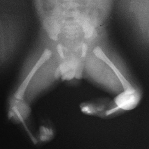
Babygram of the lower limbs in bilateral tibial hemimelia: the hips and distal femora are present, both tibiae are absent (only a small cartilaginous anlage), the fibula is present on each side, and the feet are deformed. (Chinnakkannan et al., Indian J Hum Genet 2013;19:108, Fig. 3; CC BY-NC-SA - non-commercial use only.)
Babygram of the lower limbs in bilateral tibial hemimelia: the hips and distal femora are present, both tibiae are absent (only a small cartilaginous anlage), the fibula is present on each side, and the feet are deformed. (Chinnakkannan et al., Indian J Hum Genet 2013;19:108, Fig. 3; CC BY-NC-SA - non-commercial use only.)
III.4 Treatment by Jones type
The decision sequence is: (1) is there active knee extension / a proximal tibia? (2) what is the foot quality and projected discrepancy?[69]
- Type 1a (no proximal tibia, no functioning extensor) → knee disarticulation + prosthetic fitting, the standard, giving near-normal gait and energy outcomes.
- Type 1b / Type 2 (proximal tibia present, quadriceps functioning) → reconstruction: a tibiofibular synostosis (centralising the fibula onto the tibial remnant) plus a Syme or modified Boyd foot ablation and a below-knee prosthesis.
- The Brown procedure (fibular centralisation beneath the femoral condyles) has a high failure rate from progressive knee flexion contracture (unopposed hamstrings) and requires active knee extension, so it is contraindicated in type 1a; modern practice prefers knee disarticulation for 1a.[70]
- Type 3 does well after a modified Syme/Chopart ablation; Type 4 (diastasis, rigid varus) is usually treated by a Syme ankle disarticulation.[71]
III.5 Fibular versus tibial hemimelia - the comparison the examiner wants
| Feature | Fibular hemimelia | Tibial hemimelia |
|---|---|---|
| Frequency | Commonest long-bone deficiency (7.4-20/million) | Rare (~1/million) |
| Inheritance | Sporadic | Autosomal dominant (the only AD long-bone deficiency) |
| Foot/ankle | Equinovalgus (valgus), lateral ray loss, ball-and-socket ankle | Equinovarus (varus), medial/preaxial ray loss, preaxial polydactyly |
| Bone | Fibula short/absent; tibia bowed (anteromedial) | Tibia short/absent; fibula intact, migrated proximally |
| Knee | ACL/PCL deficient, AP instability, valgus knee | Quadriceps/extensor often absent (esp. 1a), flexion contracture, absent patella |
| Hand | Uncommon | Lobster-claw hand, polydactyly characteristic |
| Defining decision | Foot function + LLD (Birch ray count) | Proximal tibia + extensor mechanism (Jones) |
Mnemonic: fibu-LAR → vaL-gus / LAteral rays; tibial → varus / medial rays.[72]
Part IV - Congenital pseudarthrosis of the tibia (CPT)
IV.1 Definition and significance
Congenital pseudarthrosis (better called congenital tibial dysplasia) is a dysplasia presenting as an anterolateral bow that progresses to fracture and a recalcitrant non-union, usually in infancy or early childhood; the pseudarthrosis is typically not present at birth but develops with weight-bearing.[73] It is rare (~1 in 150,000-250,000 births), usually unilateral, sited at the middle/distal-third junction of the tibia, with the fibula involved in about one-third of patients.[74] It remains one of the harder problems in paediatric orthopaedics to treat.[75]
The pathology is a fibrous hamartoma and a hamartomatous (pathologic) periosteum, highly cellular, fibromatosis-like tissue continuous with the periosteum, believed to impair callus formation. No neurofibroma is found in the lesion itself; the periosteum is the culprit, and resecting it is the operative key.[76]
IV.2 The neurofibromatosis-1 association
- About 50-75% of CPT patients have NF1 (one source: “up to 75%”; another: “up to 55%”; cite the range).[77]
- Conversely, only ~5% of NF1 patients develop CPT.[78]
- NF1 is autosomal dominant (~1/3,000), caused by mutations on chromosome 17 (neurofibromin), with ~50% new mutations. The histology of CPT is identical with or without NF1.[79]
- Fibrous/osteofibrous dysplasia is the occasional alternative association (present in up to ~15%), and amniotic band syndrome is a rare cause.[80]
IV.3 Classification
- Crawford (1986), radiographic: type I anterolateral bow + dense (sclerotic) medullary canal; type II bow + widened canal/tubulation defect; type III bow + cystic lesion; type IV bow + frank fracture/cyst/pseudarthrosis (established, worst prognosis).[81]
- Boyd (1982), six types; type II (anterior bow + hourglass constriction, fracture before age 2, often NF1) is the classic NF1 type and the hardest to unite.[82]
- Andersen (1976), six descriptive types (clubfoot, cystic, late, fibular, dysplastic, angulated).[83]
- The most useful practical distinction is intact versus established (fractured) pseudarthrosis: “the most relevant criterion to guide treatment is whether the tibia is fractured or intact.” None of the formal classifications reliably predicts outcome, because union is often only transient.[84]
IV.4 Clinical features and imaging
The hallmark is a distinctly anterolateral bow (bowing in any other direction is not CPT). The sequence is: the apex becomes dysplastic with a narrowed/obliterated medullary canal → cystic change → fracture (usually within the first 5-6 years, often atraumatic) → pseudarthrosis. The bone ends are atrophic and tapered (“sucked-candy”/spindle-shaped), or else sclerotic with a narrowed canal; abnormal painless mobility signals an established pseudarthrosis. Every patient is examined for café-au-lait spots/NF1 stigmata. CT quantifies healing and MRI shows the extent of the abnormal periosteum.[85]
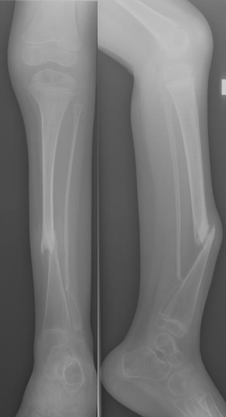
Antero-posterior and lateral radiographs of the lower leg in congenital pseudarthrosis of the tibia (neurofibromatosis-associated): anterolateral bowing with a dysplastic, tapered segment and pseudarthrosis at the middle/distal-third junction. (Kinderradiologie Olgahospital, Klinikum Stuttgart, Wikimedia Commons, CC BY-SA 3.0.)
Antero-posterior and lateral radiographs of the lower leg in congenital pseudarthrosis of the tibia (neurofibromatosis-associated): anterolateral bowing with a dysplastic, tapered segment and pseudarthrosis at the middle/distal-third junction. (Kinderradiologie Olgahospital, Klinikum Stuttgart, Wikimedia Commons, CC BY-SA 3.0.)
IV.5 Treatment
Natural history. An anterolateral bow is progressive and will fracture; an established pseudarthrosis will not heal spontaneously and will not respond to casting once fractured. Pseudarthrosis appearing before age 4 carries a worse prognosis (refracture).[86]
Non-operative (intact, pre-pseudarthrotic limb): brace to prevent fracture. As soon as an anterolateral bow is recognised, a protective orthosis is fitted: an AFO before walking, then a full-time clamshell KAFO (patella-tendon-bearing) once walking, continued to skeletal maturity. An intact bowed tibia must not be osteotomised, because surgery precipitates pseudarthrosis.[87]
Operative: the unifying principle and the three surgical pillars. Once a pseudarthrosis is established, the principle is to resect the hamartomatous periosteum/fibrous tissue, stabilise, maintain alignment and add biologic stimulation. The three pillars used to achieve and maintain union are:[88]
- Intramedullary rod (Williams rod): excise the pseudarthrosis, ream, pass a rod (often transfixing the ankle in young children), and leave it across the ankle for ≥2 years; primary IM rodding is the North-American default.
- Vascularised free fibular graft: a vascularised structural replacement from the contralateral leg, reserved largely for prior failures or large atrophic defects (union ~90% but frequently needing further surgery).
- Ilizarov / circular external fixator ± bone transport: Paley reported ~94% union after one procedure and 100% after two, refracture being the main complication.
The best modern results combine a circular fixator + an intramedullary nail + iliac-crest autograft, leaving the rod in after frame removal to guard against refracture. Adjuncts include BMP-2 (off-label and controversial, with some series showing benefit and others none) and bisphosphonates (to reduce refracture); Paley’s combined protocol pairs zoledronic acid with periosteal resection, an IM rod, a meshed periosteal autograft + iliac crest + BMP-2, and an Ilizarov frame.[89]
Refracture and amputation. Refracture is the dominant late complication (more common in younger children), hence bracing to skeletal maturity. Expect ankle/subtalar stiffness, valgus ankle and a limb-length discrepancy averaging ~5 cm. For repeated failures, severe discrepancy or a non-functional limb, **amputation (Boyd or Syme)
- prosthesis** is the last resort.[90]
Part V - Congenital angular deformities (bowing) of the tibia
The single most important teaching point of this topic: the direction of the bow tells you the diagnosis and the management.[91]
V.1 Anterolateral bowing - the dangerous one (harbinger of CPT / NF1)
Anterolateral bowing is the premonitory sign of congenital tibial dysplasia → pseudarthrosis, strongly NF1-associated, and progressive. The cardinal rule: do not perform a corrective osteotomy, which precipitates pseudarthrosis. Instead brace (clamshell/KAFO) and refer, and look for café-au-lait spots. (See Part IV.)[92]
V.2 Posteromedial bowing - the benign one
Posteromedial bowing is present at birth, sited at the middle/lower-third junction, with the posterior component usually exceeding the medial (posterior angulation up to ~90° in the newborn) and sometimes a skin dimple at the apex. It is always associated with a calcaneovalgus foot (the dorsum of the foot nearly touching the shin), reflecting an intrauterine malposition.[93]
It is benign and remodels spontaneously, rapidly in the first year and plateauing by 4-5 years, though resolution may be incomplete, leaving residual posterior bow ± ankle valgus, and there is no risk of pathologic fracture (the crucial contrast with anterolateral bowing).[94]
The price is a predictable limb-length discrepancy that grows with the child: - Pappas: median 4.1 cm (range 3.3-6.9 cm) at maturity; - the Atlas: average ~3 cm, up to 8 cm. - → quote a typical ~3-7 cm.[95]
Management is therefore: reassure the parents, stretch (± serial cast) the calcaneovalgus foot, and follow and treat the discrepancy, with contralateral epiphysiodesis for a projected discrepancy up to ~4-5 cm and lengthening beyond that (often needing a second, proximal osteotomy because the diaphyseal apex is too distal). Lengthening outcomes here are better than in fibular hemimelia. Surgery for the bow itself is reserved for insufficient remodelling after ~6 years, and ankle valgus is addressed by medial distal-tibial guided growth or a supramalleolar osteotomy.[96]
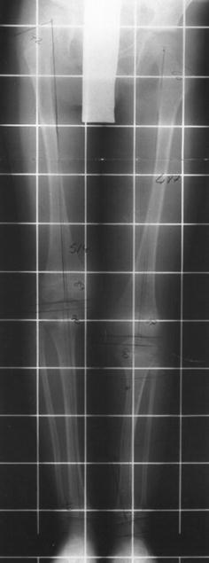
Standing antero-posterior radiograph of the lower legs (with a measurement grid) in congenital posteromedial bowing of the tibia: tibial bowing and a length discrepancy. (Kaufman et al., Strategies Trauma Limb Reconstr 2012;7:147, Fig. 1; CC BY.)
Standing antero-posterior radiograph of the lower legs (with a measurement grid) in congenital posteromedial bowing of the tibia: tibial bowing and a length discrepancy. (Kaufman et al., Strategies Trauma Limb Reconstr 2012;7:147, Fig. 1; CC BY.)
V.3 Anteromedial bowing - points to fibular hemimelia
Anteromedial bowing accompanies congenital fibular deficiency: a short/absent fibula with tibial shortening, a ball-and-socket ankle, an equinovalgus foot and absent lateral rays (± ipsilateral femoral shortening). It is managed as fibular hemimelia (Part II).[97]
V.4 The bowed infantile tibia - the wider differential
Beyond the three congenital directional bows, the differential of an angulated infantile tibia includes physiologic genu varum (bilateral, symmetric, resolving to mild valgus by age 7-8), Blount disease (developmental tibia vara at the posteromedial proximal physis, in early walkers, often obese), rickets/renal osteodystrophy, osteogenesis imperfecta (to be excluded in any bowed infant), and skeletal dysplasias.[98]
V.5 Direction-of-bow → diagnosis (summary table)
| Direction of bow | Diagnosis | Key associations | Behaviour | Action |
|---|---|---|---|---|
| Anterolateral | Congenital tibial dysplasia → CPT | NF1 (~50-75%), fibrous dysplasia (~15%) | Progressive → fractures, will not heal | Brace; NO osteotomy; refer; surgery (rod / vascularised fibula / Ilizarov + periosteal resection) once fractured |
| Posteromedial | Congenital posteromedial bowing | Calcaneovalgus foot | Benign - remodels; LLD ~3-7 cm | Stretch/cast foot; observe; treat LLD (epiphysiodesis/lengthening) |
| Anteromedial | Fibular hemimelia | Short/absent fibula, ball-and-socket ankle | Fixed deficiency, shortening | Manage as fibular deficiency |
| Lateral/varus (proximal) | Physiologic varum vs Blount vs rickets | Obesity/early walker (Blount) | Physiologic resolves; Blount progresses | Distinguish radiographically; treat Blount |
Bulgarian terminology glossary
For consistency with the Bulgarian state-examination vocabulary (and with the operative terminology of Boychev, Хирургическа ортопедия), the key terms of this topic are:
| English | Bulgarian |
|---|---|
| Proximal femoral focal deficiency / congenital femoral deficiency | Проксимален фокален дефицит на бедрената кост / вроден дефицит (недоразвитие) на бедрото |
| Longitudinal fibular deficiency (fibular hemimelia) | Вроден дефицит на малкопищялната кост (фибула) / фибуларна хемимелия |
| Longitudinal tibial deficiency (tibial hemimelia) | Вроден дефицит на голямопищялната кост (тибия) / тибиална хемимелия |
| Congenital pseudarthrosis of the tibia | Вродена псевдоартроза на подбедрицата (тибията) |
| Congenital bowing of the tibia | Вродено изкривяване (огъване) на подбедрицата |
| Anterolateral / posteromedial / anteromedial bow | Антеролатерално / постеромедиално / антеромедиално изкривяване |
| Calcaneovalgus foot | Калканеовалгусно стъпало |
| Coxa vara | Варусна деформация на бедрената шийка (coxa vara) |
| Limb-length discrepancy | Разлика в дължината на крайниците |
| Neurofibromatosis type 1 | Неврофиброматоза тип 1 (болест на фон Реклингхаузен) |
| Limb lengthening | Удължаване на крайник |
| Epiphysiodesis | Епифизиодеза |
| Amputation (Syme / Boyd) | Ампутация (по Syme / по Boyd) |
| Knee arthrodesis (fusion) | Артродеза на колянната става |
| Rotationplasty (Van Nes) | Ротационна пластика (по Ван Нес) |
| Prosthesis / prosthetic fitting | Протеза / протезиране |
Note on Bulgarian sources: Boychev’s Хирургическа ортопедия is principally an operative atlas; congenital limb deficiencies of this kind are managed in specialist paediatric centres and the indigenous terminology above follows standard Bulgarian orthopaedic usage rather than a single Boychev chapter. Where a named technique (Syme, Boyd, Van Nes, Ilizarov) is used, the Bulgarian literature retains the eponym.
Figure credits and licences
All images were independently opened and visually verified to depict the stated entity before use. Licences were confirmed against the Wikimedia Commons API or the NCBI PMC Open-Access service.
- Clinical CFD (short limb) -
pffd_clinical_short_thigh.jpg. Wikimedia Commons user “Doughty74”. CC BY-SA 3.0. https://commons.wikimedia.org/wiki/File:PFFD.jpg - CFD radiograph (both hips, infant) -
pffd_radiograph_both_hips.png. Simpson-White RW, Fernandes JA, Bell MJ. Acta Orthop 2013;84(3):323-5, Fig. 1. CC BY. https://www.ncbi.nlm.nih.gov/pmc/articles/PMC3715821/ - Fibular hemimelia radiograph -
fibular_hemimelia_radiograph.jpeg. Kinderradiologie Olgahospital, Klinikum Stuttgart (Wikimedia “Zieger M”). CC BY-SA 4.0. https://commons.wikimedia.org/wiki/File:FHemiMelie.jpeg - Tibial hemimelia (bilateral) babygram -
tibial_hemimelia_radiograph_bilateral.png. Chinnakkannan S, Das RR, Rughmini K, Ahmed S. Indian J Hum Genet 2013;19(1):108-10, Fig. 3. CC BY-NC-SA - non-commercial use only. https://www.ncbi.nlm.nih.gov/pmc/articles/PMC3722620/ - Congenital pseudarthrosis of the tibia (NF) -
tibial_pseudarthrosis_nf1.png. Kinderradiologie Olgahospital, Klinikum Stuttgart (Wikimedia “Zieger M”). CC BY-SA 3.0. https://commons.wikimedia.org/wiki/File:TP_NF.png - Congenital posteromedial bow of the tibia -
posteromedial_bow_tibia_radiograph.png. Kaufman SD, Fagg JA, Jones S, Bell MJ, Saleh M, Fernandes JA. Strategies Trauma Limb Reconstr 2012;7(3):147-53, Fig. 1. CC BY. https://www.ncbi.nlm.nih.gov/pmc/articles/PMC3482434/


Licensing note for document distribution: image 4 is CC BY-NC-SA (non-commercial) - acceptable for a personal, non-commercial study document, but it must be removed or replaced if this material is ever used commercially. The CC BY-SA images (1, 3, 5) require that any redistributed derivative carry the same share-alike licence.
No Van Nes rotationplasty photograph/radiograph is included: no cleanly-licensed real image was located (only an excluded schematic line drawing).
References
-
Sabharwal, Pediatric Lower Limb Deformities, p.368.
-
Sabharwal, p.368; Lovell & Winter, Pediatric Orthopaedics, p.4766.
-
Sabharwal, p.368.
-
Sabharwal, pp.367, 372.
-
Lovell & Winter, p.4763; Sabharwal, p.368.
-
Lovell & Winter, pp.4764, 4779 - figure given as 70-80% and, elsewhere, “up to 70%”.
-
Lovell & Winter, p.4764.
-
Sabharwal, pp.369-370; Lovell & Winter, p.4766.
-
Sabharwal, p.368.
-
Sabharwal, p.368; Lovell & Winter, p.4763.
-
Sabharwal, p.369.
-
Sabharwal, p.368; Atlas of Amputations and Limb Deficiencies, 4th ed., p.910; Lovell & Winter, p.4763.
-
Lovell & Winter, p.4767.
-
Atlas of Amputations, p.910; Lovell & Winter, p.4763.
-
Sabharwal, p.374.
-
Sabharwal, pp.374-375; Atlas of Amputations, p.911; Lovell & Winter, p.4756. Note a source disagreement on class A: Sabharwal stresses the pseudarthrosis ossifies only in some patients (p.374), whereas the Atlas states it “will ossify progressively with time” (p.911) - Sabharwal’s cautious phrasing is preferable.
-
Sabharwal, p.376.
-
Lovell & Winter, p.4757; Atlas of Amputations, p.911.
-
Sabharwal, pp.376, 378.
-
Sabharwal, p.381.
-
Sabharwal, pp.375-376.
-
Sabharwal, pp.377-378.
-
Sabharwal, pp.377-378.
-
Lovell & Winter, pp.4763-4764; Atlas of Amputations, p.912.
-
Atlas of Amputations, pp.911-912; Lovell & Winter, p.4757.
-
Sabharwal, p.372; Atlas of Amputations, p.913; Lovell & Winter, p.4766.
-
Lovell & Winter, p.4766; Atlas of Amputations, p.913.
-
Lovell & Winter, p.4763.
-
Sabharwal, pp.372-373.
-
Sabharwal, pp.373, 376.
-
Sabharwal, pp.373-374; Lovell & Winter, p.4766.
-
Sabharwal, pp.371-372.
-
Sabharwal, pp.368, 376, 382.
-
Lovell & Winter, pp.4768-4770.
-
Lovell & Winter, p.4770.
-
Sabharwal, pp.381-382.
-
Sabharwal, pp.383-384, 418-422; Lovell & Winter, p.4781.
-
Lovell & Winter, p.4770.
-
Atlas of Amputations, pp.890, 905; Sabharwal, p.378; Lovell & Winter, p.4773.
-
Lovell & Winter, pp.4771-4773.
-
Lovell & Winter, pp.4773-4774; Sabharwal, p.378; Atlas of Amputations, p.907.
-
Sabharwal, pp.379-380; Lovell & Winter, p.4780.
-
Lovell & Winter, p.4780; Atlas of Amputations, p.907.
-
Sabharwal, pp.426-427; Lovell & Winter, p.4779.
-
Sabharwal, p.382; Lovell & Winter, p.4770.
-
Sabharwal, p.432; Lovell & Winter, p.4682.
-
Sabharwal, p.432; Lovell & Winter, p.4682.
-
Sabharwal, p.432.
-
Lovell & Winter, p.4682.
-
Sabharwal, pp.433-434.
-
Sabharwal, pp.434-435; Lovell & Winter, pp.4681, 4685.
-
Sabharwal, pp.433-438.
-
Lovell & Winter, p.4682; Sabharwal, p.432.
-
Sabharwal, p.432.
-
Sabharwal, p.432.
-
Sabharwal, p.432; Lovell & Winter, pp.4681-4682.
-
Sabharwal, pp.432, 439.
-
Sabharwal, pp.432, 438; Lovell & Winter, pp.4682-4683.
-
Lovell & Winter, p.4681.
-
Lovell & Winter, pp.4682-4683; Sabharwal, pp.438-439.
-
Sabharwal, p.432; Lovell & Winter, pp.4684-4685.
-
Sabharwal, pp.432, 434, 458; Lovell & Winter, p.4685.
-
Atlas of Amputations, p.894.
-
Sabharwal, pp.460-461; Atlas of Amputations, pp.894-895; Lovell & Winter, p.4730.
-
Atlas of Amputations, pp.896-897; Lovell & Winter, pp.4730-4731; Sabharwal, pp.460-461.
-
Sabharwal, pp.461-462; Atlas of Amputations, pp.895-896; Lovell & Winter, pp.4726-4730.
-
Atlas of Amputations, pp.894-895; Lovell & Winter, p.4731.
-
Atlas of Amputations, p.897; Lovell & Winter, p.4733.
-
Atlas of Amputations, p.897; Lovell & Winter, pp.4734-4736.
-
Atlas of Amputations, pp.898-899; Lovell & Winter, p.4734.
-
Atlas of Amputations, pp.899-900.
-
Atlas of Amputations, p.894; Sabharwal, pp.432, 460-462; Lovell & Winter, pp.4681, 4731.
-
Franklin & Davidson, in Sabharwal, p.487.
-
Sabharwal, p.487.
-
Atlas of Amputations, internal p.4036.
-
Sabharwal, pp.487-488; Atlas of Amputations, p.4032.
-
Sabharwal, pp.29, 488.
-
Sabharwal, p.488.
-
Sabharwal, p.488.
-
Sabharwal, p.29.
-
Sabharwal, p.488.
-
Sabharwal, p.488; Atlas of Amputations, p.4034.
-
Sabharwal, p.488.
-
Sabharwal, p.488; Atlas of Amputations, p.4034.
-
Sabharwal, pp.487-489; Atlas of Amputations, pp.4032-4036.
-
Atlas of Amputations, pp.4032-4036.
-
Sabharwal, p.489; Atlas of Amputations, p.4036.
-
Sabharwal, pp.490-497; Atlas of Amputations, pp.4039-4057.
-
Sabharwal, pp.491-496.
-
Atlas of Amputations, pp.4052-4057; Sabharwal, p.492.
-
Sabharwal, pp.29, 505.
-
Sabharwal, pp.29, 487; Atlas of Amputations, p.4036.
-
Sabharwal (Joseph/Shah/Siddesh chapter), pp.499-501.
-
Sabharwal, pp.501-502; Atlas of Amputations, pp.4061-4071.
-
Sabharwal, p.501; Atlas of Amputations, p.4062.
-
Sabharwal, pp.503-505; Atlas of Amputations, p.4071.
-
Sabharwal, pp.28-29, 499.
-
Sabharwal, pp.29-30, 485.