Contents
- Part I - General Principles of Overuse Soft-Tissue Disorders
- Part II - Periarthritis of the Shoulder (Periarthritis humeroscapularis)
- Part III - Elbow and Forearm Tendinopathies
- Part IV - Wrist and Hand Stenosing Tenosynovitis
- Part V - Lower-Limb Tendinopathy and Enthesopathy
- References
Part I - General Principles of Overuse Soft-Tissue Disorders
The musculoskeletal soft tissues that transmit and absorb load (tendons, their sheaths and paratenon, the entheses where they anchor to bone, and the bursae that smooth their gliding) share a common vulnerability to repetitive, submaximal overload. The conditions grouped under this topic (periarthritis of the shoulder, the various tendinopathies, the enthesopathies, and bursitis) are united less by anatomy than by mechanism: load applied faster than the tissue can adapt, producing a failed healing response rather than the acute inflammation the old “-itis” names imply.
Tendon structure and biology
A tendon is a hierarchical, sparsely cellular structure built almost entirely of type I collagen embedded in a proteoglycan ground substance. Tropocollagen assembles into fibrils, fibrils into fibres, fibres into fascicles, and fascicles into the tendon, with tenocytes (specialised fibroblasts) maintaining the matrix.[1]
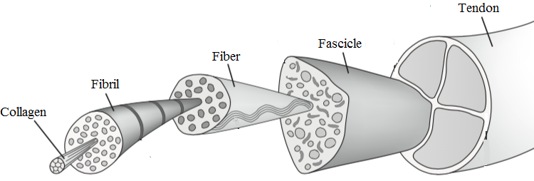
Figure 1. The hierarchical structure of tendon: collagen molecule → fibril → fibre → fascicle → tendon. Source: Blumpkin999, via Wikimedia Commons, CC0 1.0 (public domain).
Figure 1. The hierarchical structure of tendon: collagen molecule → fibril → fibre → fascicle → tendon. Source: Blumpkin999, via Wikimedia Commons, CC0 1.0 (public domain).
At rest the collagen lies in a wavy crimp pattern that straightens under tension. Healthy tendon depends on cyclic loading (mechanotransduction): physiological strain is anabolic, but continued loading of an already weakened tendon accumulates injury.[2] Tendons are relatively hypovascular, with recognised watershed zones (the supraspinatus “critical zone” and the Achilles mid-portion 2-6 cm above its insertion). They anchor to bone through a four-zone enthesis (tendon → fibrocartilage → mineralised fibrocartilage → bone). Where a tendon has no synovial sheath it is wrapped instead by paratenon.
Terminology: tendinitis, tendinosis, tendinopathy
The major conceptual shift in this field has been away from “tendinitis,” which wrongly implies inflammation, toward the recognition that most chronic tendon pain is degenerative. Following Maffulli, the umbrella clinical term is tendinopathy; tendinitis is reserved for the rare case with true inflammatory cells on histology; paratenonitis denotes inflammation of the paratenon or sheath (the old tenosynovitis/peritendinitis); and tendinosis denotes intratendinous collagen degeneration.[3] The histology of tendinosis is characteristic and counter-intuitive: disorganised, thinned collagen; mucoid (myxoid) degeneration with increased ground substance; neovascularisation accompanied by ingrowing nerves (a likely pain source); tenocyte changes (apoptosis and proliferation); and a conspicuous absence of inflammatory cells.[4]
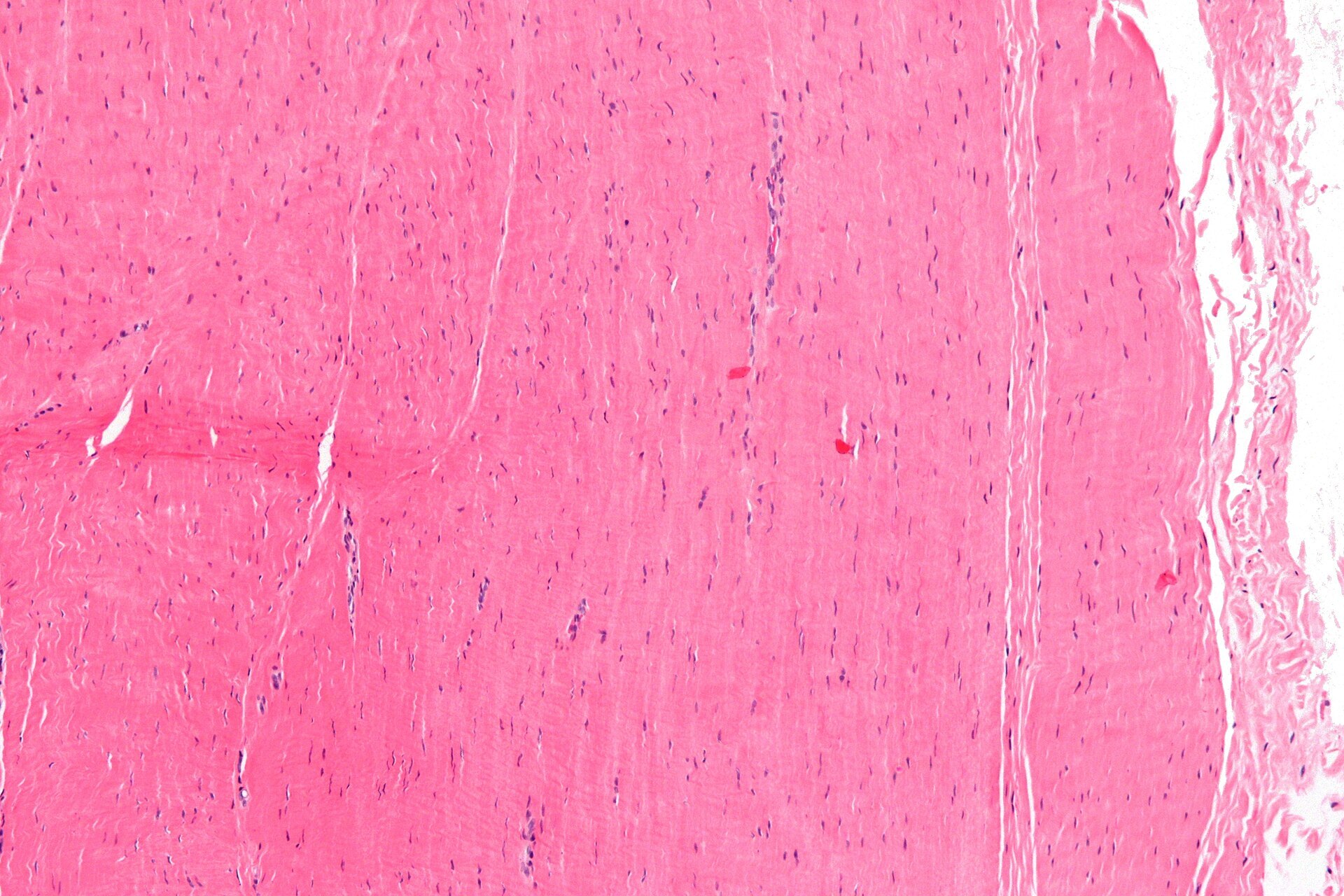
Figure 2. Normal tendon histology (H\&E): dense parallel collagen bundles with sparse interspersed tenocyte nuclei. Source: Nephron, via Wikimedia Commons, CC BY-SA 3.0.
Figure 2. Normal tendon histology (H&E): dense parallel collagen bundles with sparse interspersed tenocyte nuclei. Source: Nephron, via Wikimedia Commons, CC BY-SA 3.0.
The earliest morphological change is in the tenocytes, not the collagen.[5]
Pathogenesis and the continuum model
The driving concept is overuse with a failed healing response: repetitive load exceeds the tendon’s reparative capacity. A widely used framework is the tendinopathy continuum (Cook and Purdam): reactive tendinopathy → tendon dysrepair → degenerative tendinopathy, with the early stages more reversible than the late.[6] Several systemic and iatrogenic factors predispose to “occult” tendinopathy: diabetes mellitus, rheumatoid arthritis, gout and calcium pyrophosphate deposition, renal failure, hypothyroidism, corticosteroids (local or systemic), and the fluoroquinolone antibiotics (which alter the extracellular matrix and impair tenocyte metabolism); anabolic steroids increase rupture risk.[7]
Principles of management
Treatment of tendinopathy is built on load management and progressive exercise, the best-evidenced modality being an eccentric loading programme, which is thought to drive tendon remodelling.[8] NSAIDs have a limited role. Corticosteroid injection gives short-term relief but no durable benefit, weakens the tendon and risks rupture, and in some sites (notably tennis elbow) may leave patients worse at one year.[9] Platelet-rich plasma and autologous blood, sclerosing (polidocanol) injection of neovessels, and extracorporeal shock-wave therapy (ESWT) are used, though the high-level evidence is mixed and often disappointing. Surgery (debridement of the degenerate tissue with stimulation of a healing response) is a last resort.[10]
A bursa is a synovial-lined sac that reduces friction at points where tendon, muscle or skin moves over bone. Bursitis, its painful inflammation, may be aseptic (overuse, trauma, or crystal disease such as gout/CPPD) or septic. That distinction, made clinically and by aspiration with cell count, Gram stain and culture, governs treatment: septic bursitis needs drainage and antibiotics, whereas aseptic bursitis is managed by compression, activity modification and judicious anti-inflammatories.[11] An enthesopathy is the analogous degenerative/overload disorder of a tendon or ligament insertion (e.g. plantar fasciitis, the epicondylitides).
Part II - Periarthritis of the Shoulder (Periarthritis humeroscapularis)
The older French/American term scapulohumeral periarthritis (Duplay; Putnam) lumped together the painful, stiff, non-arthritic shoulder. Modern practice resolves it into three distinct entities: subacromial impingement/rotator-cuff disease, adhesive capsulitis (frozen shoulder), and calcific tendinitis. These remain the core of this exam topic.[12]
Subacromial impingement and rotator-cuff tendinopathy
The rotator cuff (supraspinatus, infraspinatus, subscapularis, teres minor) both rotates and, above all, stabilises the humeral head by concavity-compression; the supraspinatus passes beneath the coracoacromial arch (acromion + coracoacromial ligament + coracoid).[13]
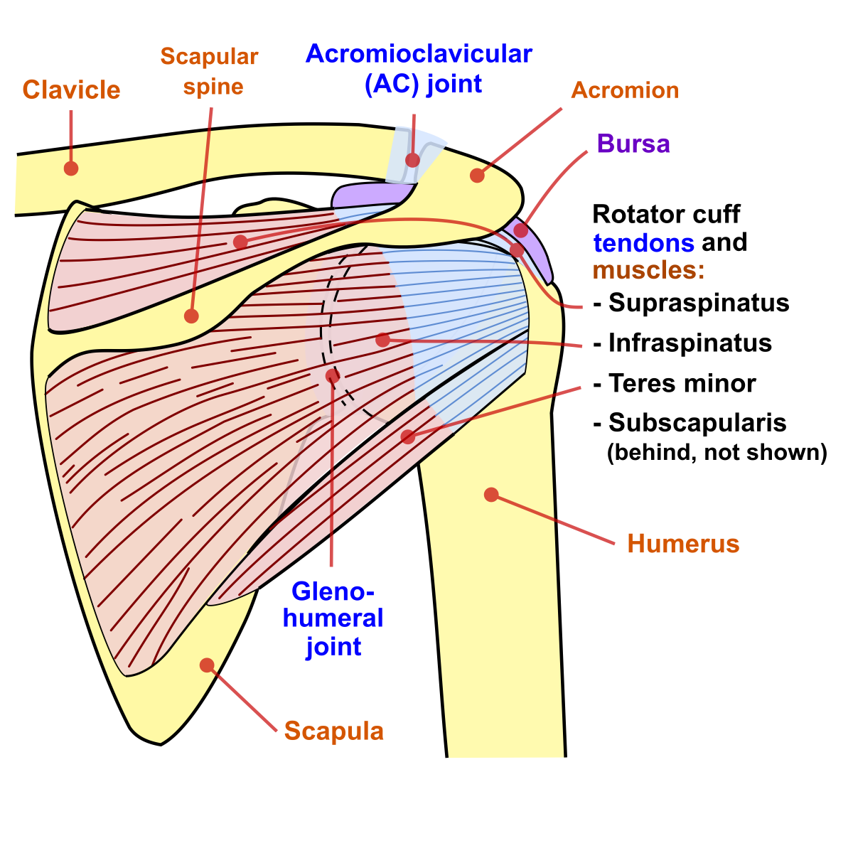
Figure 3. Posterior view of the shoulder showing the rotator cuff tendons and the subacromial bursa beneath the acromion. Source: Jmarchn, via Wikimedia Commons, CC BY-SA 3.0.
Figure 3. Posterior view of the shoulder showing the rotator cuff tendons and the subacromial bursa beneath the acromion. Source: Jmarchn, via Wikimedia Commons, CC BY-SA 3.0.
Neer’s model held that most cuff disease is a spectrum of impingement in three stages (stage I oedema/haemorrhage, stage II fibrosis and tendinitis, stage III partial or full-thickness tearing and bony change), and the Bigliani acromial morphology (type I flat, II curved, III hooked) was linked to cuff tears, though whether acromial shape is cause or consequence remains debated.[14] The modern view is multifactorial: intrinsic tendon degeneration plus extrinsic impingement and overload.
Clinically there is a painful arc of abduction (60-120°) and a battery of provocative tests: Neer’s sign (pain on forced forward flexion) confirmed by the Neer impingement test (relief after subacromial local anaesthetic), the Hawkins-Kennedy test (pain on internal rotation of the 90°-flexed arm), Jobe’s empty-can test (supraspinatus), the external-rotation strength/lag tests (infraspinatus/teres minor), and the lift-off and belly-press tests (subscapularis); objective weakness beyond pain is the most specific sign of cuff deficiency.[15]
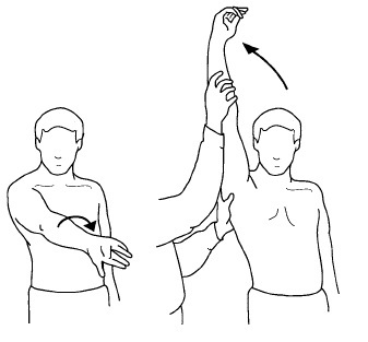
Figure 4. The Neer impingement sign: passive forward flexion of the internally rotated arm drives the greater tuberosity against the anterior acromion. Source: Nasch92, via Wikimedia Commons, CC BY-SA 4.0.
Figure 4. The Neer impingement sign: passive forward flexion of the internally rotated arm drives the greater tuberosity against the anterior acromion. Source: Nasch92, via Wikimedia Commons, CC BY-SA 4.0.
Ultrasound (≈91% sensitive and specific) and **MRI (the gold standard,
90% sensitive and specific)** characterise tears, while plain films may show a high-riding head with an acromiohumeral distance <6 mm in massive tears.[16]
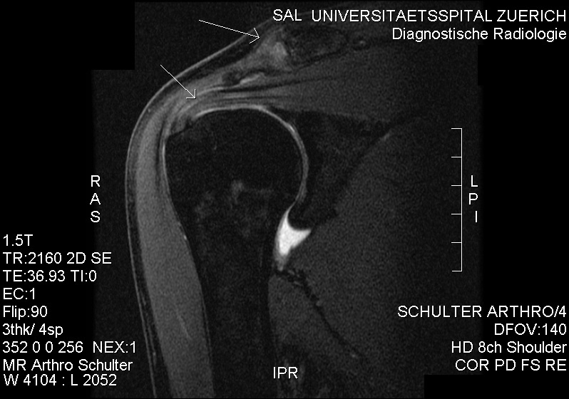
Figure 5. Coronal MR arthrogram of subacromial impingement with partial supraspinatus damage. Source: RSatUSZ, via Wikimedia Commons, CC BY-SA 3.0.
Figure 5. Coronal MR arthrogram of subacromial impingement with partial supraspinatus damage. Source: RSatUSZ, via Wikimedia Commons, CC BY-SA 3.0.
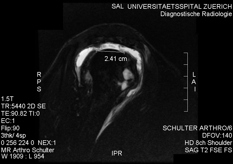
Figure 6. Sagittal MR arthrogram of a full-thickness supraspinatus tear (transmural gap). Source: RSatUSZ, via Wikimedia Commons, CC BY-SA 3.0.
Figure 6. Sagittal MR arthrogram of a full-thickness supraspinatus tear (transmural gap). Source: RSatUSZ, via Wikimedia Commons, CC BY-SA 3.0.
Most patients are managed non-operatively (activity modification, NSAIDs, a subacromial corticosteroid injection of limited durability, and above all cuff and periscapular strengthening). Operative repair is reserved for failure of 3-6 months of conservative care or an acute sizeable tear in a younger patient, with partial tears repaired when they exceed about 50% of tendon thickness.[17] The routine addition of acromioplasty to cuff repair is now controversial.[18]
Adhesive capsulitis (frozen shoulder)
Codman coined “frozen shoulder” in 1934 and Neviaser “adhesive capsulitis” in 1945. The condition is a chronic capsular fibrosis and contracture, especially of the rotator interval and coracohumeral ligament, that secondarily restricts both active and passive motion in the absence of another intrinsic disorder.[19] It affects about 2-5% of people, roughly 70% women, typically aged 40-60 (the Chinese “fifties shoulder”). Its single strongest association is diabetes mellitus (frozen shoulder in 10-20%, up to 35%, of diabetics, with worse outcomes), followed by thyroid disease and Dupuytren contracture.[20] The hallmark physical sign is loss of passive external rotation (from the contracted anterior capsule/rotator interval).[21]
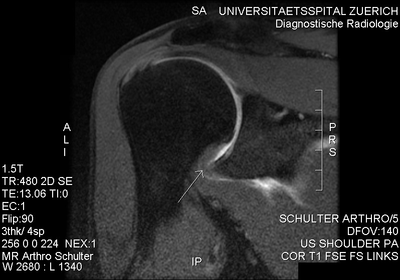
Figure 7. Coronal MR arthrogram in adhesive capsulitis: a thickened joint capsule at the inferior axillary recess (arrow). Source: RSatUSZ, via Wikimedia Commons, CC BY-SA 3.0.
Figure 7. Coronal MR arthrogram in adhesive capsulitis: a thickened joint capsule at the inferior axillary recess (arrow). Source: RSatUSZ, via Wikimedia Commons, CC BY-SA 3.0.
The classic course passes through three phases: a painful freezing phase (2-9 months), a stiff frozen phase (3-12 months), and a thawing phase over months to years. Although often described as ultimately self-limiting, a substantial minority retain measurable restriction or pain at long follow-up.[22] Diagnosis is clinical (a plain radiograph chiefly excludes arthritis). Treatment escalates from physiotherapy (gentle stretching; forceful stretching during the freezing phase is counter-productive) and intra-articular corticosteroid (the best-supported short-term measure), through hydrodilatation, to manipulation under anaesthesia and arthroscopic capsular release for refractory cases.[23]
Calcific tendinitis
Calcific tendinitis is the deposition of reactive calcium hydroxyapatite within the cuff (usually the supraspinatus), thought to follow a fibrocartilaginous metaplasia of tenocytes into chondrocytes, and it most often affects adults aged 31-60.[24] Uhthoff’s cyclical model describes a precalcific phase, a calcific phase (formative → resting → resorptive), and a postcalcific (healing) phase. The resorptive phase is the acutely painful one, as the chalky, toothpaste-like deposit is broken down (the deposit has been called “the kidney stone of the shoulder”).[25] The Gärtner radiographic classification grades the deposit from sharply circumscribed and dense (type I, formative) to cloudy and ill-defined (type III, resorptive).[26] Because the disease is self-limiting, treatment begins conservatively. Persistent symptoms are addressed with ultrasound-guided needling and lavage (barbotage), extracorporeal shock-wave therapy, and, failing those, arthroscopic removal of the deposit.[27]
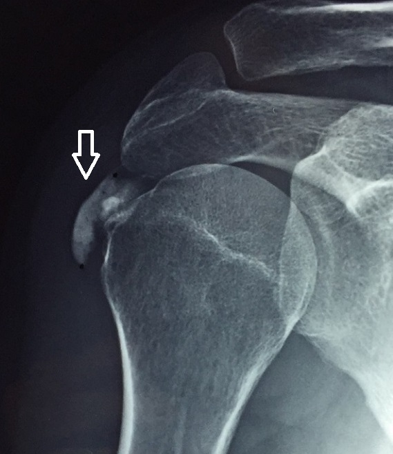
Figure 8. AP radiograph of calcific tendinitis: a calcific deposit at the supraspinatus footprint (arrow). Source: Pakos et al., annotated by Mikael Häggström, via Wikimedia Commons, CC BY 4.0.
Figure 8. AP radiograph of calcific tendinitis: a calcific deposit at the supraspinatus footprint (arrow). Source: Pakos et al., annotated by Mikael Häggström, via Wikimedia Commons, CC BY 4.0.
Part III - Elbow and Forearm Tendinopathies
Lateral epicondylitis (“tennis elbow”)
The commonest elbow tendinopathy (affecting ~3% of people, peaking at 35-55 years, lateral:medial ratio 3-6:1) is, despite its name, not an inflammatory tendinitis but an angiofibroblastic tendinosis of the extensor carpi radialis brevis (ECRB) origin (Nirschl), with the extensor digitorum communis involved in about a third.[28] Pain localises just distal and anterior to the lateral epicondyle and is provoked by resisted wrist extension (the Cozen test) and resisted middle-finger extension (the latter not specific, since it is also positive in radial tunnel syndrome, the key differential).[29] The Nirschl pathology grades (I-IV) run from reactive change to ECRB rupture.[30] It is largely self-limiting over 1-2 years, and management is conservative: activity modification, a counterforce brace, eccentric exercise. The important caution is that corticosteroid injection gives only short-term relief and may worsen the one-year outcome. Refractory cases (after 6-12 months) may have open or arthroscopic ECRB debridement (the “Nirschl procedure”).[31]
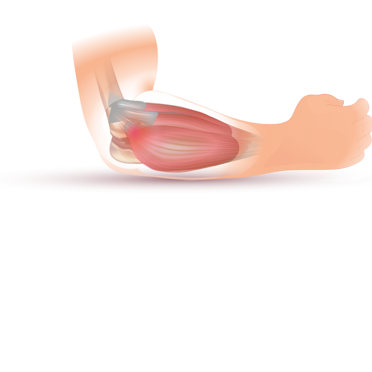
Figure 9. Lateral epicondylitis: the common extensor (ECRB) origin at the lateral epicondyle. Source: InjuryMap, via Wikimedia Commons, CC BY-SA 4.0.
Figure 9. Lateral epicondylitis: the common extensor (ECRB) origin at the lateral epicondyle. Source: InjuryMap, via Wikimedia Commons, CC BY-SA 4.0.
Medial epicondylitis (“golfer’s elbow”)
Medial epicondylitis is the same angiofibroblastic tendinosis at the flexor-pronator origin (pronator teres/FCR), less common than the lateral form, provoked by resisted forearm pronation and wrist flexion.[32] Its clinically vital feature is the association with ulnar neuropathy (in 23-60%), captured by the Gabel-Morrey classification (type I without, type II with ulnar nerve involvement), which predicts a poorer result.[33] Treatment mirrors the lateral side; surgery addresses the flexor-pronator origin and any symptomatic ulnar nerve.
Olecranon bursitis
Unlike the epicondylitides, olecranon bursitis is primarily inflammatory, of the subcutaneous bursa (which does not communicate with the joint), from pressure/trauma, gout/CPPD, or rheumatoid disease.[34] The essential distinction is septic versus aseptic (infection risk is highest with acute presentation, overlying wounds and diabetes). Aseptic cases are treated with compression and activity modification; aspiration is approached cautiously (through an anterolateral track, to avoid a chronic sinus) and corticosteroid injection carries a notable complication rate.[35]
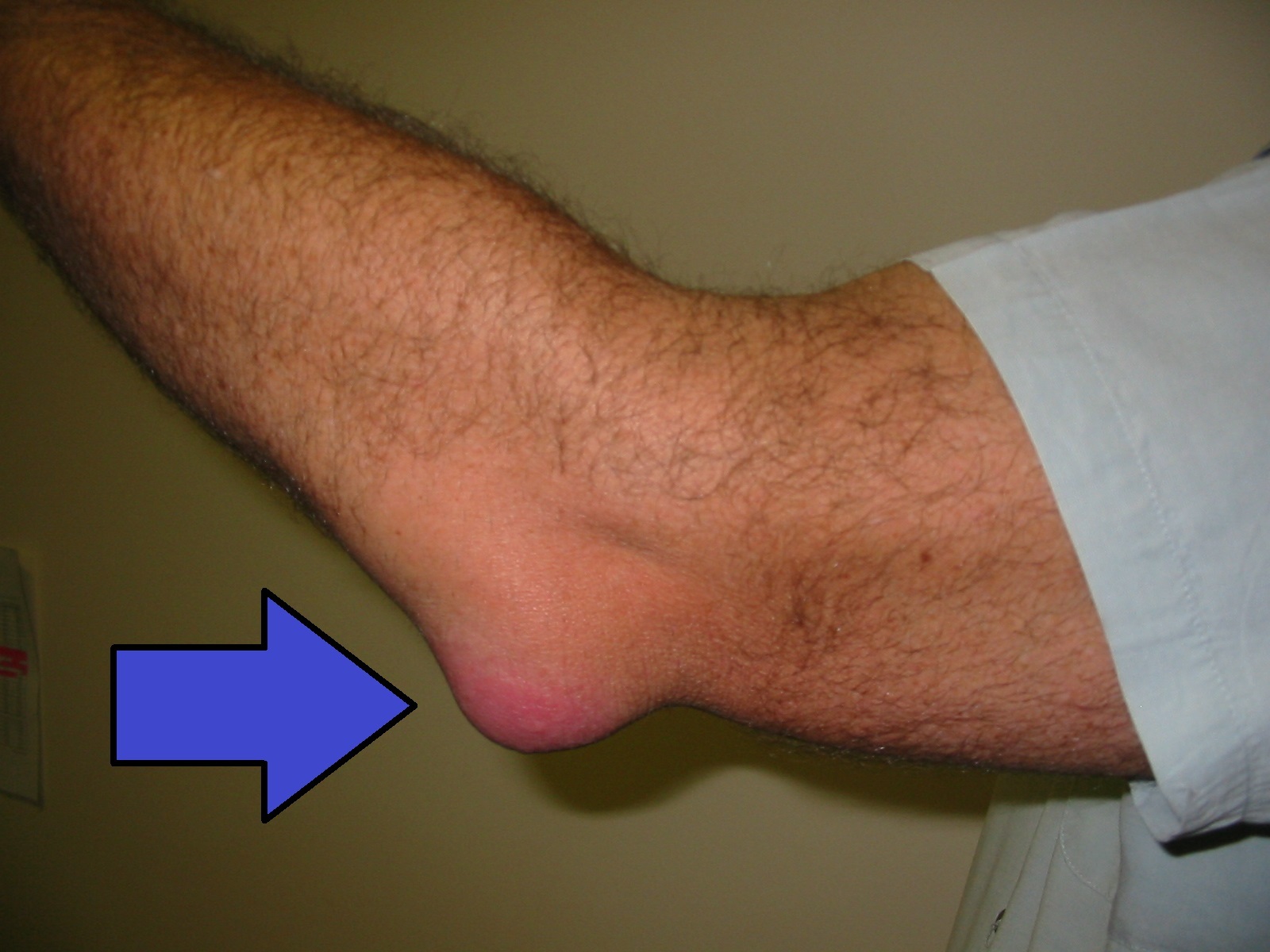
Figure 10. Olecranon bursitis: a fluctuant swelling over the olecranon (arrow). Source: NJC123, via Wikimedia Commons, CC0 1.0 (public domain).
Figure 10. Olecranon bursitis: a fluctuant swelling over the olecranon (arrow). Source: NJC123, via Wikimedia Commons, CC0 1.0 (public domain).
Part IV - Wrist and Hand Stenosing Tenosynovitis
De Quervain tenosynovitis
De Quervain disease (Fritz de Quervain, 1912) is a stenosing tenosynovitis of the first dorsal compartment (the abductor pollicis longus and extensor pollicis brevis) at the radial styloid, with sheath thickening (about three times normal) and mucopolysaccharide deposition rather than true inflammation.[36] It is far commoner in women, classically in pregnancy and the postpartum period (“mother’s wrist”).[37] An important anatomical point is a septum dividing the EPB into its own subcompartment in ~40% of wrists (more in surgical series), which raises the risk of treatment failure if not released.[38] The diagnosis rests on the Finkelstein test (the examiner ulnar-deviates the thumb and wrist) and the Eichhoff manoeuvre (the patient clasps the thumb in a fist before ulnar deviation); Finkelstein’s is considered the superior test.[39]
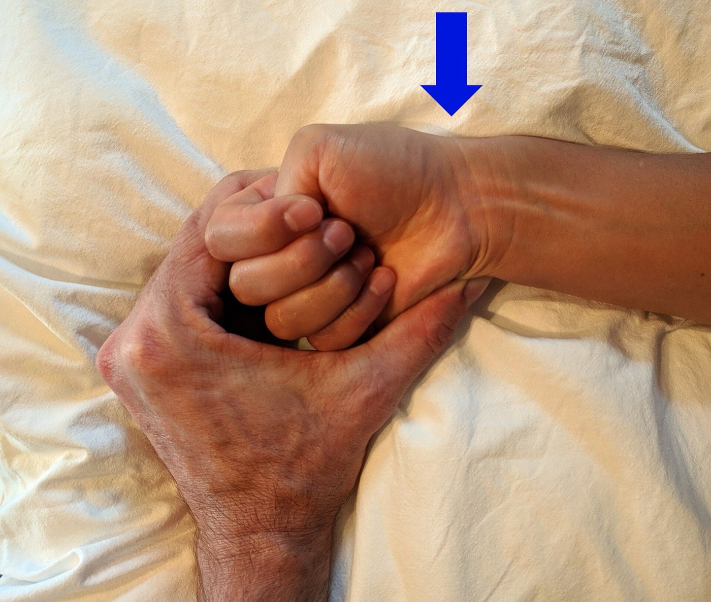
Figure 11. The Finkelstein/Eichhoff manoeuvre for de Quervain tenosynovitis; the arrow marks pain at the radial styloid (first dorsal compartment). Source: James Heilman, MD, via Wikimedia Commons, CC BY-SA 4.0.
Figure 11. The Finkelstein/Eichhoff manoeuvre for de Quervain tenosynovitis; the arrow marks pain at the radial styloid (first dorsal compartment). Source: James Heilman, MD, via Wikimedia Commons, CC BY-SA 4.0.
Treatment is a thumb-spica splint and corticosteroid injection (more effective than splinting alone, NNT ≈ 2), with surgical release of the first compartment for failures. Surgery takes care to release any separate EPB sheath, to protect the superficial radial nerve, and to incise dorsally to avoid volar tendon subluxation.[40]
Trigger finger and trigger thumb
Trigger digit is a stenosing tenosynovitis of the flexor sheath at the A1 pulley, where a mismatch between a thickened tendon and the constricted pulley causes catching, locking and pain; the A1 pulley itself develops chondrometaplasia.[41] It affects about 2% of the general population but ~20% of diabetics, women about twice as often, peaking at 50-70 years; the ring finger and thumb are most often involved.[42] Severity is graded by the Quinnell classification (I pre-triggering/pain; II demonstrable catching, actively correctable; III locking requiring passive extension; IV fixed PIP flexion contracture).[43] First-line treatment is corticosteroid injection (≈60% success versus 15-20% placebo, lower in diabetics) and/or MCP splinting; refractory or recurrent cases undergo A1 pulley release (open or percutaneous), the chief surgical hazard being injury to the radial digital nerve of the thumb, which lies very superficially and crosses obliquely.[44]
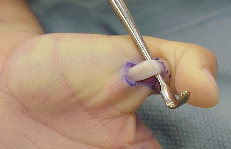
Figure 12. Open A1 pulley release for trigger digit: the flexor tendon exposed at the base of the thumb after incising the A1 pulley. Source: JoJoEMC, via Wikimedia Commons, CC BY-SA 3.0.
Figure 12. Open A1 pulley release for trigger digit: the flexor tendon exposed at the base of the thumb after incising the A1 pulley. Source: JoJoEMC, via Wikimedia Commons, CC BY-SA 3.0.
Other wrist tendinopathies
Intersection syndrome is friction tenosynovitis where the first-compartment tendons cross the radial wrist extensors about 4 cm proximal to the wrist (tenderness more proximal than de Quervain, sometimes with a “wet leather” crepitus); extensor carpi ulnaris tendinopathy/instability (Montalvan: tendinopathy, instability, rupture) presents with dorsoulnar pain and a painful snap, tested by the ECU synergy test and often coexisting with TFCC injury.[45]
Part V - Lower-Limb Tendinopathy and Enthesopathy
Patellar tendinopathy (“jumper’s knee”)
Patellar tendinopathy is tendinosis of the proximal patellar tendon at the inferior patellar pole (the deep posterior fibres), an overuse injury of jumping athletes (prevalence up to ~32-44% in elite basketball and volleyball).[46] There is anterior knee pain and tenderness at the inferior pole, classically reproduced on the single-leg decline-squat test, graded by the Blazina stages (1 pain after activity; 2 pain at the start, easing with warm-up, returning with fatigue; 3 constant pain; 4 rupture) and followed with the VISA-P questionnaire; ultrasound (grey-scale plus Doppler showing neovascularisation) is more accurate than MRI.[47] Treatment is an eccentric (decline-squat) or heavy-slow-resistance programme (a randomised trial found no advantage of surgery over 12 weeks of eccentric training), with surgery (open or arthroscopic debridement of the degenerate inferior pole) reserved for refractory structural disease.[48]
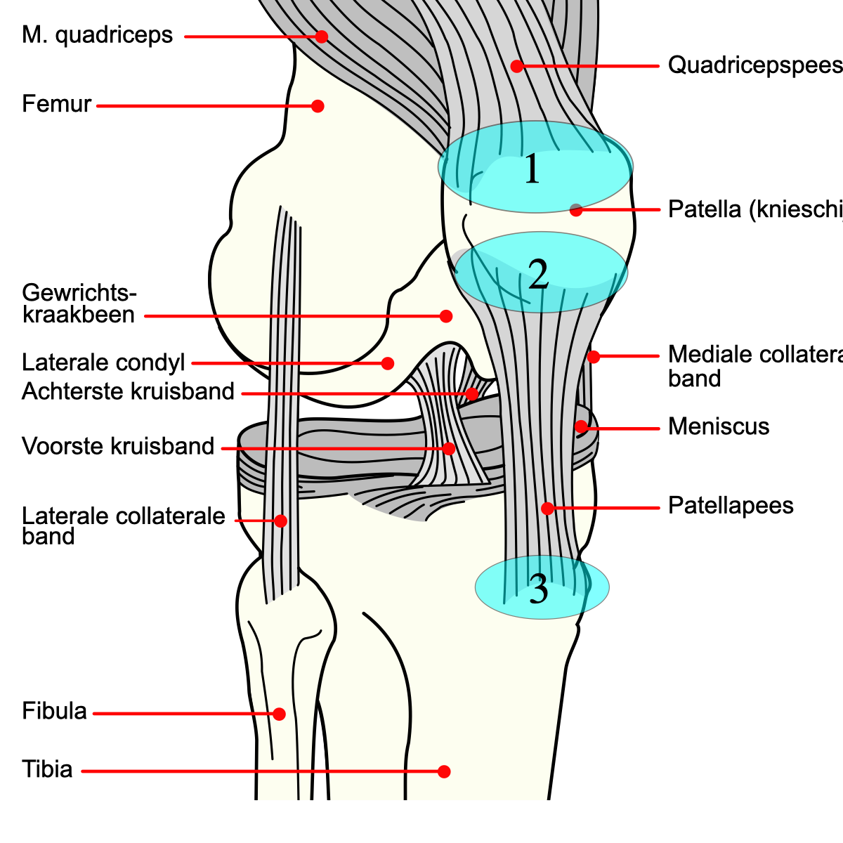
Figure 13. Patellar tendinopathy (“jumper’s knee”): the patellar tendon and the inferior-pole pain zone (diagram labelled in Dutch). Source: Mysid / .Koen, via Wikimedia Commons, CC BY-SA 3.0.
Figure 13. Patellar tendinopathy (“jumper’s knee”): the patellar tendon and the inferior-pole pain zone (diagram labelled in Dutch). Source: Mysid / .Koen, via Wikimedia Commons, CC BY-SA 3.0.
Achilles tendinopathy
Achilles tendinopathy is divided into mid-portion (non-insertional) disease, 2-6 cm above the calcaneus in its watershed zone, and insertional disease at the calcaneal attachment, the latter associated with retrocalcaneal bursitis and a Haglund deformity (“pump bump”).[49] There is painful, fusiform tendon thickening; the Silfverskiöld test assesses gastrocnemius tightness, and midline tenderness suggests insertional tendinopathy versus lateral tenderness for retrocalcaneal bursitis.[50] The mainstay is an eccentric heel-drop (Alfredson-type) programme, especially effective in mid-portion disease, supplemented by ESWT, GTN patches and, for failures, surgical debridement (± FHL transfer for insertional disease with a Haglund excision); corticosteroid injection is contraindicated because of rupture risk.[51] An acute Achilles rupture is distinguished by the Thompson (calf-squeeze) test (absent plantarflexion on squeezing the calf) and a palpable gap.[52]
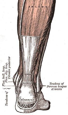
Figure 14. The Achilles tendon and its insertion on the calcaneus. Mid-portion tendinopathy occurs 2-6 cm above the insertion; insertional disease at the calcaneal attachment. From Gray’s Anatomy (1918), public domain, via Wikimedia Commons.
Figure 14. The Achilles tendon and its insertion on the calcaneus. Mid-portion tendinopathy occurs 2-6 cm above the insertion; insertional disease at the calcaneal attachment. From Gray’s Anatomy (1918), public domain, via Wikimedia Commons.
Plantar fasciitis (plantar fasciosis)
Plantar fasciitis is the commonest cause of inferior heel pain, a degenerative enthesopathy of the plantar fascia at the medial calcaneal tubercle rather than a true inflammation.[53] Its hallmark is “first-step” pain: sharp medial heel pain on the first steps in the morning or after rest, with tenderness at the fascial origin reproduced by toe-extension loading (the windlass mechanism). Risk factors include reduced ankle dorsiflexion/tight gastrocnemius, a cavus or planus foot, obesity and prolonged standing.[54] The heel spur is a marker, not the cause (present in many asymptomatic feet).[55] It is self-limiting in ~90%; treatment centres on plantar-fascia-specific and gastrocnemius stretching (the former superior to Achilles stretching alone), orthoses/heel cups, and night dorsiflexion splints, with corticosteroid injection giving only short-term relief (and risking fascial rupture/fat-pad atrophy), ESWT for chronic cases, and partial plantar fasciotomy reserved for failure of 6-12 months of conservative care.[56]
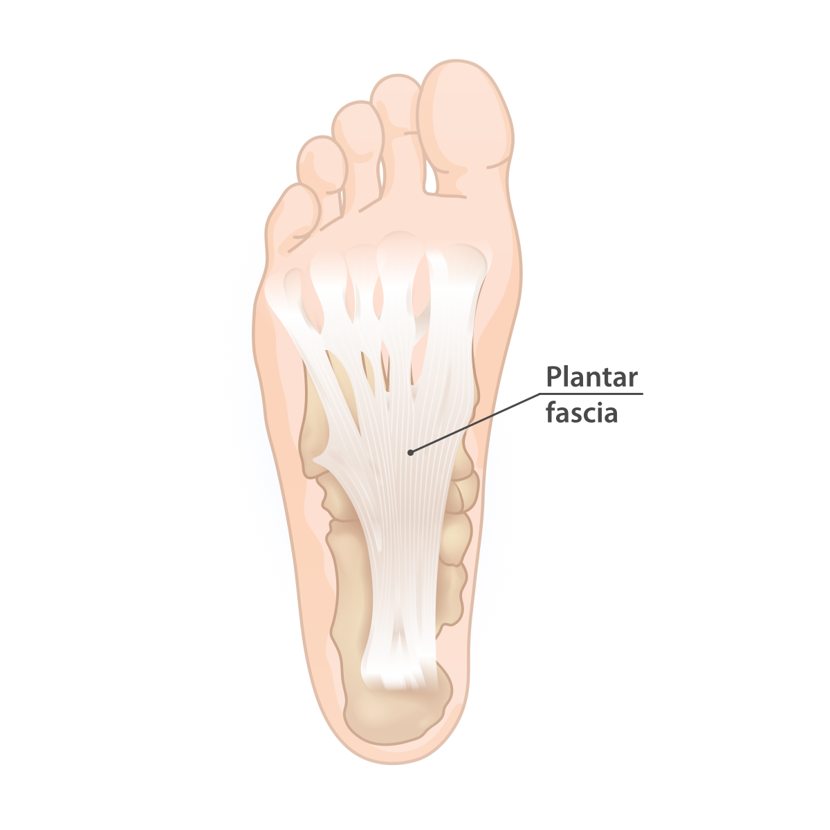
Figure 15. The plantar fascia, fanning from the medial calcaneal tubercle (the site of the enthesopathy) to the metatarsal heads. Source: InjuryMap, via Wikimedia Commons, CC BY-SA 4.0.
Figure 15. The plantar fascia, fanning from the medial calcaneal tubercle (the site of the enthesopathy) to the metatarsal heads. Source: InjuryMap, via Wikimedia Commons, CC BY-SA 4.0.
Greater trochanteric pain syndrome
Lateral hip pain in the trochanteric region, long labelled “trochanteric bursitis,” is now understood to be mostly a gluteus medius/minimus tendinopathy or tear (the “rotator cuff tear of the hip”), with bursitis usually secondary, often to a tight iliotibial band or external snapping hip.[57] Abductor tendon tears are common in ageing women (reported in up to 25% in their sixties); there is lateral trochanteric tenderness, weak abduction, inability to lie on the side, and abductor-insufficiency signs (Trendelenburg gait/sign, the hip lag sign), with MRI ~91% accurate for tears.[58] Treatment is conservative (load management, physiotherapy, NSAIDs, and cautious corticosteroid bursal injection), with surgical tendon repair or iliotibial-band lengthening (bursectomy, Z- or H-plasty) for discrete tears or recalcitrant symptoms.[59]
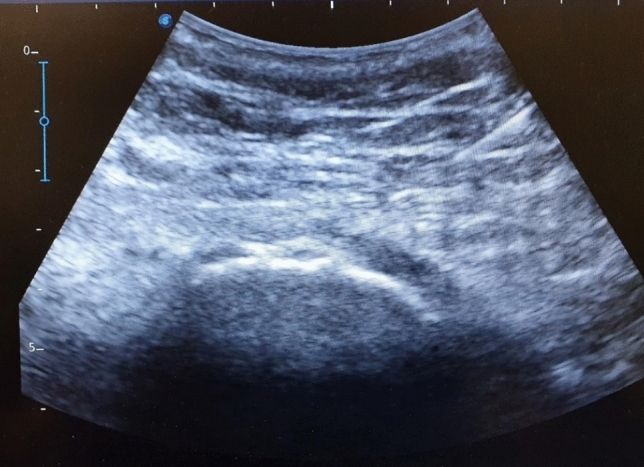
Figure 16. Ultrasound of the greater trochanteric region showing the gluteal tendon over the trochanteric cortex, the lesion underlying greater trochanteric pain syndrome. From Donati et al. (2025), PMC13241414, Fig. 2, CC BY 4.0.
Figure 16. Ultrasound of the greater trochanteric region showing the gluteal tendon over the trochanteric cortex, the lesion underlying greater trochanteric pain syndrome. From Donati et al. (2025), PMC13241414, Fig. 2, CC BY 4.0.
References
-
DeLee-Drez-Miller p. 1744. Elastic modulus ~1200-1800 MPa; ultimate tensile strength ~50-105 MPa.
-
DeLee p. 1745.
-
DeLee pp. 1744-1745; Green’s Operative Hand Surgery, ch. 56, p. 2383.
-
DeLee p. 1745; Green’s OHS p. 2383.
-
DeLee p. 1745.
-
Continuum model summarised from established teaching; the DeLee principles extract details the load/failed-healing mechanism (p. 1745) without naming the Cook-Purdam stages.
-
DeLee p. 1746 (Box 107.1); Green’s OHS p. 2383, which notes declining tendon cellularity after the third decade.
-
DeLee pp. 1751, 1942; heavy slow-resistance training is an effective alternative.
-
DeLee p. 977; Green’s OHS p. 1242-1243 (Coombes). The peritendinous corticosteroid/rupture concern is greatest for the Achilles and patellar tendons.
-
DeLee pp. 1751-1755; Green’s OHS p. 1242.
-
General principles; the DeLee olecranon-bursitis section (p. 982-984) exemplifies the septic-versus-aseptic approach.
-
Rockwood & Matsen, The Shoulder, p. 1147, which traces “periarthritis” directly to this topic’s name.
-
DeLee pp. 742, 745.
-
DeLee pp. 742, 744-745, 759; the flat/curved/hooked typing is referenced rather than numbered in the body text.
-
DeLee pp. 747-749, 768-769.
-
DeLee p. 750.
-
DeLee pp. 751-757.
-
DeLee p. 756.
-
DeLee p. 801; Rockwood & Matsen pp. 1147-1148.
-
Rockwood & Matsen pp. 1152-1155; DeLee p. 801.
-
DeLee pp. 803-804.
-
Rockwood & Matsen pp. 1155-1156; DeLee p. 802.
-
DeLee pp. 805-808; Rockwood & Matsen pp. 1158-1163. Manipulation is deferred until the inflammatory freezing phase has settled, and avoided in severe osteopenia and long-standing diabetes.
-
Rockwood & Matsen, ch. 15, p. 796.
-
Uhthoff phases from established teaching; Rockwood & Matsen (p. 796) confirms hydroxyapatite deposition followed by spontaneous resorption and the acute resorptive-phase pain; DeLee p. 798 records the “kidney stone of the shoulder” description.
-
Gärtner classification from established teaching (not present in the available chapter text).
-
Rockwood & Matsen p. 796 (barbotage and ESWT comparisons), pp. 797-799 (arthroscopic excision).
-
DeLee, ch. 61, p. 973; Green’s OHS, ch. 25, p. 1239.
-
DeLee p. 973; Green’s OHS p. 1239-1240.
-
DeLee p. 975, Table 61.1.
-
DeLee pp. 976-978; Green’s OHS pp. 1241-1243 (Coombes, Smidt).
-
DeLee p. 973; Green’s OHS p. 1240.
-
Green’s OHS pp. 1240-1242; DeLee p. 973.
-
DeLee pp. 982-983.
-
DeLee pp. 983-984.
-
Green’s OHS, ch. 56, p. 2387.
-
DeLee, ch. 71, p. 1145.
-
Green’s OHS pp. 2387-2389.
-
Green’s OHS pp. 2388, 2396; DeLee p. 1145.
-
Green’s OHS pp. 2388-2390; DeLee p. 1145.
-
Green’s OHS p. 2383.
-
Green’s OHS pp. 2383-2384.
-
Green’s OHS p. 2384, Table 56.1.
-
Green’s OHS pp. 2384-2386.
-
DeLee pp. 1146-1147; Green’s OHS pp. 2390-2391.
-
DeLee pp. 1744-1746.
-
DeLee pp. 1746-1748.
-
DeLee pp. 1751-1756 (Bahr).
-
DeLee pp. 1937-1938.
-
DeLee p. 1938.
-
DeLee pp. 1939-1942.
-
DeLee p. 1938.
-
DeLee p. 1981 (Ali).
-
DeLee pp. 1981-1983.
-
DeLee pp. 1981-1982 (Tanz, Shmokler).
-
DeLee pp. 1982-1985 (DiGiovanni).
-
DeLee pp. 1313, 1325-1326.
-
DeLee pp. 1313-1315.
-
DeLee pp. 1314-1326.
-
DeLee pp. 1744-1745; Green’s OHS p. 2383.
-
Rockwood & Matsen p. 1147.
-
DeLee pp. 742, 747-749.
-
DeLee pp. 801-808; Rockwood & Matsen pp. 1152-1163.
-
Rockwood & Matsen p. 796; Uhthoff/Gärtner from standard teaching.
-
DeLee pp. 973-978; Green’s OHS pp. 1239-1243.
-
Green’s OHS pp. 1240-1242.
-
Green’s OHS pp. 2387-2390.
-
Green’s OHS pp. 2383-2386.
-
DeLee pp. 1937-1942.
-
DeLee pp. 1981-1985.
-
DeLee pp. 1313-1326.