Contents
- Scope and orientation
- Part I - General concepts: osteonecrosis and the osteochondroses
- Part II - Legg-Calvé-Perthes disease
- Part III - The other osteochondroses
- III.1 Köhler disease - tarsal navicular
- III.2 Freiberg disease (infraction) - metatarsal head
- III.3 Sever disease - calcaneal apophysitis
- III.4 Osgood-Schlatter disease - tibial tubercle
- III.5 Sinding-Larsen-Johansson disease - inferior patella
- III.6 Panner disease versus osteochondritis dissecans of the capitellum
- III.7 Scheuermann disease - juvenile kyphosis
- III.8 Briefer entities and cross-references
- Part IV - Secondary avascular necrosis in children
- Bulgarian terminology glossary
- Figure credits and licences
Scope and orientation
This topic covers the avascular necroses of childhood. The centrepiece is Legg-Calvé-Perthes disease (LCPD), the idiopathic osteonecrosis of the immature femoral head, but the syllabus heading (“аваскуларни некрози в детска възраст”) is broader. It also takes in the other osteochondroses (Köhler, Freiberg, Osgood-Schlatter, Scheuermann, Panner and the rest) and the secondary avascular necroses that affect children: corticosteroid and chemotherapy-related, sickle-cell, Gaucher, and post-traumatic.
A few unifying ideas run through the whole topic:
- A shared final pathway. Whatever the trigger, the bone dies because its blood supply is interrupted. It is then repaired by creeping substitution, and during that repair the softened subchondral bone can fracture (the crescent sign) and collapse. Whether the joint surface ends up round or flat decides the long-term outcome.
- The osteochondroses are not all true necrosis. Some are genuine epiphyseal osteonecrosis (Perthes, Köhler, Freiberg, Panner), others are traction apophysites of overuse (Osgood-Schlatter, Sinding-Larsen-Johansson, Sever), and others are physeal disorders (Scheuermann). Knowing which category an eponym belongs to is half the exam answer.
- In the immature hip, biology favours the young. The younger child has time to remodel and an acetabulum that can still mould a soft head, which is why age at onset dominates the prognosis of Perthes.
- The treatment lever in Perthes is containment. Keep the plastic head seated in the acetabulum, and maintain movement, so it remodels spherically.
A Bulgarian terminology glossary, a viva self-test appendix, and image credits follow the clinical sections.
Part I - General concepts: osteonecrosis and the osteochondroses
I.1 Terminology
Osteonecrosis (ON) is death of a circumscribed area of bone from loss of its blood supply; the synonyms avascular necrosis (AVN), ischaemic necrosis and aseptic necrosis are used interchangeably (“aseptic” originally distinguished it from infection).[1] The most common cause overall is trauma (a displaced fracture or dislocation that tears local vessels); the most common atraumatic cause is corticosteroid use.[2] Two lesion patterns matter: subchondral / epiphyseal (juxta-articular) osteonecrosis, which is clinically important because it leads to joint-surface collapse, and medullary / metaphyseal infarcts, which are often silent.[3]
The osteochondroses are a group of disorders of the growing ossification centres that begin as degeneration or necrosis and are followed by regeneration and recalcification.[4] They are conventionally divided into three groups:[5]
- articular (epiphyseal): Perthes (femoral head), Köhler (navicular), Freiberg (metatarsal head), Kienböck (lunate), and osteochondritis dissecans;
- non-articular (apophyseal / traction apophysitis): Osgood-Schlatter (tibial tubercle), Sinding-Larsen-Johansson (inferior patella), Sever (calcaneus), Iselin (5th metatarsal base);
- physeal: Scheuermann (thoracic spine) and Blount (proximal tibia).
A genuine source disagreement worth flagging: the non-articular group is now regarded as overuse / traction injury rather than true avascular necrosis, whereas some imaging texts still classify several osteochondroses (Perthes, Freiberg) under “idiopathic osteonecrosis.”[6]
I.2 Pathophysiology of osteonecrosis
Whatever the cause, the evolution is the same: marrow necrosis and osteocyte death, a reparative reaction around the dead zone, and finally collapse of the necrotic bone with secondary arthritis.[7] The cellular timeline is well defined: haematopoietic cells die first (within ~12 hours), marrow fat cells from about day 2, and osteocytes within 2-5 days, disappearing completely by 2-4 weeks.[8]
Vascular interruption arises by four mechanisms, which map onto the causes: direct vascular interruption (trauma), intravascular occlusion (sickled cells, fat or nitrogen emboli), extravascular compression (marrow-fat hypertrophy, Gaucher-cell infiltration, marrow oedema raising intraosseous pressure) and direct cytotoxic injury (radiation, chemotherapy, corticosteroid).[9]
Repair is by “creeping substitution” (Phemister): fibrovascular tissue invades from the periphery and lays new woven bone on the dead trabeculae. In established osteonecrosis this repair front does not penetrate the central sequestrum. A fibrous reactive zone forms at the interface (the band seen on MRI), and the dead, mechanically weak bone is prone to subchondral fracture.[10] That subchondral fracture is the crescent sign, the pre-collapse-to-collapse turning point in the natural history.[11]
Part II - Legg-Calvé-Perthes disease
II.1 Definition and history
LCPD is an idiopathic osteonecrosis of the immature capital femoral epiphysis. It is a self-limiting disorder in which the head’s blood supply is interrupted, the head passes through necrosis and repair over roughly 2-4 years, and during that window the biologically plastic head can deform and predispose to premature osteoarthritis.[12] It was described almost simultaneously in 1910 by Arthur Legg (Boston, attributed it to trauma), Jacques Calvé (France, altered osteogenesis) and Georg Perthes (Germany, an “osteochondritis”); Henning Waldenström had described it in 1909 but thought it a benign tuberculosis, and later gave us the term coxa plana and the radiographic staging still used today.[13]
II.2 Epidemiology
- Incidence varies widely with geography and ethnicity, roughly 0.5-21 per 100,000 children under 15. It is highest in white northern Europeans (~10.8/100,000) and Japanese, and lowest in Black (~0.5/100,000) and Chinese populations.[14]
- Age: most commonly 4-8 years (range ~2 years to the early teens); around 80% present between 4 and 9.[15]
- Sex: boys predominate, about 4-5:1, but girls have a worse prognosis (they mature earlier, leaving less time to remodel, and tend to have more extensive disease).[16]
- Bilateral in ~10-15%, usually asynchronous. Synchronous, symmetrical bilateral changes should instead raise the suspicion of an epiphyseal dysplasia (MED/SED); these are familial, affect other epiphyses, and produce short stature.[17]
- Constitutional and environmental associations: delayed bone age (averaging ~2 years), short stature with disproportionately short distal limbs, low birth weight, exposure to second-hand smoke, lower socioeconomic status, and an excess of attention-deficit/hyperactivity. Catterall’s “susceptible child” is a small, very active boy with delayed skeletal maturation.[18] Despite occasional positive family histories, the condition is not inherited, and a strong family history of bilateral disease again points to a dysplasia.[19]
II.3 Aetiology and pathogenesis
The cause is unknown, but the central event is interruption of the blood supply to the capital epiphysis.[20] The vascular anatomy explains the vulnerability: between about ages 3 and 8 the head is supplied almost entirely by the lateral epiphyseal (posterosuperior retinacular) vessels from the medial femoral circumflex artery, which run subperiosteally along the neck and are isolated from the metaphysis by the growth plate. The anterior anastomotic network is sparse, especially in boys.[21] These vessels can be compromised by an effusion (tamponade) or by the hip held in abduction and internal rotation, which is the link to transient synovitis (about 1-7% of irritable hips later prove to be Perthes).[22]
Several theories compete. The double-infarction theory (a single infarct does not reproduce the human histology, but a second one does) is supported by experimental and human pathology.[23] A role for thrombophilia/coagulopathy (factor V Leiden, protein C/S deficiency) was proposed but is now inconsistent and largely refuted as a cause; the same caution applies to a reduced-IGF-1 hypothesis.[24] What is agreed is the cycle: interrupted supply → necrosis → revascularisation → resorption → reossification → remodelling, over about 2-4 years.[25]
II.4 Pathology and the biology of deformity
During the avascular phase the bony nucleus stops growing while the overlying articular cartilage, fed by synovial fluid, survives and thickens. Revascularisation then lays new woven bone on the dead trabeculae (creeping substitution → the “head within a head” density) while simultaneously resorbing them. A subchondral fracture occurs at the anterosuperior point of greatest stress (the clinical onset of pain), and its extent determines the size of the necrotic segment.[26] The crushed necrotic bone is resorbed and replaced by biologically plastic fibrocartilage (Catterall’s “chondrification of the infarct”); robust osteoclastic resorption outpaces a weak osteoblastic response, so the weakened head collapses under load.[27]
The deformity comes from this softened, mouldable head being loaded across the acetabular rim. While the head is contained and congruent the acetabulum acts as a mould and keeps it round; once the soft anterolateral segment extrudes, the lateral acetabular lip dents it. Extrusion beyond ~20% carries a high risk of permanent deformity.[28] Late deformity is compounded by physeal growth disturbance: central arrest gives a short neck and trochanteric overgrowth (coxa breva), while lateral arrest gives a tilted head with coxa valga.[29]
II.5 The stages (Waldenström)
The disease passes through four radiographic stages (radiographic “fragmentation” corresponds to pathological resorption):[30]
| Stage | Synonyms | Key radiographic features | Approx. duration |
|---|---|---|---|
| I. Initial / necrotic | sclerosis, condensation | Small, dense (sclerotic) epiphysis; apparent medial joint-space widening; subchondral fracture (crescent) | ~7 months |
| II. Fragmentation | resorption | Epiphysis appears fragmented (lucency + density); collapse, especially of the lateral pillar | ~8 months |
| III. Reossification | reparative, healing | New bone reappears; head/neck shape becomes set | ~18 months (longest) |
| IV. Healed / residual | remodelled | No avascular bone; residual deformity fixed; remodelling continues to maturity | - |
The modified Waldenström / Elizabethtown scheme subdivides the first three stages into early (A) and late (B), giving seven sub-stages in all, because femoral-head extrusion and deformity increase sharply at the late-fragmentation (IIB) point. This is the rationale for achieving containment before stage IIB.[31]
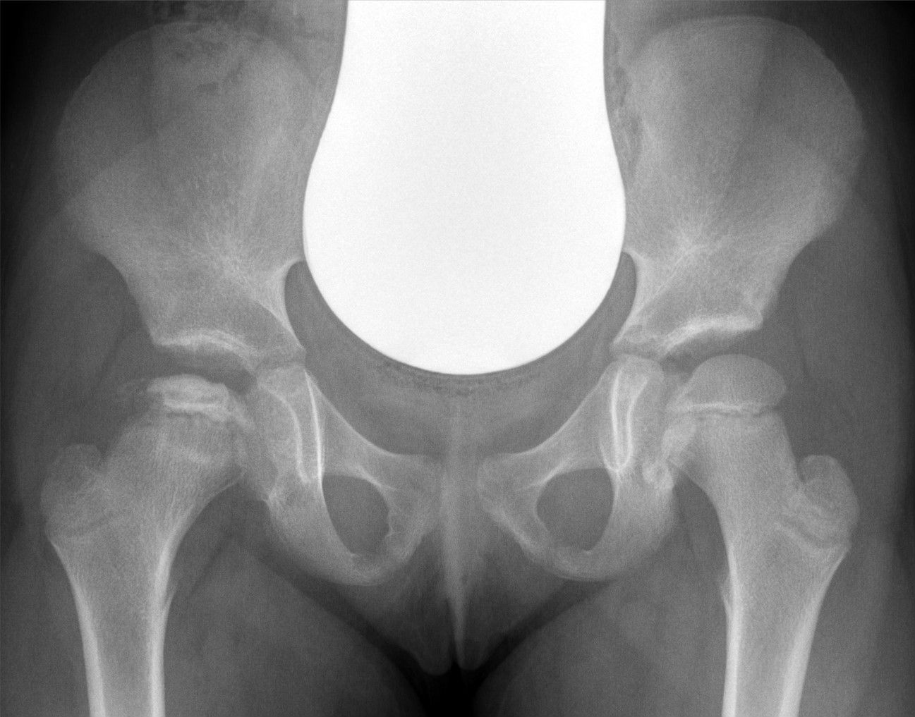
Antero-posterior pelvis of a child with Legg-Calvé-Perthes disease of the right hip: the right capital femoral epiphysis (image left) is small, sclerotic, fragmented and flattened compared with the normal, smoothly ossified left epiphysis. (Hellerhoff, Wikimedia Commons; CC BY-SA 4.0.)
Antero-posterior pelvis of a child with Legg-Calvé-Perthes disease of the right hip: the right capital femoral epiphysis (image left) is small, sclerotic, fragmented and flattened compared with the normal, smoothly ossified left epiphysis. (Hellerhoff, Wikimedia Commons; CC BY-SA 4.0.)
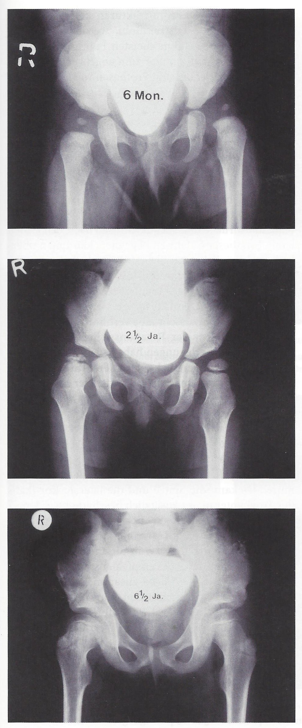
Untreated natural history of right-hip Perthes in one child at 6 months, 2½ years and 6½ years after onset (German labels: Mon. = months, Ja. = years), showing progression through fragmentation to a flattened, enlarged head. (Mehlauge, Wikimedia Commons; CC BY-SA 3.0.)
Untreated natural history of right-hip Perthes in one child at 6 months, 2½ years and 6½ years after onset (German labels: Mon. = months, Ja. = years), showing progression through fragmentation to a flattened, enlarged head. (Mehlauge, Wikimedia Commons; CC BY-SA 3.0.)
II.6 Clinical presentation
The typical patient is an otherwise healthy, often very active boy of 4-8 with the insidious onset of a limp, present for weeks to months because the symptoms are mild and intermittent.[32] Pain is mild, activity-related and relieved by rest, and is frequently referred to the thigh or knee, a classic cause of diagnostic delay.[33] Examination shows an antalgic (later Trendelenburg) gait and loss of abduction and internal rotation, with adductor spasm and a flexion/adduction posture; thigh and buttock atrophy indicate longstanding disease, and true shortening signals significant collapse and a poor prognosis.[34] The differential includes the epiphyseal dysplasias, hypothyroidism, sickle-cell and Gaucher osteonecrosis, infection, and the irritable hip of transient synovitis.[35]
II.7 Imaging
Standard views are an AP pelvis and a frog-leg (Lauenstein) lateral; radiographs may be normal for the first 2-3 months.[36] Early signs are a small, sclerotic epiphysis, apparent medial joint-space widening, lateral displacement and the subchondral fracture/crescent (present transiently in only ~25%); later come fragmentation, coxa magna, coxa plana, lateral extrusion (a broken Shenton line) and metaphyseal cysts.[37]
Catterall’s five “head-at-risk” signs identify a head destined to do badly: Gage sign (a lucent V in the lateral epiphysis/metaphysis), calcification lateral to the epiphysis, lateral subluxation, a horizontal growth plate, and a diffuse metaphyseal reaction. In untreated patients, no poor result occurred without two or more of these signs.[38] MRI is the most sensitive early test (it shows the infarct before radiographic change, and gadolinium-perfusion MRI quantifies the extent of avascularity before collapse), while dynamic arthrography demonstrates the true cartilaginous head shape and hinge abduction, the deformed head hinging on the lateral rim with medial dye pooling, a sign that the hip is no longer containable.[39] A technetium bone scan shows an early “cold” lateral wedge but has been superseded by MRI.[40]
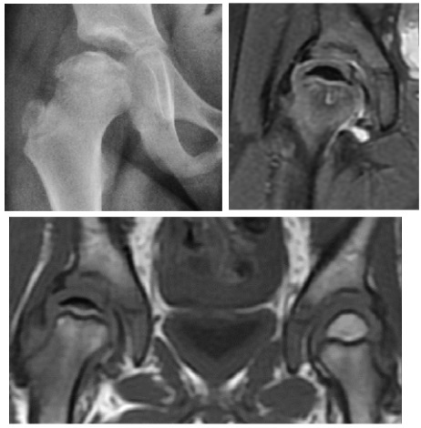
Grade-C Perthes: plain radiograph (coxa plana) with coronal proton-density fat-saturated and T1 MRI; the affected epiphysis shows low T1 signal (necrosis) against the normal contralateral side. (Ruiz Santiago et al., Radiol Res Pract 2016; CC BY 4.0.)
Grade-C Perthes: plain radiograph (coxa plana) with coronal proton-density fat-saturated and T1 MRI; the affected epiphysis shows low T1 signal (necrosis) against the normal contralateral side. (Ruiz Santiago et al., Radiol Res Pract 2016; CC BY 4.0.)
II.8 Classification systems
LCPD systems do three different jobs. Some stage the disease (Waldenström), some predict severity while it is active (Catterall, Salter-Thompson, lateral pillar), and Stulberg defines the outcome at maturity. The severity systems can only be applied reliably in the fragmentation stage.[41]
- Catterall groups I-IV grade the extent of head involvement: I = anterior head only (≤25%), II = up to ~50% with a sequestrum, III = up to ~75%, IV = whole head. Groups I-II do well, III-IV badly; reliability is only moderate (better if II and III are merged).[42]
- Salter-Thompson A/B uses the extent of the subchondral fracture: A = <50% (an intact lateral column protects the head), B = >50%. Its limitation is that the fracture line is transient.[43]
- Herring lateral pillar (A / B / B-C border / C) grades the height of the lateral pillar of the epiphysis during early fragmentation: A = full height (uniformly good), B = >50% maintained, B/C border = ~50%/poorly ossified, C = <50% maintained. It is the most reproducible classification and the strongest predictor of outcome, though up to a third of patients change category over time.[44]
- Stulberg I-V is the outcome classification, grading the healed head: I-II spherical congruent (no arthritis), III-IV aspherical congruent (mild-moderate late OA), V aspherical incongruent (severe OA before age 50). The final head shape and congruency are the dominant determinants of long-term arthritis.[45]
II.9 Prognostic factors
The dominant factor is the final head shape and congruency (Stulberg). The clinical predictors of reaching a poor shape are, in order: age at onset (the single most important; younger is better, with thresholds debated around 6 and 8 years); female sex (less remodelling time); extent of involvement (lateral pillar/Catterall); femoral-head extrusion; the head-at-risk signs; loss of motion; and hinge abduction, which is a contraindication to containment.[46]
II.10 Treatment
The principle is containment: keeping the soft head seated within the acetabulum so it remodels spherically, with restoration and maintenance of movement as the single most important and universally agreed measure. Because the head is more than half a sphere and the acetabulum only half, no method contains it through the whole gait cycle, and containment must be achieved before late fragmentation (stage IIB). Weight relief alone is ineffective.[47]
Non-operative management suits the good-prognosis hip (lateral pillar A, young children): activity modification, NSAIDs, and physiotherapy/traction/Petrie casts to recover abduction. Bracing has been largely abandoned: the Atlanta Scottish Rite and other abduction orthoses were shown to be no better than physiotherapy in several series and in the Herring trial.[48]
Operative containment uses the femoral varus (derotation) osteotomy (Axer) and/or a pelvic osteotomy (Salter innominate; shelf acetabuloplasty; triple), alone or combined. With a femoral osteotomy the neck-shaft angle should not be left in excessive varus (kept above about 105-110°), and a concurrent trochanteric epiphysiodesis reduces the complications of overgrowth and shortening.[49]
The main evidence is the Herring multicentre prospective study (children 6-12, graded by Stulberg at maturity): the two strongest predictors were the lateral pillar group and age at onset, and the benefit of surgery was concentrated in older children (over 8 years / bone age over ~6) with lateral pillar B or B/C border. Young children, and any hip in lateral pillar C, gained little from operation. An independent trial (Wiig) found the varus osteotomy significantly better than bracing or physiotherapy in children over 6 with extensive necrosis.[50]
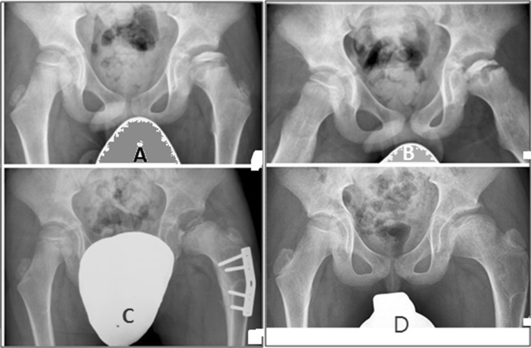
Operative containment for severe Perthes in a 7½-year-old boy (left hip, Catterall IV / lateral pillar C): (A, B) pre-operative AP and frog-leg views; (C) after a proximal-femoral varus osteotomy with a fixation plate; (D) a more spherical head at 48 months. The white shapes are gonad shields. (Elzohairy, J Orthop Traumatol 2016; CC BY 4.0.)
Operative containment for severe Perthes in a 7½-year-old boy (left hip, Catterall IV / lateral pillar C): (A, B) pre-operative AP and frog-leg views; (C) after a proximal-femoral varus osteotomy with a fixation plate; (D) a more spherical head at 48 months. The white shapes are gonad shields. (Elzohairy, J Orthop Traumatol 2016; CC BY 4.0.)
For the late, non-containable or deformed hip the aim becomes salvage: a valgus (or shelf) osteotomy for hinge abduction, a Chiari osteotomy for coverage, cheilectomy or femoral-head reduction osteotomy for impingement, and arthrodiastasis (articulated hip distraction) for the worst hips; end-stage disease eventually comes to total hip arthroplasty.[51]
II.11 Outcome
Most patients do well for decades, but the disease shortens the life of the hip. Stulberg I-II hips behave normally; III-IV develop mild-to-moderate osteoarthritis in late adulthood; V develops severe arthritis before age 50.[52] Long-term cohorts bear this out: 20-40 years after onset most patients are active and pain-free, but by the sixth and seventh decades osteoarthritis becomes common, and the long-term Iowa cohort found osteoarthritis roughly ten times the rate of the general population, with many requiring arthroplasty.[53]
Part III - The other osteochondroses
III.1 Köhler disease - tarsal navicular
Osteochondrosis of the tarsal navicular, in young children (~2-8 years, more often boys), presenting with a painful limp and medial midfoot tenderness. The radiograph shows a sclerotic, flattened, fragmented navicular. It is self-limiting; a short period in a walking cast often speeds symptom relief.[54]
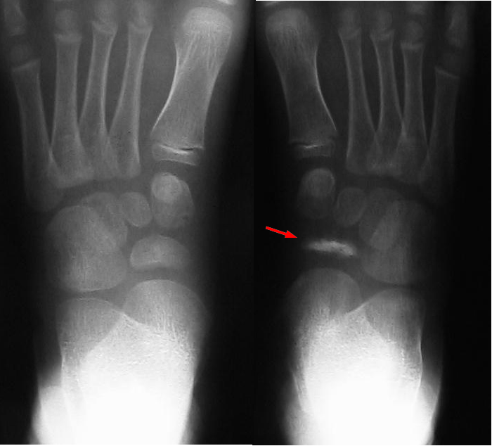
Both feet of a child with Köhler disease: the right tarsal navicular (red arrow) is sclerotic, flattened and fragmented compared with the normal rounded navicular on the left. (Kinderradiologie Olgahospital, Klinikum Stuttgart, Wikimedia Commons; CC BY-SA 4.0.)
Both feet of a child with Köhler disease: the right tarsal navicular (red arrow) is sclerotic, flattened and fragmented compared with the normal rounded navicular on the left. (Kinderradiologie Olgahospital, Klinikum Stuttgart, Wikimedia Commons; CC BY-SA 4.0.)
III.2 Freiberg disease (infraction) - metatarsal head
Osteonecrosis of a metatarsal head, most often the 2nd (the 3rd in up to a quarter of cases). It is the only osteochondrosis commoner in females (about 5:1), presenting in adolescence with forefoot pain, swelling and stiff, painful metatarsophalangeal motion.[55] Smillie staging (I-V) runs from an epiphyseal fissure, through central collapse and loose-body formation, to end-stage flattening and arthritis. Early disease is managed conservatively (stiff-soled shoe, metatarsal padding); progressive disease may need debridement or a dorsal closing-wedge osteotomy that rotates the intact plantar cartilage into the joint.[56]
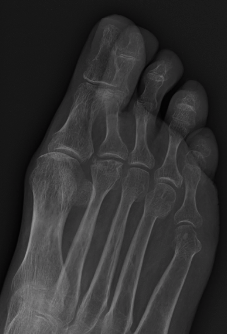
Antero-posterior forefoot in Freiberg disease: the 2nd metatarsal head is flattened, broadened and sclerotic compared with the rounded neighbouring heads. (James Heilman MD, Wikimedia Commons; CC BY-SA 4.0.)
Antero-posterior forefoot in Freiberg disease: the 2nd metatarsal head is flattened, broadened and sclerotic compared with the rounded neighbouring heads. (James Heilman MD, Wikimedia Commons; CC BY-SA 4.0.)
III.3 Sever disease - calcaneal apophysitis
A traction apophysitis of the calcaneal apophysis at the Achilles insertion, in active, skeletally immature children (~8-12 years), presenting with activity-related heel pain and local tenderness, often with a tight, weak gastrocsoleus. It is a clinical diagnosis (radiographs are not needed), is self-limiting (settling over 6-12 months), and is treated with activity modification, calf stretching and a heel raise. Despite the original name it is not inflammatory but an overuse injury; corticosteroid injection and surgery are contraindicated.[57]
III.4 Osgood-Schlatter disease - tibial tubercle
A traction apophysitis of the tibial tubercle at the patellar-tendon insertion, very common in adolescents at the growth spurt (boys ~12-15, girls ~8-12) doing running and jumping sport. The diagnosis is clinical (pain and a tender, prominent tubercle), and a radiograph is reserved for atypical presentations to exclude a tumour around the knee. It is self-limiting, resolving as the tubercle fuses, occasionally leaving a separate ossicle that can be excised in the symptomatic adult.[58]
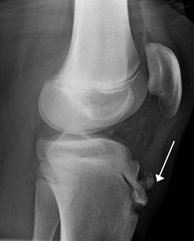
Lateral knee (open physes) in Osgood-Schlatter disease: the white arrow marks fragmentation of the tibial-tubercle apophysis / a separate ossicle at the patellar-tendon insertion. (James Heilman MD, Wikimedia Commons; CC BY-SA 3.0.)
Lateral knee (open physes) in Osgood-Schlatter disease: the white arrow marks fragmentation of the tibial-tubercle apophysis / a separate ossicle at the patellar-tendon insertion. (James Heilman MD, Wikimedia Commons; CC BY-SA 3.0.)
III.5 Sinding-Larsen-Johansson disease - inferior patella
The same traction apophysitis as Osgood-Schlatter but at the inferior pole of the patella (the proximal end of the patellar tendon). It is much less common, managed identically, and must be distinguished from an acute patellar sleeve avulsion fracture.[59]
III.6 Panner disease versus osteochondritis dissecans of the capitellum
These are two lesions of the humeral capitellum at different ages, often confused. Panner disease is an osteochondrosis of the whole capitellar ossific nucleus in younger boys (7-12, peak ~9), presenting with mild lateral elbow pain and slight loss of extension; the radiograph shows fragmentation of the entire nucleus (resembling Perthes), no loose bodies form, and it is benign and self-limiting with rest.[60] Osteochondritis dissecans (OCD) of the capitellum affects older adolescents (10-15), the thrower or gymnast, as a focal subchondral lesion that can detach into a loose body, with a far less predictable course and a real risk of late arthritis; stable lesions are rested, unstable or loose fragments are treated surgically.[61]
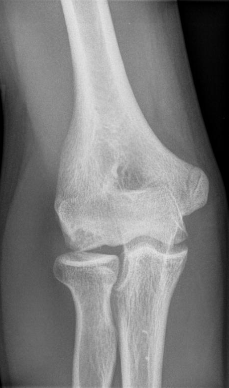
Antero-posterior elbow of a child with Panner disease: the capitellar ossification centre is irregular, fragmented and sclerotic - an osteochondrosis of the capitellum. (Kinderradiologie Olgahospital, Klinikum Stuttgart, Wikimedia Commons; CC BY-SA 3.0.)
Antero-posterior elbow of a child with Panner disease: the capitellar ossification centre is irregular, fragmented and sclerotic - an osteochondrosis of the capitellum. (Kinderradiologie Olgahospital, Klinikum Stuttgart, Wikimedia Commons; CC BY-SA 3.0.)
III.7 Scheuermann disease - juvenile kyphosis
A physeal osteochondrosis of the thoracic spine and the commonest cause of a structural, rigid kyphosis in adolescents, presenting around the pubertal growth spurt (age 10-12; the sexes are affected about equally). The cause is debated. Scheuermann’s original avascular-ring-apophysis idea is now doubted, and a mechanical (Hueter-Volkmann) growth-suppression model is favoured.[62]
The diagnostic radiographic criteria (Sorensen) are anterior wedging of more than 5° in at least three adjacent vertebrae, with Schmorl nodes, endplate irregularity and disc-space narrowing; normal thoracic kyphosis is 20-45°. The typical (thoracic, apex T7-T9) form is contrasted with the atypical thoracolumbar form, which is more often painful in adult life.[63] Clinically the kyphosis is rigid and sharply angular (unlike the smooth, correctable postural kyphosis), with tight hamstrings and pectorals; the natural history is generally benign.[64]
Management is by physiotherapy (for pain and posture, but it does not correct the bony deformity), bracing (Milwaukee or a TLSO) in the skeletally immature patient with a flexible curve over ~50-60°, and surgery (posterior instrumented fusion, with Ponte osteotomies) for a progressive thoracic curve over ~75-80° or a painful thoracolumbar curve over ~50-55°, correcting only to a high-normal kyphosis to avoid junctional problems.[65]
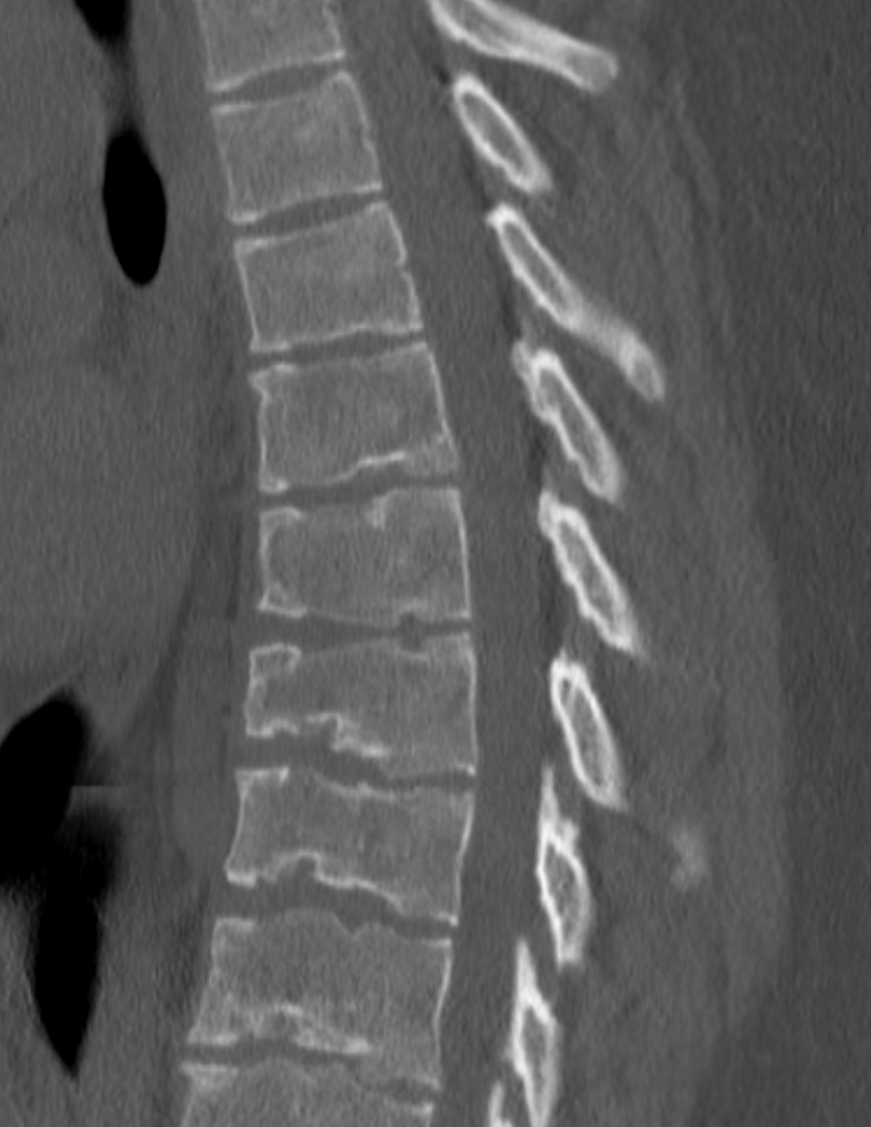
Sagittal CT of the thoracolumbar spine in Scheuermann disease: several adjacent anteriorly wedged vertebrae with irregular endplates and Schmorl nodes. (James Heilman MD, Wikimedia Commons; CC BY-SA 4.0.)
Sagittal CT of the thoracolumbar spine in Scheuermann disease: several adjacent anteriorly wedged vertebrae with irregular endplates and Schmorl nodes. (James Heilman MD, Wikimedia Commons; CC BY-SA 4.0.)
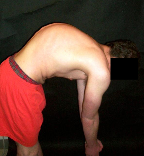
Forward-bend clinical photograph in Scheuermann disease showing a rigid, sharply angular thoracic kyphosis. (Wikimedia Commons; CC0.)
Forward-bend clinical photograph in Scheuermann disease showing a rigid, sharply angular thoracic kyphosis. (Wikimedia Commons; CC0.)
III.8 Briefer entities and cross-references
- Iselin disease: traction apophysitis of the base of the 5th metatarsal (peroneus brevis insertion); tenderness and pain on resisted eversion; treated with activity modification and peroneal strengthening.[66]
- Thiemann disease: osteochondrosis of the phalangeal epiphyses of the hand (adolescents, often familial). (Not described in the available sources; included for completeness.)
- Kienböck disease (lunate) and Blount disease (proximal tibial physis) are osteochondroses too, but occur largely outside the Perthes age group and are covered under their own syllabus topics.[67]
Part IV - Secondary avascular necrosis in children
Beyond the idiopathic osteochondroses, several systemic and mechanical insults cause osteonecrosis in children and young adults. They share the pathophysiology of Part I and differ mainly in the trigger.
IV.1 Causes
- Corticosteroid-induced: the commonest atraumatic cause. Risk rises with dose and duration (roughly a 4-5% increase in risk per additional 10 mg/day of prednisone; cumulative doses over ~2 g in 3 months are high-risk), and most cases relevant to children arise in the treatment of acute lymphoblastic leukaemia, nephrotic syndrome, transplantation and SLE. The mechanism is multifactorial: marrow-fat hypertrophy raising intraosseous pressure, osteocyte apoptosis, impaired angiogenesis and a hypercoagulable shift.[68]
- Chemotherapy / ALL: glucocorticoids (especially dexamethasone) plus methotrexate; the risk is higher in older children and adolescents (over ~10 years) and in girls, and the necrosis is often multifocal.[69]
- Sickle-cell disease: vaso-occlusion by sickled cells infarcts the femoral and humeral heads, often multifocally; osteonecrosis affects 37-50% of adults but fewer children, and a key clinical problem is distinguishing bone infarct from osteomyelitis (Salmonella and Staphylococcus). Sickle-cell trait does not cause it.[70]
- Gaucher disease: glucocerebroside-laden macrophages crowd the marrow and compress its vasculature, producing femoral head/neck necrosis and bone crises, with the classic Erlenmeyer-flask distal femur; osteonecrosis occurs in up to 60%.[71]
- Trauma: the highest-risk paediatric mechanical causes are femoral-neck fracture, traumatic hip dislocation, slipped capital femoral epiphysis (osteonecrosis in ~25%), and iatrogenic necrosis after DDH treatment (forced abduction compressing the epiphyseal vessels, cross-referenced to Topic 7).[72]
- Others: alcohol, dysbaric (Caisson) disease, radiation, SLE/antiphospholipid syndrome, and idiopathic.[73]
IV.2 Staging and the crescent sign
Three radiographic/MRI systems are used for the femoral head:[74]
- Ficat-Arlet (0/I-IV): I normal radiograph (MRI-positive), II sclerosis/cysts with a preserved contour, III crescent sign ± early flattening, IV collapse and secondary osteoarthritis.
- ARCO (0-4): adds an MRI-only stage 1 and incorporates lesion size and location.
- Steinberg (University of Pennsylvania, 0-VI): the first to make lesion size and MRI integral, subdividing each stage by extent.
Across all systems the crescent sign (a subchondral fracture) marks the irreversible pre-collapse-to-collapse boundary and is best seen on the frog-leg lateral.[75]
IV.3 Imaging and management
Radiographs are often normal early, then show mixed sclerosis → crescent → collapse → secondary arthritis. MRI is the most sensitive early test (sensitivity and specificity over 98%): the earliest sign is a low-signal band on T1 around the necrotic segment, with the double-line sign on T2 (a hyperintense inner line and hypointense outer rim).[76]
Management begins with treating the underlying cause and protecting the joint. The prognosis hinges on the crescent: caught before collapse, the joint may be preserved by core decompression (with or without grafting/biologics) or a redirecting osteotomy; once the head has collapsed, the outlook is guarded and end-stage disease comes to total hip arthroplasty (modern bearings give good survivorship even in the young).[77] The key paediatric difference is that the idiopathic immature hip (Perthes) is managed by containment, not by core decompression or arthroplasty, and the young child’s remodelling potential makes the prognosis age-dependent.[78]
Bulgarian terminology glossary
For consistency with the Bulgarian state-examination vocabulary (and the operative terminology of Boychev, Хирургическа ортопедия):
| English | Bulgarian |
|---|---|
| Avascular / aseptic necrosis (osteonecrosis) | Аваскуларна (асептична) некроза (остеонекроза) |
| Legg-Calvé-Perthes disease | Болест на Лег-Калве-Пертес (Perthes) |
| Capital femoral epiphysis | Глава(та) на бедрената кост / проксимална феморална епифиза |
| Coxa plana / coxa magna | Coxa plana (плоско бедро) / coxa magna (уголемена глава) |
| Osteochondrosis / osteochondroses | Остеохондроза / остеохондрози |
| Containment | Покриване (контейнмент) на бедрената глава |
| Femoral varus (derotation) osteotomy | Варизираща (деротационна) остеотомия на бедрото |
| Pelvic (Salter) osteotomy | Тазова остеотомия (по Salter) |
| Köhler disease | Болест на Köhler (на ладиевидната кост) |
| Freiberg disease | Болест на Freiberg (на главата на метатарзалната кост) |
| Osgood-Schlatter disease | Болест на Osgood-Schlatter (на тибиалната грапавина) |
| Sinding-Larsen-Johansson disease | Болест на Sinding-Larsen-Johansson |
| Sever disease (calcaneal apophysitis) | Болест на Sever (калканеален апофизит) |
| Panner disease | Болест на Panner (на капитулума) |
| Scheuermann disease (juvenile kyphosis) | Болест на Scheuermann (юношеска кифоза) |
| Schmorl node | Възел (херния) на Schmorl |
| Sickle-cell disease | Сърповидноклетъчна анемия (болест) |
| Gaucher disease | Болест на Gaucher |
| Slipped capital femoral epiphysis | Епифизеолиза на бедрената глава |
| Total hip arthroplasty | Тотално ендопротезиране на тазобедрената става |
Figure credits and licences
All images were independently opened and visually verified to depict the stated entity before use; licences were confirmed against the Wikimedia Commons API or the NCBI PMC Open-Access service. All are public-domain, CC0, CC BY or CC BY-SA (no non-commercial files).
- Perthes - AP pelvis (child) -
perthes_AP-pelvis_child_right-hip_Hellerhoff_CCBYSA4.jpg. Hellerhoff, Wikimedia Commons. CC BY-SA 4.0. - Perthes - untreated natural-history series -
perthes_untreated-3stage-series_Mehlauge_CCBYSA3.jpg. Mehlauge, Wikimedia Commons. CC BY-SA 3.0. - Perthes - radiograph + MRI (grade C) -
perthes_MRI-T1-PDfatsat-plus-xray_gradeC_RuizSantiago_CCBY4.jpg. Ruiz Santiago F. et al., Radiology Research and Practice 2016 (DOI 10.1155/2016/6369237), via Wikimedia Commons. CC BY 4.0. - Perthes - pre/post varus osteotomy -
perthes_pre-and-post-varus-osteotomy_Elzohairy_PMC5071238_CCBY4.jpg. Elzohairy MM, J Orthop Traumatol 2016;17(4):345-351 (DOI 10.1007/s10195-016-0412-0). CC BY 4.0. - Köhler disease -
kohler_foot_navicular-sclerosis-flattening_Olgahospital_CCBYSA4.png. Kinderradiologie Olgahospital, Klinikum Stuttgart, Wikimedia Commons. CC BY-SA 4.0. - Freiberg disease -
freiberg_foot_2nd-MT-head-flattening_Heilman_CCBYSA4.png. James Heilman MD, Wikimedia Commons. CC BY-SA 4.0. - Osgood-Schlatter disease -
osgood-schlatter_lateral-knee_tubercle-ossicle-arrow_Heilman_CCBYSA3.png. James Heilman MD, Wikimedia Commons. CC BY-SA 3.0. - Panner disease -
panner_AP-elbow_capitellum-osteochondrosis_Olgahospital_CCBYSA3.png. Kinderradiologie Olgahospital, Klinikum Stuttgart, Wikimedia Commons. CC BY-SA 3.0. - Scheuermann disease - sagittal CT -
scheuermann_sagittalCT_wedging-and-schmorl-nodes_Heilman_CCBYSA4.png. James Heilman MD, Wikimedia Commons. CC BY-SA 4.0. - Scheuermann disease - clinical photograph -
scheuermann_clinical-photo_forward-bend-kyphosis_CC0.jpg. Wikimedia Commons. CC0.
CC BY-SA images require that any redistributed derivative carry the same share-alike licence.
References
-
Osteonecrosis (Koo, Mont & Jones), pp.20, 48.
-
Osteonecrosis, pp.20, 89.
-
Osteonecrosis, p.132.
-
Morrey, The Elbow and Its Disorders, p.303.
-
Brukner & Khan, Clinical Sports Medicine, p.936.
-
Brukner & Khan, p.935; Greenspan, Orthopedic Imaging, pp.173-174.
-
Osteonecrosis, p.145.
-
Osteonecrosis, pp.146-147.
-
Osteonecrosis, pp.145-146, 160.
-
Osteonecrosis, p.148.
-
Osteonecrosis, pp.153-154, 161.
-
Feldman & Paley, Legg-Calvé-Perthes Disease, pp.9-10, 38; Pediatric & Adolescent Hip, p.170.
-
Feldman & Paley, p.9; Catterall, Legg-Calvé-Perthes Disease, pp.16-17; Lovell & Winter, p.1199.
-
Feldman & Paley, p.10; Pediatric & Adolescent Hip, p.173; Lovell & Winter, p.1201.
-
Lovell & Winter, p.1201; Catterall, pp.49-50.
-
Feldman & Paley, pp.10, 13; Lovell & Winter, pp.1201, 1220.
-
Lovell & Winter, p.1201; Wenger & Rang, p.327; Pediatric & Adolescent Hip, p.177.
-
Feldman & Paley, pp.10-12; Lovell & Winter, p.1201; Catterall, pp.19-20.
-
Lovell & Winter, p.1201; Catterall, pp.18-19.
-
Feldman & Paley, p.10; Catterall, p.18.
-
Catterall, pp.24-26; Lovell & Winter, pp.1201-1202.
-
Salter, p.382; Catterall, p.50; Lovell & Winter, p.1202.
-
Lovell & Winter, p.1202; Catterall, p.23.
-
Feldman & Paley, pp.11-12, 16; Lovell & Winter, pp.1200-1203.
-
Pediatric & Adolescent Hip, p.170; Feldman & Paley, p.38.
-
Salter, pp.378-379; Catterall, pp.59-60.
-
Catterall, pp.43-47; Pediatric & Adolescent Hip, p.170.
-
Salter, p.379; Pediatric & Adolescent Hip, p.170; Catterall, pp.86-87.
-
Lovell & Winter, pp.1209-1211; Pediatric & Adolescent Hip, p.172.
-
Lovell & Winter, pp.1206-1207; Pediatric & Adolescent Hip, p.171; Catterall, pp.59-61.
-
Feldman & Paley, pp.30-32; Pediatric & Adolescent Hip, pp.171-172.
-
Lovell & Winter, p.1221; Pediatric & Adolescent Hip, p.177.
-
Lovell & Winter, p.1221; Catterall, p.51.
-
Lovell & Winter, p.1222; Salter, p.383; Catterall, p.51.
-
Lovell & Winter, p.1225; Catterall, pp.52-55.
-
Lovell & Winter, p.1222; Feldman & Paley, p.14.
-
Catterall, pp.59-62; Lovell & Winter, pp.1206-1207; Pediatric & Adolescent Hip, p.178.
-
Catterall, pp.89-95; Lovell & Winter, p.1219.
-
Feldman & Paley, pp.14-16, 27, 42; Lovell & Winter, p.1222; Pediatric & Adolescent Hip, pp.178-179.
-
Lovell & Winter, p.1222; Wenger & Rang, p.326.
-
Feldman & Paley, p.24.
-
Feldman & Paley, pp.24-25; Catterall, pp.62-63; Lovell & Winter, pp.1212-1217.
-
Feldman & Paley, pp.27-28; Lovell & Winter, p.1217.
-
Feldman & Paley, pp.26-27; Lovell & Winter, pp.1217-1219.
-
Feldman & Paley, pp.28-29; Pediatric & Adolescent Hip, p.187; Lovell & Winter, pp.1216-1217.
-
Feldman & Paley, pp.12-13; Lovell & Winter, pp.1217-1220; Catterall, pp.92-95.
-
Lovell & Winter, pp.1226-1227; Feldman & Paley, pp.40-42; Pediatric & Adolescent Hip, pp.187-189.
-
Lovell & Winter, pp.1226-1231; Pediatric & Adolescent Hip, pp.188-189.
-
Feldman & Paley, pp.88-102; Lovell & Winter, p.1232.
-
Feldman & Paley, pp.115-116; Pediatric & Adolescent Hip, pp.188-189.
-
Catterall, pp.112-113; Lovell & Winter, pp.1233-1241; Feldman & Paley, pp.134-151, 179-181.
-
Feldman & Paley, p.29; Pediatric & Adolescent Hip, p.187.
-
Lovell & Winter, pp.1213-1216.
-
Brukner & Khan, p.945.
-
Mann’s Surgery of the Foot & Ankle, p.451.
-
Mann’s Surgery of the Foot & Ankle, pp.451-453.
-
Brukner & Khan, p.945.
-
Brukner & Khan, pp.942-943.
-
Brukner & Khan, p.943; DeLee, Drez & Miller, p.1437.
-
Morrey, pp.303-304.
-
Morrey, pp.304-309.
-
Rothman-Simeone, The Spine, pp.474-475; Lovell & Winter, pp.776-777.
-
Rothman-Simeone, p.474; Lovell & Winter, pp.776-780; Greenspan, p.1015.
-
Rothman-Simeone, pp.475-476; Lovell & Winter, pp.778-779.
-
Rothman-Simeone, pp.477-479; Lovell & Winter, pp.780-784.
-
Brukner & Khan, p.945; Mann’s Surgery of the Foot & Ankle, pp.567-568.
-
Brukner & Khan, pp.936, 939, 943.
-
Osteonecrosis, pp.89-93, 99-100.
-
Osteonecrosis, pp.90-91.
-
Osteonecrosis, pp.130-134.
-
Osteonecrosis, pp.141-142, 146.
-
Osteonecrosis, pp.20, 36; Greenspan, pp.2299-2300.
-
Osteonecrosis, pp.102-104, 137-142.
-
Osteonecrosis, pp.23-24, 201, 265.
-
Osteonecrosis, pp.153-154, 161, 176-177.
-
Osteonecrosis, pp.94, 156, 177.
-
Osteonecrosis, pp.25-28, 95, 161-163, 265-267.
-
Lovell & Winter, pp.3508-3522; Osteonecrosis, pp.91, 132.