Contents
- Introduction and scope
- Part I - Definition, the compartmental concept and natural history
- Part II - Aetiology: primary and secondary gonarthrosis
- Part III - Pathology, biomechanics and the role of alignment
- Part IV - Clinical features and examination
- Part V - Imaging, radiographic staging and alignment measurement
- Part VI - Non-operative management
- Part VII - Joint-preserving surgery: arthroscopy and osteotomy
- Part VIII - Unicompartmental and patellofemoral arthroplasty
- Part IX - Total knee arthroplasty: indications, design and technique
- Part X - Complications and outcomes of knee arthroplasty
- Bulgarian terminology (Боев / Boychev tradition) - glossary
- Image attributions
- References
Introduction and scope
Gonarthrosis is osteoarthritis of the knee: progressive loss of articular cartilage with subchondral sclerosis, cyst formation and osteophytes. It ranks among the commonest causes of pain and disability in the older adult. The examiner expects the compartmental concept (medial, lateral and patellofemoral, with medial-compartment disease and varus being the commonest pattern), the central role of limb alignment in driving and treating it, the radiographic staging (Kellgren-Lawrence and Ahlbäck) read on the right weight-bearing views, the evidence-based conservative programme, and the surgical ladder that runs from osteotomy (the realignment operation for the young, active, malaligned knee) through unicompartmental and patellofemoral arthroplasty to total knee arthroplasty (TKA), with its design choices and complications.
Two ideas run through the topic. First, alignment is destiny: a varus knee overloads the medial compartment and a valgus knee the lateral, so the whole of joint-preserving surgery is about moving the weight-bearing line off the worn compartment. Second, the operation must match the patient. Arthroscopic débridement does almost nothing for established osteoarthritis, osteotomy buys time for the young and active, unicompartmental replacement suits isolated disease, and total replacement is the durable answer for the older tricompartmental knee. Each fails in a characteristic way if mis-applied.
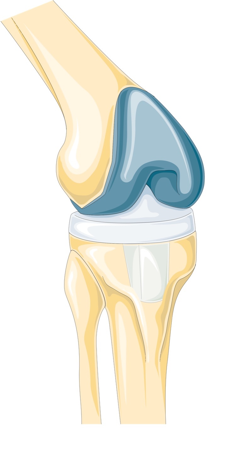
Knee joint anatomy (parasagittal section). The femoral, tibial and patellar articular surfaces (cartilage in blue) whose progressive loss defines gonarthrosis. Servier Medical Art (Laboratoires Servier), CC BY-SA 3.0, via Wikimedia Commons.
Knee joint anatomy (parasagittal section). The femoral, tibial and patellar articular surfaces (cartilage in blue) whose progressive loss defines gonarthrosis. Servier Medical Art (Laboratoires Servier), CC BY-SA 3.0, via Wikimedia Commons.
Part I - Definition, the compartmental concept and natural history
Osteoarthritis is the progressive failure of the joint, with loss of articular cartilage and the development of subchondral sclerosis, subchondral cysts, osteophytes and deformity.[1] One caveat matters: “arthrosis” is not the same as symptomatic “arthritis”. The radiographic changes are common in asymptomatic people, so the diagnosis requires the clinical picture and the radiograph to agree.[2]
The knee is described as three compartments (medial tibiofemoral, lateral tibiofemoral, and patellofemoral), each graded separately.[3] Medial-compartment osteoarthritis is the commonest pattern and is associated with varus deformity, because the medial compartment normally carries the greater share of load; genu valgum and isolated lateral disease are considerably rarer.[4]
The natural history is not one of relentless progression. In surveys of the elderly the prevalence and severity of knee osteoarthritis stay broadly constant across the seventh, eighth and ninth decades, and joint space occasionally even improves.[5] Established medial gonarthrosis, however, carries a genuinely poor prognosis: long-term follow-up shows that more than half of such knees deteriorate, worse in younger patients and when there is axial deviation or instability.[6]
Part II - Aetiology: primary and secondary gonarthrosis
Osteoarthritis is divided into primary (idiopathic) and secondary disease, though much of what is called “primary” reflects subtle predisposing factors. The secondary causes the examiner expects are:
- Prior meniscectomy: loss of the load-sharing meniscus predictably leads to arthrosis; after complete meniscectomy osteoarthritis can be expected within 5-10 years.[7]
- Malalignment (varus or valgus): the dominant mechanical driver (see Part III).
- Post-traumatic disease: intra-articular fracture, and chronic ligament insufficiency, especially anterior-cruciate (ACL) deficiency, which increases medial-compartment stress.[8]
- Inflammatory arthritis: rheumatoid arthritis (affecting 0.5-1% of the population) with synovial pannus eroding cartilage and bone, driven by IL-1 and TNF-α.[9]
- Crystal arthropathy: gout and, importantly, calcium pyrophosphate deposition (CPPD / chondrocalcinosis), which markedly worsens the longevity of an osteotomy.[10]
- Osteonecrosis: spontaneous osteonecrosis of the knee (SPONK/SONK) in those over about 55, and secondary osteonecrosis in younger patients on corticosteroids or with alcohol excess.[11]
- Osteochondritis dissecans and previous septic arthritis.[12]
The general risk factors are age, obesity (weight loss reduces the incidence of symptomatic knee osteoarthritis), female sex, heavy occupational loading, prior injury and genetic predisposition.[13]
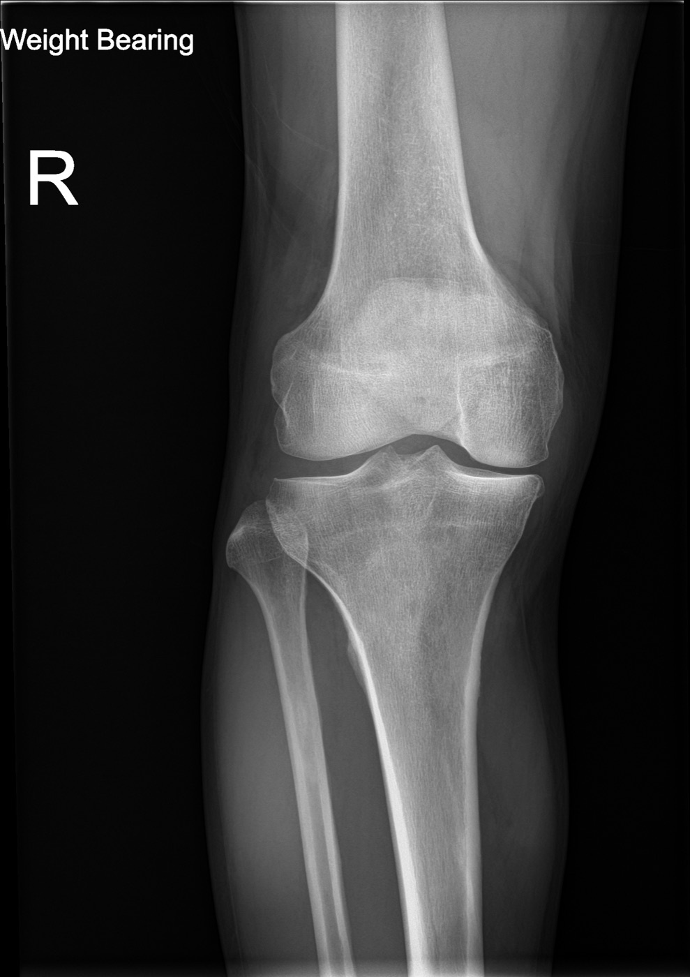
Medial-compartment gonarthrosis. Weight-bearing AP radiograph of the knee showing medial joint-space narrowing - the commonest pattern, associated with varus. Ptrump16, CC BY-SA 4.0, via Wikimedia Commons.
Medial-compartment gonarthrosis. Weight-bearing AP radiograph of the knee showing medial joint-space narrowing - the commonest pattern, associated with varus. Ptrump16, CC BY-SA 4.0, via Wikimedia Commons.
Part III - Pathology, biomechanics and the role of alignment
Cartilage is avascular, alymphatic and aneural, with limited healing capacity; full-thickness defects that reach subchondral bone heal only with inferior fibrocartilage.[14] In osteoarthritis early matrix synthesis is elevated and later reduced; aggrecan content falls, collagen is proteolytically cleaved, and degradation is driven by matrix metalloproteinases under the control of IL-1 and TNF-α.[15] Because cartilage is aneural, the pain arises elsewhere: from subchondral microfracture and remodelling, periosteal irritation, ligamentous stress, venous congestion and, when present, synovitis (which is far milder than in rheumatoid disease).[16]
The menisci are central to load transmission, carrying at least 50-70% of the load in extension and up to 85% at 90° of flexion; total medial meniscectomy reduces the femoral contact area by 50-70% and doubles the contact stress, which is why “meniscectomy is not wholly innocuous.”[17]
Alignment governs how load is distributed. The mechanical axis runs from the centre of the femoral head to the centre of the ankle and normally lies about 1.2° varus, while the anatomic (femorotibial) axis averages about 5-7° valgus.[18] Varus malalignment shifts the load onto the medial compartment and, with cartilage and bone loss, stretches the lateral structures, increasing varus further. The dynamic counterpart is the adduction moment during gait, and a low pre-operative adduction moment predicts a better, longer-lasting osteotomy result. A frank varus thrust throws the whole reaction force onto the medial compartment and accelerates degeneration.[19]
Part IV - Clinical features and examination
The typical history is of activity-related pain localised to the affected compartment (medial in the common varus knee), with stiffness, effusion, crepitus and progressive loss of motion.[20] Patellofemoral disease gives anterior knee pain and crepitus that is worse on stairs and hills, on rising from a chair and on prolonged sitting, with much less pain on level ground.[21]
On examination the gait is assessed for angular deformity, a limp, a fixed flexion deformity and, importantly, a varus thrust, which signals more advanced disease.[22] Range of motion, fixed deformity, effusion and ligamentous stability are recorded; medial laxity in a varus arthritic knee is usually secondary to medial bone loss rather than a primary ligament problem.[23] The differential diagnosis is wide and includes pes anserine bursitis, meniscal pathology, the inflammatory and crystal arthropathies, spontaneous osteonecrosis, referred pain from the hip or spine, and vascular claudication.[24]
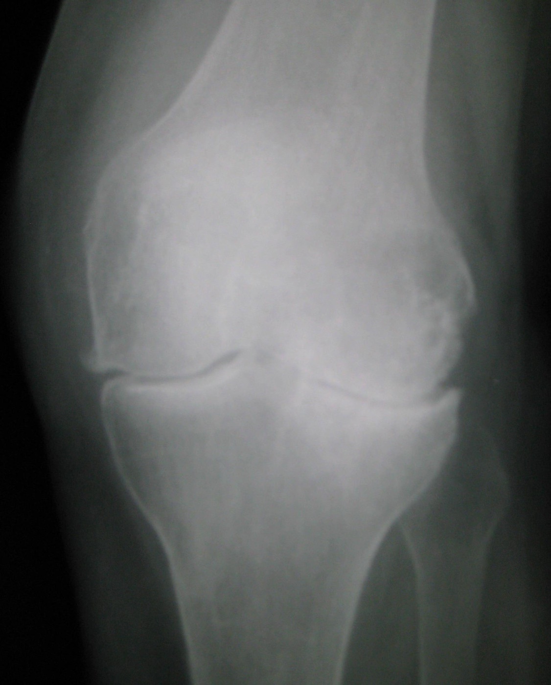
Tricompartmental knee osteoarthritis. AP radiograph of the left knee: marginal osteophytes, joint-space narrowing and subchondral sclerosis. James Heilman, MD, CC BY-SA 3.0, via Wikimedia Commons.
Tricompartmental knee osteoarthritis. AP radiograph of the left knee: marginal osteophytes, joint-space narrowing and subchondral sclerosis. James Heilman, MD, CC BY-SA 3.0, via Wikimedia Commons.
Part V - Imaging, radiographic staging and alignment measurement
The essential radiographs are the weight-bearing AP, a lateral, the skyline / Merchant (axial) patellar view, and, to assess the back of the joint where cartilage is lost first, a standing posteroanterior view in about 30-45° of flexion (the Rosenberg / “Schuss” view).[25] A full-length standing hip-knee-ankle (HKA) alignment film is necessary to measure the mechanical axis, and varus/valgus stress films are used to confirm that the opposite compartment opens up (preserved) before an osteotomy.[26]
The cardinal radiographic features are joint-space narrowing, subchondral sclerosis, subchondral cysts and osteophytes.[27] Two grading systems are quoted:
- Kellgren-Lawrence (KL): grade 0 none, 1 doubtful, 2 minimal, 3 moderate, 4 severe, read on a single AP view.[28]
- Ahlbäck: grade 0 joint space not narrowed, 1 narrowed, 2 obliterated <50%, 3 obliterated >50%.[29]
At arthroscopy the Outerbridge classification grades cartilage (I softening, II fragmentation/fissuring under half an inch [~1.3 cm], III fragmentation/fissuring over half an inch, IV erosion to bone).[30]
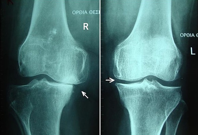
Knee osteoarthritis, radiographic grades II and III. Bilateral standing AP radiographs with arrows on the narrowed joint spaces, illustrating progressive severity. Charalampos Gkouvas (Harrygouvas), CC BY-SA 3.0, via Wikimedia Commons.
Knee osteoarthritis, radiographic grades II and III. Bilateral standing AP radiographs with arrows on the narrowed joint spaces, illustrating progressive severity. Charalampos Gkouvas (Harrygouvas), CC BY-SA 3.0, via Wikimedia Commons.
For alignment planning the HKA angle (between the femoral and tibial mechanical axes) is normally close to 0°, and the target of a realignment osteotomy is expressed by the weight-bearing line / Fujisawa point, the coordinate at which the line crosses the tibial plateau (0% medial to 100% lateral). For unloading medial-compartment disease the target is typically the 62% point (range 50-75%), i.e. a deliberate slight overcorrection into valgus.[31]
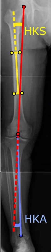
Lower-limb alignment (HKA and HKS angles). Long-leg standing radiograph annotated with the hip-knee-ankle (blue) and hip-knee-shaft (yellow) axes used to plan realignment. Mikael Häggström, CC BY 2.0, via Wikimedia Commons.
Lower-limb alignment (HKA and HKS angles). Long-leg standing radiograph annotated with the hip-knee-ankle (blue) and hip-knee-shaft (yellow) axes used to plan realignment. Mikael Häggström, CC BY 2.0, via Wikimedia Commons.
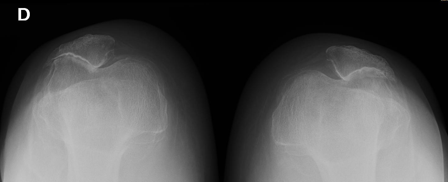
Severe patellofemoral osteoarthritis (axial / skyline view). Lateral-facet joint-space loss, osteophytes and subluxation of both patellae. Jmarchn, CC BY 4.0, via Wikimedia Commons.
Severe patellofemoral osteoarthritis (axial / skyline view). Lateral-facet joint-space loss, osteophytes and subluxation of both patellae. Jmarchn, CC BY 4.0, via Wikimedia Commons.
Part VI - Non-operative management
Conservative care is first-line and, with knowledge of the benign natural history of many knees, can maintain comfort indefinitely in many patients.[32]
- Weight loss: each pound lost removes 3-4 pounds of joint load, and weight loss reduces the incidence of symptomatic knee osteoarthritis.[33]
- Exercise and physiotherapy, chiefly quadriceps strengthening: muscle atrophies rapidly in the painful knee (up to 30% in a week), and randomised trials show strengthening improves pain and function; low-impact aerobic conditioning (cycling, swimming) is encouraged and high-impact activity avoided.[34]
- Assistive devices: a cane held in the opposite hand offloads the affected knee by roughly half.[35]
- Bracing: a valgus-producing unloader brace is significantly more effective than a simple sleeve for medial-compartment disease, and lateral heel wedges are used adjunctively.[36]
- Analgesics and NSAIDs: acetaminophen (paracetamol), up to 4 g/day, is the recommended first-line simple analgesic; NSAIDs give a further 10-20% improvement but carry significant gastrointestinal (3300 deaths/year in the US attributable) and renal risk, so they are used judiciously, and up to two-thirds of patients do well without them long-term.[37]
- Intra-articular corticosteroid: gives real but short-lived relief (significant at 1-2 weeks, indistinguishable from placebo by 4 weeks); a useful stopgap for an acute flare, not injected more often than every 4-6 weeks.[38]
- Viscosupplementation (hyaluronic acid): reported response rates of 70-80%, but the evidence is genuinely conflicting (some trials show no advantage over placebo or over corticosteroid), so its efficacy remains unresolved.[39]
- Glucosamine and chondroitin: symptomatic benefit and, in two long trials, slowed joint-space narrowing; sold as unregulated supplements.[40]
Part VII - Joint-preserving surgery: arthroscopy and osteotomy
Arthroscopic débridement - what it does not do
Arthroscopic lavage and débridement do not work for osteoarthritis. The landmark Moseley sham-controlled trial (180 patients randomised to lavage, débridement or a placebo skin-incision procedure) found no difference in pain at one or two years between the real procedures and the sham.[41] The narrow remaining role is mechanical: removal of a loose body or an unstable meniscal flap causing locking or catching. Even then arthroscopy is palliative rather than curative, and should not include extensive débridement of fibrillated cartilage.[42]
Osteotomy about the knee
The principle of osteotomy is to transfer the weight-bearing forces from the arthritic compartment to a healthier one, buying years of function in a patient too young and active for replacement.[43] Patient selection is the single most important factor: the ideal candidate is a thin, active patient in the fifth or sixth decade with localised unicompartmental pain, a stable knee, full extension and flexion of at least 90°, and no patellofemoral or inflammatory disease.[44] Rheumatoid disease and an arthritic or previously meniscectomised opposite compartment are contraindications; obesity, age over 60 and a flexion arc under 90° are relative contraindications.[45]
- High tibial osteotomy (HTO) corrects varus for medial-compartment disease, using either a lateral closing wedge (Coventry; quicker to unite, but it violates the proximal tibiofibular joint and risks the peroneal nerve) or a medial opening wedge (a single cut away from the nerve, intra-operatively adjustable, but needs a bone graft and unites more slowly).[46] The mechanical axis is deliberately overcorrected to the Fujisawa 62% point, roughly 3-5° of valgus, because an adduction moment persists during gait; undercorrection is the usual cause of failure.[47]
- Distal femoral osteotomy (DFO) corrects valgus for lateral-compartment disease (a supracondylar osteotomy is preferred over a varus-producing tibial osteotomy, which would leave an oblique joint line); the target here is a neutral mechanical axis, and overcorrection into varus is contraindicated.[48]
The complications of osteotomy are peroneal nerve injury (the most feared, lower with the opening wedge), intra-articular fracture, compartment syndrome, nonunion, loss of correction and patella baja.[49] Survivorship is good in the medium term and declines thereafter. Meta-analysis gives about 75% good results at 5 years and 60% at about 8 years, with 10-year survival (avoiding arthroplasty) around 70%, and far better results when the alignment is adequately corrected (Coventry: 94% at 10 years when corrected to ≥8° valgus versus 63% when undercorrected).[50] A well-placed osteotomy can be converted to a TKA later, though the conversion is technically more demanding.[51]
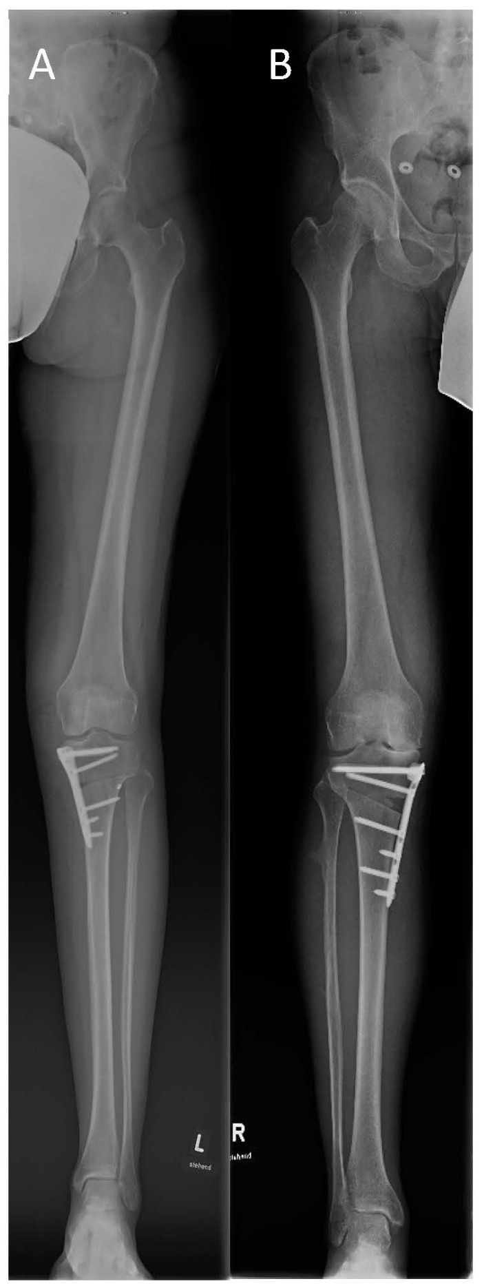
Medial open-wedge high tibial osteotomy. Post-operative full-length radiographs with angular-stable plates (LOQTEQ® and TomoFix™) and the corrected mechanical axis. Park et al., J Pers Med 2023;13(3):472, Fig. 2, CC BY 4.0.
Medial open-wedge high tibial osteotomy. Post-operative full-length radiographs with angular-stable plates (LOQTEQ® and TomoFix™) and the corrected mechanical axis. Park et al., J Pers Med 2023;13(3):472, Fig. 2, CC BY 4.0.
Part VIII - Unicompartmental and patellofemoral arthroplasty
Unicompartmental knee arthroplasty (UKA)
UKA resurfaces a single tibiofemoral compartment while preserving both cruciate ligaments and the other compartments, giving near-normal kinematics, a faster recovery and a higher early success rate than osteotomy.[52] The classic Kozinn & Scott criteria select a lower-demand patient over about 60, weighing under ~82 kg, with at least 90° of flexion, a flexion contracture under 5°, a deformity of no more than 10° varus or 15° valgus that corrects to neutral, an intact ACL, and no eburnated bone in the patellofemoral or opposite compartment.[53] Inflammatory arthritis is a formal contraindication, and absence of the ACL is a major one. Designs are fixed-bearing or mobile (meniscal) bearing (Oxford); the polyethylene must be at least 8 mm thick; and only about 10% of UKAs are lateral.[54] The cardinal technical rule is the opposite of osteotomy: do not overcorrect. A slight residual deformity (about 3° varus for a medial UKA) is left, because overcorrection drives osteoarthritis in the unreplaced compartment.[55] Modern 10-year survivorship is about 90-98% (Oxford mobile-bearing series ~97%), although community registries record higher revision rates; the characteristic mode of failure is progression of arthritis in an unreplaced compartment.[56]
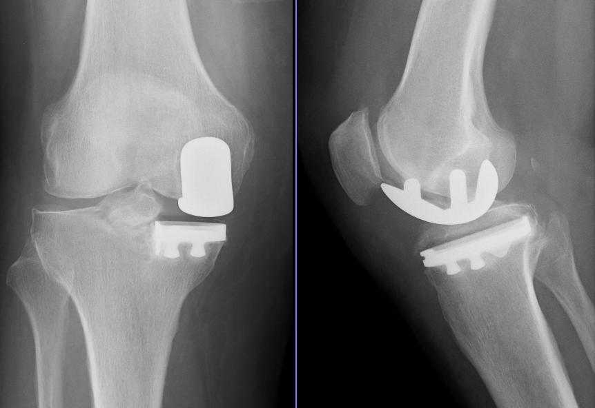
Medial unicompartmental knee arthroplasty. AP and lateral radiographs of a medial UKA (femoral runner and tibial baseplate, medial compartment only). MBq, public domain, via Wikimedia Commons.
Medial unicompartmental knee arthroplasty. AP and lateral radiographs of a medial UKA (femoral runner and tibial baseplate, medial compartment only). MBq, public domain, via Wikimedia Commons.
Patellofemoral arthroplasty (PFA)
Isolated patellofemoral osteoarthritis affects about 11% of men and 24% of women over 55 with symptomatic knee disease, often on a background of trochlear dysplasia.[57] PFA is best reserved for the younger patient (under about 55) with isolated anterior-compartment disease and no tibiofemoral or inflammatory involvement; even focal tibiofemoral chondromalacia compromises the result.[58] Results are good or excellent in roughly 85%. The main modes of failure are patellar instability/maltracking and, in the long term, progression of tibiofemoral arthritis (a quarter need conversion, usually to TKA, by 15 years).[59]
Part IX - Total knee arthroplasty: indications, design and technique
TKA is the gold-standard treatment for end-stage tri- or bicompartmental osteoarthritis failing conservative care, “one of the most successful operations in medical history,” with survival as high as 98% at 15 years.[60] The contraindications are active infection, an extensor-mechanism that does not work, and a neuropathic (Charcot) joint (the last two relative in modern practice).[61]
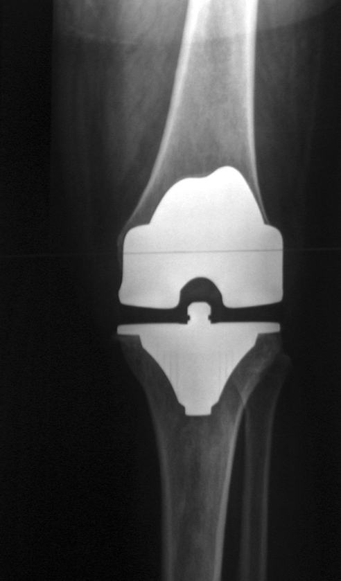
Total knee arthroplasty. Post-operative AP and lateral radiographs of a posterior-stabilised TKA (femoral and tibial components, polyethylene insert, patellar button on the lateral view). Frédéric Jacquot (fpjacquot), CC BY-SA 3.0, via Wikimedia Commons.
Total knee arthroplasty. Post-operative AP and lateral radiographs of a posterior-stabilised TKA (femoral and tibial components, polyethylene insert, patellar button on the lateral view). Frédéric Jacquot (fpjacquot), CC BY-SA 3.0, via Wikimedia Commons.
Design choices
- Cruciate-retaining (CR) versus posterior-stabilised (PS). The posterior cruciate guides femoral rollback and resists posterior subluxation. CR designs retain it (preserving a central stabiliser and bone); PS designs sacrifice it and substitute a cam-and-post mechanism that engages at about 70° of flexion to reproduce rollback, making deformity correction easier and the technique more reproducible. Both give excellent and broadly equivalent survivorship (modern series 92-100% at 10-12 years), and there is no consistent difference in ultimate range of motion.[62]
- Fixed versus mobile bearing. Mobile (rotating-platform or meniscal) bearings combine surface conformity with backside mobility to reduce contact stress; wear is very low, but they can spin out or dislocate.[63]
- Cemented versus cementless fixation. Cemented fixation is the gold standard against which others are measured, with large series showing roughly 92% survival versus 61-72% cementless at 10 years, because cement compensates for small incongruities at the bone cut.[64]
- The patella and the resurfacing debate. Surgeons divide into “always”, “never” and “selectively” resurface camps; the evidence trends toward resurfacing, with the two largest randomised trials showing markedly less residual anterior knee pain after resurfacing (Waters & Bentley: 25% versus 5%).[65]
- The constraint ladder. From least to most constrained: CR → PS → constrained condylar (CCK) → rotating hinge; increasing constraint is used as collateral stability and bone stock are lost.[66]
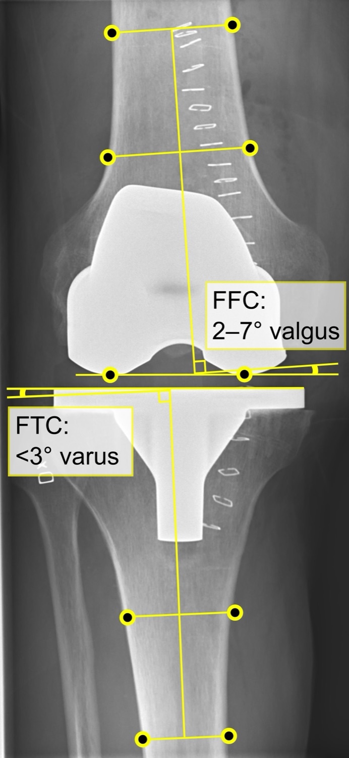
Total-knee-arthroplasty alignment. Long-leg AP radiograph of a normal TKA annotated with the mechanical axis and the femoral (FFC) and tibial (FTC) component angles. Mikael Häggström, CC0 1.0, via Wikimedia Commons.
Total-knee-arthroplasty alignment. Long-leg AP radiograph of a normal TKA annotated with the mechanical axis and the femoral (FFC) and tibial (FTC) component angles. Mikael Häggström, CC0 1.0, via Wikimedia Commons.
Alignment and technique
The goal is a neutral mechanical axis with symmetric load. The distal femoral cut is made in 5-7° of valgus (the difference between the anatomic and mechanical axes), the tibial cut at a right angle to its mechanical axis with a small posterior slope, and a varus tibial cut is never made (it causes uneven loading and early failure).[67] Femoral rotation is set from the transepicondylar axis, Whiteside’s line and the posterior condyles, erring toward external rotation to balance the flexion gap and track the patella.[68] The two classic balancing philosophies are measured resection (resect a fixed amount, preserve the joint line) and gap balancing (release ligaments first, then make the flexion and extension gaps equal).[69] Exposure is usually through a medial parapatellar arthrotomy (alternatives: subvastus, midvastus; a rectus snip or tibial-tubercle osteotomy for the stiff knee). Patellar tracking is checked with the “no-thumb” test, with a lateral release needed in fewer than 10% of cases.[70]
Part X - Complications and outcomes of knee arthroplasty
- Periprosthetic joint infection (PJI). Predominantly gram-positive (Staphylococcus aureus ~35%). The diagnosis rests on ESR/CRP plus aspiration (a synovial differential >65% neutrophils or a leucocyte count >1.7 × 10³/µL is sensitive and specific). For an acute infection with a well-fixed implant, débridement with liner exchange (DAIR) may be tried but its success is variable and often poor, particularly for Staphylococcus aureus; for chronic infection the two-stage exchange with an antibiotic-loaded cement spacer is the gold standard, curing ~90% (best with antibiotic-loaded cement at reimplantation).[71]
- Aseptic loosening, wear and osteolysis. The commonest mode of late mechanical failure; polyethylene wear particles drive an osteolytic foreign-body response, and the tibial component characteristically fails into varus. Wear is worsened by screw holes (conduits for debris) and thin or oxidised polyethylene (minimum insert thickness ~8 mm).[72]
- Instability: flexion instability, extension instability and genu recurvatum, from imbalance of the flexion and extension gaps; managed by rebalancing or increasing constraint.[73]
- Stiffness / arthrofibrosis. The strongest predictor of post-operative motion is pre-operative motion; manipulation under anaesthesia is used at about 6-12 weeks, and component malrotation (especially femoral internal rotation) is a treatable cause.[74]
- Extensor-mechanism and patellofemoral problems: maltracking and subluxation (often from component internal rotation), patellar clunk syndrome (a fibrous nodule, typically in posterior-stabilised designs), patellar fracture, and extensor-mechanism (patellar/quadriceps tendon) disruption, where prevention is far better than repair.[75]
- Periprosthetic fracture: supracondylar femoral fractures (risk factors: anterior notching, osteopenia), classified by Rorabeck-Lewis and treated by locked plating, retrograde nailing or, with a loose component, revision; tibial fractures are classified by Felix.[76]
- Peroneal (common fibular) nerve palsy: about 0.3-0.6%, typically after correction of a valgus or flexion-contracture deformity; the first step is to release the dressing and flex the knee.[77]
- Venous thromboembolism: deep-vein thrombosis occurs in 50-84% of unprophylaxed knees (predominantly calf vein), but fatal pulmonary embolism is rare (~0.2-0.6%); chemical and mechanical prophylaxis are standard.[78]
- Wound complications: skin necrosis and dehiscence, minimised by using the most lateral prior incision and by gastrocnemius flap coverage when needed.[79]
Outcomes are reported with the Knee Society Score and similar measures; modern survivorship is around 90-98% at 10-20 years, with infection, aseptic loosening, instability and wear the leading reasons for revision.[80] Revision TKA climbs the constraint ladder (CCK or rotating hinge) and manages bone loss with augments, stems and, for massive defects, distal femoral replacement or an allograft-prosthetic composite.[81]
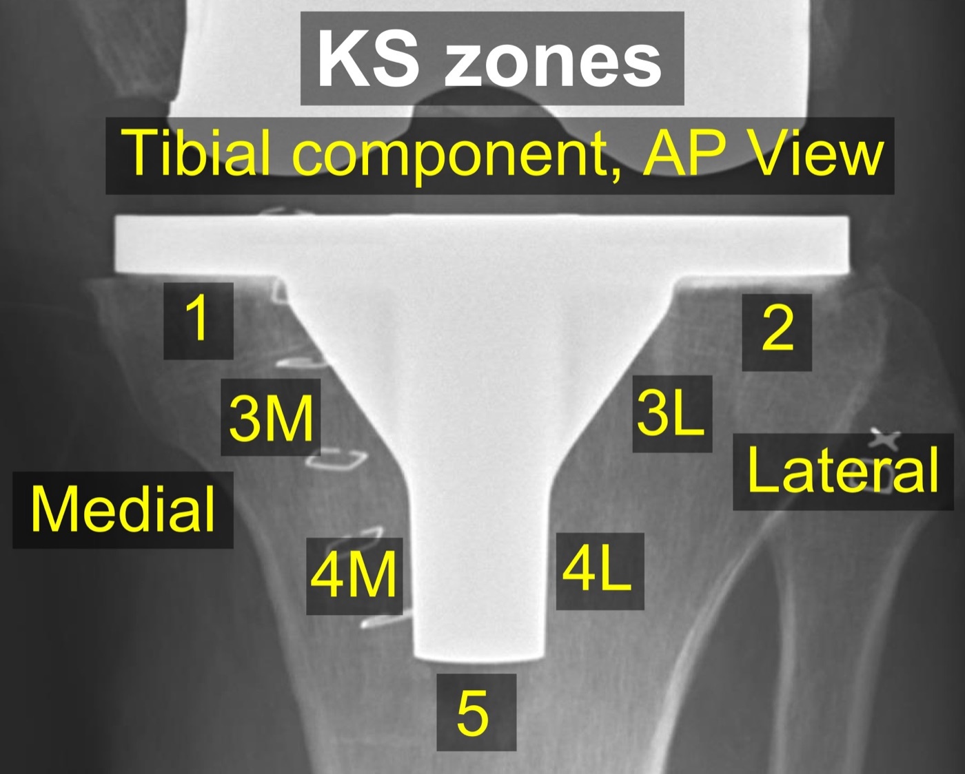
Knee Society radiographic zones. AP radiograph of a TKA tibial component with the 2015 Knee Society zones used to document radiolucent lines and loosening. Mikael Häggström, CC0 1.0, via Wikimedia Commons.
Knee Society radiographic zones. AP radiograph of a TKA tibial component with the 2015 Knee Society zones used to document radiolucent lines and loosening. Mikael Häggström, CC0 1.0, via Wikimedia Commons.
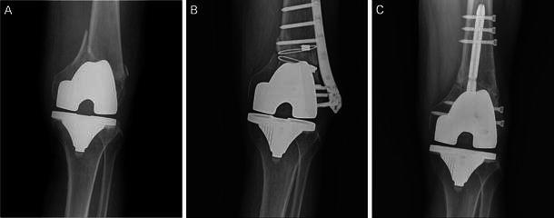
Periprosthetic supracondylar femoral fracture after TKA. A fracture above the femoral component, here treated by plate fixation - a characteristic late complication. Gondalia et al., J Orthop Traumatol 2014;15(3):201-207, Fig. 1, CC BY.
Periprosthetic supracondylar femoral fracture after TKA. A fracture above the femoral component, here treated by plate fixation - a characteristic late complication. Gondalia et al., J Orthop Traumatol 2014;15(3):201-207, Fig. 1, CC BY.
To tie the topic together: gonarthrosis is compartmental and alignment-driven, medial and varus most often. Stage it on weight-bearing and long-leg films, treat it first with weight loss, quadriceps strengthening, a cane in the opposite hand and an unloader brace, and reserve arthroscopy for true mechanical symptoms. Realign the young malaligned knee with an osteotomy (overcorrect the mechanical axis for an HTO, neutral for a DFO), resurface isolated disease with a unicompartmental or patellofemoral implant (here, do not overcorrect), and replace the tricompartmental knee with a cemented total knee. Then watch for infection, loosening, instability and the extensor mechanism.
Bulgarian terminology (Боев / Boychev tradition) - glossary
The following Bulgarian equivalents bridge the international literature with the terminology of the Bulgarian school of orthopaedics (in the tradition of Бойчо Бойчев / Boycho Boychev) used in the state examination.
| English term | Bulgarian term (Cyrillic) | Transliteration |
|---|---|---|
| Gonarthrosis (knee osteoarthritis) | Гонартроза (остеоартроза на коляното) | Gonartroza (osteoartroza na kolyanoto) |
| Medial compartment | Медиален компартмент | Medialen kompartment |
| Patellofemoral joint | Пателофеморална става | Patelofemoralna stava |
| Varus / valgus deformity | Варусна / валгусна деформация | Varusna / valgusna deformatsiya |
| Mechanical axis | Механична ос | Mehanichna os |
| Joint-space narrowing | Стесняване на ставната цепка | Stesnyavane na stavnata tsepka |
| Osteophyte | Остеофит | Osteofit |
| Subchondral sclerosis | Субхондрална склероза | Subhondralna skleroza |
| Subchondral cyst | Субхондрална киста | Subhondralna kista |
| Meniscus | Мениск | Menisk |
| Articular cartilage | Ставен хрущял | Staven hrushtyal |
| High tibial osteotomy | Висока тибиална остеотомия | Visoka tibialna osteotomiya |
| Distal femoral osteotomy | Дистална феморална остеотомия | Distalna femoralna osteotomiya |
| Opening / closing wedge | Отварящ / затварящ клин | Otvaryasht / zatvaryasht klin |
| Total knee arthroplasty | Тотално колянно ендопротезиране | Totalno kolyanno endoprotezirane |
| Unicompartmental arthroplasty | Еднокомпартментно протезиране | Ednokompartmentno protezirane |
| Posterior cruciate ligament | Задна кръстна връзка | Zadna krastna vrazka |
| Polyethylene insert | Полиетиленов инлей | Polietilenov inley |
| Cemented fixation | Циментна фиксация | Tsimentna fiksatsiya |
| Periprosthetic fracture | Перипротезна фрактура | Periprotezna fraktura |
| Aseptic loosening | Асептично разхлабване | Aseptichno razhlabvane |
| Periprosthetic infection | Перипротезна инфекция | Periprotezna infektsiya |
Image attributions
(Figure attributions and licences are listed in the figure MANIFEST and inserted with each image. All images are openly licensed [CC0 / Public Domain / CC BY / CC BY-SA] or used under their stated terms; any non-commercial [NC] item is flagged as such and must not be used in a commercial product.)
References
-
Insall & Scott, Surgery of the Knee (4th ed.), p. 351.
-
Insall & Scott, p. 351.
-
Insall & Scott, p. 372.
-
Master Techniques in Knee Arthroplasty (Lotke/Lonner), p. 383; Insall & Scott, p. 1333.
-
Insall & Scott, p. 351.
-
Insall & Scott, p. 1343.
-
Insall & Scott, p. 342, p. 1323.
-
Insall & Scott, p. 1323, p. 1348.
-
Insall & Scott, p. 346.
-
Insall & Scott, p. 351, p. 1348.
-
Insall & Scott, p. 351.
-
Insall & Scott, p. 1323, p. 1338.
-
Insall & Scott, p. 351-352, p. 355.
-
Insall & Scott, p. 329, p. 343-344.
-
Insall & Scott, p. 344.
-
Insall & Scott, p. 351-352.
-
Insall & Scott, p. 335.
-
Insall & Scott, p. 1326; Lotke, p. 383.
-
Insall & Scott, p. 1345, p. 1348.
-
Insall & Scott, p. 1323, p. 1347.
-
Lotke, p. 366.
-
Insall & Scott, p. 1347-1348.
-
Insall & Scott, p. 1348.
-
Insall & Scott, p. 351; Lotke, p. 367.
-
Insall & Scott, p. 1325, p. 1349; Lotke, p. 395.
-
Insall & Scott, p. 1325, p. 1349; Lotke, p. 384.
-
Insall & Scott, p. 351.
-
Insall & Scott, p. 372-373.
-
Insall & Scott, p. 373.
-
Insall & Scott, p. 373, p. 1664.
-
Insall & Scott, p. 1327, p. 1349-1350; Lotke, p. 384.
-
Insall & Scott, p. 351.
-
Insall & Scott, p. 352.
-
Insall & Scott, p. 352-353.
-
Insall & Scott, p. 353.
-
Insall & Scott, p. 353.
-
Insall & Scott, p. 356-358.
-
Insall & Scott, p. 359-360.
-
Insall & Scott, p. 360-361.
-
Insall & Scott, p. 362-363.
-
Insall & Scott, p. 375.
-
Insall & Scott, p. 379.
-
Insall & Scott, p. 1323.
-
Insall & Scott, p. 1323-1324.
-
Insall & Scott, p. 1324.
-
Insall & Scott, p. 1328-1332; Lotke, p. 383.
-
Lotke, p. 384; Insall & Scott, p. 1346, p. 1338.
-
Insall & Scott, p. 1333-1334; Lotke, p. 395-398.
-
Insall & Scott, p. 1337, p. 1364-1365.
-
Insall & Scott, p. 1338; Lotke, p. 392.
-
Insall & Scott, p. 1325, p. 1368.
-
Insall & Scott, p. 1430, p. 1443.
-
Lotke, p. 349; Insall & Scott, p. 1444-1445.
-
Insall & Scott, p. 1433, p. 1443, p. 1448.
-
Insall & Scott, p. 1438, p. 1448.
-
Insall & Scott, p. 1448, p. 1443.
-
Insall & Scott, p. 1462.
-
Insall & Scott, p. 1465.
-
Insall & Scott, p. 1468.
-
Insall & Scott, p. 1648, p. 1544.
-
Insall & Scott, p. 1678.
-
Insall & Scott, p. 1544-1546, p. 1554, p. 1561.
-
Insall & Scott, p. 1580, p. 1584.
-
Insall & Scott, p. 1648-1649, p. 1514.
-
Insall & Scott, p. 1596, p. 1608-1609.
-
Insall & Scott, p. 1516-1517, p. 1547.
-
Insall & Scott, p. 1500-1503.
-
Insall & Scott, p. 1481-1483.
-
Insall & Scott, p. 1479-1492.
-
Insall & Scott, p. 1493-1495, p. 1512-1513, p. 1604.
-
Insall & Scott, p. 1806-1812.
-
Insall & Scott, p. 1754-1761, p. 1768.
-
Insall & Scott, p. 1747-1751.
-
Insall & Scott, p. 1752; Lotke, p. 445-448.
-
Insall & Scott, p. 1771-1774.
-
Insall & Scott, p. 1762-1766; Lotke, p. 408.
-
Insall & Scott, p. 1746.
-
Insall & Scott, p. 1738-1742.
-
Insall & Scott, p. 1743-1744.
-
Insall & Scott, p. 1544, p. 1568-1570.
-
Insall & Scott, p. 1516, p. 1200; Lotke, p. 418.