Contents
- Scope and orientation
- Part I - The arch, definitions and the flexible/rigid assessment
- Part II - Flexible flatfoot (and accessory navicular)
- Part III - Congenital vertical talus
- Part IV - Tarsal coalition
- Part V - Adult acquired flatfoot deformity (PTTD)
- Part VI - Hereditary joint laxity (hypermobility)
- Bulgarian terminology glossary
- Figure credits and licences
Scope and orientation
This topic has two halves. The first is the flatfoot (pes planus) in all its forms: the common, benign flexible flatfoot of childhood; the rigid congenital flatfoot of congenital vertical talus; the tarsal coalition that causes a rigid (peroneal spastic) flatfoot in the adolescent; and the adult acquired flatfoot deformity driven by posterior tibial tendon dysfunction. The second half is hereditary joint laxity (hypermobility): how it is scored, when it becomes a symptomatic syndrome, and the heritable connective-tissue disorders behind it.
A few organising ideas run through both halves:
- A flatfoot is a combination, not one deformity: hindfoot valgus, a sagging medial arch, and forefoot supination/abduction, with or without a tight heel cord.
- Flexible versus rigid is the central distinction. A flexible foot restores its arch on tiptoe and on the Jack toe-raise test; a rigid one does not, and rigidity points to a coalition, a vertical talus, or arthritis.
- Most paediatric flatfoot is normal and needs only reassurance; treatment is for the symptomatic foot, the tight heel cord, or the rigid/structural deformity.
- Laxity is a graded trait. Hypermobility is common and usually benign, but a small subset have a symptomatic syndrome or a serious heritable disorder (Marfan, Ehlers-Danlos, osteogenesis imperfecta) whose recognition can save a life.
A Bulgarian glossary, a viva self-test appendix, and image credits follow the clinical sections.
Part I - The arch, definitions and the flexible/rigid assessment
A flatfoot combines hindfoot (heel) valgus, forefoot supination relative to the hindfoot, a midfoot abducted or straight with a depressed medial longitudinal arch, and a variable element of tight tendo-Achilles. Mosca stresses that the forefoot is supinated, not pronated, and that the term “subluxation” is a misnomer in the ordinary flatfoot (a true peritalar dislocation occurs only in congenital vertical talus).[1] A useful localising rule: in a flatfoot the apex of the deformity is plantar, so the problem is in the foot itself, whereas a cavus foot points dorsally toward a neurological cause.[2] The arch is set chiefly by the bone-ligament complex (Harris & Beath); ligamentous laxity is the primary variable, and muscle activity serves propulsion and balance rather than static support.[3]
The flexible-versus-rigid distinction is the main examination. In a flexible foot, standing on tiptoe converts the valgus heel to varus and reconstitutes the arch, and the Jack toe-raise test (passive dorsiflexion of the great toe tightens the plantar fascia through the windlass mechanism) elevates the arch and inverts the subtalar joint. A rigid foot does neither.[4] One pitfall: a solid talocalcaneal coalition can develop secondary midtarsal hypermobility that mimics subtalar motion, so subtalar motion is assessed with the ankle locked in neutral dorsiflexion.[5]
The tight heel cord is the element that most often makes a flatfoot painful, and it is characterised by the Silfverskiöld test: dorsiflexion is measured with the knee extended and again flexed (with the subtalar joint held neutral). If dorsiflexion improves with the knee flexed, the gastrocnemius alone is tight (treat by gastrocnemius recession); if it remains limited, the whole triceps surae is tight (treat by tendo-Achilles lengthening). Normal is at least 10° of dorsiflexion.[6]
Imaging, when needed, must be weight-bearing, since an unloaded flatfoot looks better than it is. The measures that matter are the lateral talo-first-metatarsal (Meary) angle (normally a straight line, 0°, breaking plantar-apex in a flatfoot), the calcaneal pitch (reduced), and the talonavicular coverage (the navicular abducts and dorsiflexes on the talar head).[7]
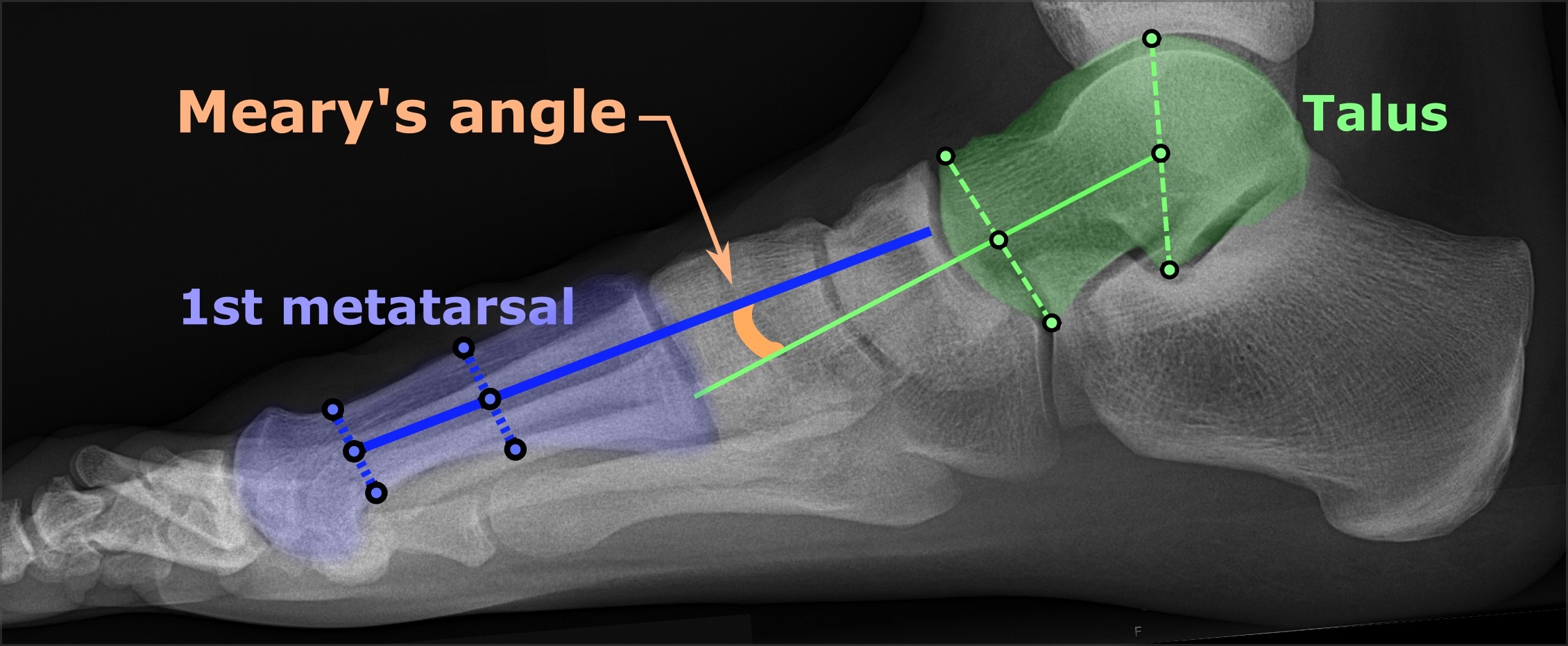
Measurement of the lateral talo-first-metatarsal (Meary) angle on a weight-bearing radiograph of a normal foot - the talar and first-metatarsal axes are normally colinear; the angle breaks (apex plantar) in a flatfoot. (Mikael Häggström, Wikimedia Commons; CC0.)
Measurement of the lateral talo-first-metatarsal (Meary) angle on a weight-bearing radiograph of a normal foot - the talar and first-metatarsal axes are normally colinear; the angle breaks (apex plantar) in a flatfoot. (Mikael Häggström, Wikimedia Commons; CC0.)
Part II - Flexible flatfoot (and accessory navicular)
II.1 Physiologic flexible flatfoot
Flexible flatfoot is an anatomic variation, not a deformity: hindfoot valgus, forefoot supination and a low arch, with no heel-cord contracture, that corrects on tiptoe and on the Jack test.[8] It is very common. Flatfoot is present in roughly 45% of preschoolers and ~15% of older children, the arch developing spontaneously through the first decade, and it is more prevalent in obese children and those with generalised laxity.[9] The natural history is benign: flatfeet that remain flat function and feel no worse than average-arched feet, and low arches do not increase injury risk.[10]
Treatment is reassurance. Imaging is unnecessary for the asymptomatic child, and randomised data show that shoe modifications and inserts do not change arch development; orthoses can ease activity-related aching within a few months but alter nothing structurally.[11]
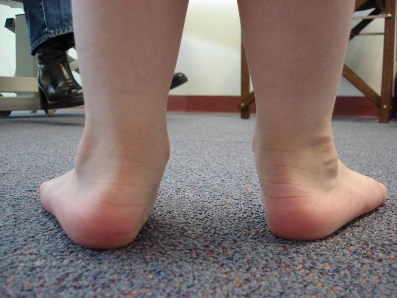
Standing posterior (rear) view of a child with flexible flatfoot: bilateral hindfoot (heel) valgus. (FA RenLis, Wikimedia Commons; CC BY-SA 3.0.)
Standing posterior (rear) view of a child with flexible flatfoot: bilateral hindfoot (heel) valgus. (FA RenLis, Wikimedia Commons; CC BY-SA 3.0.)
II.2 Flexible flatfoot with a tight heel cord (the symptomatic subtype)
The same flexible foot plus a contracted gastrocnemius or whole triceps surae is reclassified as a deformity, because the tight heel cord forces the calcaneus to dorsiflex around a fixed, plantar-flexed talus, concentrating weight-bearing stress under the talar head.[12] This causes pain under the talar head or in the sinus tarsi, and a rigid arch support makes it worse: inserts must be flat and cushioned.[13] Non-operative care is heel-cord stretching and soft inserts. The operation, reserved for the recalcitrant painful foot, is the calcaneal lengthening osteotomy (Evans/Mosca) with medial soft-tissue plication, plus a gastrocnemius recession or tendo-Achilles lengthening chosen by the Silfverskiöld test, and a plantar-flexion medial-cuneiform (Cotton) osteotomy if forefoot supination persists.[14] The lengthening osteotomy is favoured (between ages 6 and 10) because it corrects all components of the deformity while preserving subtalar motion. Subtalar arthroereisis is more controversial, with complication rates of roughly 4-19%, frequent removal, and insufficient evidence on a Cochrane review, and arthrodesis is a salvage of last resort.[15]
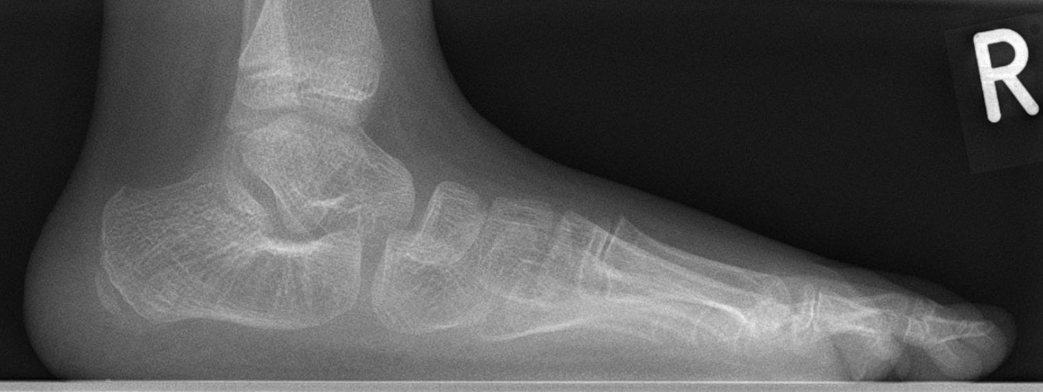
Weight-bearing lateral radiograph of a child’s foot with pes planus: a plantar-flexed talus, low calcaneal pitch and a flattened arch. (Hellerhoff, Wikimedia Commons; CC BY-SA 4.0.)
Weight-bearing lateral radiograph of a child’s foot with pes planus: a plantar-flexed talus, low calcaneal pitch and a flattened arch. (Hellerhoff, Wikimedia Commons; CC BY-SA 4.0.)
II.3 Accessory navicular
An accessory navicular (os tibiale externum) is a secondary ossicle at the navicular tuberosity, at the insertion of the tibialis posterior, and is associated with a flexible flatfoot. The Geist types are: I, a small sesamoid within the tendon; II, a larger ossicle joined to the navicular by a synchondrosis (the symptomatic type, where shear across the synchondrosis causes pain); III, a fused cornuate navicular. Symptomatic lesions are managed conservatively first; the operation is simple excision (often equivalent to the historical Kidner procedure of excision plus tibialis-posterior advancement).[16]
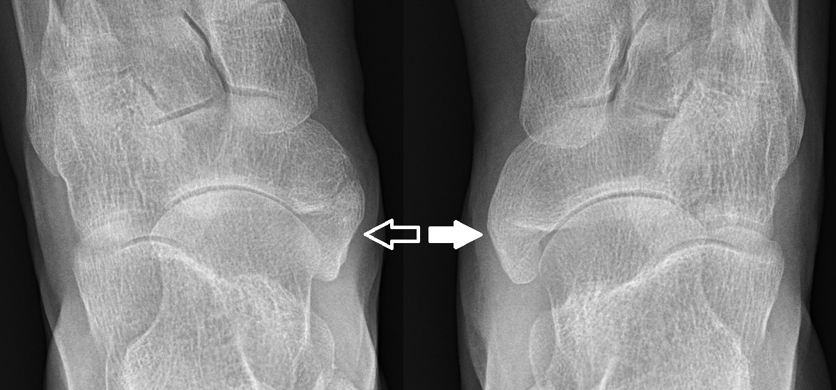
Dorsoplantar radiograph of both feet: a separate accessory navicular ossicle (one foot) and a cornuate navicular (other foot), arrowed. (Mikael Häggström, Wikimedia Commons; CC0.)
Dorsoplantar radiograph of both feet: a separate accessory navicular ossicle (one foot) and a cornuate navicular (other foot), arrowed. (Mikael Häggström, Wikimedia Commons; CC0.)
Part III - Congenital vertical talus
Congenital vertical talus (CVT) is the rigid “rocker-bottom” congenital flatfoot: a fixed dorsolateral dislocation of the talonavicular joint with a verticalised, plantar-flexed talus, an everted subtalar joint, and fixed equinus. None of the components corrects on manipulation, and the prominent talar head is palpable in the sole.[17] Incidence is about 1 in 10,000; roughly half are idiopathic and half are associated with neuromuscular or genetic conditions (arthrogryposis, myelomeningocele, trisomies, with vertical talus in ~10% of myelomeningocele), so every CVT warrants a neuromuscular work-up. About half are bilateral, and 12-20% have a positive family history (HOXD10/CDMP-1).[18] The milder, reducible variant is the oblique talus, which sits between flexible flatfoot and true CVT on a continuum.[19]
Imaging is by stress lateral radiographs. On the maximum-plantar-flexion lateral, the first-metatarsal/navicular axis stays dorsally translated and the talus does not reduce, confirming the fixed dorsal dislocation that defines CVT (in an oblique talus it partially reduces). On the maximum-dorsiflexion view the talus stays vertical.[20] Untreated CVT always causes disability, the rigid talar head becoming a painful weight-bearing surface.[21]
Treatment is now the reverse-Ponseti (Dobbs) method: serial casts that plantar-flex and invert the foot (the opposite of clubfoot casting, with counter-pressure on the medial talar head), followed by a percutaneous tendo-Achilles tenotomy and a limited talonavicular capsulotomy with retrograde pinning of the reduced joint; a tibialis-anterior transfer is added in children over two. This has largely replaced extensive soft-tissue release, with better long-term motion and alignment and higher outcome scores.[22] Salvage for the older or recurrent foot includes naviculectomy and, eventually, arthrodesis.[23]
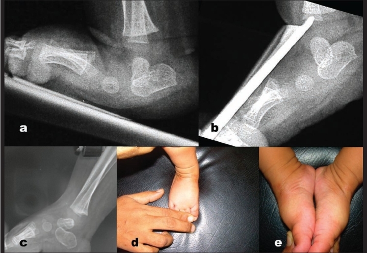
Congenital vertical talus in an infant: (a) lateral radiograph with a vertical, plantar-flexed talus and talonavicular dislocation; (b) the forced-plantarflexion stress lateral showing the talus stays vertical (fixed dislocation); (c) post-correction film; (d, e) clinical images. (Mahajan et al., Indian J Orthop 2008; CC BY.)
Congenital vertical talus in an infant: (a) lateral radiograph with a vertical, plantar-flexed talus and talonavicular dislocation; (b) the forced-plantarflexion stress lateral showing the talus stays vertical (fixed dislocation); (c) post-correction film; (d, e) clinical images. (Mahajan et al., Indian J Orthop 2008; CC BY.)
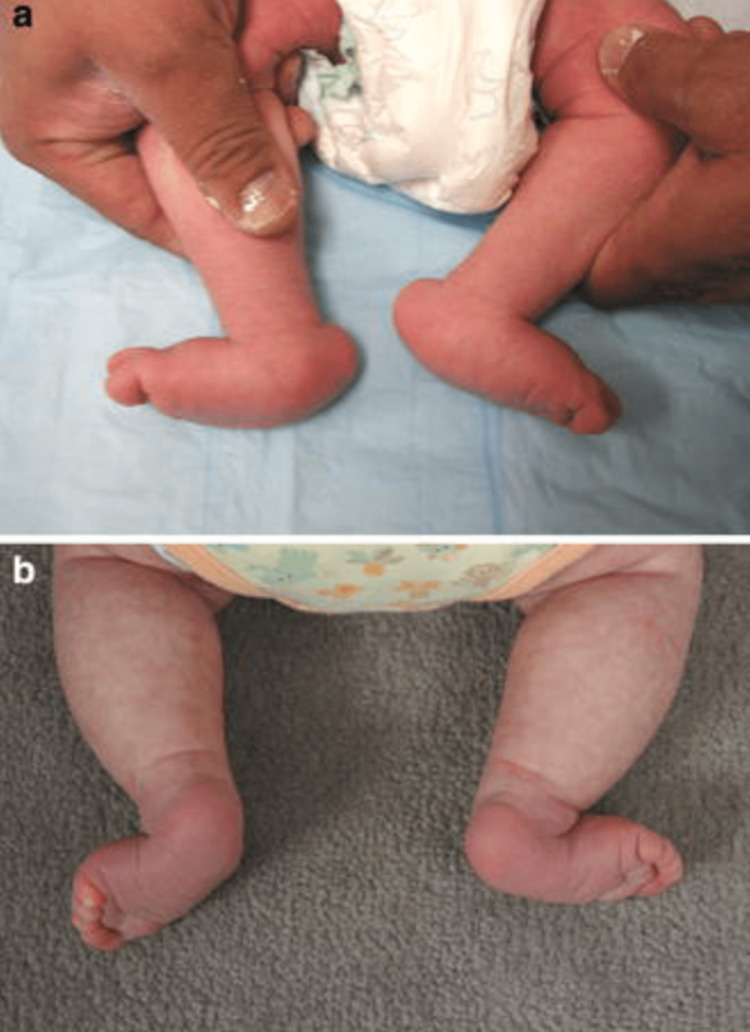
Clinical appearance of bilateral congenital vertical talus: the convex plantar surface (“rocker-bottom” foot) with a prominent talar head. (Cureus 2023;15(9):e45867; CC BY 4.0.)
Clinical appearance of bilateral congenital vertical talus: the convex plantar surface (“rocker-bottom” foot) with a prominent talar head. (Cureus 2023;15(9):e45867; CC BY 4.0.)
Part IV - Tarsal coalition
A tarsal coalition is a congenital fibrous, cartilaginous or bony bridge between two tarsal bones (a failure of mesenchymal segmentation), and it is the commonest cause of a rigid (peroneal spastic) flatfoot in the adolescent.[24] The two common types, calcaneonavicular and talocalcaneal (middle-facet), make up about 90% in roughly equal proportion; overall prevalence is about 1-2% (higher in cadaver series), about half are bilateral, and inheritance is autosomal dominant.[25] Symptoms begin as the bar ossifies (calcaneonavicular at about 8-12 years, talocalcaneal at about 12-16), with activity-related hindfoot pain, recurrent ankle sprains and a rigid, valgus hindfoot; only about a quarter of coalitions ever become symptomatic.[26]
Imaging: the calcaneonavicular bar shows the “anteater nose” sign (an elongated anterior calcaneal process) on the oblique view; the talocalcaneal coalition shows the “C-sign” and a talar beak (a traction spur, not arthritis) on the lateral, with CT the definitive study for type, size and posterior-facet health.[27]
Treatment is conservative first (a period in a boot or cast relieves about a third). The painful calcaneonavicular bar is resected with fat or extensor-digitorum-brevis interposition (80-90% good results); a talocalcaneal coalition is resected if it is “resectable” by the Wilde criteria (under 50% of the posterior-facet area, no facet narrowing, and hindfoot valgus under 16°); otherwise a calcaneal lengthening (with heel-cord lengthening) corrects the valgus and relieves pain even with the coalition left in place. Arthrodesis is reserved for established subtalar arthritis.[28]
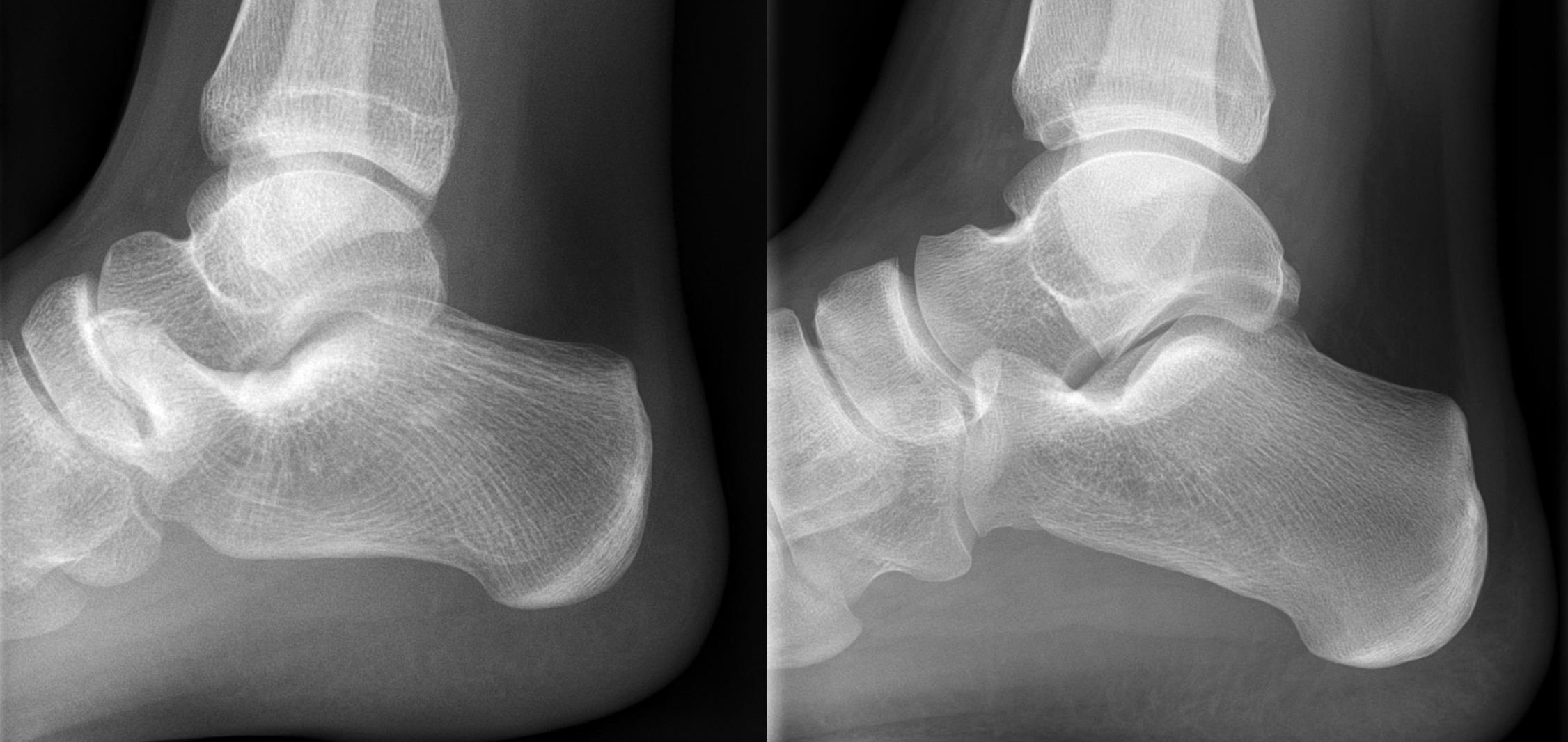
Calcaneonavicular coalition: the affected side (left) shows an elongated anterior calcaneal process - the “anteater nose” sign - compared with the normal side. (Hellerhoff, Wikimedia Commons; CC BY-SA 3.0.)
Calcaneonavicular coalition: the affected side (left) shows an elongated anterior calcaneal process - the “anteater nose” sign - compared with the normal side. (Hellerhoff, Wikimedia Commons; CC BY-SA 3.0.)
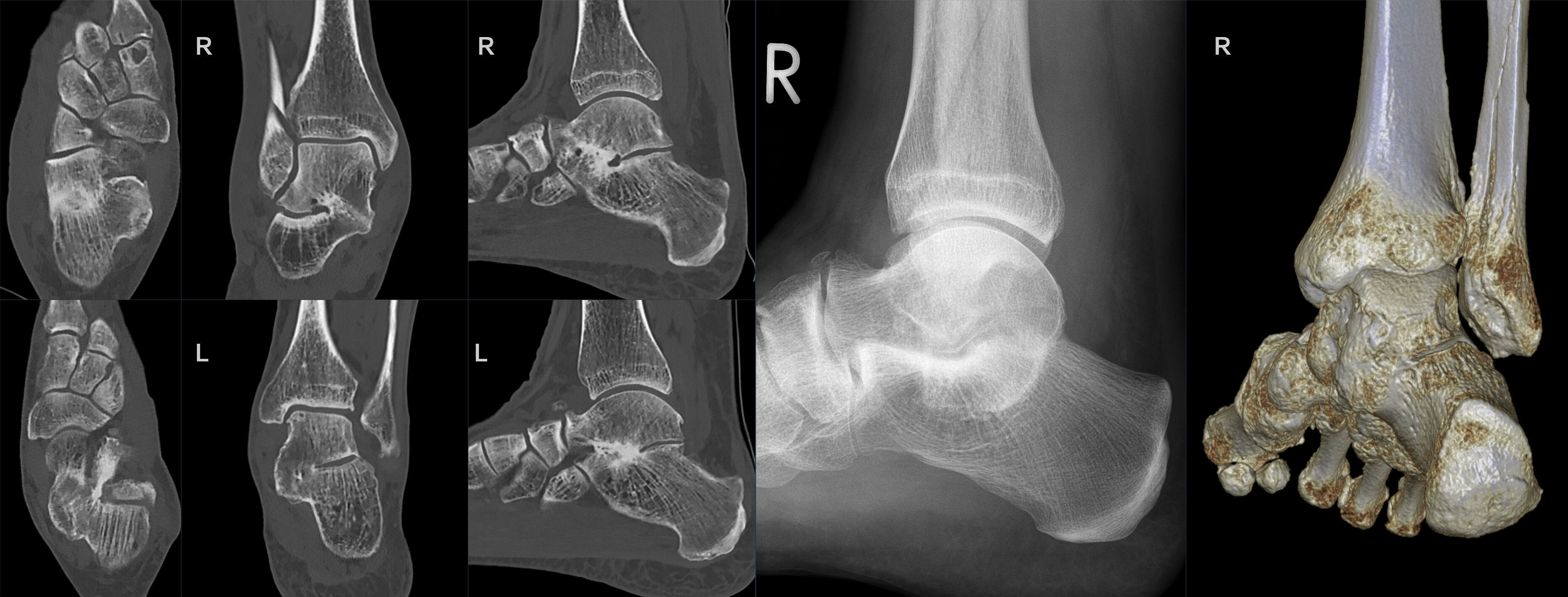
Bilateral talocalcaneal (middle-facet) coalition on CT: coronal and sagittal reconstructions, a lateral radiograph and a 3-D rendering show the bony bridge at the sustentaculum tali. (Hellerhoff, Wikimedia Commons; CC BY-SA 4.0.)
Bilateral talocalcaneal (middle-facet) coalition on CT: coronal and sagittal reconstructions, a lateral radiograph and a 3-D rendering show the bony bridge at the sustentaculum tali. (Hellerhoff, Wikimedia Commons; CC BY-SA 4.0.)
Part V - Adult acquired flatfoot deformity (PTTD)
V.1 Definition and pathomechanics
Adult acquired flatfoot deformity (AAFD) is a progressive collapse of the medial longitudinal arch in the adult, most often driven by posterior tibial tendon dysfunction (PTTD), together with attrition of the spring (calcaneonavicular) ligament. The cascade runs from hindfoot valgus to forefoot abduction (a “too many toes” appearance) and talonavicular uncoverage, then compensatory forefoot varus, then arch collapse, and finally ankle valgus with deltoid failure.[29]
The posterior tibial tendon is the chief dynamic invertor and arch supporter and the antagonist of peroneus brevis; it has a hypovascular zone about 14 mm long, ~40 mm proximal to its insertion, behind the medial malleolus, and an excursion of only 1-2 cm, so even ~1 cm of elongation renders it incompetent.[30] When it fails, the unopposed peroneus brevis drives valgus, the Achilles pull shifts lateral to the subtalar axis and becomes an evertor that accelerates the deformity, and the plantar ligaments fatigue.[31]
V.2 Aetiology, presentation and examination
The typical patient is a middle-aged or older woman, often with obesity, hypertension, diabetes or steroid exposure; a younger group has a seronegative enthesopathy.[32] The presentation evolves from medial pain and swelling along the tendon to lateral subfibular impingement pain as the heel collapses into valgus. The main examination findings are the “too many toes” sign (forefoot abduction seen from behind) and the single-limb heel-rise test, in which the patient cannot rise onto the toes, or the heel fails to invert.[33]
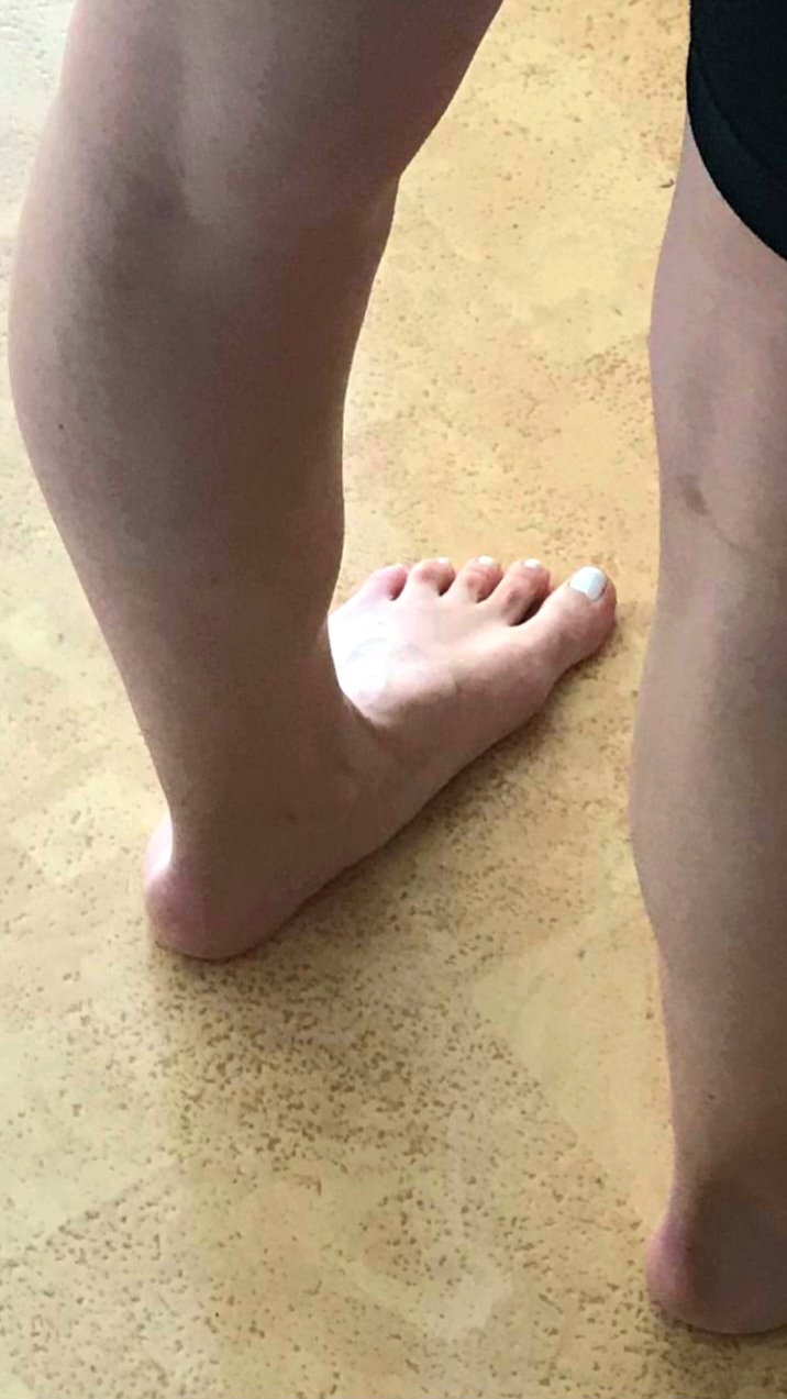
Clinical appearance of an adult acquired flatfoot: the medial longitudinal arch has collapsed to the floor. (AnnaAndersson00, Wikimedia Commons; CC0.)
Clinical appearance of an adult acquired flatfoot: the medial longitudinal arch has collapsed to the floor. (AnnaAndersson00, Wikimedia Commons; CC0.)
V.3 Classification (Johnson & Strom / Myerson)
| Stage | Tendon / deformity | Key findings | Management |
|---|---|---|---|
| I | Tenosynovitis, no deformity | Medial pain/swelling; normal alignment; heel inverts on rise | Immobilisation/NSAIDs; tenosynovectomy if refractory |
| II | PTT elongation/rupture, flexible flatfoot | Hindfoot valgus, forefoot abduction; cannot do single heel rise; passively correctable | Joint-sparing reconstruction (below) |
| III | Rigid flatfoot ± arthritis | Fixed forefoot varus (≥10-15°), subtalar will not reduce; lateral pain | Triple arthrodesis |
| IV (Myerson) | Ankle valgus | Valgus talar tilt + deltoid attenuation on the standing ankle film | Deltoid reconstruction ± fusion / TAA |
Myerson sub-stages stage II by the dominant deformity (hindfoot valgus, flexible vs fixed forefoot supination, forefoot abduction, medial-ray instability), and these map onto the procedures chosen.[34]
V.4 Imaging and treatment
Weight-bearing radiographs show the increased lateral Meary angle, reduced calcaneal pitch, and talonavicular uncoverage (lateral subluxation on the AP); a standing AP ankle view is essential to detect stage IV; MRI/ultrasound assess the tendon and spring ligament.[35]
- Stage I: rest/immobilisation, NSAIDs (avoid steroid injection), orthosis; tenosynovectomy if refractory.[36]
- Stage II (flexible): an orthosis (UCBL/AFO) non-operatively, then joint-sparing reconstruction: an FDL (flexor digitorum longus) transfer with a medializing calcaneal osteotomy (Koutsogiannis), adding a lateral column lengthening (Evans) for forefoot abduction/talonavicular uncoverage above ~30-40%, a Cotton (medial-cuneiform) osteotomy for residual forefoot varus, spring-ligament repair, and a gastrocnemius recession/TAL for equinus.[37]
- Stage III (rigid): triple arthrodesis (with adjuncts for residual forefoot or equinus deformity).[38]
- Stage IV (ankle valgus): deltoid ligament reconstruction for the flexible ankle; ankle arthrodesis, tibiotalocalcaneal/pantalar fusion, or total ankle replacement for the rigid/arthritic ankle.[39]
V.5 Other causes of acquired adult flatfoot
Beyond PTTD: Charcot neuroarthropathy (diabetic midfoot collapse, the most important; the Eichenholtz stages of fragmentation→coalescence→reconstruction and the Brodsky anatomical pattern, producing a rocker-bottom foot prone to ulceration); trauma (Lisfranc injury, navicular or calcaneal malunion); inflammatory arthropathy (rheumatoid disease, largely through ligament/joint destruction rather than direct tendon failure); an adult-presenting tarsal coalition; and iatrogenic over-resection.[40]
Part VI - Hereditary joint laxity (hypermobility)
VI.1 Definitions
Joint hypermobility is a graded trait distributed normally through the population, with hypermobile people simply at one extreme.[41] Generalised joint hypermobility is defined by a scoring cut-off; it becomes the joint hypermobility syndrome / benign joint hypermobility syndrome (BJHS) only when laxity is accompanied by musculoskeletal symptoms with no other rheumatological disease.[42] The modern framework reclassifies this spectrum into hypermobility spectrum disorders (HSD) and hypermobile Ehlers-Danlos syndrome (hEDS), recognising that the old “BJHS” and “EDS type III” overlap so much they are treated as one entity.[43]
VI.2 Scoring
The Beighton score (0-9) uses five manoeuvres, scored on each side where paired:[44]
- passive dorsiflexion of the little finger beyond 90° (1 point each hand);
- passive apposition of the thumb to the flexor forearm (1 each);
- elbow hyperextension beyond 10° (1 each);
- knee hyperextension beyond 10° (1 each);
- forward trunk flexion with the knees straight and the palms flat on the floor (1 point).
Most clinicians require an adult score of about 4-6 of 9; laxity declines with age (use a lower threshold in the elderly) and is greater in females.[45] The historical Carter & Wilkinson criteria (3 of 5) preceded it, and the Brighton criteria add extra-articular minor features (a marfanoid habitus, skin and eye signs, dislocations, soft-tissue rheumatism) to diagnose the syndrome, since the Beighton score alone would miss most syndromic patients.[46]
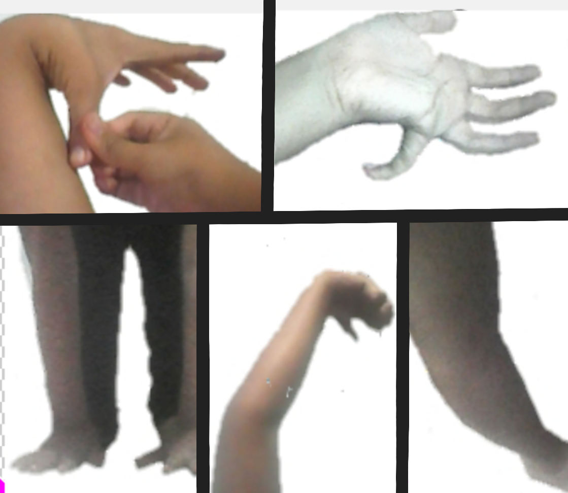
The Beighton manoeuvres in a person scoring 9/9: thumb-to-forearm, little-finger hyperextension, elbow and knee hyperextension, and palms flat to the floor. (Rollcloud, Wikimedia Commons; CC0.)
The Beighton manoeuvres in a person scoring 9/9: thumb-to-forearm, little-finger hyperextension, elbow and knee hyperextension, and palms flat to the floor. (Rollcloud, Wikimedia Commons; CC0.)
VI.3 Epidemiology and clinical features
Hypermobility is commoner in females, declines with age, and is more prevalent in some non-European (notably African) populations, often without symptoms; familial cases are usually autosomal dominant.[47] The orthopaedic manifestations are recurrent instability and dislocations (patella, shoulder, hip), pes planovalgus, genu recurvatum, scoliosis, chronic widespread joint pain and soft-tissue rheumatism, impaired proprioception, and a tendency to premature osteoarthritis (the hip relatively spared).[48] Management is principally physiotherapy and proprioceptive training (patients have “loose joints and tight muscles,” so naive range-of-motion programmes can worsen symptoms), with surgery used sparingly because lax tissue gives a higher failure rate (“lax joints are best left lax”).[49]
The red flags that mandate a search for a serious heritable disorder are cardiovascular signs (aortic dilatation, mitral prolapse), ectopia lentis, a marfanoid habitus, marked skin fragility/hyperextensibility, bone fragility with blue sclerae, and disproportionate short stature with multiple dislocations.[50]
VI.4 The major heritable connective-tissue disorders
- Ehlers-Danlos syndromes (EDS): the triad of articular hypermobility, skin hyperextensibility and tissue fragility. The 2017 classification recognises 13 subtypes, ~90% being the classical (COL5A1/COL5A2) and hypermobile types; the vascular type (COL3A1) is the dangerous one, with a risk of fatal arterial, bowel or uterine rupture. Surgery is hazardous (sutures cut out, wounds gape, bleeding), and stabilisation operations fail.[51]
- Marfan syndrome: autosomal dominant, due to fibrillin-1 (FBN1) with increased TGF-β signalling. The revised Ghent criteria centre on ectopia lentis, aortic root aneurysm and family history, with a systemic score (wrist and thumb signs, arachnodactyly, pectus, scoliosis, protrusio, pes planus). Scoliosis is often the presenting feature, and recognising the syndrome saves lives through cardiac referral (aortic dissection); bracing is less effective and spinal surgery carries higher complication rates and dural ectasia.[52]
- Osteogenesis imperfecta: a type I collagen (COL1A1/COL1A2) disorder of bone fragility with ligamentous laxity and blue sclerae, graded by the Sillence classification (type I mild, II lethal perinatal, III severe survivable, IV moderate, plus newer types). Management is cyclical bisphosphonates and realignment osteotomy with telescoping intramedullary rods.[53]
- Briefly: Down syndrome (atlantoaxial and hip/patellar instability, planovalgus); homocystinuria (mimics Marfan but with downward lens dislocation and a thrombotic tendency); Loeys-Dietz (TGF-β receptor, aggressive aneurysms); Stickler (type II collagen, early arthritis, Pierre-Robin); Larsen (filamin B, multiple dislocations, congenital knee dislocation, cervical instability).[54]
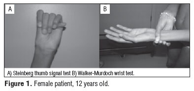
Marfan systemic signs on real clinical photographs: (A) the Steinberg thumb sign and (B) the Walker-Murdoch wrist sign. (Cipriano et al., via Wikimedia Commons; CC BY 4.0.)
Marfan systemic signs on real clinical photographs: (A) the Steinberg thumb sign and (B) the Walker-Murdoch wrist sign. (Cipriano et al., via Wikimedia Commons; CC BY 4.0.)
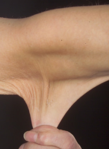
Skin hyperextensibility in Ehlers-Danlos syndrome: the forearm skin lifts away abnormally far when pinched. (Whitaker et al., PMC3504533, Wikimedia Commons; CC BY 2.5.)
Skin hyperextensibility in Ehlers-Danlos syndrome: the forearm skin lifts away abnormally far when pinched. (Whitaker et al., PMC3504533, Wikimedia Commons; CC BY 2.5.)
Bulgarian terminology glossary
For consistency with the Bulgarian state-examination vocabulary (and the operative terminology of Boychev, Хирургическа ортопедия):
| English | Bulgarian |
|---|---|
| Flatfoot / pes planus | Плоско ходило (стъпало) / pes planus |
| Flexible flatfoot | Гъвкаво (мобилно) плоско ходило |
| Hindfoot valgus | Валгус на задното стъпало (петата) |
| Medial longitudinal arch | Медиален надлъжен свод |
| Congenital vertical talus | Вроден вертикален талус (рокер-ботъм стъпало) |
| Tarsal coalition | Тарзална коалиция (сраствания на ходилните кости) |
| Calcaneonavicular / talocalcaneal | Калканеонавикуларна / талокалканеарна |
| Accessory navicular | Допълнителна (акцесорна) ладиевидна кост (os tibiale externum) |
| Posterior tibial tendon dysfunction | Дисфункция на сухожилието на задния тибиален мускул |
| Adult acquired flatfoot deformity | Придобито плоско ходило при възрастни |
| Triple arthrodesis | Тройна артродеза |
| Calcaneal (medializing) osteotomy | Медиализираща калканеална остеотомия |
| Joint hypermobility / laxity | Ставна хипермобилност / халтавост (лабилност) |
| Hereditary joint laxity | Наследствена ставна халтавост |
| Beighton score | Скор на Beighton |
| Ehlers-Danlos syndrome | Синдром на Ehlers-Danlos |
| Marfan syndrome | Синдром на Marfan |
| Osteogenesis imperfecta | Незавършена остеогенеза (osteogenesis imperfecta) |
| Ligamentous laxity | Лигаментна (връзкова) хлабавост |
Figure credits and licences
All images were independently opened and visually verified to depict the stated entity before use; licences were confirmed against the Wikimedia Commons API or the NCBI PMC Open-Access service. All are CC0/public-domain, CC BY or CC BY-SA (no non-commercial files).
- Meary-angle measurement (normal foot) -
flatfoot_meary_angle_method_CC0.jpg. Mikael Häggström. CC0. - Flexible flatfoot, child (rear view) -
flatfoot_clinical_children_rear.jpg. FA RenLis, Wikimedia Commons. CC BY-SA 3.0. - Pes planus, paediatric weight-bearing lateral -
flatfoot_xray_lateral_standing_child7y.jpg. Hellerhoff, Wikimedia Commons. CC BY-SA 4.0. - Accessory & cornuate navicular (annotated) -
accessory_navicular_dorsoplantar_annotated_CC0.jpg. Mikael Häggström. CC0. - Congenital vertical talus, radiographs -
cvt_vertical_talus_lateral_and_stress_PMC.jpg. Mahajan R et al., Indian J Orthop 2008;42(3):347 (PMC2739479). CC BY. - Congenital vertical talus, clinical rocker-bottom -
cvt_rocker_bottom_clinical_infant_PMC.jpg. Cureus 2023;15(9):e45867 (PMC10518251). CC BY 4.0. - Calcaneonavicular coalition (anteater sign) -
coalition_calcaneonavicular_anteater_lateral.jpg. Hellerhoff, Wikimedia Commons. CC BY-SA 3.0. - Talocalcaneal coalition (CT + radiograph) -
coalition_talocalcaneal_CT_and_xray.jpg. Hellerhoff, Wikimedia Commons. CC BY-SA 4.0. - Adult acquired flatfoot, clinical -
flatfoot_clinical_adult_medial_CC0.jpg. AnnaAndersson00. CC0. - Beighton manoeuvres (9/9 montage) -
beighton_full_montage_9of9_CC0.png. Rollcloud, Wikimedia Commons. CC0. - Marfan thumb & wrist signs -
marfan_thumb_and_wrist_signs_combined.jpg. Cipriano GFB et al. (Braz J Phys Ther/SciELO), via Wikimedia Commons. CC BY 4.0. - Ehlers-Danlos skin hyperextensibility -
eds_skin_hyperextensible_human.png. Whitaker JK et al. (PMC3504533), Wikimedia Commons. CC BY 2.5.
CC BY/CC BY-SA images require attribution (and, for SA, that any redistributed derivative keep the same licence); the CC0 images are public-domain.
References
-
Mosca, Principles & Management of Pediatric Foot & Ankle, pp.33-34, 37; Lovell & Winter, p.4441.
-
Mosca, p.33.
-
Lovell & Winter, p.4444; Mosca, p.30.
-
Mosca, pp.38-41; Lovell & Winter, pp.4445-4446.
-
Mosca, p.39; Lovell & Winter, p.4574.
-
Mosca, p.44; Lovell & Winter, pp.4446-4447.
-
Mosca, pp.45-46; Lovell & Winter, pp.4447-4449.
-
Mosca, pp.109-110; Lovell & Winter, p.4451.
-
Lovell & Winter, pp.4441-4443.
-
Mosca, p.110; Lovell & Winter, pp.4449-4450.
-
Mosca, p.110; Lovell & Winter, pp.4451-4452.
-
Mosca, pp.109-111; Lovell & Winter, p.4452.
-
Mosca, pp.110-111.
-
Mosca, p.112.
-
Lovell & Winter, pp.4454-4456, 4464-4472; Mosca, pp.61-63.
-
Mosca, pp.45-46, 60-61 and historical references p.281; Lovell & Winter, p.4454 - types I-III and the Kidner procedure are standard descriptions; the dedicated clinical pages were outside the extracted text.
-
Lovell & Winter, pp.4415, 4418; Mosca, pp.105-108.
-
Lovell & Winter, pp.4416-4417.
-
Mosca, pp.105-107; Lovell & Winter, p.4421.
-
Lovell & Winter, pp.4422-4428; Mosca, pp.105-106.
-
Lovell & Winter, p.4429.
-
Lovell & Winter, pp.4429-4437; Mosca, pp.105, 108.
-
Lovell & Winter, pp.4437-4438; Mosca, pp.84, 108-109.
-
Mosca, pp.30, 119; Lovell & Winter, pp.4571-4572.
-
Lovell & Winter, pp.4572-4573.
-
Lovell & Winter, pp.4583-4584; Mosca, pp.119-121.
-
Lovell & Winter, pp.4579-4582; Mosca, pp.121-124.
-
Lovell & Winter, pp.4584-4593, 4599-4600; Mosca, pp.122-125.
-
Mann’s Surgery of the Foot & Ankle, pp.598-601; Myerson, Reconstructive Foot & Ankle Surgery, p.208.
-
Mann’s, pp.591-596.
-
Mann’s, pp.593-594.
-
Mann’s, pp.595-596.
-
Mann’s, pp.598-601.
-
Mann’s, pp.600-601; Myerson, pp.208-210. The newer RAM (Roger A. Mann) classification keeps the Johnson & Strom structure and is descriptive, not yet validated - Mann’s p.601.
-
Mann’s, pp.601-605.
-
Mann’s, pp.611-615.
-
Mann’s, pp.615-636; Myerson, pp.211-219.
-
Mann’s, pp.644-647; Myerson, pp.221-222.
-
Mann’s, pp.637-644; Myerson, pp.222-224.
-
Mann’s, pp.594-596, 601, 647-653. The Eichenholtz/Brodsky detail for Charcot is standard knowledge; a dedicated Charcot chapter was outside the extracted sources.
-
Beighton, Grahame & Bird, Hypermobility of Joints, ch.1 p.18; ch.2 p.27.
-
Beighton, ch.1 pp.17-18.
-
Beighton, ch.1 p.21; Lovell & Winter, p.818.
-
Beighton, ch.2 p.28.
-
Beighton, ch.2 p.29.
-
Beighton, ch.2 pp.28-31.
-
Beighton, ch.2 pp.29-30, 37-38; ch.9 pp.184-186.
-
Beighton, ch.9 pp.176-187.
-
Beighton, ch.9 pp.179-180; Lovell & Winter, pp.820-821.
-
Beighton, ch.2 pp.38-39; ch.9 pp.181, 188-189.
-
Beighton, ch.9 pp.167-180; Lovell & Winter, p.818.
-
Beighton, ch.9 pp.188-189; Lovell & Winter, pp.814-818.
-
Lovell & Winter, pp.821-824; Beighton, ch.9 p.189.
-
Lovell & Winter, pp.814, 821-831, 884-888, 904-907, 921-922; Beighton, ch.9 pp.188-198; ch.10 p.209.