Contents
- Introduction and scope
- Part I - Definition and the concept of impingement
- Part II - Pathomechanics and morphology
- Part III - Acetabular version, coverage and the radiographic metrics
- Part IV - Epidemiology and natural history
- Part V - Clinical features and examination
- Part VI - Imaging and diagnosis
- Part VII - Treatment
- Part VIII - Outcomes and complications
- Bulgarian terminology (Боев / Boychev tradition) - glossary
- Image attributions
Introduction and scope
Femoroacetabular impingement (FAI) is a dynamic, motion-induced abnormal contact between the proximal femur and the acetabular rim that, repeated over years, damages the labrum and articular cartilage and is now recognised as a leading cause of “idiopathic” osteoarthritis of the non-dysplastic hip.[1] The examiner expects the three morphological patterns (cam, pincer and the commonest, mixed), the way each injures the joint (the cam “outside-in” chondral shear versus the pincer “inside-out” labral crush), the radiographic vocabulary (the alpha angle, the centre-edge angle, and the signs of acetabular retroversion: the crossover, posterior-wall and ischial-spine signs), the clinical syndrome (groin pain in a young active adult, a positive anterior impingement test), and the treatment logic of hip preservation: reshape the bone, repair the labrum, and do it before the cartilage passes the point of no return.
Two ideas run through the topic. First, FAI is not a radiological diagnosis: the same bony shapes are present in a large fraction of asymptomatic people, so the diagnosis requires symptoms, clinical signs and imaging together. Treat the patient, not the X-ray.[2] Second, what determines the outcome is stability and cartilage status, not the size of the bump: over-resecting a retroverted or borderline-dysplastic rim converts impingement into instability, while operating on a hip that has already lost its joint space simply hastens the way to a replacement.
Part I - Definition and the concept of impingement
FAI describes abnormal abutment of the femoral head-neck junction against the acetabular rim during terminal hip motion, classically flexion combined with internal rotation and adduction.[3] The problem is a dynamic one: the overload occurs with movement. That distinguishes it from developmental dysplasia, where a deficient socket is statically overloaded at rest.[4] The same hip can sit at either end of this spectrum, and the hardest patient to read is the borderline-dysplastic, mildly retroverted hip, in which the pain may arise from impingement, from instability, or from both.[5]
The modern concept was formalised by Reinhold Ganz and the Bern group in 2003, building on their 2001 description of a technique for safe surgical dislocation of the hip without avascular necrosis, which for the first time allowed the joint to be inspected dynamically and the damage to be seen directly.[6] Earlier descriptions existed: the French empreinte iliaque of 1899, and Smith-Petersen’s 1936 acetabuloplasty for the impinging neck of slipped epiphysis, protrusio and coxa plana. Stulberg’s 1975 “pistol-grip” deformity and Murray’s “tilt deformity” had already linked an aspherical head to so-called primary osteoarthritis.[7] Ganz’s contribution was to tie the morphology, the chondrolabral damage and the surgical results together.
The FAI-arthritis link is strong. When hips coming to arthroplasty in patients under 50 years are examined after excluding dysplasia, slipped epiphysis and Perthes disease, about 97% show cam, pincer or mixed morphology, and FAI-type deformity is found in 96-99% of “primary” osteoarthritic hips replaced before 55.[8] FAI is best thought of not as a disease in itself but as a process by which a structurally abnormal hip fails.[9]
Part II - Pathomechanics and morphology
The organising classification is cam (a femoral-side abnormality), pincer (an acetabular-side abnormality), and mixed, the commonest, where both coexist.[10] An equivalent mechanical framing is inclusion-type (cam) versus impaction-type (pincer) damage.[11]
Cam morphology
A cam is an aspherical femoral head with loss of the normal head-neck offset: a bony “bump” or convexity at the anterosuperior head-neck junction, the “pistol-grip” deformity of Stulberg.[12] In flexion the non-spherical segment is forced into a congruent acetabulum (the inclusion mechanism), generating an “outside-in” shear that delaminates the anterosuperior acetabular cartilage from the subchondral bone beginning at the chondrolabral junction; the labrum is relatively spared until late.[13] So cam impingement damages cartilage more than labrum, and because the weight-bearing dome stays intact until the head migrates into the acetabular defect, radiographic joint-space narrowing appears late, by which time a relatively young adult may already have end-stage arthrosis.[14]
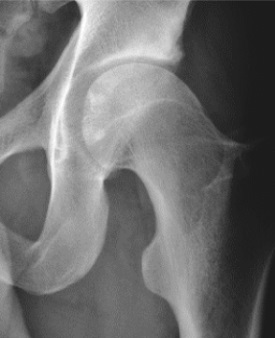
Cam morphology. AP hip showing loss of the normal head-neck offset with a focal convexity at the anterosuperior junction (the “pistol-grip” appearance). From Ruiz Santiago et al., Radiol Res Pract 2016;2016:6369237, Fig. 1d (CC BY 4.0).
Cam morphology. AP hip showing loss of the normal head-neck offset with a focal convexity at the anterosuperior junction (the “pistol-grip” appearance). From Ruiz Santiago et al., Radiol Res Pract 2016;2016:6369237, Fig. 1d (CC BY 4.0).
The typical patient is a young athletic man; cam asphericities are roughly three to five times commoner in males.[15] The deformity is thought to form during adolescence: the anterolateral physis extends onto the neck in response to high-impact loading around the time of physeal closure, so that cam is essentially a silent, subclinical slipped epiphysis in many cases, with frank slipped epiphysis and Perthes disease as its extreme forms.[16] There is a heritable component, with a sibling relative risk of about 2.8 for cam (and 2.0 for pincer).[17]
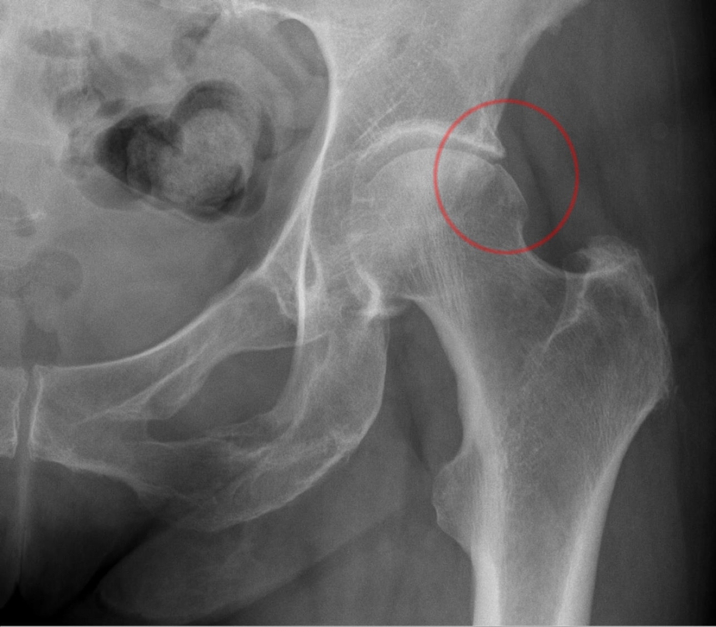
Combined-type FAI. Single-hip AP radiograph; the circle marks a focal bony prominence at the antero-superior head-neck junction (a dominant cam with a minor pincer component). Hellerhoff, Wikimedia Commons, CC BY-SA 3.0.
Combined-type FAI. Single-hip AP radiograph; the circle marks a focal bony prominence at the antero-superior head-neck junction (a dominant cam with a minor pincer component). Hellerhoff, Wikimedia Commons, CC BY-SA 3.0.
Pincer morphology
A pincer is acetabular over-coverage, so that the rim abuts the femoral neck.[18] Over-coverage may be focal (cranial, from true acetabular retroversion) or global (from coxa profunda, protrusio acetabuli, or generalised retroversion, in which the impingement is circumferential).[19] The mechanism is impaction: the rim crushes the labrum first in a linear pattern, with cartilage damage initially confined to a narrow rim strip. Over time a subset of hips lever the head on the rim, producing a “contre-coup” cartilage lesion in the posteroinferior acetabulum (“inside-out” injury).[20] Pincer impingement therefore damages the labrum more than the cartilage, the reverse of cam, with intralabral cyst formation and, over years, labral ossification that deepens the rigid socket and worsens the over-coverage.[21]
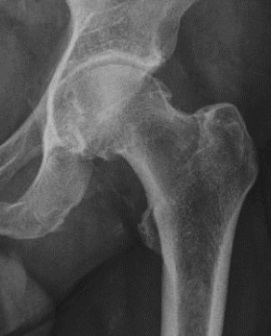
Pincer morphology. AP hip showing acetabular over-coverage of the femoral head. From Ruiz Santiago et al., Radiol Res Pract 2016;2016:6369237, Fig. 1c (CC BY 4.0).
Pincer morphology. AP hip showing acetabular over-coverage of the femoral head. From Ruiz Santiago et al., Radiol Res Pract 2016;2016:6369237, Fig. 1c (CC BY 4.0).
The classic pincer patient is a middle-aged, active woman. Over-coverage is present in 10-15% of hips and is bilateral in roughly three-quarters, and protrusio in particular predominantly affects women, in whom the higher medial load preserves the superolateral joint space even at end stage.[22] One distinction matters here, between global over-coverage and true retroversion: a truly retroverted acetabulum is over-covered anteriorly but deficient posteriorly, a different mechanical problem in which aggressive anterior rim resection risks leaving the head posteriorly uncovered and unstable.[23]
Mixed impingement
Mixed (combined cam-plus-pincer) impingement is the commonest pattern. In the Bern series, 47% of cam hips had an associated acetabular deformity and 63% of pincer hips an abnormal head shape, and large screening cohorts find that isolated pincer accounts for only about 10% of cases, with combined signs in over 90%.[24] Because femoral and acetabular deformities so often coexist, and because both are frequently bilateral even when only one hip hurts, pure cam and pure pincer are the exception, and an operation that addresses only one side commonly fails.
Part III - Acetabular version, coverage and the radiographic metrics
A precise vocabulary of angles and signs underpins both diagnosis and the safe limits of surgery.
Lateral centre-edge angle (LCE) of Wiberg, on a well-centred AP pelvis, is the angle between a vertical line through the femoral-head centre and a line to the lateral edge of the sourcil. Below 20° indicates dysplasia, 20-25° is borderline, roughly 25-40° is normal, and over 40° indicates over-coverage (pincer); some authors flag over-coverage already above 35°.[25]
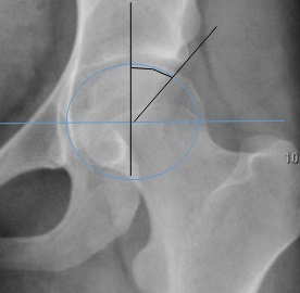
Lateral centre-edge angle of Wiberg. Construction on an AP hip: the angle between the vertical through the head centre and the line to the lateral acetabular edge. From Ruiz Santiago et al., Radiol Res Pract 2016;2016:6369237, Fig. 6b (CC BY 4.0).
Lateral centre-edge angle of Wiberg. Construction on an AP hip: the angle between the vertical through the head centre and the line to the lateral acetabular edge. From Ruiz Santiago et al., Radiol Res Pract 2016;2016:6369237, Fig. 6b (CC BY 4.0).
Anterior centre-edge angle (ACE) of Lequesne, measured on the false-profile view (standing, the pelvis rotated about 65° from the AP), quantifies anterior coverage; the normal value is around 20-25°, with anterior over-coverage above and undercoverage below.[26]
Tönnis angle (acetabular index / sourcil angle) is the inclination of the weight-bearing sourcil; normal 0-10°, over 10° dysplastic, negative values over-covered.[27]
Signs of acetabular retroversion are read on a properly rotated, non-tilted AP pelvis:
- the crossover (figure-of-eight) sign: the anterior wall line crosses lateral to the posterior wall line in the cranial acetabulum, instead of the two converging at the lateral sourcil;[28]
- the posterior wall sign: the posterior wall lies medial to the head centre, indicating deficient posterior coverage;[29]
- the ischial spine sign: a prominent ischial spine projecting into the pelvis.[30]
The crossover and posterior-wall signs together signify global retroversion, and the combination of crossover, positive posterior-wall and ischial-spine signs identifies the hip that needs reorientation rather than simple rim trimming.[31] One trap is that the crossover sign can be a false positive from pelvic tilt or a prominent anterior inferior iliac spine in an otherwise normally-versed acetabulum. Siebenrock showed that as little as 6° of pelvic rotation distorts the version assessment, so technique matters and CT is used when in doubt.[32]
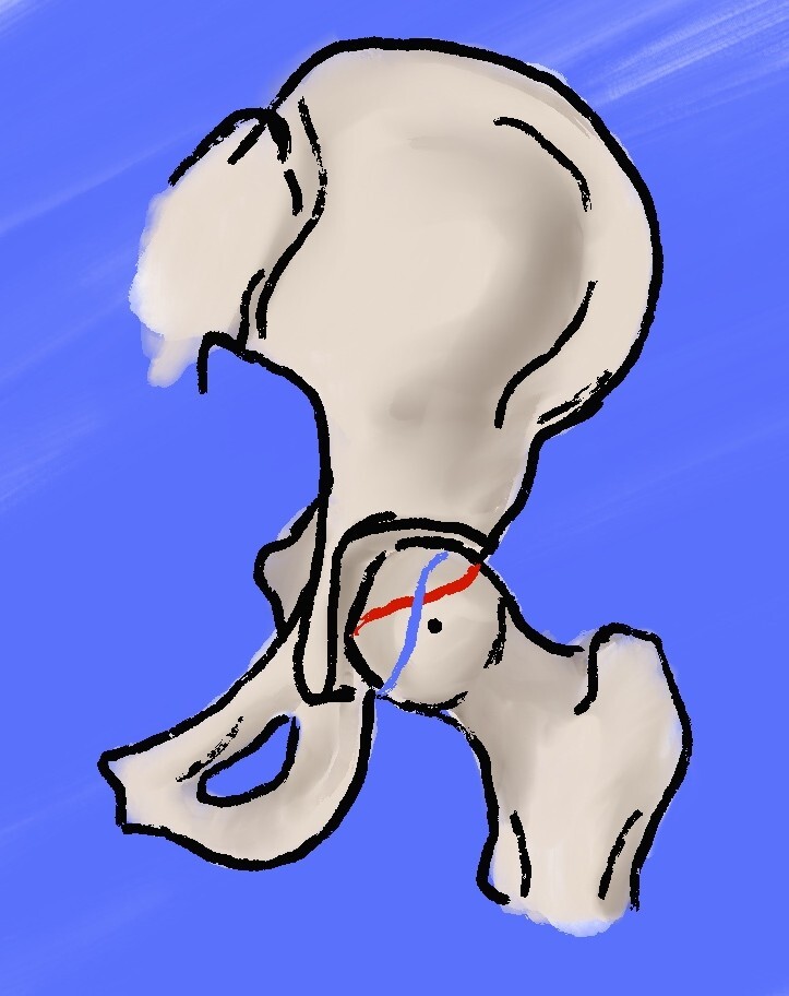
Crossover sign of acetabular retroversion (schematic). The anterior acetabular wall (red) crosses lateral to the posterior wall (blue) in the cranial acetabulum. Beckenrandgeschichten, Wikimedia Commons, CC BY-SA 4.0.
Crossover sign of acetabular retroversion (schematic). The anterior acetabular wall (red) crosses lateral to the posterior wall (blue) in the cranial acetabulum. Beckenrandgeschichten, Wikimedia Commons, CC BY-SA 4.0.
Coxa profunda and protrusio are read against the ilioischial line: in coxa profunda the acetabular fossa floor touches or crosses the line; in protrusio the femoral head itself crosses it.[33] Coxa profunda may, however, be a normal variant, particularly in women.[34]
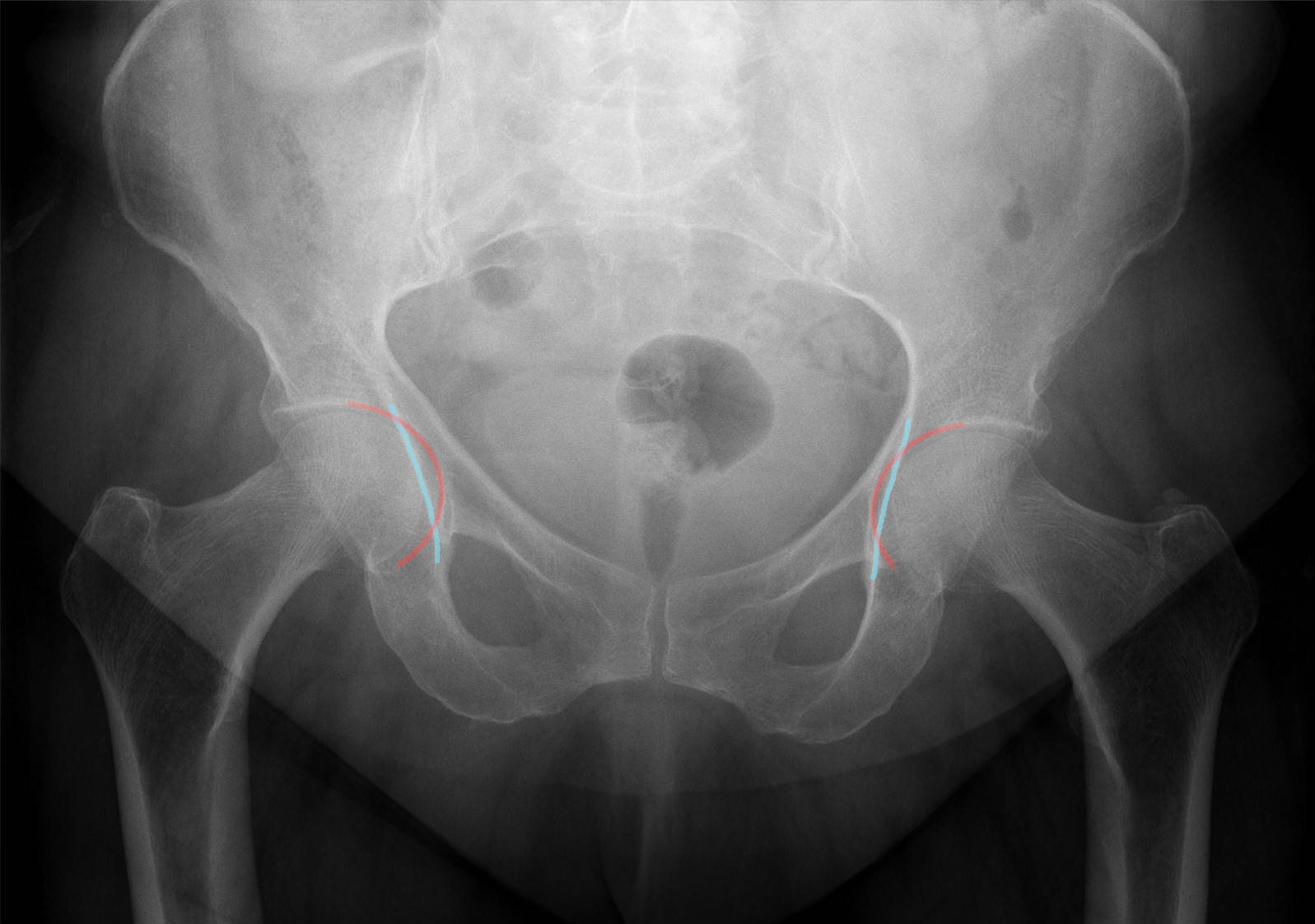
Coxa profunda (annotated). AP pelvis with the acetabular floor overlapping the ilioischial line bilaterally - the radiographic coxa profunda sign of a deep socket. Hellerhoff, Wikimedia Commons, CC BY-SA 4.0.
Coxa profunda (annotated). AP pelvis with the acetabular floor overlapping the ilioischial line bilaterally - the radiographic coxa profunda sign of a deep socket. Hellerhoff, Wikimedia Commons, CC BY-SA 4.0.
Femoral version completes the picture: normal anteversion is about 15° in the adult, falling from 30-40° at birth. Relative femoral retroversion (anteversion below 15°) rotates a cam into the rim earlier in flexion, hence pain on sitting or rising from a car, while excessive anteversion predisposes to anterior instability and posterior extra-articular impingement.[35]
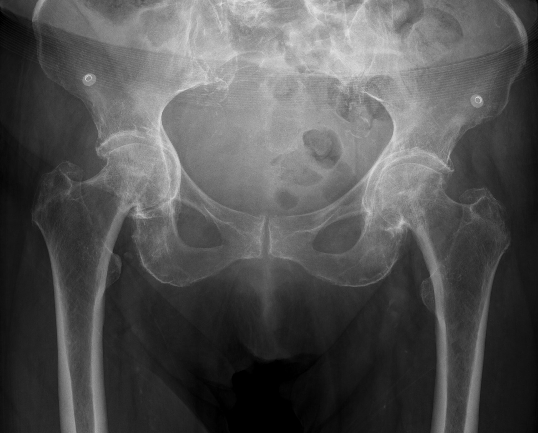
Coxa profunda and protrusio acetabuli. AP pelvis with coxa profunda on the left and protrusio acetabuli (head medial to the ilioischial line) on the right - the over-coverage end of the pincer spectrum. Hellerhoff, Wikimedia Commons, CC BY-SA 4.0.
Coxa profunda and protrusio acetabuli. AP pelvis with coxa profunda on the left and protrusio acetabuli (head medial to the ilioischial line) on the right - the over-coverage end of the pincer spectrum. Hellerhoff, Wikimedia Commons, CC BY-SA 4.0.
The labrum and the chondrolabral junction
The acetabular labrum is a horseshoe of fibrocartilage, mean thickness about 5 mm, attached to the bony rim and continuous with the transverse ligament; its base meets the articular cartilage through a transition or “watershed” zone.[36] It carries free nerve endings (both proprioceptive and nociceptive) and functions as a suction seal that increases the acetabular surface area by more than 25%, maintains the intra-articular fluid film and distributes load evenly.[37] Its periphery is vascularised from the capsule, but the central two-thirds are relatively avascular, which is why repair requires a bleeding bony bed and why the watershed zone is the plane in which the labrum is detached and re-attached during rim surgery.[38]
Extra-articular impingement
Beyond the rim, impingement can be extra-articular: subspine (AIIS) impingement from a prominent anterior inferior iliac spine (graded by Hetsroni into types 1-3), ischiofemoral impingement between the ischium and the lesser trochanter (with quadratus femoris oedema), trochanteric-pelvic impingement in coxa vara, and iliopsoas impingement, in which a tight tendon scars to the anterior capsule and damages the labrum at the 3-o’clock position.[39]
Part IV - Epidemiology and natural history
FAI most often becomes symptomatic in the second and third decades; cam disease presents in the young athletic man, pincer disease in the middle-aged woman, and mixed disease somewhere between.[40] The key epidemiological fact is that the morphology is very common in people without symptoms. Asymptomatic cam is found in roughly 24-30% of men and about 11% of women, in 37-55% of athletes versus about 23% of the general population, and asymptomatic pincer in up to two-thirds of hips.[41] Among male collegiate American-football players, 95% had a radiographic cam or pincer deformity; male basketball players show a ten-fold excess of cam; and 75% of ice-hockey players had an alpha angle over 55° compared with 42% of skiers. High-impact sport during skeletal immaturity shapes the deformity.[42]
This high background prevalence is why FAI cannot be diagnosed from an X-ray alone, and why prophylactic surgery on an asymptomatic hip is not recommended.[43] Even within one symptomatic patient, the contralateral hip has a cam in about 78% but is painful in only about 26%.[44]
When FAI does progress, cam morphology carries the clearest osteoarthritis risk. An alpha angle over 60° raises the adjusted odds of end-stage osteoarthritis to about 3.7, and an angle over 83° to about 9.7; a large cohort found each degree of alpha above 65° added roughly 5% to the osteoarthritis risk.[45] The pincer-osteoarthritis link is weaker and disputed: some prospective data suggest pincer over-coverage does not drive osteoarthritis and may even be protective, whereas dysplasia clearly does.[46] Radiographic progression to arthrosis is highly variable, quoted between 18% and 73% over ten years, and there is a “tipping point” of cartilage loss beyond which no osteotomy will change the natural history.[47]
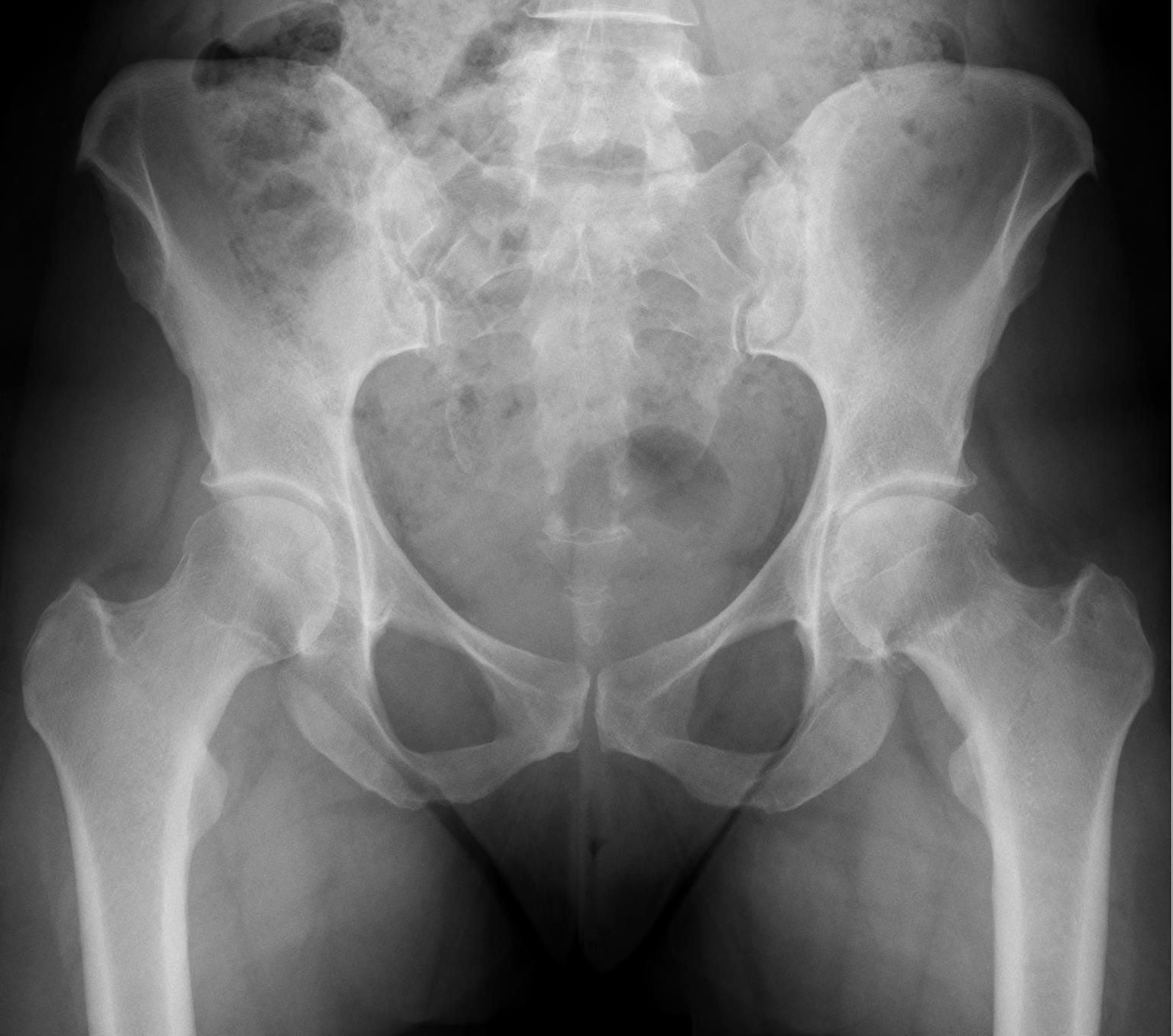
Cam impingement with secondary osteoarthritis. AP pelvis: a prominent head-neck convexity (cam) with central joint-space narrowing - the cam deformity having progressed to coxarthrosis. Hellerhoff, Wikimedia Commons, CC BY-SA 4.0.
Cam impingement with secondary osteoarthritis. AP pelvis: a prominent head-neck convexity (cam) with central joint-space narrowing - the cam deformity having progressed to coxarthrosis. Hellerhoff, Wikimedia Commons, CC BY-SA 4.0.
Part V - Clinical features and examination
The history is of insidious anterior groin pain in an active young adult, often pointed out with the hand cupped over the trochanter and thumb in the groin, the “C-sign.”[48] Groin pain is moderate or severe in up to 85%, frequently with lateral hip, buttock or anterior-thigh pain, and is worse with sitting than with walking: deep flexion provokes it, so patients struggle to get in and out of a car, to sit in low chairs, or to put on shoes and socks.[49] Mechanical symptoms (clicking, catching, locking or giving way) point to a labral tear.[50] The onset is insidious in more than half of patients (with about a quarter each reporting a traumatic or an acute non-traumatic onset), and there is typically a long delay, of order 30 months, before diagnosis; pain radiating below the knee should prompt a search for a spinal cause.[51]
On examination the cardinal finding is reduced range of motion, especially loss of internal rotation in flexion: mean internal rotation at 90° of flexion is about 9-15° in symptomatic FAI versus around 30° in normal hips, and flexion is commonly limited to 90-100°.[52] Both hips are examined, as the morphology is often bilateral. The key provocative tests are:
- the anterior impingement test (FADIR): flexion, adduction and internal rotation reproduce anterosuperior groin pain. Described by Klaue and colleagues, it is highly sensitive (positive in up to 99%) but not specific, being positive in almost any irritable hip;[53]
- the posterior impingement test: extension with external rotation, reproducing deep posterior pain;[54]
- the FABER (Patrick) test: flexion-abduction-external rotation, with the knee-to-table distance almost always greater on the symptomatic side (abnormal in up to 97%);[55]
- the log-roll test: passive rotation of the femur in the socket, which is insensitive but the most specific sign of intra-articular pathology;[56]
- the Stinchfield (resisted straight-leg-raise) test and Drehmann sign (obligate external rotation as the hip is flexed, also seen in slipped epiphysis and Perthes).[57]
A Trendelenburg gait suggests advanced disease or an associated dysplasia, and tests of capsular laxity (the external-rotation dial and axial-traction apprehension tests) help identify the borderline-dysplastic hip in which over-resection would be dangerous.[58] The main differentials are athletic pubalgia (sports hernia), adductor strain, snapping or inflamed iliopsoas, femoral-neck stress fracture, trochanteric pathology, hip dysplasia and referred lumbar pain. A diagnostic intra-articular local-anaesthetic injection is useful here: relief confirms an intra-articular source, while little relief redirects attention to extra-articular or spinal causes.[59]
Part VI - Imaging and diagnosis
Plain radiographs are the workhorse. A typical series is a well-centred AP pelvis plus a lateral (a Dunn view at 45° or 90°, a cross-table lateral or a frog-leg lateral) and, for anterior coverage, a false-profile view.[60] Correct AP technique is essential to avoid a spurious crossover sign: the beam is centred between the symphysis and the spines, the legs are internally rotated about 15°, and tilt and rotation are controlled by keeping the coccyx centred over and a defined distance above the symphysis.[61] The different views profile the head-neck junction at different points of the “clock face” (the AP at 12 o’clock, the 45° Dunn at 1, the frog-leg at 2, the cross-table at 3), and the AP view often underestimates or misses a cam, which is why a lateral is mandatory.[62]
The alpha angle of Nötzli is the principal cam metric: a best-fit circle is drawn around the head, and the angle is measured between the neck axis and the line from the head centre to where the head contour first leaves the circle anteriorly. Normal is about 42-45°; over 50-55° is abnormal, and symptomatic cam hips average 66-74°.[63] Because the angle is highly dependent on rotation and on the imaging plane, a cam can be missed on one view and obvious on another; radial CT or MR sequences that sweep around the neck axis give a 360° assessment.[64] The complementary metric is the head-neck offset and offset ratio: the anterior offset falls from about 11.6 mm normally to 7.2 mm in cam disease, and an offset ratio below 0.15 has a 95% positive predictive value for FAI.[65]
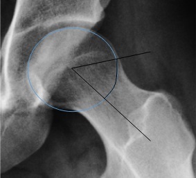
Alpha angle (45° Dunn view). A best-fit circle on the femoral head; the alpha angle lies between the neck axis and the line to the point where the head contour exits the circle. From Ruiz Santiago et al., Radiol Res Pract 2016;2016:6369237, Fig. 6g (CC BY 4.0).
Alpha angle (45° Dunn view). A best-fit circle on the femoral head; the alpha angle lies between the neck axis and the line to the point where the head contour exits the circle. From Ruiz Santiago et al., Radiol Res Pract 2016;2016:6369237, Fig. 6g (CC BY 4.0).
Cross-sectional imaging refines the picture. CT with three-dimensional reconstruction gives the clearest image of the bony deformity and quantifies femoral and acetabular version, distinguishing a focal cranial rim lesion from true global retroversion and correcting the false-positive crossover sign.[66] MRI, or MR arthrography (MRA), is used for the labrum and cartilage: MRA is the traditional test of choice for the labrum, with sensitivity and accuracy of 92-100% and 93-96%, although modern 3-tesla non-contrast MRI now performs as well and is in fact more sensitive than MRA for chondral injury (86-100% versus 58-79%).[67] dGEMRIC and T2/T1-rho mapping detect early biochemical cartilage degeneration before gross damage appears.[68]
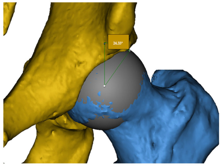
3-D CT reconstruction of the hip. Surface rendering of the acetabulum (yellow) and femur (blue) with a best-fit sphere on the head and the lateral centre-edge angle measured - the modality that best displays bony morphology and version. İsmailoğlu et al., J Clin Med 2025;14:7220, Fig. 1 (CC BY 4.0).
3-D CT reconstruction of the hip. Surface rendering of the acetabulum (yellow) and femur (blue) with a best-fit sphere on the head and the lateral centre-edge angle measured - the modality that best displays bony morphology and version. İsmailoğlu et al., J Clin Med 2025;14:7220, Fig. 1 (CC BY 4.0).
The diagnostic algorithm rests on three things together: a symptomatic hip, consistent clinical signs and confirmatory imaging, supported where needed by a diagnostic injection.[69] Asymptomatic morphology, however striking, is not by itself an indication to operate.
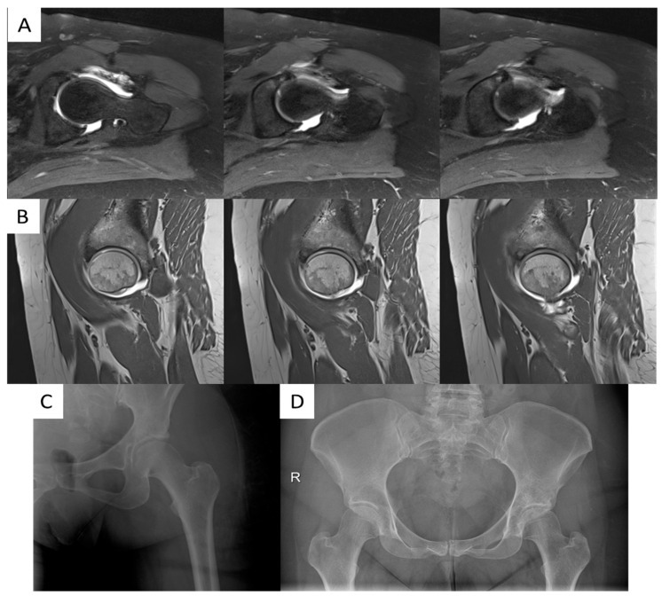
MR arthrogram of an acetabular labral tear. Axial T2 (A) and sagittal T1 (B) MR-arthrogram images show an antero-superior labral tear with subchondral cystic change. Minelli et al., J Clin Med 2025;14:8298, Fig. 5 (CC BY 4.0).
MR arthrogram of an acetabular labral tear. Axial T2 (A) and sagittal T1 (B) MR-arthrogram images show an antero-superior labral tear with subchondral cystic change. Minelli et al., J Clin Med 2025;14:8298, Fig. 5 (CC BY 4.0).
Part VII - Treatment
The aim of hip preservation is an impingement-free functional arc of motion, achieved by reshaping the bone and repairing the chondrolabral damage, so as to relieve pain and delay or avoid arthritis.[70]
Non-operative management
First-line care is activity and sport modification, anti-inflammatories and physiotherapy, and the patient must understand that its goal is pain relief, not improved range of motion. Physiotherapy targets core, abductor and peri-articular strength and neuromuscular control; attempting to push range of motion tends to make symptoms worse.[71] Intra-articular corticosteroid or hyaluronic-acid injections give only temporary relief.[72] About 89% of mild cases improve with a safe range of motion and avoidance of provocative positions, but patients managed conservatively must be followed, because the preservation options narrow once significant cartilage damage develops; an asymptomatic hip is not operated on prophylactically.[73]
Indications and contraindications for surgery
Surgery is for the symptomatic patient with confirmed FAI who has failed a trial of conservative care: typically several months of pain limiting daily life, with positive signs, confirmatory imaging and a positive response to a diagnostic injection.[74] The dominant contraindication is established osteoarthritis. Tönnis grade III is an absolute contraindication and grade II a relative one, and a joint-space width below 2 mm is an ominous sign: about 86% of such hips convert to arthroplasty within five years, versus 16% with preserved space.[75] The other recurring warning is the borderline-dysplastic, retroverted hip: trimming an anteriorly over-covered but posteriorly deficient socket creates iatrogenic instability, so a retroverted dysplastic rim must never be simply resected.[76]
Open surgical hip dislocation (the Ganz/Bern technique)
The safe surgical dislocation described by Ganz in 2001 remains the most versatile, “most powerful” approach, giving 360° access to the head and acetabulum and allowing combined femoral or pelvic osteotomy.[77] Through a Gibson or Kocher-Langenbeck approach in the lateral position, a trochanteric flip (“digastric” or trochanteric-slide) osteotomy of about 15 mm is raised carrying the gluteus medius, minimus and vastus lateralis, while the short external rotators and piriformis are left intact on the stable trochanter.[78] The safety of the technique rests on protecting the deep branch of the medial femoral circumflex artery: the dissection stays proximal to the piriformis tendon, the rotators are preserved, and a Z-shaped capsulotomy is fashioned anterior to the lesser trochanter; the retinacular vessels entering at about 11 o’clock are identified and protected, if necessary on an extended retinacular soft-tissue flap.[79] Done correctly, the risk of avascular necrosis is essentially nil.[80] It is the approach of choice for severe or complex deformity, global over-coverage, posterior pathology, relative neck lengthening, and the residual deformities of slipped epiphysis and Perthes disease.[81]
Arthroscopic FAI surgery
Hip arthroscopy is now the dominant approach. Almost all FAI surgery is now performed arthroscopically, avoiding the trochanteric osteotomy, allowing day-case surgery and faster return to sport.[82] On a traction table with a well-padded perineal post, the central compartment (under traction) is addressed first, treating the labrum, cartilage and ligamentum teres, and then the peripheral compartment (traction released, hip flexed) for the cam.[83] Acetabular rim trimming is performed with or without taking the labrum down (the labrum is separated in the watershed zone and re-attached with suture anchors), and the femoral cam is recontoured by osteochondroplasty with a burr to restore the head-neck concavity.[84] The two cardinal limits are mechanical: resect less than about one centimetre of rim to avoid instability, and keep femoral neck resection under 30% of its diameter, beyond which the neck is weakened and may fracture.[85] Adequacy is judged by direct vision and dynamic motion (aiming for impingement-free flexion of about 100° and 15° of internal rotation), and the capsule is closed to prevent iatrogenic instability.[86] The commonest reason a procedure fails is incomplete bony correction, particularly failure to carry the cam resection far enough distally; arthroscopy cannot achieve relative neck lengthening, trochanteric transfer or femoral osteotomy.[87]
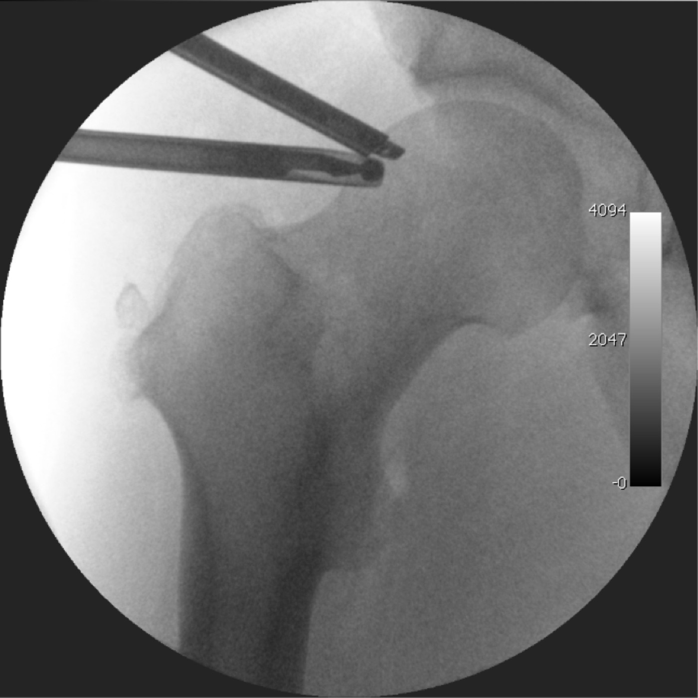
Arthroscopic cam resection. Intra-operative fluoroscopy during arthroscopic osteochondroplasty: the arthroscope (above) and the high-speed burr (below) at the femoral head-neck junction. Gileshugo, Wikimedia Commons, CC BY-SA 3.0.
Arthroscopic cam resection. Intra-operative fluoroscopy during arthroscopic osteochondroplasty: the arthroscope (above) and the high-speed burr (below) at the femoral head-neck junction. Gileshugo, Wikimedia Commons, CC BY-SA 3.0.
Mini-open and combined approaches
A combined arthroscopic and limited-open osteochondroplasty (Clohisy/Laude) treats the labrum arthroscopically and then reshapes the anterior head-neck junction under direct vision through a small anterior (Smith-Petersen or Hueter) approach, avoiding a trochanteric osteotomy; its limitation is access to the anterior head-neck only, and the lateral femoral cutaneous nerve is at risk.[88] Across the open, mini-open and arthroscopic routes, outcomes appear comparable and open surgery is not demonstrably superior, but there is no randomised head-to-head comparison with matched criteria.[89]
Labral management
The principle has shifted from débridement towards preservation: wherever tissue quality allows, the labrum is repaired or re-fixed rather than excised, to recreate the suction seal.[90] Suture anchors are placed about 2 mm off the rim, roughly one anchor per centimetre of detachment, with care not to invert or over-tension the labrum.[91] Débridement is reserved for irreparable, degenerate tissue, since subtotal labrectomy accelerates arthritis (Espinosa).[92] An irreparable but functionally important labrum, in a young or very active patient, may be reconstructed with a graft (iliotibial band, fascia lata, hamstring or ligamentum teres).[93] The outcome data favour repair: refixation gave 90% good/excellent results versus 67% for débridement (Larson), and at open surgery refixation outperformed resection (94% versus 76%) at two years (Espinosa).[94]
The retroverted acetabulum: PAO and reverse PAO
When the acetabulum is truly retroverted with posterior deficiency, the correct operation is reorientation, not rim resection. The Bernese (Ganz) periacetabular osteotomy (PAO) redirects the socket through a juxta-articular cut that leaves the posterior column intact; performed in reverse as an anteverting PAO, it corrects retroversion.[95] The candidate is identified by crossover, positive posterior-wall and ischial-spine signs with preserved anterior cartilage; a positive posterior-wall sign is in fact a prerequisite, since posterior impingement is otherwise a side-effect of the rotation.[96] The danger that justifies the more complex operation is plain: an isolated anterior rim resection in a truly retroverted acetabulum leaves the head posteriorly uncovered and unstable, and PAO gives higher survivorship than rim trimming in this setting.[97] Siebenrock’s anteverting-PAO series reported about 96% good/excellent results with no osteoarthritis or osteonecrosis at follow-up.[98]
Femoral correction and the secondary hip
On the femoral side, osteochondroplasty restores sphericity (templated to a normal alpha angle), and version abnormalities are addressed by derotational or valgus proximal femoral osteotomy, with relative neck lengthening and trochanteric advancement for the high trochanter and extra-articular impingement.[99] Extra-articular sources are treated specifically: subspine/AIIS decompression, lesser-trochanter resection for ischiofemoral impingement, and iliopsoas fractional lengthening for a snapping or impinging tendon.[100] FAI secondary to slipped epiphysis, which almost inevitably leaves a cam, is corrected at the appropriate level, with the modified Dunn procedure (capital realignment through a surgical dislocation, avascular-necrosis risk about 3% in expert hands) for severe deformity; residual Perthes deformity may need a femoral head reduction osteotomy through an extended retinacular flap.[101]
Part VIII - Outcomes and complications
Outcomes are reported with the modified Harris Hip Score, the Hip Outcome Score, the Non-Arthritic Hip Score and the Merle d’Aubigné score. Across series, both open surgical dislocation and arthroscopy give roughly 68-96% good or excellent results at two to ten years, with conversion to arthroplasty in about 9-34% depending on cartilage status and follow-up length.[102] In athletes, arthroscopy returns about 90% to sport (higher in professionals), with a shorter recovery than open surgery.[103] The consistent predictors of a good result are preserved cartilage (joint space ≥ 2 mm), a labral repair rather than débridement, younger age and a low Tönnis grade; the predictors of failure are advanced osteoarthritis, intra-operative cartilage delamination, joint space below 2 mm and increasing age.[104]
The complications are largely the mirror image of the surgical principles:
- Iatrogenic instability or dislocation from over-resecting the rim (or releasing the capsule): the reason rim resection stays under a centimetre, the capsule is closed, and the retroverted dysplastic hip is not simply trimmed.[105]
- Femoral neck fracture from over-resecting the cam beyond 30% of the neck diameter, mitigated by protected weight-bearing.[106]
- Avascular necrosis from injury to the medial femoral circumflex artery, close to nil when the Ganz technique is respected.[107]
- Heterotopic ossification, reduced by removing all bone debris and by anti-inflammatory prophylaxis (NSAIDs or indomethacin).[108]
- Nerve injury: the lateral femoral cutaneous nerve in anterior approaches, pudendal and perineal injury from arthroscopic traction (minimised by a padded post and short traction time), and the sciatic, femoral and obturator nerves in PAO (about 2% nerve palsy, mostly recovering).[109]
- Under-resection (residual impingement), the commonest reason for revision, and, conversely, over-resection; plus adhesions, fluid extravasation, and venous thromboembolism.[110]
The overall complication rate of hip arthroscopy is about 0.5-6.4%.[111]
To tie the topic together: FAI is dynamic abnormal contact, a cam that shears cartilage, a pincer that crushes the labrum, most often both. Diagnose it from the symptomatic patient, the loss of internal rotation in flexion and the alpha and centre-edge angles, never from the X-ray alone. Preserve the hip by reshaping the bone within safe limits (under a centimetre of rim, under a third of the neck), repairing rather than excising the labrum, and reorienting rather than resecting the truly retroverted socket; and counsel that the result is governed by how much cartilage remains.
Bulgarian terminology (Боев / Boychev tradition) - glossary
The following Bulgarian equivalents connect the international literature with the terminology of the Bulgarian school of orthopaedics (in the tradition of Бойчо Бойчев / Boycho Boychev) used in the state examination.
| English term | Bulgarian term (Cyrillic) | Transliteration |
|---|---|---|
| Femoroacetabular impingement | Феморо-ацетабуларен импинджмънт | Femoro-atsetabularen impindzhmant |
| Cam impingement (cam deformity) | Кам импинджмънт (кам деформитет) | Kam impindzhmant (kam deformitet) |
| Pincer impingement | Пинсер (клещовиден) импинджмънт | Pinser (kleshtoviden) impindzhmant |
| Mixed (combined) type | Смесен (комбиниран) тип | Smesen (kombiniran) tip |
| Femoral head | Бедрена глава | Bedrena glava |
| Femoral neck | Бедрена шийка | Bedrena shiyka |
| Head-neck offset | Отстъп на прехода глава-шийка | Otstap na prehoda glava-shiyka |
| Acetabular labrum | Ацетабуларен лабрум (устна) | Atsetabularen labrum (ustna) |
| Acetabular over-coverage | Свръхпокритие на ацетабулума | Svrahpokritie na atsetabuluma |
| Acetabular retroversion | Ретроверзия на ацетабулума | Retroverziya na atsetabuluma |
| Coxa profunda | Coxa profunda (дълбок ацетабулум) | Coxa profunda (dalbok atsetabulum) |
| Protrusio acetabuli | Протрузия на ацетабулума | Protruziya na atsetabuluma |
| Alpha angle | Алфа ъгъл | Alfa agal |
| Centre-edge angle (Wiberg) | Централно-ръбов ъгъл (на Wiberg) | Tsentralno-rabov agal (na Wiberg) |
| Crossover sign | Знак на кръстосване | Znak na krastosvane |
| Cartilage delamination | Деламинация (отлепване) на хрущяла | Delaminatsiya (otlepvane) na hrushtyala |
| Labral tear | Разкъсване на лабрума | Razkasvane na labruma |
| Osteochondroplasty | Остеохондропластика | Osteohondroplastika |
| Acetabular rim trimming | Ремоделиране на ацетабуларния ръб | Remodelirane na atsetabularniya rab |
| Periacetabular osteotomy | Периацетабуларна остеотомия | Periatsetabularna osteotomiya |
| Surgical hip dislocation | Хирургична луксация на тазобедрената става | Hirurgichna luksatsiya na tazobedrenata stava |
| Avascular necrosis | Аваскуларна некроза | Avaskularna nekroza |
Image attributions
(Figure attributions and licences are listed in the figure MANIFEST and inserted with each image. All images are openly licensed [CC0 / CC BY / CC BY-SA] or used under their stated terms; any non-commercial [NC] item is flagged as such and must not be used in a commercial product.)
References
-
Hip Arthroscopy & Hip Joint Preservation Surgery (Nho/Leunig), p. 168, p. 701; Berry, Surgery of the Hip, p. 453.
-
Byrd, Operative Hip Arthroscopy, p. 230-231, p. 249; DeLee, Orthopaedic Sports Medicine, p. 1272.
-
Nho/Leunig, p. 701; Berry, p. 453, p. 457; Campbell’s, p. 425.
-
Berry, p. 453; Nho/Leunig, p. 176, p. 181.
-
Campbell’s, p. 427; Berry, p. 459-460.
-
Nho/Leunig, p. 168, p. 701, p. 703; Berry, p. 462.
-
Nho/Leunig, p. 168, p. 703; Campbell’s, p. 416; Berry, p. 736-737.
-
Campbell’s, p. 424; Nho/Leunig, p. 169.
-
Nho/Leunig, p. 702-703.
-
Nho/Leunig, p. 168, p. 703; Berry, p. 427, p. 453; Campbell’s, p. 425.
-
Nho/Leunig, p. 703-704.
-
Nho/Leunig, p. 703; Berry, p. 428, p. 738; Campbell’s, p. 416, p. 422.
-
Berry, p. 454; Nho/Leunig, p. 168, p. 589, p. 703; Campbell’s, p. 425.
-
Nho/Leunig, p. 168, p. 589.
-
Nho/Leunig, p. 705; DeLee, p. 1270.
-
Nho/Leunig, p. 168, p. 170, p. 704-705; Berry, p. 453.
-
Nho/Leunig, p. 169, p. 705; Berry, p. 453.
-
Nho/Leunig, p. 701; Berry, p. 428; Campbell’s, p. 425.
-
Nho/Leunig, p. 703-704, p. 594; Berry, p. 428, p. 454.
-
Nho/Leunig, p. 168-169, p. 591; Berry, p. 429, p. 431, p. 454-455; Campbell’s, p. 425.
-
Campbell’s, p. 425; Berry, p. 431; Nho/Leunig, p. 591.
-
Campbell’s, p. 425; Nho/Leunig, p. 705, p. 594, p. 669.
-
Nho/Leunig, p. 704, p. 594.
-
Nho/Leunig, p. 703, p. 592; Berry, p. 427, p. 453, p. 730; Campbell’s, p. 426.
-
Campbell’s, p. 420, p. 426; Berry, p. 456, p. 459, p. 468; Nho/Leunig, p. 655.
-
Campbell’s, p. 421; Berry, p. 468, p. 728; Nho/Leunig, p. 653, p. 655.
-
Campbell’s, p. 421; Berry, p. 432-433, p. 467.
-
Campbell’s, p. 420; Berry, p. 457; Nho/Leunig, p. 592, p. 656.
-
Campbell’s, p. 420-421; Berry, p. 457; Nho/Leunig, p. 594, p. 656.
-
Campbell’s, p. 420; Berry, p. 457; Nho/Leunig, p. 594, p. 656.
-
Berry, p. 457, p. 462; Campbell’s, p. 427.
-
Campbell’s, p. 420-421; Berry, p. 457; Nho/Leunig, p. 656, p. 658.
-
Campbell’s, p. 421; Berry, p. 429, p. 459; Nho/Leunig, p. 653-654.
-
Campbell’s, p. 421.
-
Nho/Leunig, p. 595, p. 598, p. 705.
-
Nho/Leunig, p. 182, p. 186; Berry, p. 429, p. 728.
-
Nho/Leunig, p. 186.
-
Nho/Leunig, p. 186, p. 714; Berry, p. 728, p. 730, p. 735.
-
Nho/Leunig, p. 591-597; Campbell’s, p. 435; Berry, p. 732.
-
DeLee, p. 1270-1272; Berry, p. 453.
-
The Pediatric and Adolescent Hip, p. 256-257.
-
The Pediatric and Adolescent Hip, p. 255-256.
-
Byrd, p. 230-231, p. 249; DeLee, p. 1272.
-
Berry, p. 455; Nho/Leunig, p. 590.
-
Campbell’s, p. 425; The Pediatric and Adolescent Hip, p. 257.
-
The Pediatric and Adolescent Hip, p. 256-257; Campbell’s, p. 425.
-
Nho/Leunig, p. 168-169.
-
Berry, p. 455-456; DeLee, p. 1272.
-
Byrd, p. 251-252; Berry, p. 455; The Pediatric and Adolescent Hip, p. 257.
-
DeLee, p. 1273; Byrd, p. 278.
-
Byrd, p. 252; The Pediatric and Adolescent Hip, p. 257; Berry, p. 455.
-
Byrd, p. 231; Berry, p. 456-457; DeLee, p. 1273; The Pediatric and Adolescent Hip, p. 258.
-
Byrd, p. 231, p. 253; DeLee, p. 1273; Berry, p. 456-457; The Pediatric and Adolescent Hip, p. 258.
-
Byrd, p. 253; DeLee, p. 1273-1274.
-
Byrd, p. 253; DeLee, p. 1273.
-
DeLee, p. 1273.
-
Byrd, p. 253; DeLee, p. 1273-1274.
-
Berry, p. 456, p. 458; Byrd, p. 253-254.
-
Berry, p. 460; Byrd, p. 231, p. 254; DeLee, p. 1280.
-
DeLee, p. 1274-1276; The Pediatric and Adolescent Hip, p. 259; Berry, p. 457-458.
-
Byrd, p. 233; DeLee, p. 1274; Berry, p. 456-457; The Pediatric and Adolescent Hip, p. 259.
-
DeLee, p. 1276; Berry, p. 428.
-
Berry, p. 428, p. 456, p. 459; Campbell’s, p. 422; DeLee, p. 1275.
-
Berry, p. 428, p. 459-460; The Pediatric and Adolescent Hip, p. 262.
-
Berry, p. 428; Campbell’s, p. 422.
-
DeLee, p. 1278; Byrd, p. 231, p. 234; The Pediatric and Adolescent Hip, p. 260.
-
Berry, p. 429, p. 431, p. 437; DeLee, p. 1276-1277; The Pediatric and Adolescent Hip, p. 260.
-
DeLee, p. 1278; Berry, p. 431; The Pediatric and Adolescent Hip, p. 261-262.
-
DeLee, p. 1272; Byrd, p. 254; Berry, p. 459.
-
Berry, p. 461.
-
The Pediatric and Adolescent Hip, p. 262-263; Berry, p. 460.
-
The Pediatric and Adolescent Hip, p. 263, p. 272.
-
The Pediatric and Adolescent Hip, p. 263; Byrd, p. 249.
-
Berry, p. 461; The Pediatric and Adolescent Hip, p. 263.
-
Byrd, p. 250; Campbell’s, p. 423, p. 426; Berry, p. 735, p. 738; Nho/Leunig, p. 632.
-
Berry, p. 459, p. 734; Byrd, p. 231, p. 234; Campbell’s, p. 435.
-
Nho/Leunig, p. 711; Berry, p. 738; The Pediatric and Adolescent Hip, p. 247-248.
-
Nho/Leunig, p. 712; Campbell’s, p. 427-428.
-
Nho/Leunig, p. 712-714; Campbell’s, p. 427-428; The Pediatric and Adolescent Hip, p. 264-266.
-
The Pediatric and Adolescent Hip, p. 264, p. 268.
-
Berry, p. 739; Campbell’s, p. 427; Nho/Leunig, p. 711.
-
Campbell’s, p. 435; The Pediatric and Adolescent Hip, p. 266; Berry, p. 733.
-
Byrd, p. 232, p. 256-257.
-
Berry, p. 728-731; Byrd, p. 237, p. 243.
-
Nho/Leunig, p. 714, p. 721, p. 722; Campbell’s, p. 428.
-
Nho/Leunig, p. 723; Berry, p. 731, p. 741; Byrd, p. 241, p. 258.
-
Nho/Leunig, p. 711; Byrd, p. 257, p. 265; Campbell’s, p. 435.
-
Campbell’s, p. 429-432; Berry, p. 461; Nho/Leunig, p. 720.
-
Berry, p. 461; Byrd, p. 265.
-
Berry, p. 731; Byrd, p. 246.
-
Berry, p. 733; Byrd, p. 237.
-
Byrd, p. 191, p. 234; Berry, p. 731.
-
Berry, p. 731, p. 733-734; Nho/Leunig, p. 714.
-
Berry, p. 735, p. 742.
-
Nho/Leunig, p. 625, p. 628, p. 642.
-
Berry, p. 462; Campbell’s, p. 426, p. 432.
-
Nho/Leunig, p. 642; The Pediatric and Adolescent Hip, p. 272; Campbell’s, p. 426-427.
-
Nho/Leunig, p. 642-643; Berry, p. 463.
-
Nho/Leunig, p. 714, p. 641; Campbell’s, p. 435, p. 439; Berry, p. 460, p. 470.
-
Campbell’s, p. 435; Byrd, p. 254, p. 258.
-
The Pediatric and Adolescent Hip, p. 246-248, p. 264, p. 266, p. 268.
-
Berry, p. 462-463; Campbell’s, p. 429, p. 435.
-
Byrd, p. 246; Berry, p. 733.
-
Berry, p. 735, p. 742; Campbell’s, p. 426, p. 429; Byrd, p. 249, p. 255.
-
Byrd, p. 231, p. 257; Berry, p. 734; Nho/Leunig, p. 721.
-
Nho/Leunig, p. 714, p. 722; Campbell’s, p. 428; Byrd, p. 245, p. 265.
-
The Pediatric and Adolescent Hip, p. 264, p. 268; Byrd, p. 264.
-
Nho/Leunig, p. 715; Campbell’s, p. 432; Byrd, p. 245.
-
Berry, p. 734; Campbell’s, p. 430, p. 432, p. 445; Byrd, p. 246, p. 256; Nho/Leunig, p. 643-644.
-
Nho/Leunig, p. 711; Byrd, p. 257, p. 265; Berry, p. 734.
-
Berry, p. 735; The Pediatric and Adolescent Hip, p. 266.