Contents
- Part I - Definition, History, Epidemiology, and Diathesis
- Part II - Pathophysiology
- Part III - Fascial Anatomy and the Cords
- Part IV - Clinical Features and Staging
- Part V - Treatment
- References
Part I - Definition, History, Epidemiology, and Diathesis
Dupuytren disease is the most common heritable fibrotic disorder. It is a chronic, progressive fibrosis of the palmar and digital subcutaneous tissues that produces nodules and cords and, in a minority, the flexion contractures of the fingers and thumb that give the condition its clinical name.[1] The disease and the contracture are not the same thing: about nine of ten patients with palmar Dupuytren disease have no contracture at all, and the Dupuytren contracture is only one expression of a wider process.[2] The mechanism is a “two-hit” interaction of genetic predisposition and environmental stressors.[3]
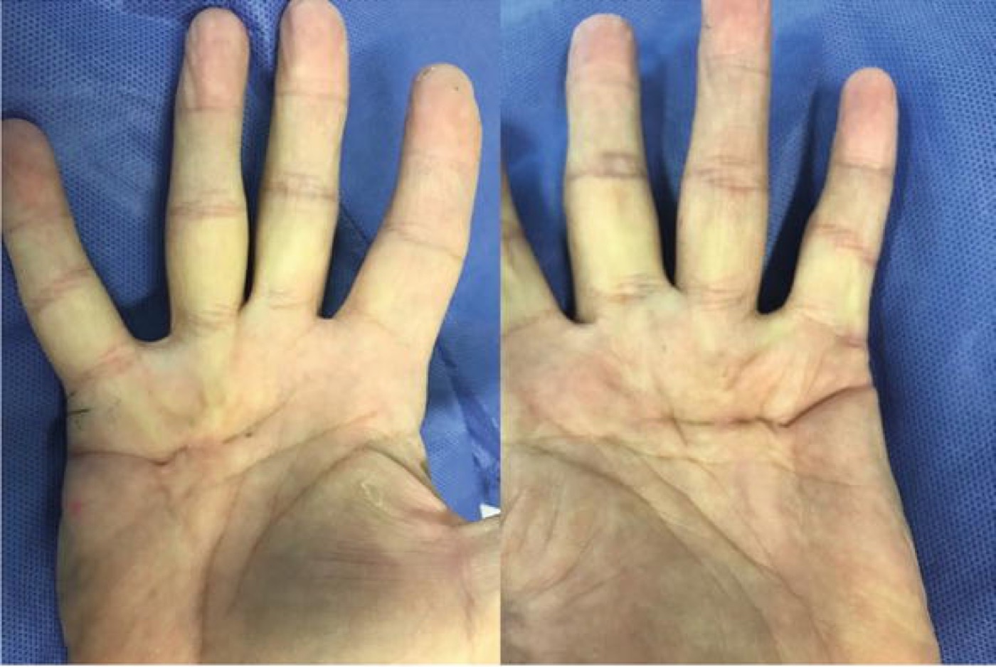
Figure 1. Dupuytren disease: palmar cords producing flexion contractures of the ring and little fingers. From Johnson, Pavano & Rodner (2018), IntechOpen, CC BY 3.0.
Figure 1. Dupuytren disease: palmar cords producing flexion contractures of the ring and little fingers. From Johnson, Pavano & Rodner (2018), IntechOpen, CC BY 3.0.
Baron Guillaume Dupuytren described his open fasciotomy in 1831, though Plater had given a clinical description as early as 1614 and Cooper performed a percutaneous fasciotomy in 1822.[4] The central cell, the myofibroblast, was identified in Dupuytren tissue by Gabbiani in 1972, and collagenase was approved by the US FDA in 2010.[5]
The disease is most prevalent in older Caucasian men with a family history, with a Caucasian prevalence rising from about 12% at age 55 to 21% at 65 and 29% at 75; onset is rare before 40, and the male predominance is in the order of 6-10:1, women lagging by roughly 10-15 years (their onset is delayed, plausibly by oestrogen’s suppression of myofibroblast differentiation, until after the menopause).[6] Disease begins on the ulnar side of the hand at the distal palmar crease, and the ring and little fingers are most commonly affected; about half of patients eventually develop bilateral disease.[7] The common teaching that it is a “Viking disease” is, on modern genetic evidence, a myth. It is better regarded simply as a disease of Caucasian ancestry (as common in Bosnia as in Norway), its frequency in other populations tracking the proportion of Caucasian heritage.[8]
Hueston’s concept of a Dupuytren diathesis identifies an aggressive, recurrence-prone phenotype, classically marked by a family history, early onset (before 50), bilateral disease, knuckle pads, and ectopic disease.[9] The recognised ectopic (“Dupuytren spectrum”) manifestations are Ledderhose disease (plantar fibromatosis, in ~5%), Peyronie disease (penile, ~3%), and Garrod knuckle pads (dorsal PIP nodules, in ~20%).[10]
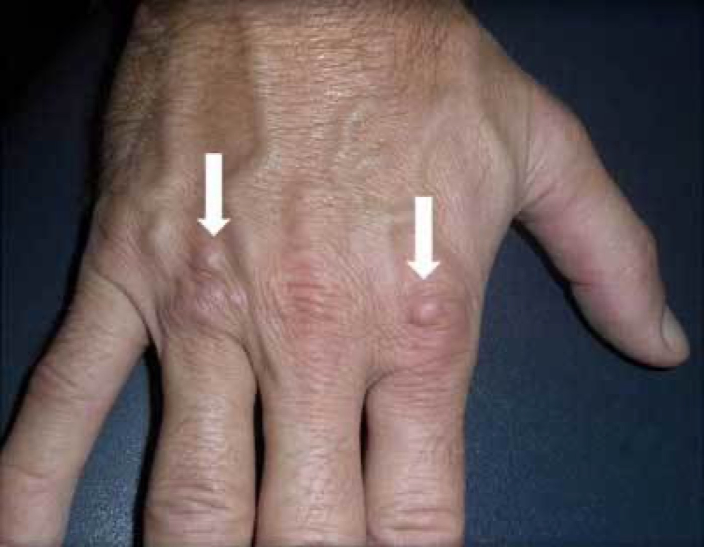
Figure 2. Garrod knuckle pads - dorsal nodules over the finger joints, an ectopic manifestation. From De Keersmaeker & Vanhoenacker (2016), J Belg Soc Radiol, PMC6102944, CC BY 4.0.
Figure 2. Garrod knuckle pads - dorsal nodules over the finger joints, an ectopic manifestation. From De Keersmaeker & Vanhoenacker (2016), J Belg Soc Radiol, PMC6102944, CC BY 4.0.
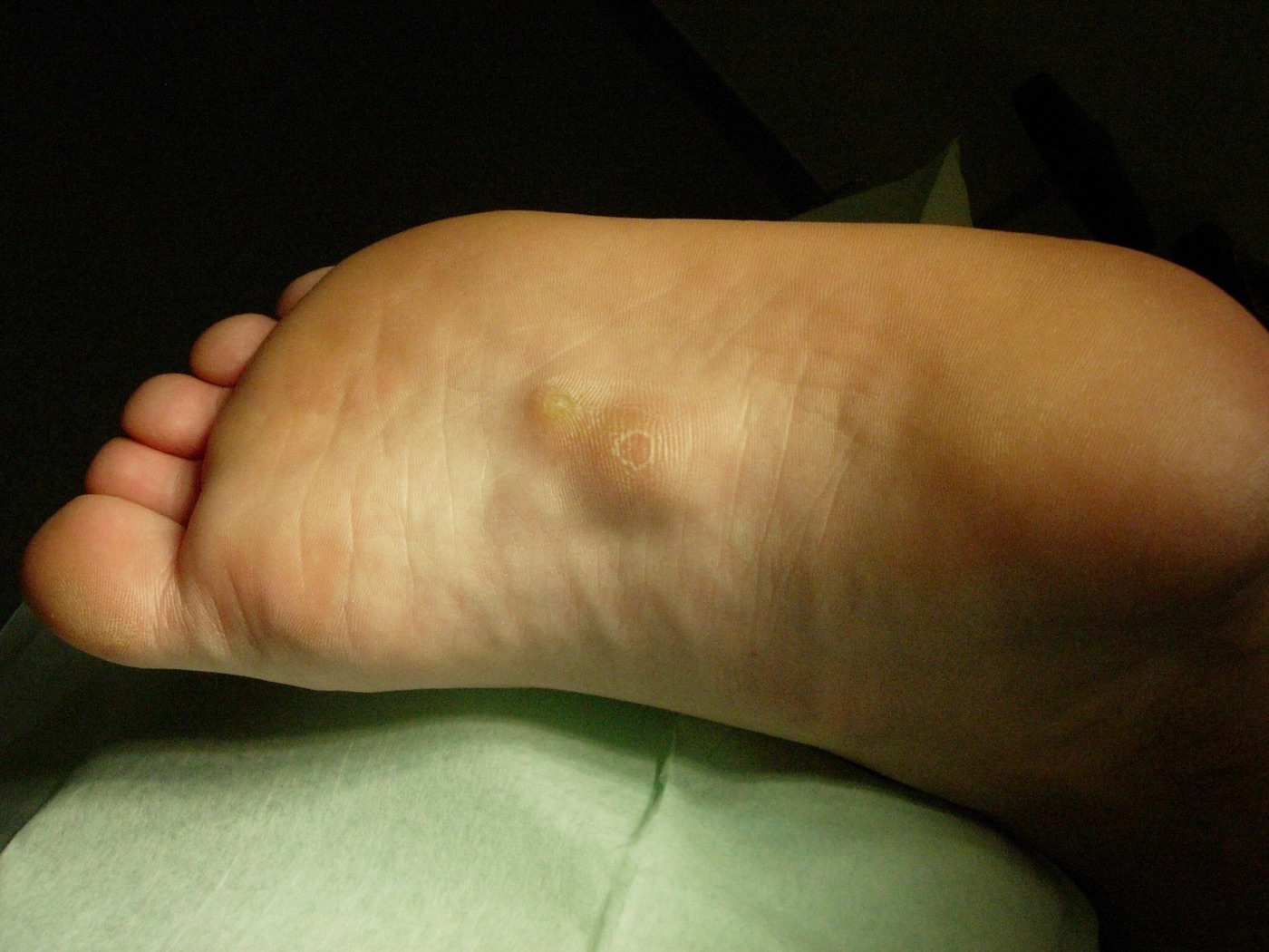
Figure 3. Ledderhose disease - plantar fibromatosis, the foot counterpart of Dupuytren. Source: Herecomesdoc, via Wikimedia Commons, CC BY-SA 3.0.
Figure 3. Ledderhose disease - plantar fibromatosis, the foot counterpart of Dupuytren. Source: Herecomesdoc, via Wikimedia Commons, CC BY-SA 3.0.
Part II - Pathophysiology
The myofibroblast is the central effector cell: a contractile fibroblast bearing smooth-muscle α-actin and stress fibres, which deposits and remodels collagen to create and then shorten the Dupuytren cords.[11] Its full phenotype requires the combination of mechanical tension and growth factors (notably TGF-β, with bFGF). In normal wound healing myofibroblasts disappear once healing is complete, but here they persist, and that persistence is a hallmark of the disease.[12] Contraction is transmitted from the matrix through cell-membrane integrins to the cytoskeleton, the cells acting together as a syncytium.
The classic Luck histological staging captures the disease’s evolution and maps onto the clinical lesions:[13]
- Proliferative stage, the nodule: mitotic, randomly oriented fibroblasts and myofibroblasts.
- Involutional stage, the active cord: cells and collagen align along lines of tension, myofibroblasts predominate.
- Residual stage, the mature cord: relatively acellular, dense parallel collagen.
The biochemical signature is a shift in collagen: normal palmar fascia is almost entirely type I (with under 5% type III), whereas Dupuytren tissue contains a much higher proportion of type III collagen (30-40% in nodules, 20-30% in cords), together with abnormal collagen cross-linking; the type III:I ratio is highest in the proliferative stage, and proliferative histology carries roughly double the recontracture rate of involutional and more than triple that of residual disease.[14]
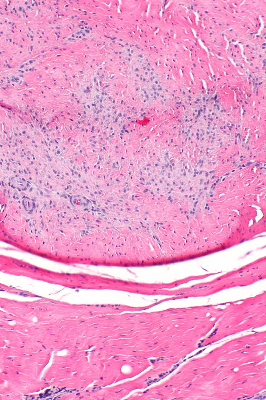
Figure 4. Histology of palmar fibromatosis (H\&E): a cellular nodule of spindle (myo)fibroblasts in collagen. Source: Nephron, via Wikimedia Commons, CC BY-SA 3.0.
Figure 4. Histology of palmar fibromatosis (H&E): a cellular nodule of spindle (myo)fibroblasts in collagen. Source: Nephron, via Wikimedia Commons, CC BY-SA 3.0.
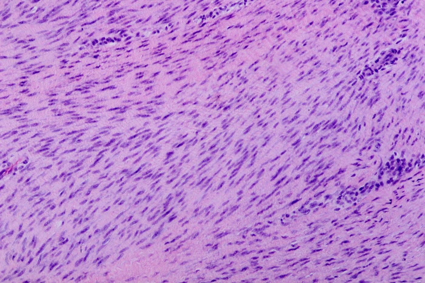
Figure 5. Proliferative-stage spindle-cell (fibroblast/myofibroblast) proliferation. Source: Patho, via Wikimedia Commons, CC BY-SA 3.0.
Figure 5. Proliferative-stage spindle-cell (fibroblast/myofibroblast) proliferation. Source: Patho, via Wikimedia Commons, CC BY-SA 3.0.
Part III - Fascial Anatomy and the Cords
Understanding the cords requires the normal anatomy, because a cord is the diseased, contracted version of a normal band.[15] The palmar aponeurosis sends four pretendinous bands distally toward the fingers (there is no central band to the thumb); in the digit, the neurovascular bundle is bordered by the Grayson ligament volar to it and the Cleland ligament dorsal to it, with the spiral band and lateral digital sheet alongside.[16]
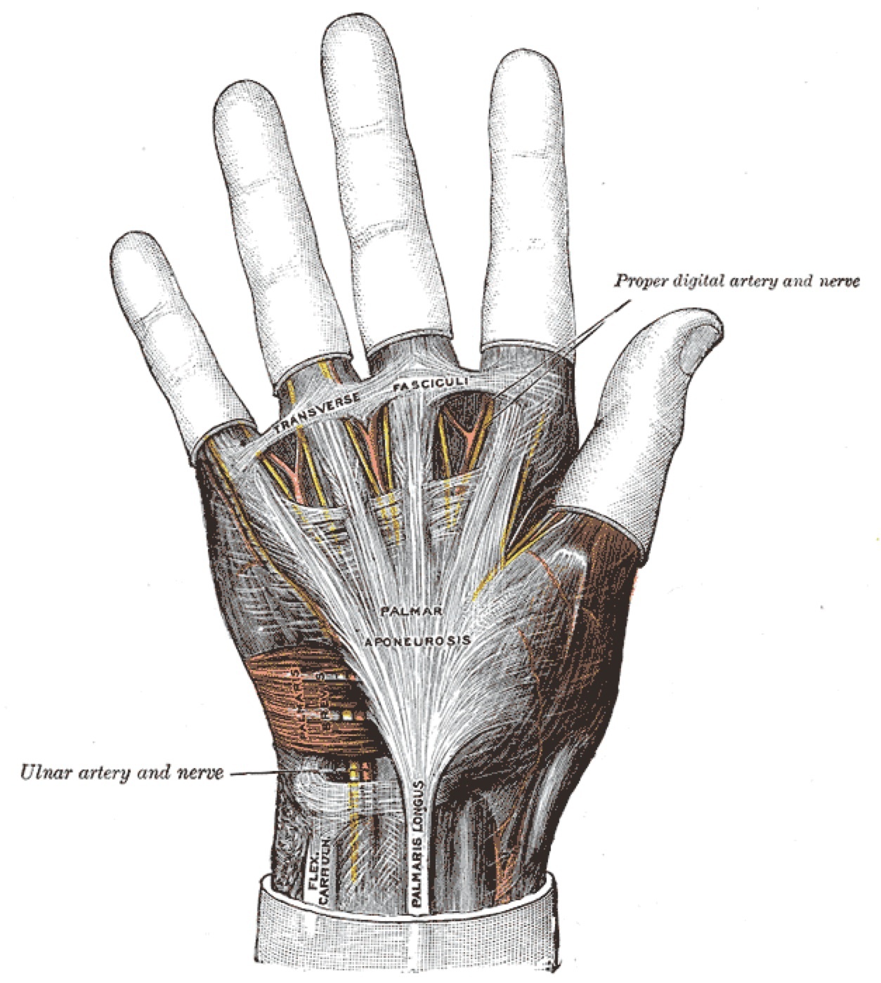
Figure 6. The palmar aponeurosis and its pretendinous slips (Gray’s plate 425). Henry Gray, Anatomy of the Human Body (1918), Plate 425 - public domain, via Wikimedia Commons.
Figure 6. The palmar aponeurosis and its pretendinous slips (Gray’s plate 425). Henry Gray, Anatomy of the Human Body (1918), Plate 425 - public domain, via Wikimedia Commons.
The relevant diseased cords are:
- Pretendinous cord, from the pretendinous band; the usual cause of MCP joint contracture.[17]
- Central cord, the commonest cord producing PIP joint contracture (most PIP contractures involve several cords).[18]
- Lateral cord, from the lateral digital sheet; may contribute to PIP and DIP contracture.
- Spiral cord, the surgically dangerous one. It forms when **four
structures (pretendinous band + spiral band + lateral digital sheet
- Grayson ligament)** become diseased; as it contracts it draws the neurovascular bundle proximally, towards the midline, and superficially, placing the digital nerve directly in the surgeon’s path. Spiral neurovascular bundles are found in as many as half of operated hands, most often on the ulnar side of the ring and little fingers.[19]
- Natatory cord, from the natatory ligament; causes web-space (digital abduction) contracture.
- Retrovascular cord, dorsal to the bundle; a cause of residual PIP flexion or DIP contracture.
- Abductor digiti minimi cord, a small-finger cord that, like the spiral cord, can displace the neurovascular bundle.[20]
Part IV - Clinical Features and Staging
The earliest sign is usually a palmar nodule, a firm, 0.5-1.5 cm subcutaneous mass fixed to the dermis and the first change noticed by about 60% of patients. Skin pitting and tethering (from dermal fixation) follow, and then the cords, which feel like taut strings under tension.[21] Nodules are usually painless, though a minority are tender. The contracture is a passive extension deficit, most often at the MCP and PIP joints (nodules characteristically appear over the MCP and PIP but not the DIP joints).[22] Differential diagnoses of a palmar nodule include the soft-tissue sarcomas and the (benign) calcifying aponeurotic fibroma.[23]
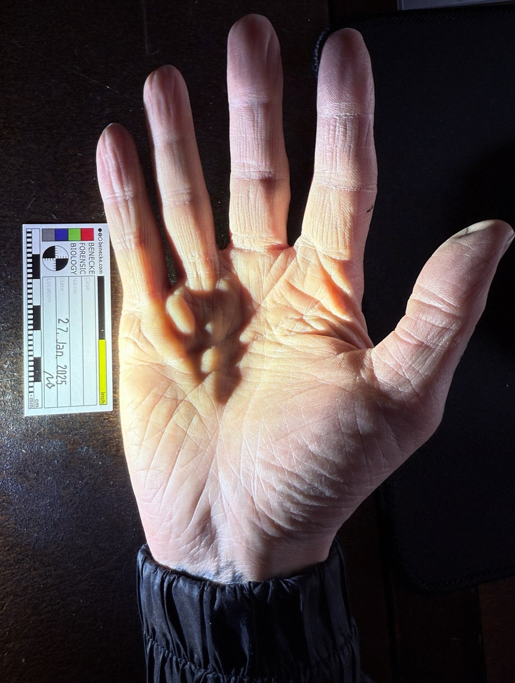
Figure 7. Palmar nodules and cords of Dupuytren disease (scale in cm). Source: Mark Benecke, via Wikimedia Commons, CC BY-SA 4.0.
Figure 7. Palmar nodules and cords of Dupuytren disease (scale in cm). Source: Mark Benecke, via Wikimedia Commons, CC BY-SA 4.0.
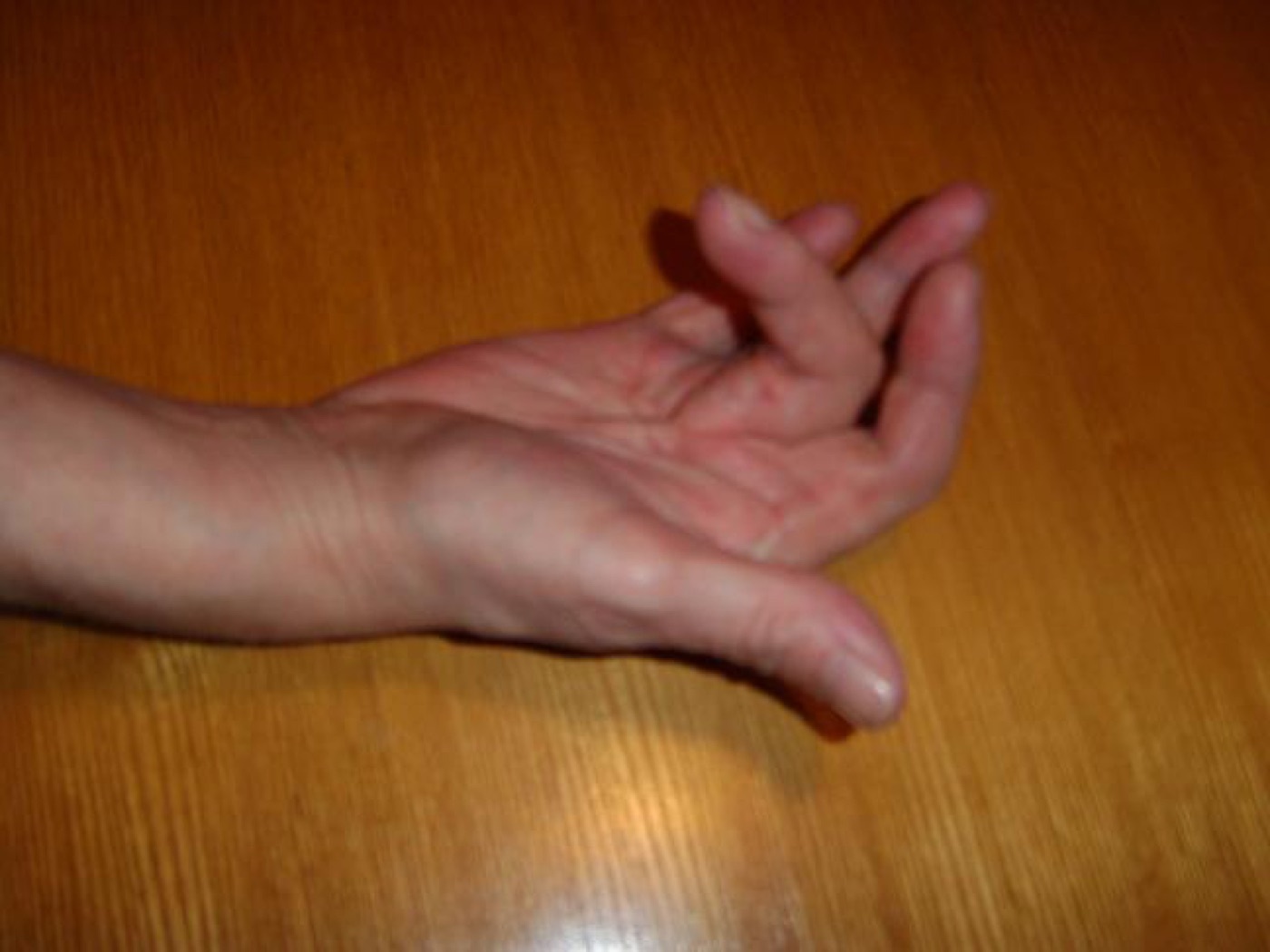
Figure 8. Established contracture: the hand cannot lie flat (a positive Hueston tabletop test). Source: Smartie77, via Wikimedia Commons, CC BY-SA 3.0.
Figure 8. Established contracture: the hand cannot lie flat (a positive Hueston tabletop test). Source: Smartie77, via Wikimedia Commons, CC BY-SA 3.0.
The bedside screen is the Hueston tabletop test: the patient cannot lay the palm and fingers flat on a table, which marks the lower end of the treatment window.[24] Severity is summarised by the Tubiana staging, which groups the combined MCP-plus-PIP flexion deformity of each ray into 45° bands (stage 0 none, 1 = 0-45°, 2 = 45-90°, 3 = 90-135°, 4 = >135°), the DIP being excluded.[25] Because the angular deformity produced per millimetre of cord shortening rises as the cord nears the joint axis, PIP contractures progress more than twice as fast as MCP contractures, and small-finger disease faster still.[26]
Part V - Treatment
Treatment is palliative, not curative; the long-term aim is to minimise both the deformity and the cumulative morbidity of repeated interventions over a lifetime.[27] Intervention is generally considered for an MCP contracture of about 30° or any significant PIP contracture (the PIP being treated earlier because it corrects less well), or for functional impairment.[28]
Non-surgical measures
These are limited. Intralesional corticosteroid softens and flattens nodules but with frequent reactivation; radiotherapy in early proliferative disease can slow progression; and post-treatment splinting has no demonstrated long-term benefit, a point on which Green’s (“less is more”) and Campbell’s (routine three-month night splinting) disagree.[29]
Minimally invasive
Percutaneous needle aponeurotomy (PNF/PNA) divides a palpable, tensionable cord through the skin with a fine needle, under local anaesthesia in the office; it is least expensive and lowest-morbidity, best suited to MCP cords with a cord that separates from the skin, and is repeatable, though recurrence is high.[30]
Collagenase Clostridium histolyticum (CCH) is injected into the cord to enzymatically lyse type I and III collagen, followed by finger manipulation one to a few days later; it corrects MCP joints far more reliably than PIP joints (in the CORD trials, contracture reduced to 0-5° in 77% of MCP versus 40% of PIP).[31] Its complications are mostly minor (skin tears in up to 40%, swelling, ecchymosis, lymphadenopathy) but include the serious flexor tendon and pulley rupture.[32] Randomised comparisons find PNA roughly equivalent to CCH at a fraction of the cost.[33]
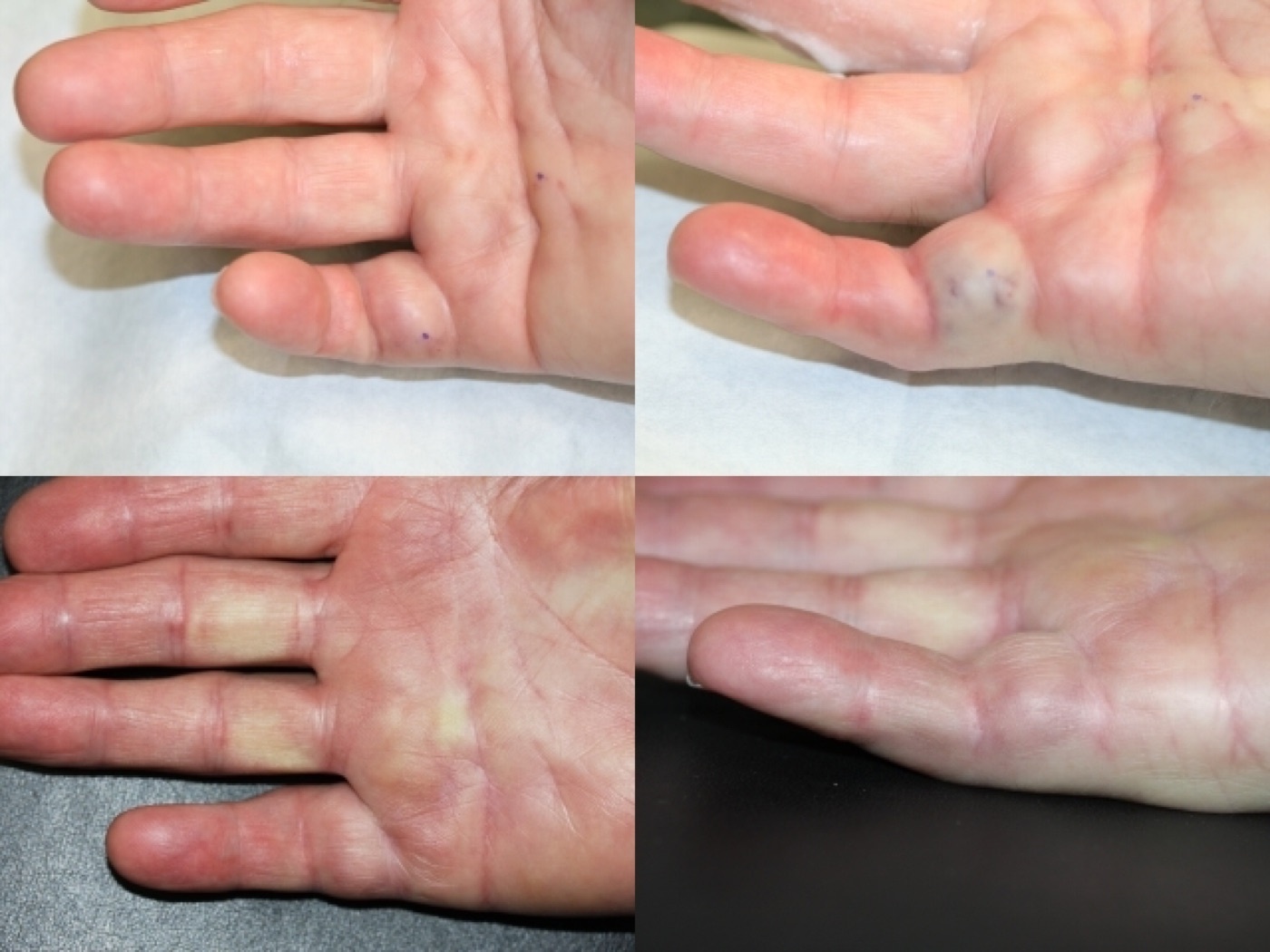
Figure 9. Collagenase (Xiaflex) treatment of a Dupuytren cord: before, next-day ecchymosis after manipulation, and after. Source: Denkler, via Wikimedia Commons (public domain).
Figure 9. Collagenase (Xiaflex) treatment of a Dupuytren cord: before, next-day ecchymosis after manipulation, and after. Source: Denkler, via Wikimedia Commons (public domain).
Surgical fasciectomy
- Limited (regional/selective) fasciectomy, which excises only the diseased fascia, is the standard operation, giving the greatest correction and the best long-term results.[34] A longitudinal incision is converted to Z-plasties (or a Bruner zig-zag is used) to lengthen the skin.
- Segmental fasciectomy removes only short cord segments, with similar outcomes.[35]
- Dermofasciectomy with a full-thickness skin graft replaces the skin and diseased soft tissue as a unit (the “firebreak” graft). It has the lowest recurrence rate and is reserved for recurrent disease, strong diathesis, or skin involvement.[36]
- The open-palm (McCash) technique leaves the transverse palmar wound to heal by secondary intention, reducing haematoma; complete (radical) fasciectomy has been abandoned for its morbidity.[37]
A fixed PIP contracture may persist after the cord is removed; capsular (volar plate/checkrein) release gives a dramatic intra-operative gain but, by Green’s account, no predictable long-term benefit and a quadrupled risk of complex regional pain syndrome; gentle correction with flexor-sheath release is safer.[38]
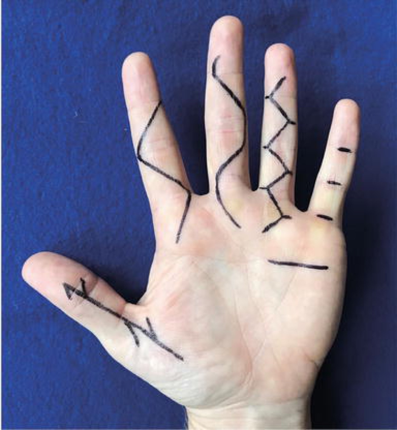
Figure 10. Incision and closure options for Dupuytren surgery - longitudinal with Z-plasty, Brunner zig-zag, V-Y, and transverse (McCash open-palm). From Johnson, Pavano & Rodner (2018), IntechOpen, CC BY 3.0.
Figure 10. Incision and closure options for Dupuytren surgery - longitudinal with Z-plasty, Brunner zig-zag, V-Y, and transverse (McCash open-palm). From Johnson, Pavano & Rodner (2018), IntechOpen, CC BY 3.0.
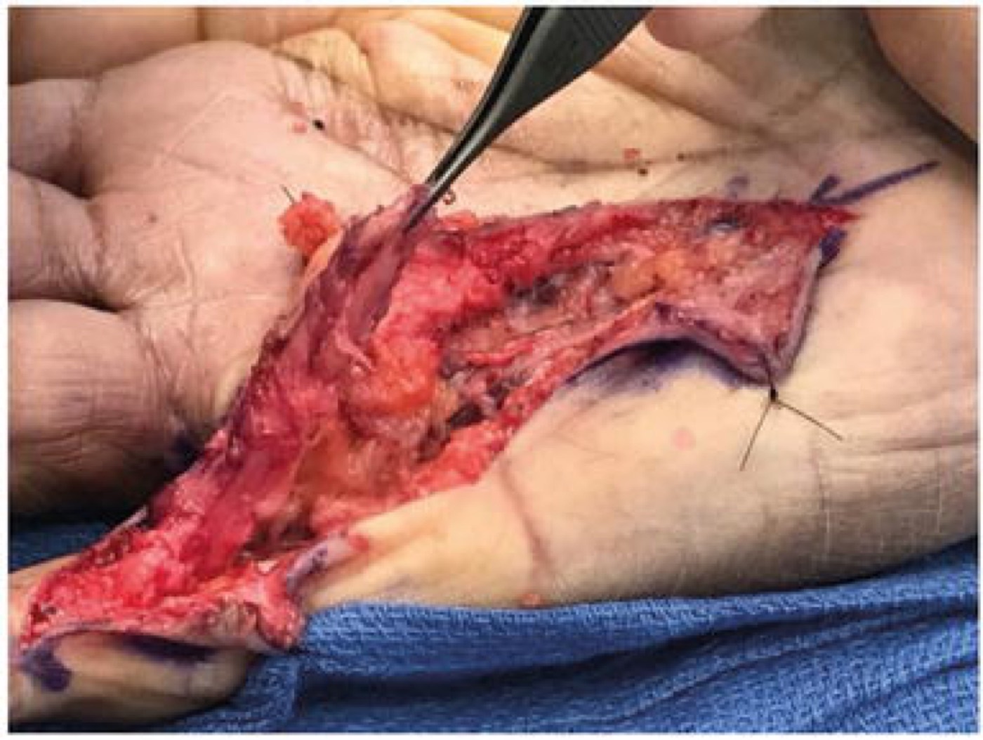
Figure 11. Limited fasciectomy: the diseased cord elevated through a Brunner incision. From Johnson, Pavano & Rodner (2018), IntechOpen, CC BY 3.0.
Figure 11. Limited fasciectomy: the diseased cord elevated through a Brunner incision. From Johnson, Pavano & Rodner (2018), IntechOpen, CC BY 3.0.
Complications and recurrence
The most significant complication of any cord procedure is digital nerve (or artery) injury, occurring in at least 3% of primary fasciectomies and over five times as often in revision surgery, the spiral cord being the chief hazard.[39] Other problems are haematoma, skin-flap necrosis, infection, stiffness, and the “flare reaction” (a prolonged painful swelling, twice as common in women, leaving permanent stiffness in about 5%); CRPS occurs in well under 1%.[40] Recurrence is the rule rather than the exception: the modalities rank consistently, with dermofasciectomy the most durable, then fasciectomy, then the minimally invasive techniques (needle aponeurotomy ≈ collagenase).[41]
References
-
Green’s Operative Hand Surgery, ch. 4 (Eaton), p. 179; Campbell’s Operative Orthopaedics, ch. 75 (Calandruccio), p. 4301.
-
Green’s OHS p. 186.
-
Green’s OHS p. 179.
-
Green’s OHS p. 193 (Table 4.2). Goyrand performed the first fasciectomy in 1834.
-
Green’s OHS p. 193.
-
Green’s OHS pp. 179, 182; Campbell’s p. 4301 (McFarlane: men 33-63 years, women 46-70).
-
Green’s OHS p. 179; Campbell’s p. 4301.
-
Green’s OHS p. 179 - a notable revision of the classic teaching that Campbell (p. 4301) still frames as “white northern European.”
-
Green’s OHS p. 180; Campbell’s p. 4301.
-
Campbell’s p. 4301; Green’s OHS pp. 180, 184.
-
Green’s OHS pp. 182-183; Campbell’s p. 4301.
-
Green’s OHS pp. 182-183; Campbell’s p. 4301.
-
Green’s OHS p. 185; Campbell’s pp. 4302-4303.
-
Green’s OHS pp. 182, 185.
-
Campbell’s p. 4303.
-
Green’s OHS p. 183; Campbell’s p. 4303. The Cleland ligament is generally spared by the disease.
-
Campbell’s p. 4303; Green’s OHS p. 190.
-
Green’s OHS p. 190; Campbell’s p. 4303.
-
Campbell’s pp. 4303-4304; Green’s OHS pp. 190-191.
-
Campbell’s pp. 4303-4304; Green’s OHS p. 192.
-
Green’s OHS p. 184; Campbell’s p. 4301.
-
Campbell’s pp. 4301-4302; Green’s OHS p. 184.
-
Green’s OHS p. 184.
-
Green’s OHS pp. 192-193.
-
Green’s OHS p. 185.
-
Green’s OHS p. 185.
-
Green’s OHS p. 193; Campbell’s p. 4305.
-
Campbell’s pp. 4309-4310 (PIP ≥15°, MCP ≥30°); Green’s OHS p. 193 frames this as a “window of opportunity” with the best correction obtained at 20-40° of bend rather than a fixed trigger.
-
Green’s OHS pp. 196, 199; Campbell’s pp. 4305-4306, 4312.
-
Green’s OHS pp. 193, 199-200; Campbell’s p. 4307.
-
Campbell’s pp. 4305-4306; Green’s OHS pp. 193-194.
-
Campbell’s pp. 4306-4307; Green’s OHS p. 193.
-
Campbell’s p. 4308 (Strömberg: 79% vs 76% straight fingers, CCH ~3× the cost); Green’s OHS treats the two as having similar outcomes (p. 193).
-
Campbell’s p. 4309; Green’s OHS p. 194.
-
Green’s OHS pp. 194-195.
-
Green’s OHS pp. 196-197; Campbell’s p. 4311.
-
Green’s OHS pp. 192, 196; Campbell’s pp. 4311, 4316.
-
Green’s OHS p. 198; Campbell’s p. 4309 - PIP contractures >60° correct only to about half regardless of capsulotomy.
-
Green’s OHS p. 193; Campbell’s p. 4311.
-
Green’s OHS p. 193; Campbell’s p. 4304.
-
Green’s OHS p. 193 (Table 4.3: dermofasciectomy ~2%/year, local fasciectomy 5-10%/year, PNF and CCH 10-20%/year); Campbell’s pp. 4307-4317 (cumulative figures - ~50% after fasciectomy with 15% reoperation). Green’s cautions that “recurrence” has no standard definition and is better expressed as a rate.
-
Green’s OHS pp. 179, 186.
-
Green’s OHS pp. 182-185.
-
Green’s OHS p. 183; Campbell’s p. 4303.
-
Campbell’s pp. 4303-4304; Green’s OHS pp. 190-191.
-
Campbell’s p. 4301; Green’s OHS p. 180.
-
Green’s OHS p. 179; Campbell’s p. 4301.
-
Green’s OHS pp. 185, 192-193.
-
Campbell’s pp. 4309-4310; Green’s OHS p. 193.
-
Green’s OHS pp. 193-197; Campbell’s pp. 4305-4311.
-
Green’s OHS p. 193; Campbell’s p. 4311.