Contents
- Introduction and scope
- Part I - The intervertebral disc and the degenerative cascade
- Part II - Cervical degenerative disease: spondylosis and radiculopathy
- Part III - Cervical spondylotic myelopathy
- Part IV - Lumbar disc herniation and sciatica
- Part V - Lumbar spinal stenosis
- Part VI - Spondylolisthesis
- Part VII - Spinal fusion and instrumentation
- Part VIII - The spine in ankylosing spondylitis
- Bulgarian terminology (Боев / Boychev tradition) - glossary
- Image attributions
- References
Introduction and scope
Degenerative disease of the spine (disc degeneration, cervical spondylosis with radiculopathy or myelopathy, lumbar disc herniation, spinal stenosis and spondylolisthesis) is the commonest reason adults consult a spine surgeon, and the rigid spine of ankylosing spondylitis is its dangerous counterpoint. The examiner expects the degenerative cascade of the intervertebral disc; the cervical syndromes (the root-level patterns of radiculopathy, and the upper-motor-neuron picture of cervical spondylotic myelopathy); the lumbar syndromes (disc herniation and sciatica with the traversing-versus-exiting-root rule, the surgical emergency of cauda equina, neurogenic versus vascular claudication in stenosis, and the two spondylolistheses); the principles of decompression and fusion; and the ankylosing-spondylitis spine, with its bamboo spine, brittle transverse fracture and fixed kyphosis to be corrected.
Two ideas run through the topic. First, the level of the lesion is read from the neurology: a compressed cervical or lumbar root produces a stereotyped motor, sensory and reflex pattern, and knowing which root a herniation strikes (the traversing root for a paracentral disc, the exiting root for a foraminal one) localises the disease before imaging confirms it. Second, most degenerative spine disease is benign and self-limiting. The great majority of disc herniations and back-pain episodes resolve without surgery, so the decisions that matter are recognising the few that need urgent surgery (cauda equina, progressive myelopathy, the unstable ankylosed-spine fracture) and selecting the right operation for the rest.
Part I - The intervertebral disc and the degenerative cascade
The disc has three parts: the gelatinous nucleus pulposus (an aggrecan-and-water matrix, about 80% water in the young adult, held in a type-II collagen network), the surrounding annulus fibrosus (some twenty concentric lamellae of type-I collagen), and the cartilaginous endplates.[1] It is one of the largest avascular structures in the body, nourished by diffusion across the endplate, which makes it vulnerable to age-related failure.[2] In the healthy disc only the outer annulus is innervated (by the sinuvertebral nerve).[3]
With degeneration, the nucleus loses proteoglycan and water, type-I collagen replaces type II, matrix metalloproteinases and inflammatory cytokines predominate, and sensory nerves and vessels grow into the normally aneural inner disc; this ingrowth is the substrate of discogenic pain.[4] The Kirkaldy-Willis degenerative cascade describes three overlapping phases of a motion segment: dysfunction (annular tears, facet synovitis, ~15-45 years), instability (disc resorption, facet laxity, segmental subluxation, ~35-70 years) and restabilization (disc-height loss, osteophytes, autofusion, >60 years). Disc herniation is a complication of the early phases and stenosis of the later ones.[5] Genetics is the dominant determinant of disc degeneration (roughly half to three-quarters of the variance in twin studies).[6] Degenerative findings are very common in asymptomatic people: a major lumbar MRI finding in about a third of the pain-free, and over 70% of people harbour an asymptomatic disc prolapse. Imaging must therefore be correlated with symptoms, and provocative discography is now recommended against for diagnosing discogenic pain.[7]
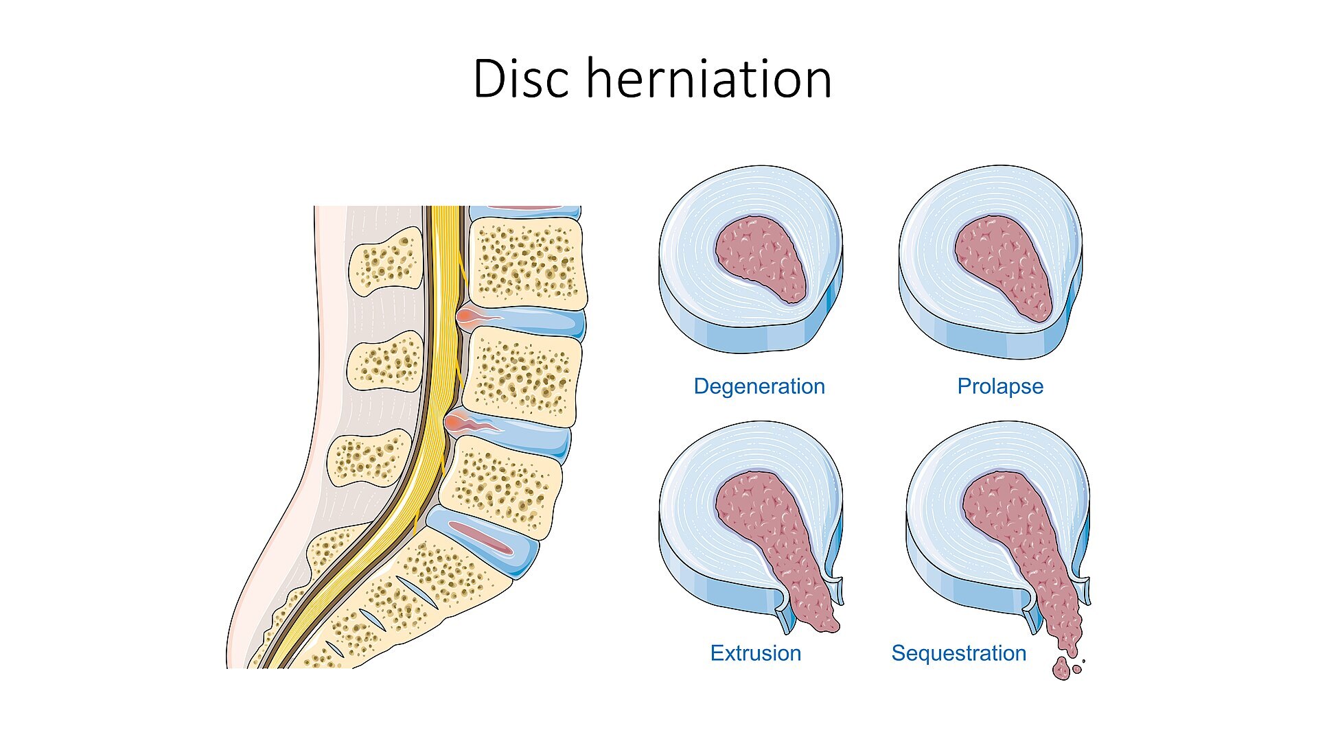
Disc herniation grades. Sagittal view (left) and axial panels showing degeneration, prolapse (protrusion), extrusion and sequestration. Laboratoires Servier, SMART Servier Medical Art, Wikimedia Commons, CC BY-SA 3.0.
Disc herniation grades. Sagittal view (left) and axial panels showing degeneration, prolapse (protrusion), extrusion and sequestration. Laboratoires Servier, SMART Servier Medical Art, Wikimedia Commons, CC BY-SA 3.0.
Part II - Cervical degenerative disease: spondylosis and radiculopathy
Cervical spondylosis is age-related degeneration of the discs and facet joints with osteophyte (“spondylotic bar”) formation, uncovertebral (joint of Luschka) hypertrophy and ligamentum flavum buckling; the most affected levels are C5-C6 then C6-C7.[8] A soft disc herniation (a younger patient) is distinguished from a hard disc / osteophytic bar (older). It produces overlapping syndromes of axial neck pain, radiculopathy and myelopathy.[9]
Cervical radiculopathy is root compression by a soft disc or a foraminal osteophyte. The root-level syndromes are: C5 (deltoid weakness, lateral-arm sensory, biceps reflex), C6 (biceps and wrist extensors, thumb, brachioradialis reflex), C7 (triceps and wrist flexors, middle finger, the most commonly involved root) and C8 (intrinsics and finger flexors, ulnar border, no reflex).[10] The cervical root exits above its same-numbered pedicle, so a C6-C7 disc compresses the C7 root.[11] The provocative tests are the Spurling sign (extension, rotation and axial compression toward the painful side; about 95% sensitive and 94% specific), the shoulder-abduction relief sign, and Lhermitte’s sign.[12] The natural history is favourable, since most resolve with nonoperative care. When disease is intractable or progressive, surgery is by anterior cervical discectomy and fusion (ACDF), posterior foraminotomy for a lateral soft disc, or cervical disc arthroplasty. The recognised long-term cost of fusion is adjacent-segment disease (about a 25% incidence within ten years).[13]
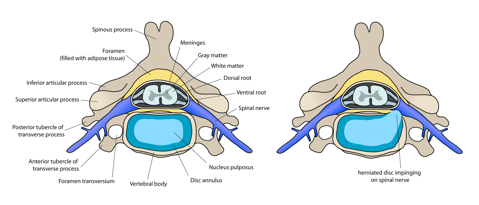
Cervical disc and the radiculopathy mechanism. Axial diagram: normal disc anatomy (left) and a herniated disc impinging on the spinal nerve root (right). debivort, Wikimedia Commons, CC BY-SA 3.0.
Cervical disc and the radiculopathy mechanism. Axial diagram: normal disc anatomy (left) and a herniated disc impinging on the spinal nerve root (right). debivort, Wikimedia Commons, CC BY-SA 3.0.
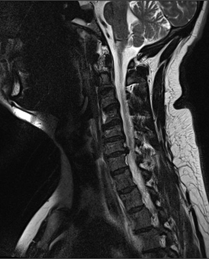
Cervical disc herniation. Sagittal T2 MRI showing a C6-C7 disc herniation indenting the cord/thecal sac. Wikimedia Commons, CC BY-SA 3.0.
Cervical disc herniation. Sagittal T2 MRI showing a C6-C7 disc herniation indenting the cord/thecal sac. Wikimedia Commons, CC BY-SA 3.0.
Part III - Cervical spondylotic myelopathy
Cervical spondylotic myelopathy (CSM) is the commonest cause of spinal-cord dysfunction in adults over 55. It arises from static canal narrowing (a normal mid-sagittal canal is about 17 mm; stenosis below 13 mm, with a Pavlov/Torg ratio ≤ 0.8 indicating a developmentally narrow canal) compounded by dynamic compression during flexion and extension.[14] The clinical picture is insidious: gait disturbance (the earliest sign), loss of hand dexterity and weakness, with upper-motor-neuron signs below the level (hyperreflexia, the Hoffmann sign, the inverted radial reflex, Babinski response, clonus and Lhermitte’s sign) and a “myelopathy hand” with a positive finger-escape sign.[15] Disability is graded by the Nurick scale (0-5) and the modified JOA score (out of 17), and a T2 cord signal change (myelomalacia) on MRI predicts poorer recovery.[16] CSM tends to deteriorate stepwise and is a surgical disorder. Decompression is anterior (ACDF or corpectomy, for one-to-two-level disease, a kyphotic spine, or OPLL) or posterior (laminectomy with fusion, or laminoplasty, for multilevel disease but only when sagittal alignment is neutral or lordotic, since a posterior approach fails in a kyphotic spine).[17] Ossification of the posterior longitudinal ligament (OPLL) is an important cause of myelopathy and complicates anterior surgery (the dura may be ossified).[18]
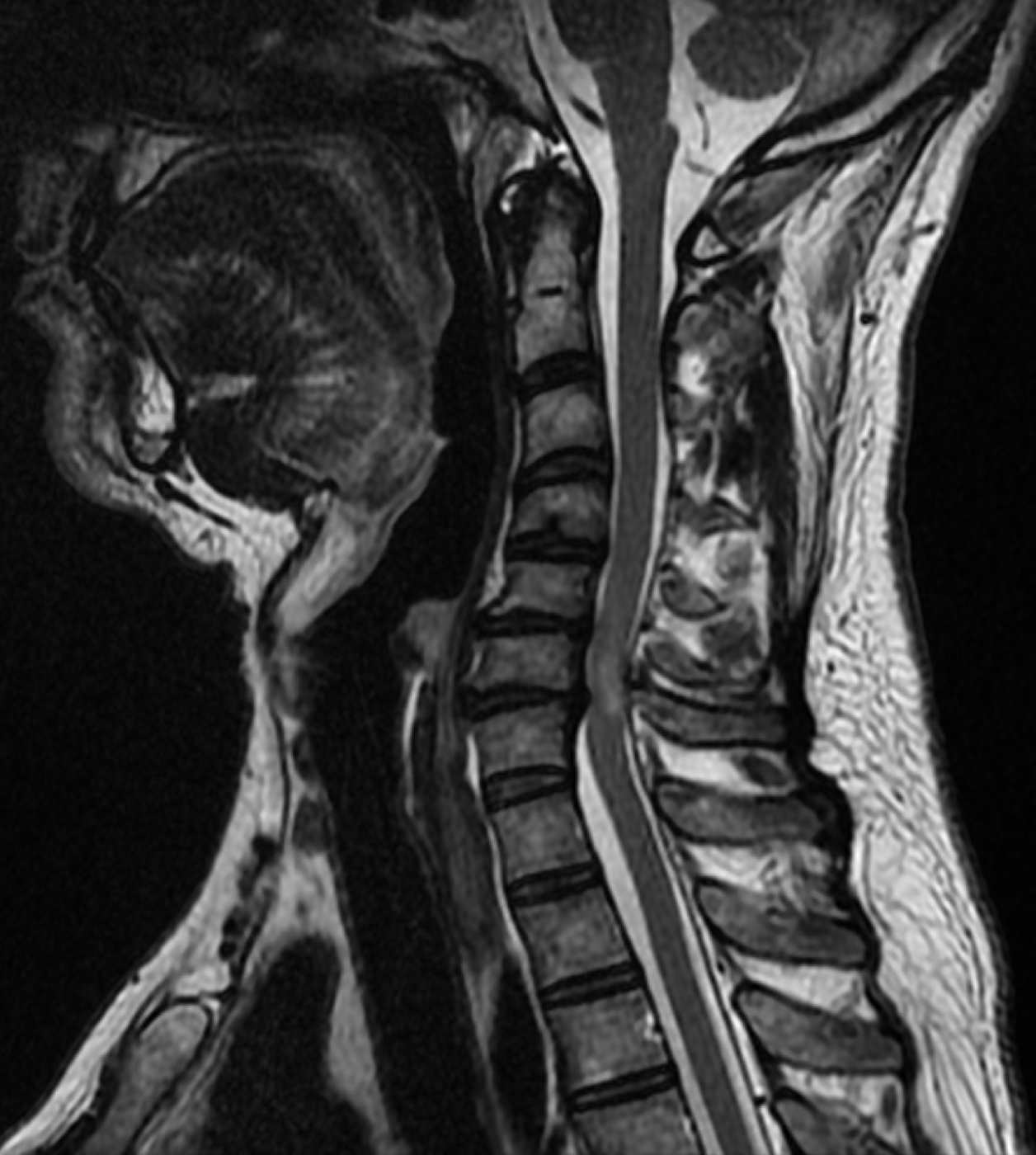
Cervical spondylotic myelopathy. Sagittal T2 cervical MRI with cord compression at a lower-cervical level. Jmarchn, Wikimedia Commons, CC BY-SA 3.0.
Cervical spondylotic myelopathy. Sagittal T2 cervical MRI with cord compression at a lower-cervical level. Jmarchn, Wikimedia Commons, CC BY-SA 3.0.
Part IV - Lumbar disc herniation and sciatica
A herniation is graded by morphology (bulge, protrusion, extrusion and sequestration (free fragment)), and, because the posterior longitudinal ligament is deficient laterally, the commonest site is posterolateral (paracentral); over 95% occur at L4-L5 or L5-S1.[19] The cardinal rule of root involvement: a paracentral herniation compresses the traversing (lower) root (an L4-L5 paracentral disc strikes L5), while a foraminal/far-lateral herniation compresses the exiting root (an L4-L5 far-lateral disc strikes L4).[20] The lumbar root syndromes are: L4 (quadriceps, knee jerk, medial leg), L5 (extensor hallucis longus and foot dorsiflexion, dorsum of the foot, no reliable reflex) and S1 (plantarflexion, ankle jerk, lateral and plantar foot).[21]
The diagnosis rests on the neurology and the tension signs: the straight-leg-raise (Lasègue) test (positive between about 35° and 70°), the highly specific crossed straight-leg raise (considered pathognomonic of a herniation), and the femoral stretch test for the upper lumbar roots.[22] Radicular pain is not purely mechanical: compression of a non-inflamed root gives painless weakness, and pain requires a chemical/inflammatory response in which TNF-α from the nucleus pulposus is a key mediator.[23] Cauda equina syndrome (saddle anaesthesia, urinary retention or incontinence, bilateral sciatica and progressive deficit) is a surgical emergency requiring urgent decompression.[24] Otherwise the natural history is benign (most herniations resolve; the Weber and SPORT trials show surgical and nonoperative outcomes converge by years, though surgery relieves leg pain faster), and the operation for a persistent radiculopathy is microdiscectomy.[25]
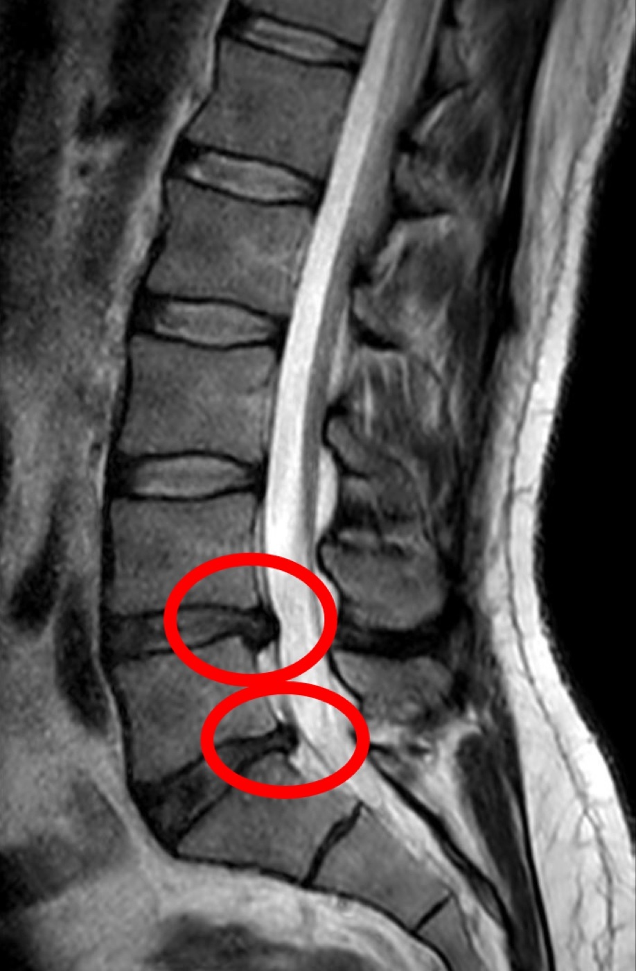
Lumbar disc herniation. Sagittal T2 MRI: a left paramedian L4-L5 herniation (circled) compressing the traversing L5 root. Miguel Tremblay, Wikimedia Commons, CC0.
Lumbar disc herniation. Sagittal T2 MRI: a left paramedian L4-L5 herniation (circled) compressing the traversing L5 root. Miguel Tremblay, Wikimedia Commons, CC0.
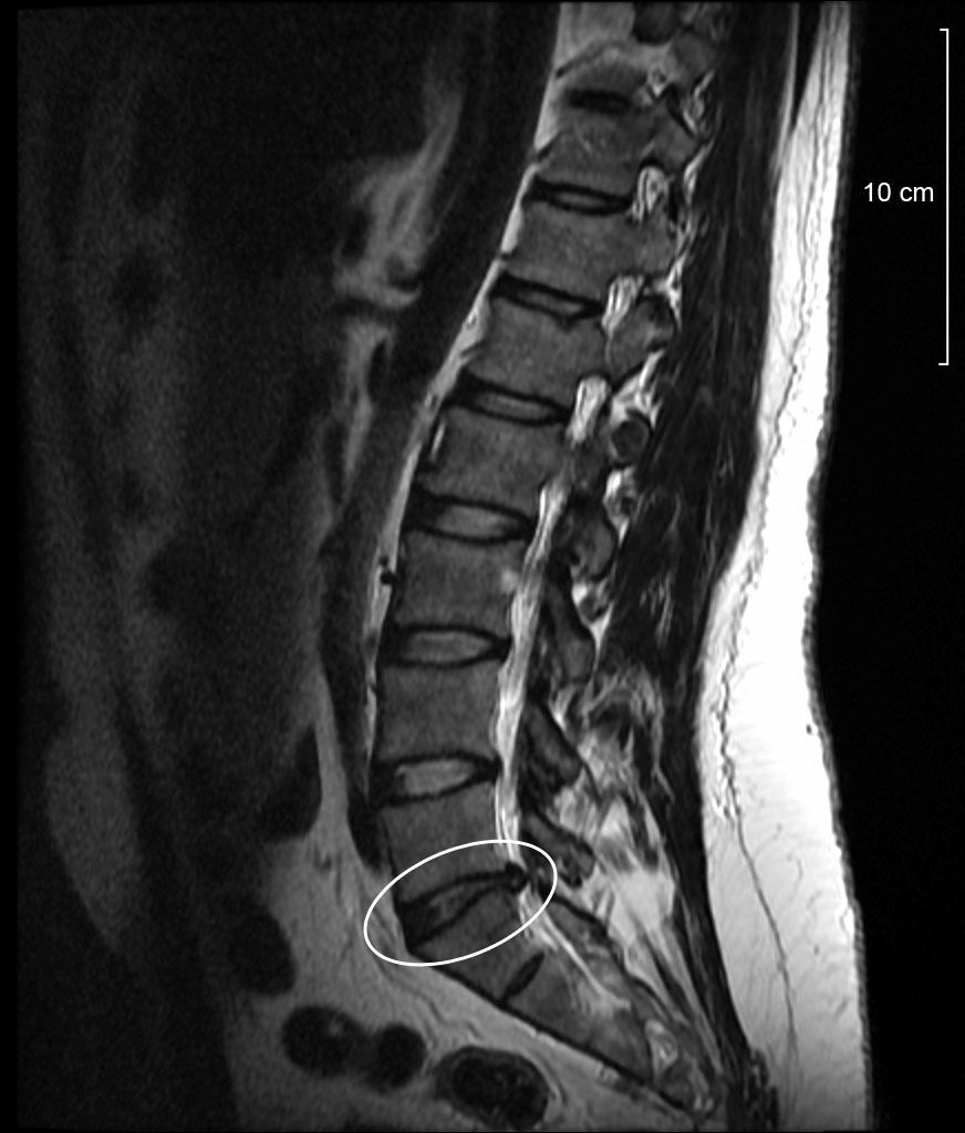
Lumbar disc protrusion. Sagittal T2 MRI with a focal lower-lumbar disc protrusion (circled) compressing the nerve root. Wikimedia Commons, public domain.
Lumbar disc protrusion. Sagittal T2 MRI with a focal lower-lumbar disc protrusion (circled) compressing the nerve root. Wikimedia Commons, public domain.
Part V - Lumbar spinal stenosis
Lumbar spinal stenosis is narrowing of the central canal, lateral recess or foramen, congenital (short pedicles, symptomatic in the fourth decade) or, far more often, acquired/degenerative (a disc bulge in front plus facet hypertrophy and infolded ligamentum flavum behind), typically presenting in the sixties at L4-L5.[26] Its hallmark is neurogenic claudication: buttock and leg pain on standing and walking, relieved by flexion. The distinction from vascular claudication is high-yield. The stenotic patient leans on a shopping cart (the “shopping-cart sign”), prefers walking uphill (flexed, painless) to downhill, tolerates a bicycle, and has normal pulses, whereas vascular claudication is posture-independent, occurs at a fixed walking distance, is worse uphill, and comes with absent pulses.[27] MRI is the modality of choice (with the usual caveat that asymptomatic stenosis is common in the elderly).[28] About half of mild-to-moderate cases do well without surgery. When conservative care fails, decompressive laminectomy is the gold standard (preserving at least half of each facet to avoid iatrogenic instability), and fusion is added when there is instability, a degenerative scoliosis or resection of more than half the facets; the SPORT trial confirmed the surgical benefit.[29]
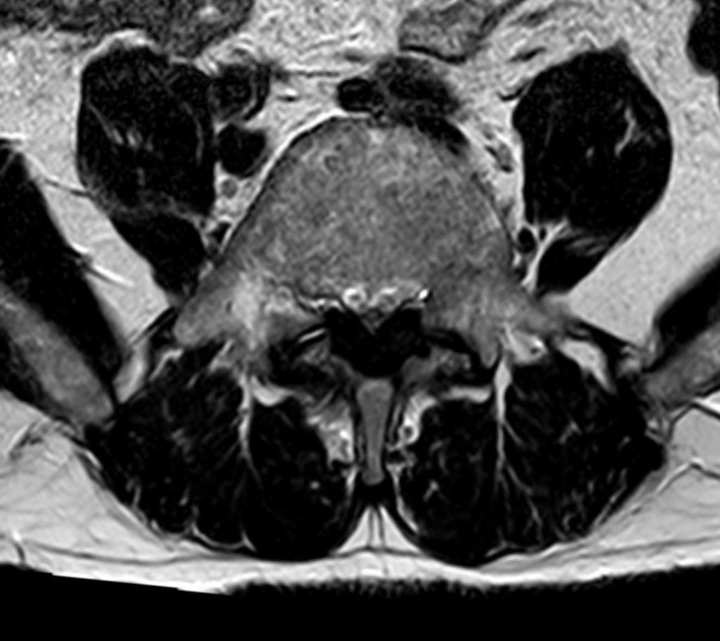
Lumbar central-canal stenosis. Axial T2 MRI: ligamentum-flavum hypertrophy narrowing the central canal. Hellerhoff, Wikimedia Commons, CC BY-SA 3.0.
Lumbar central-canal stenosis. Axial T2 MRI: ligamentum-flavum hypertrophy narrowing the central canal. Hellerhoff, Wikimedia Commons, CC BY-SA 3.0.
Part VI - Spondylolisthesis
Degenerative spondylolisthesis is a forward slip with an intact neural arch, commonest at L4-L5, four-to-five times more frequent in (older) women, arising from facet and disc degeneration. It presents like stenosis (neurogenic claudication, often an L5 radiculopathy) and is best seen on a standing lateral radiograph, being frequently missed on supine MRI. Its landmark evidence is Herkowitz’s trial showing decompression plus fusion is far superior to decompression alone (96% versus 44% satisfactory).[30] Isthmic spondylolisthesis is due to a pars interarticularis defect (spondylolysis), commonest at L5-S1, present in 4-6% of the population; root pain arises in the lateral recess (the Gill lesion), and cauda equina is rare because the canal is effectively widened.[31] Both are graded by the Meyerding system (I 0-25%, II 25-50%, III 50-75%, IV 75-100%, V = spondyloptosis).[32] Treatment is conservative for most; surgery (decompression with fusion, occasionally direct pars repair in the young) is reserved for refractory pain, progressive slip or neurological deficit, and complete reduction of a high-grade slip carries a high neurological risk.[33]
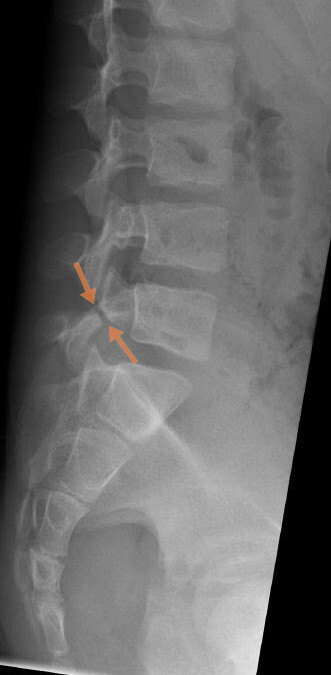
Spondylolysis. Lateral lumbar radiograph: a pars interarticularis defect (arrows). Kinderradiologie Olgahospital, Klinikum Stuttgart, Wikimedia Commons, CC BY-SA 4.0.
Spondylolysis. Lateral lumbar radiograph: a pars interarticularis defect (arrows). Kinderradiologie Olgahospital, Klinikum Stuttgart, Wikimedia Commons, CC BY-SA 4.0.
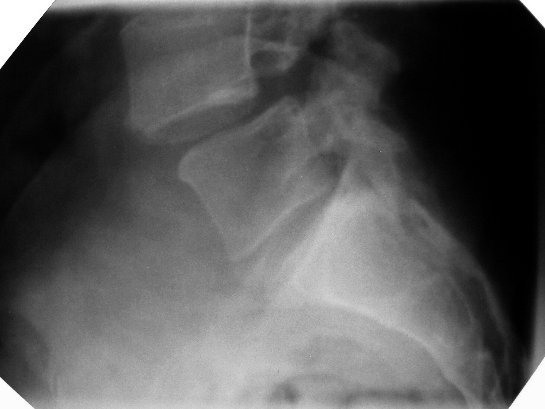
Spondylolisthesis. Lateral radiograph: a high-grade forward slip of L5 on S1. Wikimedia Commons, CC BY-SA 3.0.
Spondylolisthesis. Lateral radiograph: a high-grade forward slip of L5 on S1. Wikimedia Commons, CC BY-SA 3.0.
Part VII - Spinal fusion and instrumentation
Fusion is indicated for instability, spondylolisthesis, deformity or after decompression that removes the stabilising structures. It may be achieved posterolaterally (intertransverse fusion) or by interbody techniques (ALIF, PLIF, TLIF, and the lateral XLIF), usually supplemented by pedicle-screw instrumentation, which raises the fusion rate though its effect on the clinical result is debated.[34] The lumbar vertebral body carries about 80% of the load, the rationale for interbody support.[35] The recognised long-term price of fusion is adjacent-segment degeneration (symptomatic in a minority of fused patients), and disc arthroplasty has been introduced in both the cervical and lumbar spine to preserve motion and, in principle, reduce it.[36]
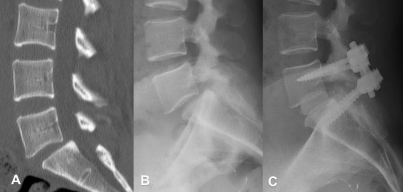
Spondylolisthesis and its fusion. (A) Sagittal CT of the slip, (B) standing lateral radiograph, (C) post-operative pedicle-screw fixation and fusion. Donnally, StatPearls / Wikimedia Commons, CC BY 4.0.
Spondylolisthesis and its fusion. (A) Sagittal CT of the slip, (B) standing lateral radiograph, (C) post-operative pedicle-screw fixation and fusion. Donnally, StatPearls / Wikimedia Commons, CC BY 4.0.
Part VIII - The spine in ankylosing spondylitis
Ankylosing spondylitis (AS) is a seronegative, HLA-B27-associated spondyloarthropathy (about 90% of patients are B27-positive) that ascends from the sacroiliac joints through the spine.[37] Enthesitis at the discovertebral junction produces squaring of the vertebral bodies and syndesmophytes that bridge the segments while the facets ankylose, giving the radiographic “bamboo spine” and leaving a spine that is rigid, osteoporotic and brittle.[38] Clinically there is inflammatory back pain, progressive loss of spinal mobility (a reduced Schober test, chest expansion under about 2.5 cm) and a fixed kyphotic deformity with loss of horizontal gaze.[39]
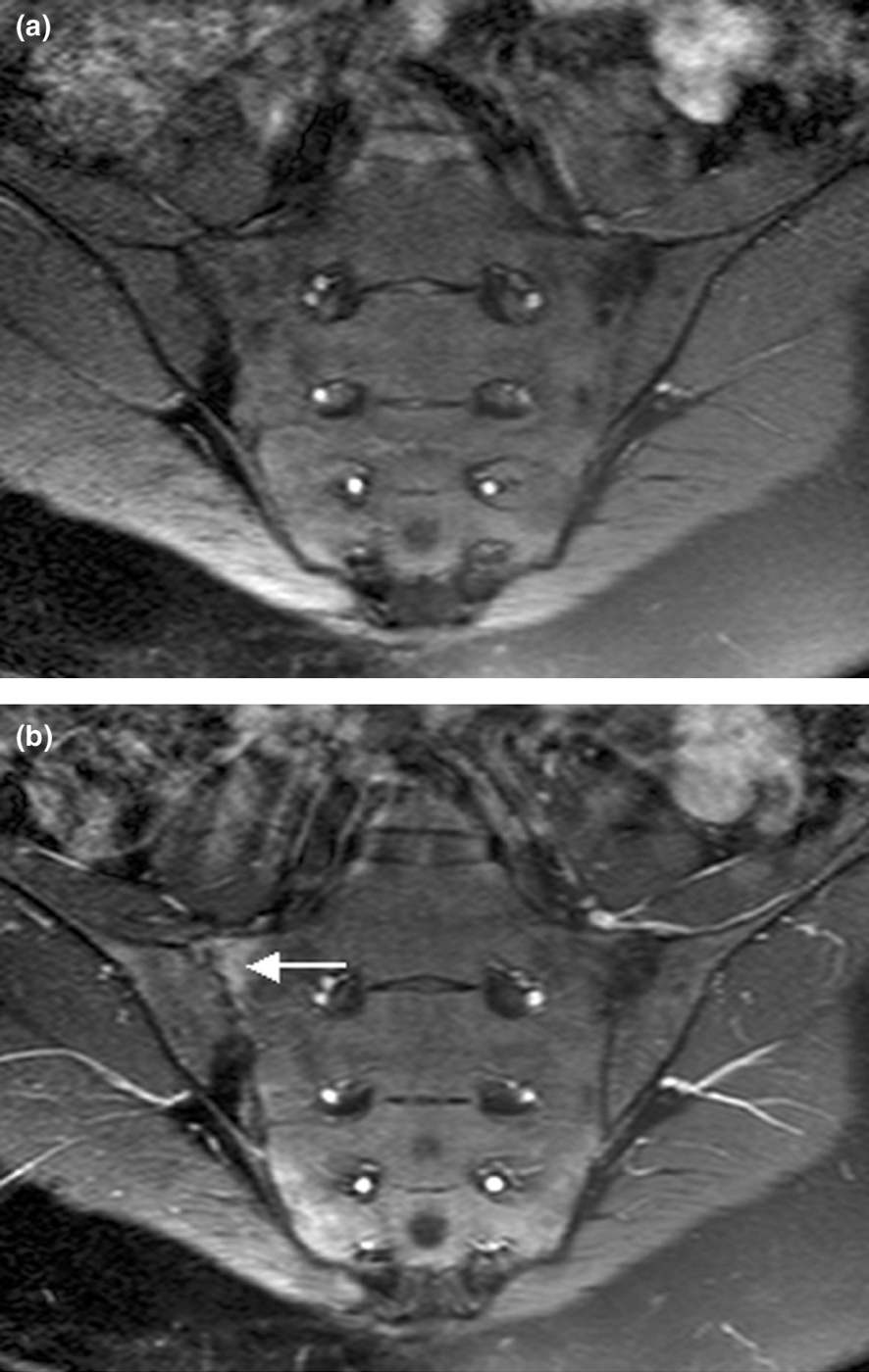
Active sacroiliitis. Semi-coronal T1 MRI of the sacroiliac joints, before (a) and after (b) contrast, with enhancement at the inflamed joint (arrow). McQueen et al., Arthritis Res Ther 2006;8:207, CC BY 2.0.
Active sacroiliitis. Semi-coronal T1 MRI of the sacroiliac joints, before (a) and after (b) contrast, with enhancement at the inflamed joint (arrow). McQueen et al., Arthritis Res Ther 2006;8:207, CC BY 2.0.
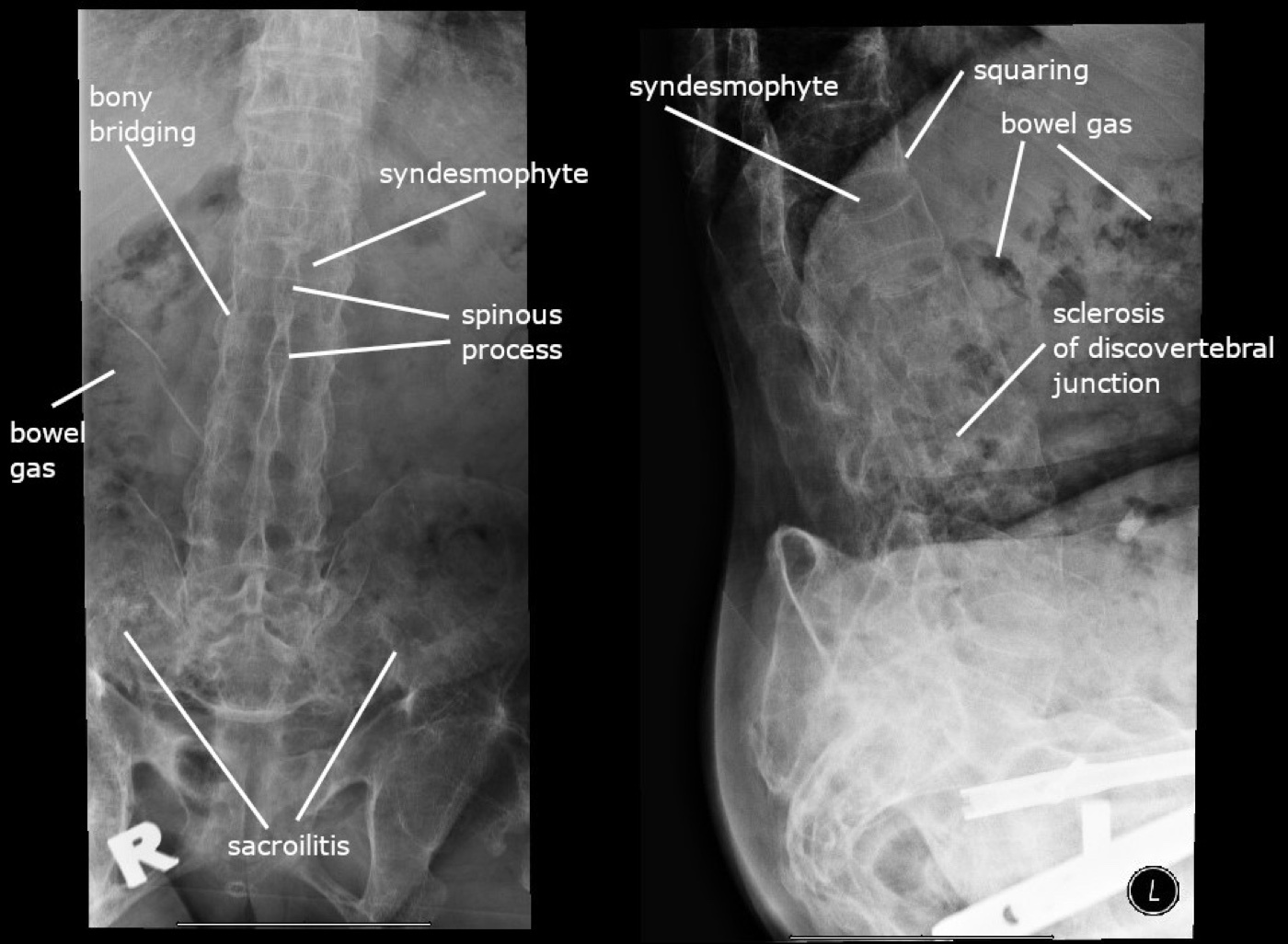
The radiographic signs of the ankylosing-spondylitis spine. Annotated lumbosacral radiographs: syndesmophytes, bony bridging, squaring of the vertebral bodies, discovertebral sclerosis and sacroiliitis. Wikimedia Commons, CC BY-SA 4.0.
The radiographic signs of the ankylosing-spondylitis spine. Annotated lumbosacral radiographs: syndesmophytes, bony bridging, squaring of the vertebral bodies, discovertebral sclerosis and sacroiliitis. Wikimedia Commons, CC BY-SA 4.0.
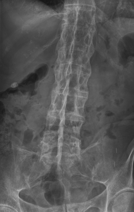
“Bamboo spine.” Radiograph showing continuous marginal syndesmophytes bridging adjacent vertebrae in advanced ankylosing spondylitis. Stevenfruitsmaak, Wikimedia Commons, CC BY-SA 3.0.
“Bamboo spine.” Radiograph showing continuous marginal syndesmophytes bridging adjacent vertebrae in advanced ankylosing spondylitis. Stevenfruitsmaak, Wikimedia Commons, CC BY-SA 3.0.
The high-stakes complication is spinal fracture: the ankylosed spine behaves like a long bone and fractures with trivial trauma, typically a transverse, three-column, highly unstable injury (the “chalk-stick” or “carrot-stick” fracture), with about 60-75% at the cervical or cervicothoracic region.[40] These fractures carry a high rate of neurological injury and epidural haematoma and are easily missed on plain films (CT and MRI are needed). The cardinal management rule is to immobilise the patient in their pre-injury position, not flat, followed by stabilisation, usually with a long posterior instrumented construct (anterior-only constructs are prone to failure).[41]
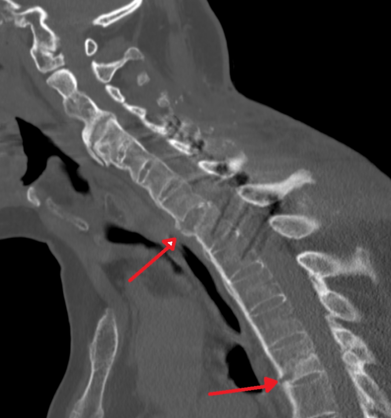
The unstable fracture of the ankylosed spine. Sagittal CT showing two transverse three-column fractures (arrows, C7 and T5) through a bamboo spine. James Heilman MD, Wikimedia Commons, CC BY-SA 4.0.
To tie the topic together: degenerative spine disease declares its level through the neurology, in the cervical root and myelopathy syndromes, the lumbar root syndromes with the traversing-versus-exiting-root rule, and neurogenic claudication distinguished from vascular by its posture-dependence. Most of it is benign and treated conservatively; the surgical priorities are the emergencies (cauda equina, progressive myelopathy) and choosing the right decompression and fusion. The ankylosed spine of ankylosing spondylitis is the opposite problem, a brittle column that fractures unstably with trivial trauma and must never be straightened to lie flat.
Bulgarian terminology (Боев / Boychev tradition) - glossary
The following Bulgarian equivalents bridge the international literature with the terminology of the Bulgarian school of orthopaedics (in the tradition of Бойчо Бойчев / Boycho Boychev) used in the state examination.
| English term | Bulgarian term (Cyrillic) | Transliteration |
|---|---|---|
| Intervertebral disc | Междупрешленен диск | Mezhdupreshlenen disk |
| Nucleus pulposus / annulus fibrosus | Пулпозно ядро / фиброзен пръстен | Pulpozno yadro / fibrozen prasten |
| Disc herniation | Дискова херния | Diskova herniya |
| Sciatica | Ишиас (лумбоишиалгия) | Ishias (lumboishialgiya) |
| Cervical spondylosis | Цервикална спондилоза | Tservikalna spondiloza |
| Radiculopathy | Радикулопатия | Radikulopatiya |
| Cervical spondylotic myelopathy | Цервикална спондилотична миелопатия | Tservikalna spondilotichna mielopatiya |
| Spinal stenosis | Стеноза на гръбначния канал | Stenoza na grabnachniya kanal |
| Neurogenic claudication | Неврогенно клаудикацио | Nevrogenno klaudikatsio |
| Spondylolysis | Спондилолиза | Spondiloliza |
| Spondylolisthesis | Спондилолистеза | Spondilolisteza |
| Pars interarticularis | Pars interarticularis (междуставна част) | Pars interarticularis |
| Cauda equina syndrome | Синдром на cauda equina (конска опашка) | Sindrom na cauda equina |
| Straight-leg-raise (Lasègue) | Тест на Lasègue (повдигане на изпънат крак) | Test na Lasègue |
| Laminectomy / decompression | Ламинектомия / декомпресия | Laminektomiya / dekompresiya |
| Spinal fusion (arthrodesis) | Спинална фузия (артродеза) | Spinalna fuziya (artrodeza) |
| Pedicle screw | Педикуларен винт | Pedikularen vint |
| ACDF (anterior cervical discectomy & fusion) | Предна цервикална дискектомия и фузия | Predna tservikalna diskektomiya i fuziya |
| Osteophyte | Остеофит | Osteofit |
| Ankylosing spondylitis | Анкилозиращ спондилит (болест на Бехтерев) | Ankilozirasht spondilit (bolest na Behterev) |
| Sacroiliitis | Сакроилиит | Sakroiliit |
| Syndesmophyte / bamboo spine | Синдесмофит / “бамбуков гръбнак” | Sindesmofit / “bambukov grabnak” |
| Kyphotic deformity | Кифотична деформация | Kifotichna deformatsiya |
| Pedicle subtraction osteotomy | Педикуларно-субтракционна остеотомия | Pedikularno-subtraktsionna osteotomiya |
Image attributions
(Figure attributions and licences are listed in the figure MANIFEST and inserted with each image. All images are openly licensed [CC0 / Public Domain / CC BY / CC BY-SA] or used under their stated terms; any non-commercial [NC] item is flagged as such and must not be used in a commercial product.)
References
-
Rothman-Simeone, The Spine (6th ed.), p. 121-124.
-
Rothman-Simeone, p. 122.
-
Rothman-Simeone, p. 122, p. 870.
-
Rothman-Simeone, p. 124-126.
-
Rothman-Simeone, p. 870, p. 1088; Campbell’s Operative Orthopaedics, p. 1952-1953.
-
Rothman-Simeone, p. 127, p. 872.
-
Rothman-Simeone, p. 875, p. 128, p. 879.
-
Rothman-Simeone, p. 708.
-
Rothman-Simeone, p. 708; Campbell’s, p. 1919.
-
Rothman-Simeone, p. 715; Campbell’s, p. 1917-1918.
-
Campbell’s, p. 1909.
-
Rothman-Simeone, p. 714; Campbell’s, p. 1918.
-
Rothman-Simeone, p. 773, p. 794.
-
Rothman-Simeone, p. 711, p. 788.
-
Rothman-Simeone, p. 716-717.
-
Rothman-Simeone, p. 787-788.
-
Rothman-Simeone, p. 788-789, p. 795-796.
-
Rothman-Simeone, p. 790, p. 792.
-
Rothman-Simeone, p. 911, p. 914; Campbell’s, p. 1974.
-
Rothman-Simeone, p. 911-912; Campbell’s, p. 1952.
-
Campbell’s, p. 1973-1974.
-
Rothman-Simeone, p. 917-918.
-
Rothman-Simeone, p. 157-161, p. 913.
-
Rothman-Simeone, p. 931-932.
-
Rothman-Simeone, p. 921-923; Campbell’s, p. 1977.
-
Rothman-Simeone, p. 1088-1092.
-
Rothman-Simeone, p. 1092, p. 1126-1127.
-
Rothman-Simeone, p. 1094, p. 1102.
-
Rothman-Simeone, p. 1107-1112.
-
Rothman-Simeone, p. 1125-1130.
-
Rothman-Simeone, p. 1278-1279.
-
Rothman-Simeone, p. 1284-1285.
-
Rothman-Simeone, p. 1280-1284.
-
Rothman-Simeone, p. 885-894.
-
Rothman-Simeone, p. 887.
-
Rothman-Simeone, p. 895-897, p. 1119.
-
Rothman-Simeone, p. 694.
-
Rothman-Simeone, p. 694; Campbell’s, p. 1939.
-
Rothman-Simeone, p. 695.
-
Rothman-Simeone, p. 695-696.
-
Rothman-Simeone, p. 695-697; Campbell’s, p. 1940.
-
Rothman-Simeone, p. 702; Campbell’s, p. 1939-1940.
-
Rothman-Simeone, p. 697-699, p. 703.
-
Rothman-Simeone, p. 697-698.