Contents
- Scope and orientation
- Part I - Definition, terminology, epidemiology and aetiology
- Part II - Pathoanatomy and natural history
- Part III - Classification
- Part IV - Clinical diagnosis by age, and screening
- Part V - Imaging
- Part VI - Treatment by age (to reduction)
- Part VII - Osteotomies
- Part VIII - Complications, residual dysplasia and adult sequelae
- Bulgarian terminology glossary
- Figure credits and licences
Scope and orientation
Developmental dysplasia of the hip (DDH) is the modern name for a spectrum of abnormal hip development. It runs from a shallow but located dysplastic acetabulum, through subluxation, to frank dislocation. The syllabus title, вродена и предизвикана дисплазия, covers both the classic congenital form and the induced (positional/packaging) dysplasia produced by abnormal postnatal posture such as swaddling.
A few organising ideas carry the whole topic:
- The disease is dynamic. A hip that is normal at birth can become dysplastic, and an unstable newborn hip can normalise on its own. This is why the field abandoned “congenital dislocation of the hip” for “developmental dysplasia of the hip.”
- Diagnosis is age-dependent. The newborn is examined for instability (Ortolani and Barlow). After a few weeks those signs vanish and the examiner relies on limited abduction, and the walking child shows a Trendelenburg gait. Imaging follows the same clock: ultrasound before the femoral ossific nucleus appears, radiographs after it.
- Treatment is also age-dependent and escalates predictably: Pavlik harness in the first months, closed reduction in the older infant, open reduction in the toddler, and bony osteotomy (femoral and/or pelvic) for the older child or residual dysplasia.
- The stakes are long-term. Untreated dysplasia, and especially subluxation, is a leading cause of early secondary osteoarthritis of the hip. The whole treatment effort aims at a concentrically reduced, well-covered hip at skeletal maturity.
A Bulgarian terminology glossary, a viva self-test appendix, and image credits follow the clinical sections.
Part I - Definition, terminology, epidemiology and aetiology
I.1 Terminology
“Congenital dislocation of the hip” (CDH) was the historic term, but it implies that the abnormality is present and detectable at birth. In reality many affected hips are normal at birth and only later subluxate or dislocate, while many unstable newborn hips resolve spontaneously. To convey this developmental nature, the term developmental dysplasia of the hip (DDH), introduced in the 1960s, was adopted from the 1980s onward.[1] The second “D” is intentionally ambiguous and covers both dysplasia and dislocation.[2]
The spectrum runs: instability → dysplasia (shallow but located acetabulum, Shenton line intact) → subluxation (the head is partly displaced, Shenton line broken) → dislocation (the head is completely out of the acetabulum).[3] In the newborn two functional terms are used: a dislocatable hip (located but can be pushed out, Barlow-positive) and a reducible/dislocated hip (out but reduces in, Ortolani-positive).[4]
Two important sub-categories sit outside the common multifactorial (“typical”) form:
- Teratologic dislocation. The femoroacetabular relationship is abnormal before birth, the hip is fixed and irreducible with limited movement in the newborn, and there are usually severe secondary changes (a true inverted limbus, for example). It is associated with neuromuscular and syndromic disorders such as arthrogryposis and myelomeningocele, although an idiopathic antenatal (irreducible) form occurs in around 2% of DDH newborns.[5]
- “Induced” / positional (packaging) dysplasia. This is produced by abnormal mechanical position. Postnatal swaddling with the hips extended and adducted promotes dislocation, whereas diapering in flexion and abduction is protective; the antenatal analogue is intrauterine crowding (breech, oligohydramnios).[6]
I.2 Epidemiology
- Frank dislocation occurs in roughly 1-2 per 1000 live births (Tönnis cites ~1.55/1000). Neonatal instability (a dislocatable or reducible hip on screening) is considerably more common, about 0.2-1.9% in Scandinavia/UK/North America and 2-5% in Germany, but the great majority resolve.[7]
- Spontaneous resolution: Barlow showed that more than 60% of clinically unstable newborn hips normalise within the first week, rising to ~88% by 3 months without treatment. Because the individual outcome cannot be predicted, all clinically unstable hips are treated.[8]
- Sex: a strong female predominance, with about 80% of cases in girls (reported ratios from roughly 4:1 up to 8:1).[9]
- Side: the left hip predominates (up to about twice as often as the right) because in the common left-occiput-anterior fetal lie the left hip is held adducted against the maternal spine. Unilateral and bilateral disease are otherwise of similar frequency.[10]
- Risk associations (with numbers): breech presentation is found in 17-23% of children with DDH (versus 2-4% of the general population); a positive family history is present in up to one in three; firstborn status (a tighter primigravid uterus) adds risk.[11]
- Ethnic / geographic variation is wide: very high rates in Native Americans (Apache/Navajo ~5%) and Laplanders (2-5%); very low in southern Chinese and black Africans (one series found no cases in 16,678 black African newborns). Cultural swaddling practice accounts for much of the spread.[12]
- Associated “packaging” deformities: torticollis, metatarsus adductus, calcaneovalgus foot and oligohydramnios all cluster with DDH, so any infant with one of these warrants a hip examination.[13]
I.3 Aetiology and risk factors
DDH is multifactorial. Tönnis grouped the causes into two broad strands: a ligamentous-laxity type (girls, hereditary plus hormonal) and a mechanical/compressive type (more equal sex ratio, associated with other moulding deformities).[14]
- Ligamentous laxity (hormonal): maternal and fetal hormones, particularly relaxin, produce capsular and pelvic-girdle laxity; this may also explain a mild seasonal variation. Source nuance: the Pediatric & Adolescent Hip argues that newborn capsular laxity is more likely a result than a cause of instability, noting that DDH is not a feature of the true hyperlaxity syndromes (Down, Ehlers-Danlos, Marfan).[15]
- Mechanical / intrauterine: breech (the single most important factor, especially frank breech with extended hips), primigravida/firstborn, oligohydramnios and twinning/crowding. The fetal lie explains the left-sided predominance.[16]
- Genetic: polygenic/multifactorial. Wynne-Davies’ recurrence figures are worth remembering: with healthy parents and one affected child the risk to a further child is about 6%; with one affected parent about 12%; with one affected parent and one affected child about 36%.[17]
- Postnatal swaddling: the induced/positional contribution (see I.1).
For screening, the risk factors are ranked: the primary factors are a positive family history and breech presentation; secondary factors include female sex, torticollis, metatarsus adductus, oligohydramnios and persistent hip asymmetry.[18]
Part II - Pathoanatomy and natural history
II.1 Pathoanatomy
The acetabulum is shallow with a deficient, elongated lateral rim. Acetabular dysplasia is regarded as largely secondary to eccentric femoral-head pressure in late gestation and infancy, but it is the change that ultimately governs the outcome. Source nuance: Tönnis’ CT data found that, rather than a uniform increase in acetabular anteversion, the anterior rim is shortened and reduced in depth.[19]
The proximal femur shows increased anteversion and often coxa valga, both of which reduce head coverage and stability, though Tönnis doubted that coxa valga is a true primary deformity rather than a positional/weight-bearing effect.[20]
The obstacles to reduction are a classic examination list:
- Inverted limbus / neolimbus. The neolimbus of Ortolani is a hypertrophied ridge of acetabular cartilage over which the head glides (producing the Ortolani sign); it is present in the great majority of perinatal cases. With progression the deformed labrum can fold inward (a true inverted limbus), but a fixed inverted limbus is largely confined to teratologic dislocation and must not be routinely excised, as it contributes to acetabular development.[21]
- Hypertrophied, elongated ligamentum teres filling the acetabulum.[22]
- Transverse acetabular ligament, contracted and narrowing the inferior entrance (sectioned at open reduction).[23]
- Pulvinar: fibrofatty tissue filling the acetabular floor.[24]
- Capsular constriction (the iliopsoas “hourglass”). The iliopsoas tendon crosses the capsule anterior to the head, producing an hourglass constriction that obstructs reduction. The anteromedial capsule is the most significant single block.[25]
- A hypertrophied, redundant capsule, sometimes adherent to the ilium.[26]
With weight-bearing the primary acetabulum is gradually obliterated and a false acetabulum forms on the lateral ilium; the femoral head loses sphericity.[27]
II.2 Natural history
- The newborn unstable hip is unpredictable: it may resolve, remain dysplastic, subluxate or dislocate. For this reason all clinically unstable hips are treated.[28]
- Complete dislocation in the long term depends on whether a false acetabulum forms and on bilaterality. With no/poor false acetabulum there is a >50% chance of a reasonable outcome, whereas a well-formed false acetabulum carries <25% good outcomes and more osteoarthritis. Bilateral dislocation produces hyperlordosis and low-back pain; unilateral dislocation produces limb-length inequality, a valgus knee and a Trendelenburg gait.[29]
- Subluxation is the most damaging state in the long term. Radiographic subluxation from untreated or incompletely treated DDH invariably leads to degenerative joint disease, because of the abnormal stresses on a malaligned joint.[30]
- Timing of osteoarthritis (Wedge & Wasylenko): the rate of deterioration tracks the severity of subluxation and the age at diagnosis. Severe subluxation produces pain in the second decade, moderate in the third to fourth, and minimal subluxation in the fifth. A fully dislocated hip, paradoxically, often becomes symptomatic later than a subluxated one.[31]
The overall message is that DDH/dysplasia/subluxation is a major cause of premature secondary osteoarthritis of the hip, and roughly half of adults with dysplastic-OA have contralateral radiographic dysplasia.[32]
Part III - Classification
Clinical/spectrum descriptors (stable, subluxatable, dislocatable, dislocated-reducible) apply to the newborn examination.[33]
Radiographic dislocation grade (Tönnis I-IV) is based on the position of the femoral-head ossific nucleus relative to Hilgenreiner’s horizontal line and Perkins’ vertical line: grade I, the centre lies medial to Perkins’ line and below Hilgenreiner’s; grade II, up to the level of the acetabular margin; grade III, at the level of the lateral acetabular margin; grade IV, the head has migrated above the acetabular margin.[34]
Graf ultrasound types (I-IV) classify the infant hip morphologically by the alpha and beta angles (detailed in Part V).[35]
The IHDI grade (International Hip Dysplasia Institute) is a contemporary scheme for the ossifying/older infant hip: it grades the position of the mid-point of the proximal femoral metaphysis relative to Hilgenreiner’s line and Perkins’ line into grades I-IV, and is useful once radiographs replace ultrasound. (This system post-dates the monographs used here and is included as standard current usage.)
Adult grading of the dysplastic/dislocated hip (for residual disease presenting in adulthood):
- Crowe I-IV, by the proximal migration of the head as a percentage of head height (roughly: I <50%, II 50-75%, III 75-100%, IV >100% = complete/high dislocation).
- Hartofilakidis A/B/C: A = dysplasia (head within the true acetabulum), B = low dislocation (head on a false acetabulum overlapping the rim of the true one), C = high dislocation (head migrated onto the ilium with no contact with the true acetabulum).[36]
Part IV - Clinical diagnosis by age, and screening
IV.1 The newborn and young infant (0-3 months): instability
- Ortolani test (“clunk of reduction”). With the infant supine and relaxed, the hip is flexed and the examiner abducts the thigh while lifting the greater trochanter forward. A dislocated but reducible head re-enters the acetabulum with a palpable clunk (the head riding back over the neolimbus). LeDamany (1912) and Ortolani (1936) described the sign.[37]
- Barlow test (provocation). A located hip is flexed and adducted while a gentle posteriorly directed force is applied; a dislocatable head slips out over the posterior rim.[38]
- A positive Ortolani or Barlow is pathological; the distinction between the two matters little in practice because treatment is identical.[39]
- Clicks are not clunks. High-pitched soft-tissue clicks (from the iliopsoas, trochanter or knee) are common in normal newborns and have no diagnostic significance. Confusing a benign click with the clunk of instability is a major source of over-diagnosis.[40]
- Why the signs disappear: as the early ligamentous laxity resolves and adaptive contractures form, the head can no longer be provoked in and out. After the first few weeks Ortolani and Barlow become negative even in a truly dislocated hip, and one must rely on limited abduction.[41]
IV.2 The older infant (≈3-12 months)
- Limited or asymmetric hip abduction is the most common physical finding once the instability tests have vanished, and the most useful late sign; it reflects adductor contracture with a subluxated or dislocated hip.[42]
- Galeazzi (Allis) sign: with hips and knees flexed, the knee on the dislocated side sits lower (apparent femoral shortening); it fails in bilateral disease.[43]
- Asymmetric thigh/gluteal skin folds: an adjunct only, with low specificity (also common in normal infants).[44]
- Klisic test: a high-riding greater trochanter.[45]
IV.3 The walking child
- Trendelenburg gait/sign (abductor insufficiency) and a waddling gait.[46]
- Increased lumbar lordosis with low-back pain, especially in bilateral disease.[47]
- Limb-length inequality, toe-walking and compensatory scoliosis in unilateral dislocation.[48]
- Bilateral pitfall: because Galeazzi and skin-fold asymmetry depend on a side-to-side difference, symmetric bilateral dislocation is easily missed; the clue is a symmetric waddle with hyperlordosis.[49]
IV.4 Screening
Every newborn should have a clinical hip examination, with referral for any abnormal or equivocal finding.[50] Beyond clinical screening, practice divides between selective ultrasound (imaging only infants with risk factors or abnormal/equivocal examination) and universal ultrasound (adopted across much of Europe). The cost-effectiveness of universal ultrasound is not clearly established, and its principal downside is the over-treatment of immature hips that would have resolved spontaneously. Most authorities reserve imaging for infants with the primary risk factors (breech, positive family history) or clinical instability.[51]
Part V - Imaging
V.1 Ultrasound
Ultrasound is the imaging modality of choice in the first months of life, before the femoral ossific nucleus appears (around 4-6 months), after which the ossifying nucleus shadows the medial acetabulum and degrades the image.[52]
Graf static technique
Performed in a standard coronal plane, the method uses three landmark lines:[53]
- the baseline, running along the lateral wall of the ilium;
- the bony roof line, tangential to the bony acetabular roof;
- the cartilage roof line, from the bony rim through the centre of the labrum.
From these come two angles:[54]
- the alpha (α) angle, the bony roof angle (baseline vs bony roof line), where higher is better;
- the beta (β) angle, the cartilaginous roof angle (baseline vs cartilage roof line).
α determines the type; β gives finer differentiation. A useful cross-check against the radiograph is that α + the acetabular index ≈ 90°.[55]
| Graf type | α (bony roof) | β / qualifier | Meaning |
|---|---|---|---|
| I (mature) | ≥ 60° | Ia β<55° / Ib β>55° | Normal, any age |
| IIa | 50-59° | age ≤ 3 mo | Physiologically immature (treat if lagging for age) |
| IIb | 50-59° | age > 3 mo | Dysplastic (too old for this α) |
| IIc | 43-49° | β < 77° | Severely dysplastic but still centred |
| D | 43-49° | β > 77° | “About to decentre” - earliest dislocation |
| III | < 43° | perichondrium pushed cranially | Dislocated |
| IV | < 43° | cartilaginous roof pushed caudally | Dislocated |
A mature type I (α ≥ 60°) should be reached by the end of 12 weeks. The distinction between types III and IV is morphological (the direction of the displaced perichondrium/cartilaginous roof), not a matter of α measurement.[56]
Harcke dynamic technique
The Harcke method adds real-time stress assessment during Ortolani/Barlow manoeuvres, evaluating femoral-head coverage and stability, so ultrasound uniquely provides both morphology (α/β) and dynamic information.[57] Its main pitfall, shared with all neonatal ultrasound, is over-diagnosis of immature hips.
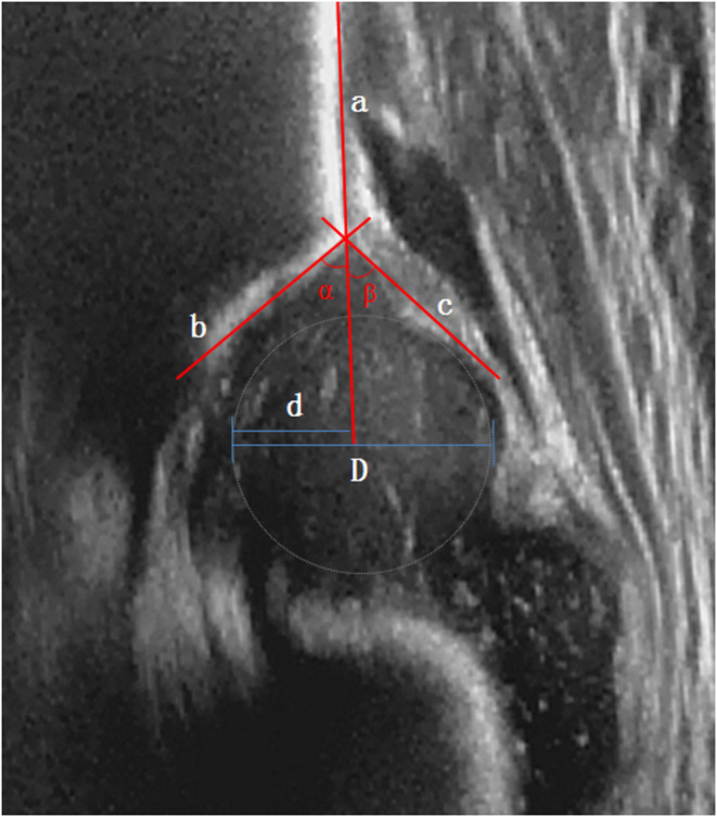
Coronal infant hip ultrasound, normal Graf type I, with the Graf construction: the baseline (a), the bony-roof line (b) and the cartilage-roof line (c) give the α (bony) and β (cartilaginous) angles; d/D mark femoral-head coverage. The acetabulum is deep and the head well covered. (Liu et al., Front Pediatr 2022;10:914545, Fig. 1; CC BY 4.0.)
Coronal infant hip ultrasound, normal Graf type I, with the Graf construction: the baseline (a), the bony-roof line (b) and the cartilage-roof line (c) give the α (bony) and β (cartilaginous) angles; d/D mark femoral-head coverage. The acetabulum is deep and the head well covered. (Liu et al., Front Pediatr 2022;10:914545, Fig. 1; CC BY 4.0.)
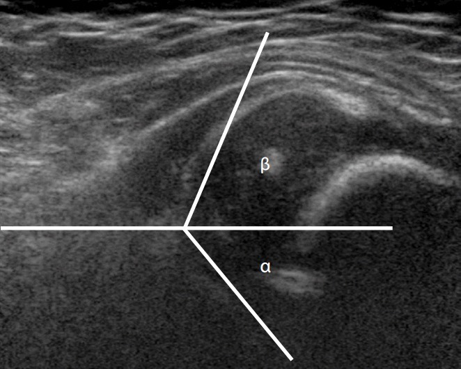
Coronal infant hip ultrasound of a dysplastic hip (Graf ≈ IIc; α 46°, β 65°) with the α/β construction lines: the bony roof is shallow and the femoral head is laterally decentred. (Kang & Koo, Ultrasonography 2017;36(4), Fig. 7; CC BY-NC 3.0 - non-commercial.)
Coronal infant hip ultrasound of a dysplastic hip (Graf ≈ IIc; α 46°, β 65°) with the α/β construction lines: the bony roof is shallow and the femoral head is laterally decentred. (Kang & Koo, Ultrasonography 2017;36(4), Fig. 7; CC BY-NC 3.0 - non-commercial.)
V.2 Radiography
Plain films become useful from about 4-6 months, once the ossific nucleus appears; before that the largely cartilaginous hip limits radiographic diagnosis. An AP pelvis is taken with the thighs neutral, since lateral rotation distorts every measurement.[58]
Reference lines and indices:
- Hilgenreiner’s line: horizontal, joining the two triradiate cartilages.[59]
- Perkins’ (Ombrédanne-Perkins) line: vertical, dropped from the lateral edge of the acetabular roof; with Hilgenreiner’s line it creates four quadrants, and the ossific centre of a normal hip lies in the lower-inner quadrant.[60]
- Acetabular index (AC angle): Hilgenreiner’s line versus the tangent to the bony acetabular roof; it falls with age. Useful cut-offs: roughly <30° in the newborn, <25° at 1 year, <20° at 2 years; a side-to-side difference of more than ~5° is suspicious.[61]
- Shenton’s (Ménard’s) line: the smooth arc from the medial femoral neck to the superior border of the obturator foramen; it is broken in subluxation/dislocation.[62]
- Centre-edge (CE) angle of Wiberg: for the older child/adult; in the adult >25° is normal, with a lower threshold accepted in younger children. Wiberg regarded <25° as inadequate coverage, and a value <20° predicts osteoarthritis.[63]
- Reimers’ migration percentage: the proportion of the head uncovered by the bony acetabulum; normal is near 0% in young children, and subluxation/dislocation give high values (it estimates uncoverage better than a falling CE angle).[64]
- Von Rosen view: an AP pelvis in maximum abduction and internal rotation; the projected femoral-shaft line normally crosses the acetabulum, but in dislocation it points above the acetabulum.[65]
- The acetabular teardrop: invisible in newborns; its later widening or deformity signals abnormal loading.[66]
Arthrography remains the reference for assessing soft-tissue obstacles and the quality of reduction. A concentric (anatomic) reduction shows the labrum lying flat with a sharp border and the head fully seated against the acetabular floor. A medial dye pool (greater than about 6 mm) indicates an incomplete, non-concentric reduction, and the “rose-thorn” appearance reflects the free border of the labrum.[67] After closed reduction, the concentricity of reduction inside the spica is confirmed with CT or MRI, the latter also assessing femoral-head vascularity.[68]
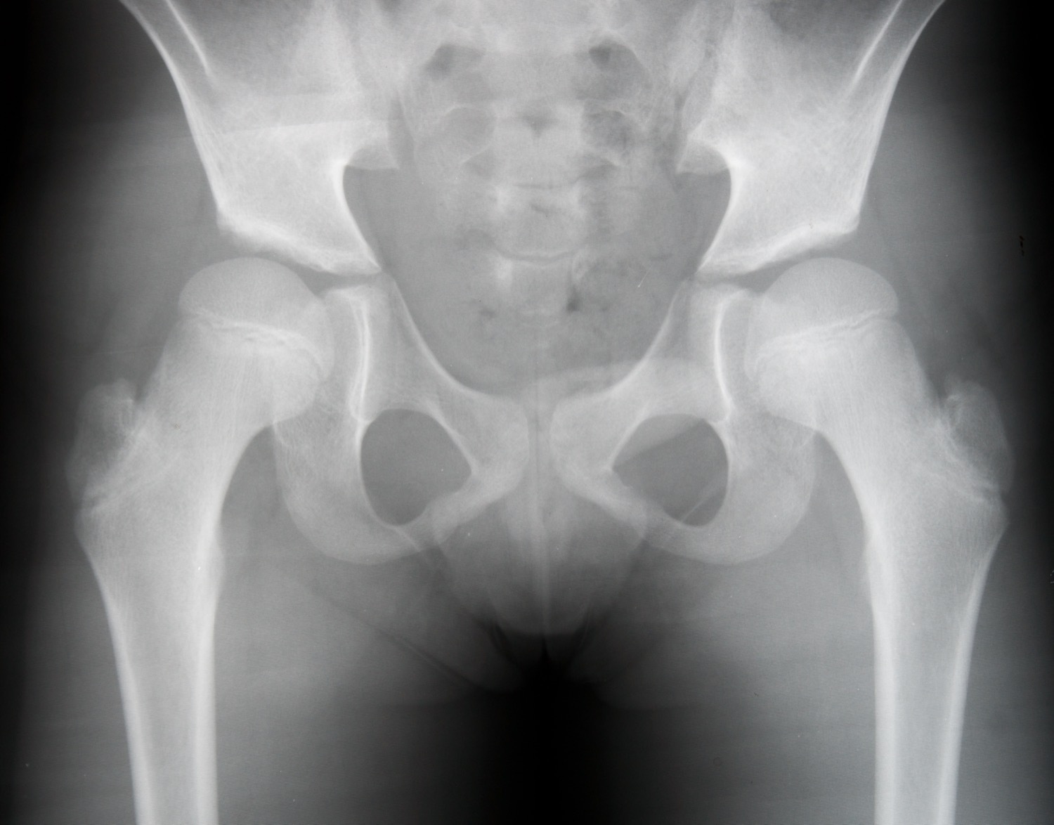
Antero-posterior pelvis of a child with developmental dysplasia of the right hip (pre-operative): the right proximal femur is small and high-riding with a shallow, steep acetabular roof, compared with the better-seated left hip. (Wikimedia Commons, “Login jetable”; CC0.)
Antero-posterior pelvis of a child with developmental dysplasia of the right hip (pre-operative): the right proximal femur is small and high-riding with a shallow, steep acetabular roof, compared with the better-seated left hip. (Wikimedia Commons, “Login jetable”; CC0.)
Part VI - Treatment by age (to reduction)
The goal at every age is to obtain and maintain a concentric (anatomic) reduction, providing the best environment for the femoral head and acetabulum to develop.[69]
VI.1 Newborn to ~6 months: the Pavlik harness
- Indication and principle. The Pavlik harness is first-line for the dislocatable and the reducible dislocated hip. It holds the hip in flexion (around 100-110°, with ≥110° favoured to prevent redislocation) and allows the legs to fall into gentle, gradual abduction under their own weight, never forced abduction. Forced wide abduction is the route to avascular necrosis.[70]
- Protocol and monitoring. The harness is worn full-time and the hip is checked, with ultrasound, within the first weeks. The single most important rule: if the hip is not reduced by about three weeks, stop. Persisting with an unreduced hip in the harness damages the acetabulum.[71]
- Success. Around 95% of unstable hips stabilise with abduction treatment; success is lower for the frankly dislocated hip and for Graf type IV.[72]
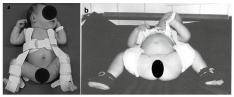
Pavlik harness on an infant. Panel (a): the harness applied - the anterior chest strap with the leg stirrups holding the hips in flexion and gentle abduction (the “human position”). Panel (b): a Daimler bandage, for comparison. Faces are anonymised in the source. (Gahleitner et al., Medicina (Kaunas) 2022;58:206, Fig. 1; CC BY 4.0.)
VI.2 About 6-18 months: closed reduction
Beyond six months the Pavlik harness succeeds in fewer than half of cases, so treatment moves to closed (or open) reduction under anaesthesia.[79]
- Adductor (± psoas) tenotomy is usually performed with the reduction to widen the safe zone and reduce the risk of growth disturbance; the psoas is released when it is blocking concentric reduction.[80]
- The “safe zone” of Ramsey is the arc between the adduction at which the hip redislocates and the abduction at which it risks AVN; the aim is to widen this zone by tenotomy rather than to force abduction.[81]
- Arthrography confirms a concentric reduction. If the head is “docked” against an interposed, blunted labrum, or a medial dye pool persists, the reduction is not anatomic and must be rejected.[82]
- The spica cast is applied in the “human position”: hyperflexion with moderate abduction. The extreme-abduction Lorenz/frog-leg position must be avoided, as it compresses the medial femoral circumflex vessels and causes AVN.[83]
- Preliminary traction is now largely abandoned: the contemporary view (and a large multicentre study) is that it does not reduce AVN. Source disagreement: Tönnis still presents data that preliminary traction lowered severe AVN; modern texts nonetheless prefer femoral shortening over traction in the older child.[84]
- The reduction is confirmed with CT/MRI, and casting continues (changed as needed) for roughly 12 weeks, followed by an abduction orthosis until the acetabular index normalises.[85]
- Acceptability rule: only an anatomic, stable reduction is accepted - one that holds through most of the range and dislocates only at the extreme of adduction/extension. A reduction that holds only in wide forced abduction is unacceptable and is converted to open reduction.[86]
VI.3 About 18 months to 3 years (and any failed closed reduction): open reduction
- Indications: failed closed reduction, a reduction stable only in extreme abduction, soft-tissue interposition, or late presentation; a hip presenting after 18 months usually needs open reduction. The old idea of delaying open reduction until the ossific nucleus appears (to lower AVN) has been disproved.[87]
- Obstacles cleared at open reduction: the contracted iliopsoas and adductor, the ligamentum teres, the transverse acetabular ligament, the pulvinar, a true inverted limbus (rare), and, most importantly, the constricted anteromedial capsule, addressed by capsulorrhaphy.[88]
- Approaches:
- Anterior (Smith-Petersen / “bikini”): the workhorse, giving wide access to all blocks, allowing capsulorrhaphy (which adds immediate stability) and a simultaneous pelvic osteotomy through the same incision, at the cost of more blood loss and some stiffness.[89]
- Medial (Ludloff / anteromedial): ideal for the child under 18 months, since it reaches the inferomedial obstacles with minimal dissection and blood loss and allows both hips in one sitting. It cannot achieve capsulorrhaphy (stability depends on the cast), is harder in older children, and historically carries a higher reported AVN risk (though the long-term Weinstein-Ponseti series found a rate, ~14%, comparable to the anterior approach).[90]
- Femoral shortening is added in the older child (especially over 3 years) to de-tension the reduction; this markedly lowers growth disturbance compared with preliminary traction, which in children over 3 years gives growth-disturbance rates over 50% and redislocation over 30%. A pelvic osteotomy is added according to acetabular dysplasia (Part VII).[91]
VI.4 Bilateral disease and the upper age limit
Bilateral surgery is staged (typically a few weeks apart).[92] For the upper age limit of attempting reduction, Tönnis regards closed reduction as appropriate up to about 3 years, with reconstruction still feasible to about 7 years.[93] The classic teaching is to attempt reduction of a unilateral dislocation up to about 8 years, but to set a lower limit (~4-6 years) for bilateral dislocation, because a symmetric, untreated bilateral dislocation is comparatively well tolerated while bilateral reconstruction is more morbid. Beyond that, untreated bilateral hips may be left, individualised to symptoms.
Part VII - Osteotomies
VII.1 Femoral osteotomy
- Varus derotation osteotomy (VDRO): an intertrochanteric osteotomy that corrects excessive anteversion (and adds varus as needed) to redirect the head into the acetabulum. After a successful reduction, excess anteversion usually corrects spontaneously, so a femoral osteotomy is reserved for the hip that fails to improve within 2-3 years and has residual acetabular dysplasia; concentricity is first confirmed on an abduction-internal-rotation film. A redirecting femoral osteotomy can stimulate acetabular development, but only if done before about age 4, and there is no acetabular benefit after age 8.[94]
- Femoral shortening osteotomy: used with open reduction in the older child to de-tension the reduction and lower AVN; far safer than preliminary traction in children over 3 years.[95]
VII.2 Pelvic osteotomies - the governing rule
The choice of pelvic osteotomy follows one rule:
- Redirectional osteotomies (which reorient the whole acetabulum) require a concentric, congruent joint with good motion and released contractures;
- Reshaping (acetabuloplasty) osteotomies require an open triradiate cartilage to act as the hinge;
- Salvage osteotomies are for the incongruent joint and do not need a concentric reduction.[96]
Redirectional (need a congruent joint)
- Salter (single innominate) osteotomy: a single cut through the ilium to the sciatic notch; the acetabular fragment is hinged on the pubic symphysis and rotated to improve anterolateral coverage (about 15° laterally and 25° anteriorly). Typical age ~2-9 years, and the symphysis must still be flexible.[97]
- Triple innominate osteotomy (Steel; Tönnis variant): cuts the ilium, pubis and ischium so the acetabular fragment is completely free (no symphyseal hinge), giving greater correction; used in the older child/adolescent with a closed triradiate cartilage.[98]
Reshaping / acetabuloplasty (need an open triradiate)
- Pemberton (pericapsular) osteotomy: an incomplete iliac cut hinging on the triradiate cartilage, which reduces the diameter of the acetabular opening and deepens the socket; ideal when the acetabulum is large relative to the head. It is intrinsically stable and needs no hardware, but risks premature triradiate closure if the cut is too close to the joint.[99]
- Dega osteotomy: an incomplete cut hinging on the intact posterior iliac cortex (rather than the triradiate cartilage), giving anterolateral coverage.[100] Source nuance: Pemberton and Dega are classically taught to decrease acetabular volume, but at least one MRI study found acetabular volume increased after Dega.[101]
Salvage (for the incongruent joint)
- Shelf (Staheli slotted acetabular augmentation): a bone graft placed over the capsule at the uncovered head, which becomes load-bearing through fibrous metaplasia of the capsule (bone over capsule, not cartilage); used for asymmetric incongruity with an intact capsule.[102]
- Chiari medial displacement osteotomy: the distal fragment is displaced medially, medialising the joint centre and improving abductor function. It does not require a concentric reduction, making it a true salvage option for the persistently subluxated hip, though posterior coverage is limited.[103]
Adolescent/adult reorientation
- Bernese periacetabular osteotomy (PAO, Ganz): a powerful multiplanar reorientation for symptomatic dysplasia in the patient with a closed triradiate cartilage and a congruent, concentrically reducible joint. It reorients and medialises the acetabular fragment while leaving the posterior column intact for early weight-bearing, and it is contraindicated in the immature pelvis (it would damage the triradiate). The femoral head’s blood supply (the deep branch of the medial femoral circumflex artery) is protected during the approach.[104]
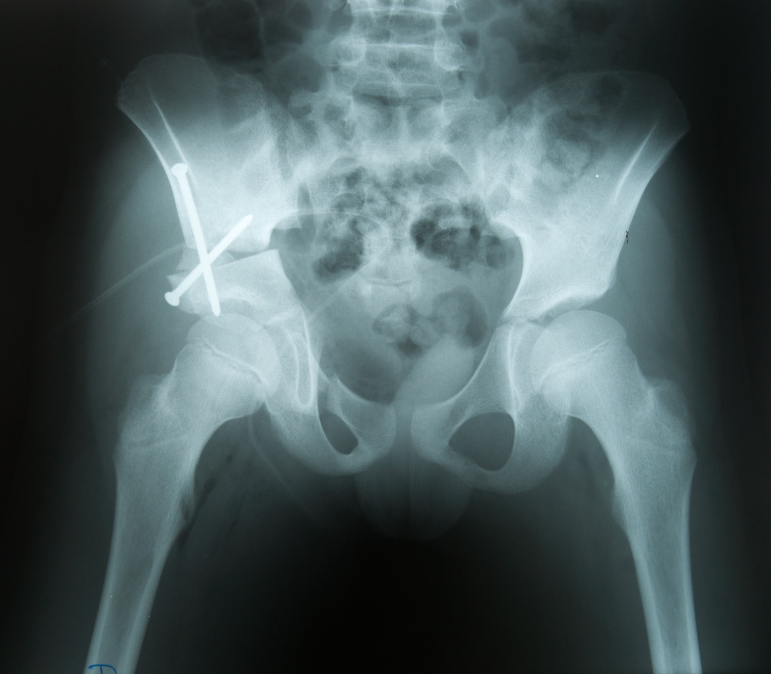
Post-operative antero-posterior pelvis after a pelvic (innominate) osteotomy of the right hip for DDH, with K-wire and screw fixation across the right ilium; the right femoral head is now better covered. (Wikimedia Commons, “Login jetable”; CC0.)
Post-operative antero-posterior pelvis after a pelvic (innominate) osteotomy of the right hip for DDH, with K-wire and screw fixation across the right ilium; the right femoral head is now better covered. (Wikimedia Commons, “Login jetable”; CC0.)
Part VIII - Complications, residual dysplasia and adult sequelae
VIII.1 Avascular necrosis (AVN) - the major iatrogenic complication
- Mechanism and risk factors: the head is most vulnerable in the first 12-18 months (mostly cartilage). The prime cause is forced/extreme positioning: extreme abduction (the Lorenz/frog-leg position) and forced internal rotation compress the medial femoral circumflex vessels, and maximal forced abduction can completely arrest epiphyseal blood flow. Using the femoral head as a “dilating sound” to force reduction is to be avoided. Younger children have a lower overall rate of growth disturbance but a higher proportion of the severe form.[105]
- Classifications:
- Kalamchi-MacEwen (groups I-IV): based on the degree of physeal/growth-plate involvement (group I confined to the ossific nucleus; group IV severe total involvement).[106]
- Bucholz-Ogden: based on the pattern of vascular injury.[107]
- Salter’s radiographic criteria for diagnosing growth disturbance of the head after DDH treatment.[108]
- As many as 25% of hips fit no classification.[109] Source nuance: Lovell’s authors caution that coxa magna is often benign post-reduction hyperaemia rather than true AVN.
- Growth-arrest sequelae: trochanteric overgrowth (→ abductor lurch, treated by greater-trochanter epiphysiodesis if the child is young, or later distal transfer), coxa magna, coxa breva, and corresponding acetabular deformity.[110]
VIII.2 Redislocation, residual dysplasia and the remodelling window
- Acetabular remodelling potential is time-limited: it is greatest in the first 18 months after reduction and continues to about 4 years, provided the reduction was achieved before about age 4; thereafter a pelvic osteotomy is increasingly needed.[111]
- Triggers for a secondary pelvic osteotomy: failure of the acetabular index to improve within 18-36 months of reduction, persistent dysplasia after age 5, or an acetabular index that plateaus above about 28°.[112]
VIII.3 Residual dysplasia and the adult hip
Even mild residual dysplasia, especially with any subluxation, leads to early secondary osteoarthritis, so every treated hip is followed to skeletal maturity (recurrent dysplasia can appear after apparent normalisation).[113] The adult dysplastic hip is graded by the CE angle and acetabular index, and the dislocated hip by Crowe or Hartofilakidis (Part III).
Total hip arthroplasty in the dysplastic hip is technically demanding. The acetabulum is small, shallow and anteverted (favouring a small cup placed at the true acetabulum/high hip centre, sometimes with structural graft), and the femur is small and excessively anteverted. For a high (Crowe IV / Hartofilakidis C) dislocation, a subtrochanteric shortening (derotation) osteotomy is used to bring the head down to the true acetabulum without over-stretching the sciatic nerve.[114]
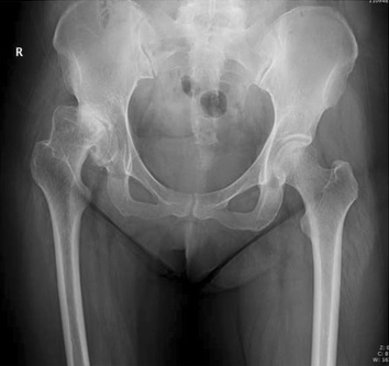
Antero-posterior pelvis of an adult with residual hip dysplasia (right hip, Crowe II): a shallow acetabulum with a steep roof and an incompletely covered, subluxated femoral head - the substrate for early secondary osteoarthritis. (Zhen et al., BMC Musculoskelet Disord 2017;18:142; CC BY 4.0, via Wikimedia Commons.)
Antero-posterior pelvis of an adult with residual hip dysplasia (right hip, Crowe II): a shallow acetabulum with a steep roof and an incompletely covered, subluxated femoral head - the substrate for early secondary osteoarthritis. (Zhen et al., BMC Musculoskelet Disord 2017;18:142; CC BY 4.0, via Wikimedia Commons.)
Bulgarian terminology glossary
For consistency with the Bulgarian state-examination vocabulary (and the operative terminology of Boychev, Хирургическа ортопедия):
| English | Bulgarian |
|---|---|
| Developmental dysplasia of the hip (DDH) | Вродена дисплазия на тазобедрената става / дисплазия на развитието на тазобедрената става |
| Congenital dislocation of the hip | Вродено изкълчване (луксация) на тазобедрената става |
| Dysplasia / subluxation / dislocation | Дисплазия / сублуксация / луксация (изкълчване) |
| Teratologic dislocation | Тератологична (фиксирана, антенатална) луксация |
| Acetabulum / acetabular roof | Ацетабулум (вертлужна кухина) / покрив на ацетабулума |
| Femoral head | Глава на бедрената кост |
| Acetabular index | Ацетабуларен индекс |
| Centre-edge angle (Wiberg) | Централно-ръбов ъгъл (на Wiberg) |
| Ortolani / Barlow test | Тест (симптом) на Ortolani / на Barlow |
| Galeazzi sign | Признак на Galeazzi |
| Limited abduction | Ограничена абдукция |
| Trendelenburg sign/gait | Признак / походка на Trendelenburg |
| Hip ultrasound (Graf) | Ехография (ултразвук) на тазобедрената става по Graf |
| Pavlik harness | Стремена (превръзка) на Pavlik |
| Closed / open reduction | Закрито / открито (оперативно) наместване (репозиция) |
| Spica cast | Тазобедрена гипсова превръзка (спика) |
| Femoral / pelvic osteotomy | Бедрена / тазова остеотомия |
| Avascular necrosis | Аваскуларна (асептична) некроза |
| Limb-length discrepancy | Разлика в дължината на крайниците |
| Secondary osteoarthritis (coxarthrosis) | Вторична коксартроза |
Figure credits and licences
All images were independently opened and visually verified to depict the stated entity before use. Licences were confirmed against the Wikimedia Commons API or the NCBI PMC Open-Access service.
- Normal Graf hip ultrasound -
normal_graf_us_pmc9126495_ccby.jpg. Liu B, Hu X, Li L, Gao S. Front Pediatr 2022;10:914545, Fig. 1. CC BY 4.0. https://pmc.ncbi.nlm.nih.gov/articles/PMC9126495/ - Dysplastic hip ultrasound (Graf ≈ IIc) -
dysplastic_us_graf_pmc5494873_ccbync.jpg. Kang YR, Koo J. Ultrasonography 2017;36(4), Fig. 7. CC BY-NC 3.0 - non-commercial use only. https://pmc.ncbi.nlm.nih.gov/articles/PMC5494873/ - Paediatric DDH, pre-operative AP pelvis -
cand_xray_dysplasia_preop.jpg. Wikimedia Commons, “Login jetable”. CC0 1.0. https://commons.wikimedia.org/wiki/File:Dysplasie_de_hanche_-_avant_op%C3%A9ration.jpg - Pavlik harness (and Daimler bandage) -
pavlik_daimler_pmc8874506_ccby_2panel.jpg. Gahleitner M, et al. Medicina (Kaunas) 2022;58(2):206, Fig. 1. CC BY 4.0. https://pmc.ncbi.nlm.nih.gov/articles/PMC8874506/ - Post-operative pelvic osteotomy, AP pelvis -
cand_xray_dysplasia_postop.jpg. Wikimedia Commons, “Login jetable”. CC0 1.0. https://commons.wikimedia.org/wiki/File:Dysplasie_de_hanche_-_hanche_droite_op%C3%A9r%C3%A9e.jpg - Adult residual hip dysplasia, AP pelvis -
ddh_appelvis_adult_commons_ccby.jpg. Zhen P, et al. BMC Musculoskelet Disord 2017;18:142. CC BY 4.0 (via Wikimedia Commons). https://commons.wikimedia.org/wiki/File:X-ray_of_hip_dysplasia_in_adult.jpg


Licensing note for distribution: image 2 is CC BY-NC 3.0 (non-commercial) - fine for a personal, non-commercial study document, but it must be removed or replaced if this material is ever used commercially. Images 1, 4 and 6 are CC BY (attribution); images 3 and 5 are CC0 (public domain).
No hip arthrogram was included: no example with a verifiable free licence was located.
References
-
The Pediatric and Adolescent Hip (Weinstein & Holt), pp.75-76; Wenger & Rang, Art and Practice of Children’s Orthopaedics, p.256.
-
Wenger & Rang, p.256.
-
Pediatric & Adolescent Hip, pp.75, 79; Tönnis, Congenital Dysplasia and Dislocation of the Hip, p.80.
-
Wenger & Rang, p.614; Pediatric & Adolescent Hip, p.82.
-
Pediatric & Adolescent Hip, pp.75-78.
-
Tönnis, p.488; Wenger & Rang, p.257.
-
Tönnis, p.59 and incidence chapter.
-
Pediatric & Adolescent Hip, pp.79, 81.
-
Pediatric & Adolescent Hip, p.81; Tönnis, pp.59-60.
-
Tönnis, pp.59-60; Pediatric & Adolescent Hip, p.81.
-
Pediatric & Adolescent Hip, pp.81, 84.
-
Tönnis, p.59; Pediatric & Adolescent Hip, p.81; Wenger & Rang, p.257.
-
Pediatric & Adolescent Hip, pp.81-82; Wenger & Rang, pp.257-258.
-
Tönnis, pp.68-69.
-
Tönnis, p.68; Pediatric & Adolescent Hip, p.82.
-
Tönnis, pp.79-80; Pediatric & Adolescent Hip, pp.80-81.
-
Tönnis, p.80.
-
Pediatric & Adolescent Hip, pp.82-83; Wenger & Rang, p.257.
-
Tönnis, pp.81, 96-97.
-
Tönnis, p.97.
-
Tönnis, pp.92-94; Pediatric & Adolescent Hip, pp.78, 91.
-
Tönnis, pp.93-95.
-
Tönnis, pp.92-93.
-
Tönnis, p.92.
-
Tönnis, pp.95-96; Pediatric & Adolescent Hip, p.91.
-
Tönnis, pp.95-96.
-
Tönnis, pp.95-96.
-
Pediatric & Adolescent Hip, p.79.
-
Pediatric & Adolescent Hip, pp.79-80; Tönnis, p.95.
-
Pediatric & Adolescent Hip, p.79; Salter, Textbook of Disorders and Injuries of the Musculoskeletal System, pp.270, 306-307.
-
Pediatric & Adolescent Hip, pp.80-81.
-
Pediatric & Adolescent Hip, pp.80-81; Salter, p.270.
-
Tönnis, p.80; Pediatric & Adolescent Hip, p.82.
-
Tönnis, pp.100-116.
-
Graf, Hip Sonography, pp.52-55; Tönnis, p.234.
-
Crowe and Hartofilakidis numeric definitions are standard adult-reconstruction classifications; in the present sources they appear by reference in Surgery of the Hip (Berry) rather than in the paediatric chapters. Tönnis uses an analogous low→high (precotyloid → supracotyloid → iliac) descriptive grade - Tönnis, pp.101-102.
-
Pediatric & Adolescent Hip, pp.86-88.
-
Pediatric & Adolescent Hip, p.88.
-
Pediatric & Adolescent Hip, pp.83, 86.
-
Pediatric & Adolescent Hip, p.87.
-
Pediatric & Adolescent Hip, p.81.
-
Pediatric & Adolescent Hip, p.84.
-
Pediatric & Adolescent Hip, p.84.
-
Pediatric & Adolescent Hip, pp.84-85.
-
Pediatric & Adolescent Hip, p.84.
-
Pediatric & Adolescent Hip, p.84.
-
Pediatric & Adolescent Hip, pp.80, 83.
-
Pediatric & Adolescent Hip, p.80.
-
Pediatric & Adolescent Hip, pp.80, 84.
-
Pediatric & Adolescent Hip, p.85.
-
Pediatric & Adolescent Hip, pp.84-85.
-
Graf, p.44; Pediatric & Adolescent Hip, pp.84-85.
-
Graf, pp.46-49.
-
Graf, pp.46, 53.
-
Graf, pp.52-53.
-
Graf, pp.52-54.
-
Pediatric & Adolescent Hip, pp.84-85.
-
Tönnis, p.129.
-
Tönnis, p.108.
-
Tönnis, p.108.
-
Surgery of the Hip (Berry), radiographic-measures section; Tönnis, pp.116-118.
-
Tönnis, p.109.
-
Tönnis, p.119; Surgery of the Hip (Berry), CE-angle section.
-
Tönnis, p.110.
-
Tönnis, p.126.
-
Tönnis, pp.110-111.
-
Pediatric & Adolescent Hip, pp.91-92, 95-96; Surgery of the Hip (Berry), arthrography section.
-
Pediatric & Adolescent Hip, p.92.
-
Pediatric & Adolescent Hip, pp.92-93.
-
Tönnis, p.270; Pediatric & Adolescent Hip, pp.93-94.
-
Pediatric & Adolescent Hip, p.94; Tönnis, pp.270-271.
-
Pediatric & Adolescent Hip, p.93; Lovell & Winter, DDH chapter.
-
Tönnis, pp.270-271.
-
Pediatric & Adolescent Hip, p.94.
-
Pediatric & Adolescent Hip, p.94.
-
Pediatric & Adolescent Hip, p.94.
-
Pediatric & Adolescent Hip, p.94.
-
Tönnis, pp.266-267; Pediatric & Adolescent Hip, p.94.
-
Pediatric & Adolescent Hip, p.94.
-
Pediatric & Adolescent Hip, p.95.
-
Pediatric & Adolescent Hip, p.95; Tönnis, p.271.
-
Pediatric & Adolescent Hip, pp.95-96.
-
Pediatric & Adolescent Hip, p.96; Lovell & Winter, DDH chapter.
-
Pediatric & Adolescent Hip, p.95; Lovell & Winter, DDH chapter; Tönnis, pp.262, 272.
-
Pediatric & Adolescent Hip, pp.96-97.
-
Pediatric & Adolescent Hip, pp.95-96; Lovell & Winter, DDH chapter.
-
Pediatric & Adolescent Hip, pp.97, 99; Lovell & Winter, DDH chapter.
-
Pediatric & Adolescent Hip, pp.95-96.
-
Pediatric & Adolescent Hip, pp.97-98.
-
Pediatric & Adolescent Hip, p.98.
-
Pediatric & Adolescent Hip, pp.98-99.
-
Pediatric & Adolescent Hip, pp.97-98.
-
Tönnis, pp.262-263.
-
Pediatric & Adolescent Hip, pp.99-100; Surgery of the Hip (Berry), femoral-osteotomy section.
-
Pediatric & Adolescent Hip, pp.98-99.
-
Pediatric & Adolescent Hip, pp.103-106; Surgery of the Hip (Berry), pelvic-osteotomy section.
-
Pediatric & Adolescent Hip, pp.103-104; Surgery of the Hip (Berry).
-
Pediatric & Adolescent Hip, p.104.
-
Pediatric & Adolescent Hip, pp.104-105.
-
Pediatric & Adolescent Hip, p.105.
-
Surgery of the Hip (Berry), Dega section.
-
Pediatric & Adolescent Hip, pp.105-106.
-
Pediatric & Adolescent Hip, pp.105-106.
-
Surgery of the Hip (Berry), PAO section; Lovell & Winter, DDH/adolescent-hip chapter.
-
Lovell & Winter, DDH chapter.
-
Lovell & Winter, DDH chapter (after Kalamchi & MacEwen, J Bone Joint Surg Am 1980;62:876).
-
Lovell & Winter, DDH chapter.
-
Lovell & Winter, DDH chapter (after Salter, Kostuik & Dallas, Can J Surg 1969).
-
Lovell & Winter, DDH chapter.
-
Lovell & Winter, DDH chapter.
-
Surgery of the Hip (Berry), remodelling section.
-
Pediatric & Adolescent Hip, pp.98, 103; Surgery of the Hip (Berry).
-
Surgery of the Hip (Berry).
-
Surgery of the Hip (Berry), dysplastic-THA section.