Contents
- Introduction and scope
- Part I - Definition, terminology and pathology
- Part II - Aetiology: primary and secondary coxarthrosis
- Part III - Clinical features and examination
- Part IV - Imaging and radiographic staging
- Part V - Non-operative management
- Part VI - Joint-preserving surgery
- Part VII - Total hip arthroplasty: the Charnley principle and indications
- Part VIII - Surgical approaches
- Part IX - Fixation: cemented and cementless
- Part X - Bearing surfaces
- Part XI - Complications of total hip arthroplasty
- Part XII - Outcomes and survivorship
- Bulgarian terminology (Боев / Boychev tradition) - glossary
- Image attributions
- References
Introduction and scope
Coxarthrosis is osteoarthritis of the hip: the mechanical and biological failure of the joint, with progressive loss of articular cartilage, subchondral sclerosis and cysts, osteophytes, and disabling pain and stiffness. Total hip arthroplasty is its definitive treatment and one of the most successful operations in surgery.[1] The examiner expects the distinction between primary and secondary disease (and the modern understanding that much “primary” hip osteoarthritis is in fact secondary to subtle dysplasia or impingement); the clinical syndrome and radiographic staging, meaning the four cardinal X-ray features, the Kellgren-Lawrence and Tönnis grades, and the migration patterns; the ladder of treatment from conservative care through joint-preserving osteotomy to replacement; and a sound grasp of total hip arthroplasty, covering the Charnley low-friction principle, the surgical approaches, the choice of fixation (cemented versus cementless) and bearing surface, and the complications and long-term results.
Two threads run through the topic. First, osteoarthritis is the failure of a joint rather than a single disease: a 1994 consensus defined it as the result of mechanical and biological events that destabilise the normal balance between cartilage degradation and synthesis, set off by genetic, developmental, metabolic and traumatic factors.[2] Second, arthroplasty is a time-limited reconstruction. The implant has a finite life, so the ideal candidate is the older patient with end-stage disease and disabling pain that has failed conservative care. The young patient is steered first toward preservation, because every replacement done early commits the patient to revision later.[3]
Part I - Definition, terminology and pathology
Coxarthrosis = osteoarthritis of the hip = degenerative joint disease. It is the commonest form of arthritis and a leading cause of disability in older people.[4] Although traditionally called “non-inflammatory,” its inflammatory component is now recognised. The 1994 workshop definition captures it well: osteoarthritis results from mechanical and biological events that destabilise the coupling of degradation and synthesis in articular cartilage and subchondral bone, producing softening, fibrillation, ulceration and loss of cartilage, sclerosis and eburnation of bone, osteophytes and subchondral cysts, presenting clinically with pain, tenderness, limited movement, crepitus and variable inflammation without systemic effects.[5]
The pathological cascade begins in the cartilage: chondrocytes attempt repair but matrix breakdown outpaces synthesis, the surface fibrillates and ulcerates, and cartilage is progressively lost down to eburnated (polished) subchondral bone. The bone responds with subchondral sclerosis, subchondral cysts (geodes) formed by synovial-fluid intrusion or microfracture, and marginal osteophytes, while the synovium and capsule become inflamed and fibrotic.[6] The driver is mechanical. Any process that concentrates contact stress, whether incongruity, malalignment, instability or over-coverage, accelerates the degeneration, which is why the abnormal hip wears out.
Part II - Aetiology: primary and secondary coxarthrosis
Hip osteoarthritis is divided into primary (idiopathic) and secondary forms, although the boundary has blurred. Much disease once called primary is now attributed to subtle femoroacetabular impingement or acetabular dysplasia, so that a truly idiopathic hip is less common than the label suggests.[7] The principal secondary causes are:
- Developmental dysplasia of the hip (DDH), where an undercovered, unstable socket concentrates load on the anterolateral rim and labrum. This is the commonest structural cause of premature hip osteoarthritis, and the head migrates superolaterally.[8]
- Femoroacetabular impingement (cam and pincer), dynamic rim contact damaging the chondrolabral junction (the subject of Topic 20).
- Sequelae of childhood hip disease, namely Legg-Calvé-Perthes disease and slipped capital femoral epiphysis, both of which leave an aspherical head and a cam-type deformity.[9]
- Avascular necrosis (osteonecrosis) of the femoral head, with collapse and secondary arthrosis.
- Post-traumatic causes: acetabular or femoral-head fracture, hip dislocation, malunion with incongruity.
- Inflammatory arthropathy: rheumatoid arthritis, ankylosing spondylitis and psoriatic arthritis (which, with protrusio, drive the head medially), and crystal arthropathy.[10]
- Septic sequelae, a previously infected hip.
- Metabolic and haematological disease: haemochromatosis, ochronosis (alkaptonuria), haemophilia, Gaucher disease and sickle-cell disease, Paget’s disease, and the epiphyseal dysplasias.[11]
- Neuropathic (Charcot) arthropathy.
Recognised risk factors include increasing age, obesity, female sex, genetic predisposition, heavy occupational or athletic loading, and previous injury or hip disease.[12]
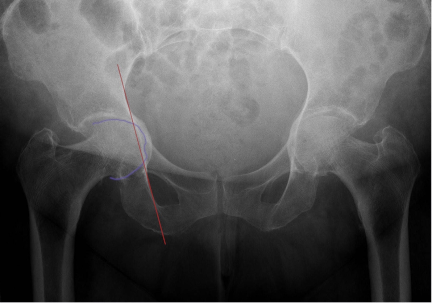
Protrusio acetabuli (a secondary-OA pattern). AP pelvis with the right femoral head migrating medial to the ilioischial line (outlined). Medial migration is the pincer/inflammatory pattern, in contrast to the superolateral migration of dysplasia. Hellerhoff, via Wikimedia Commons, CC BY-SA 4.0.
Protrusio acetabuli (a secondary-OA pattern). AP pelvis with the right femoral head migrating medial to the ilioischial line (outlined). Medial migration is the pincer/inflammatory pattern, in contrast to the superolateral migration of dysplasia. Hellerhoff, via Wikimedia Commons, CC BY-SA 4.0.
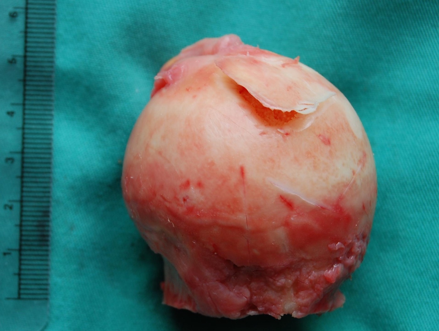
Avascular necrosis as a cause of secondary OA. Excised femoral head with a loose flap of articular cartilage overlying necrotic subchondral bone, the gross correlate of osteonecrosis progressing to arthrosis. Steven Fruitsmaak, via Wikimedia Commons, CC BY-SA 3.0.
Avascular necrosis as a cause of secondary OA. Excised femoral head with a loose flap of articular cartilage overlying necrotic subchondral bone, the gross correlate of osteonecrosis progressing to arthrosis. Steven Fruitsmaak, via Wikimedia Commons, CC BY-SA 3.0.
Part III - Clinical features and examination
The cardinal symptom is groin pain, often radiating to the anterior thigh and, classically, referred to the knee, so a hip cause must always be sought in a patient presenting with knee pain.[13] Early disease produces start-up and activity-related pain relieved by rest. As it advances, rest and night pain appear and should prompt exclusion of sepsis or tumour.[14] The patient describes stiffness, difficulty with shoes and socks, a limp, and shrinking walking distance, and may localise the pain with the hand cupped over the trochanter, the C-sign.[15]
On examination, loss of internal rotation is the earliest finding, followed by loss of extension and abduction, a progressive fixed flexion and adduction deformity with apparent leg shortening, an antalgic (coxalgic) gait, and a Trendelenburg sign from abductor dysfunction.[16] The differential includes referred lumbar or sacroiliac pain, trochanteric bursitis, and a femoral-neck stress fracture; an intra-articular anaesthetic injection confirms an intra-articular source when the picture is mixed.[17]
Part IV - Imaging and radiographic staging
The diagnosis is radiographic. A weight-bearing AP pelvis with a lateral (and, for the dysplastic or impinging hip, a false-profile view) shows the four cardinal features of osteoarthritis: joint-space narrowing, subchondral sclerosis, subchondral cysts and osteophytes.[18] The pattern of femoral-head migration is informative. Superolateral migration is the commonest and is typical of dysplasia/instability, while medial migration (protrusio) accompanies pincer over-coverage and inflammatory disease, and concentric narrowing suggests an inflammatory arthropathy.[19]
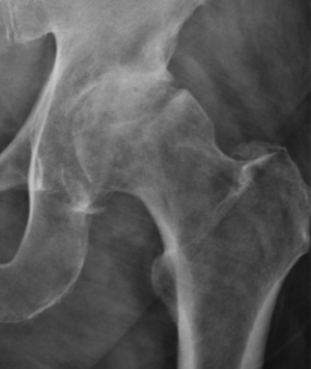
Hip osteoarthritis - the cardinal radiographic features. AP hip showing superolateral joint-space narrowing, subchondral sclerosis of the acetabular roof and marginal osteophytes. Ruiz Santiago F et al., Radiol Res Pract 2016; via Wikimedia Commons, CC BY 4.0.
Hip osteoarthritis - the cardinal radiographic features. AP hip showing superolateral joint-space narrowing, subchondral sclerosis of the acetabular roof and marginal osteophytes. Ruiz Santiago F et al., Radiol Res Pract 2016; via Wikimedia Commons, CC BY 4.0.
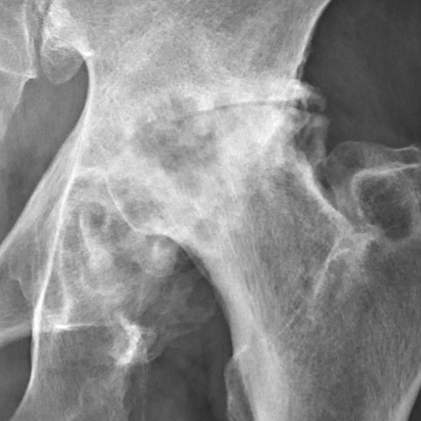
Severe (Tönnis grade 3) coxarthrosis. Near-complete loss of the superior joint space, dense sclerosis, large osteophytes and subchondral cysts. Mikael Häggström, via Wikimedia Commons (CC0).
Severe (Tönnis grade 3) coxarthrosis. Near-complete loss of the superior joint space, dense sclerosis, large osteophytes and subchondral cysts. Mikael Häggström, via Wikimedia Commons (CC0).
Two grading systems are used. The Kellgren-Lawrence grade (the classic 0-4 epidemiological scale) is based on marginal osteophytes, joint-space narrowing with subchondral sclerosis, subchondral pseudocysts and altered bone-end shape.[20] The Tönnis grade is widely used in hip-preservation work:[21]
- Grade 0: no changes.
- Grade 1: increased sclerosis of head and acetabulum, slight joint-space narrowing, slight osteophytes.
- Grade 2: small cysts in head or acetabulum, marked joint-space narrowing, slight loss of head sphericity.
- Grade 3: large cysts, severe narrowing up to complete loss of the joint space, and severe deformity of the head.
The Tönnis grade carries direct surgical weight. A low grade predicts success with joint preservation, whereas advanced disease (effectively grade 3) is an indication for replacement rather than osteotomy or arthroscopy. CT and MRI are reserved for assessing osteonecrosis, version, and the cartilage and labrum.[22]
Part V - Non-operative management
Conservative treatment is the first step for all but end-stage disease. It rests on patient education, weight loss, activity modification, and a walking aid: a cane used in the opposite hand reduces hip-joint loading by 20-30%.[23] Physiotherapy maintains range and strengthens the periarticular and abductor muscles. The pharmacological options are paracetamol and NSAIDs (more effective than paracetamol for pain and function, but requiring monitoring of renal, hepatic and haematological function), and intra-articular corticosteroid injection for flares; viscosupplementation has a limited and temporary role.[24] Conservative care carries its own risks (gastrointestinal, renal and cardiac toxicity of NSAIDs), and excessive delay of surgery in a hip that has failed it may yield a worse final result and compromise the bone stock available for reconstruction.[25]
Part VI - Joint-preserving surgery
In the younger patient with a correctable deformity and preserved cartilage, joint preservation is preferred to replacement.
Femoral osteotomy (intertrochanteric varus or valgus, with flexion/extension and derotation as needed) realigns the head within the acetabulum to increase the weight-bearing surface area and shift load onto better-preserved cartilage. On Pauwels’ principles a varus osteotomy increases the abductor lever arm and lowers the joint reaction force, while Bombelli added sagittal-plane correction to enlarge the contact area.[26] The varus osteotomy is the more predictable, and the surgeon must minimise distortion of the proximal femur so as not to compromise a future replacement.[27]
Pelvic and periacetabular osteotomy reorients the acetabulum in dysplasia, whether redirectional (the Bernese PAO), reshaping or salvage, to recover coverage and offload the rim (covered in Topics 7 and 20).[28]
Hip arthrodesis (fusion) remains an option for the very young manual labourer with isolated unilateral disease. It relieves pain durably and allows heavy work, but at a long-term cost: at twenty years and beyond, satisfaction is high yet up to 60% develop low-back, ipsilateral-knee or contralateral-hip pain, and many are eventually converted to arthroplasty.[29] Resection arthroplasty (Girdlestone) is now reserved as a salvage procedure, chiefly for the unreconstructable infected hip.[30]
Part VII - Total hip arthroplasty: the Charnley principle and indications
The Charnley low-friction arthroplasty
John Charnley’s low-friction arthroplasty, more precisely a low-frictional-torque arthroplasty, is the conceptual foundation of the modern operation.[31] Its principle is that a small femoral head (Charnley’s was 22.225 mm) articulating in a thick-walled socket minimises frictional torque at the implant-bone interface, reducing the tendency of the components to loosen.[32] Charnley showed from pendulum experiments that the natural joint works by boundary lubrication and that an artificial joint therefore needs intrinsically slippery, wear-resistant surfaces.[33] His first bearing, PTFE (Teflon), had a uniquely low friction but catastrophic wear and was abandoned in 1961 after producing florid granulomas; he replaced it in 1962 with high-molecular-weight polyethylene, whose friction was higher but whose wear resistance was hundreds of times better.[34] Three further elements completed the concept: acrylic cement to distribute load into the bone (used as a grout, not a glue); a greater-trochanteric osteotomy with medialisation of the hip centre to restore the abductor lever arm, for which Charnley preferred the term “reconstruction”; and clean-air (laminar-flow) theatres to control infection.[35] The small head also acted as a stability “safety valve,” able to subluxate and relocate under trauma rather than transmitting the force to the cement.[36]
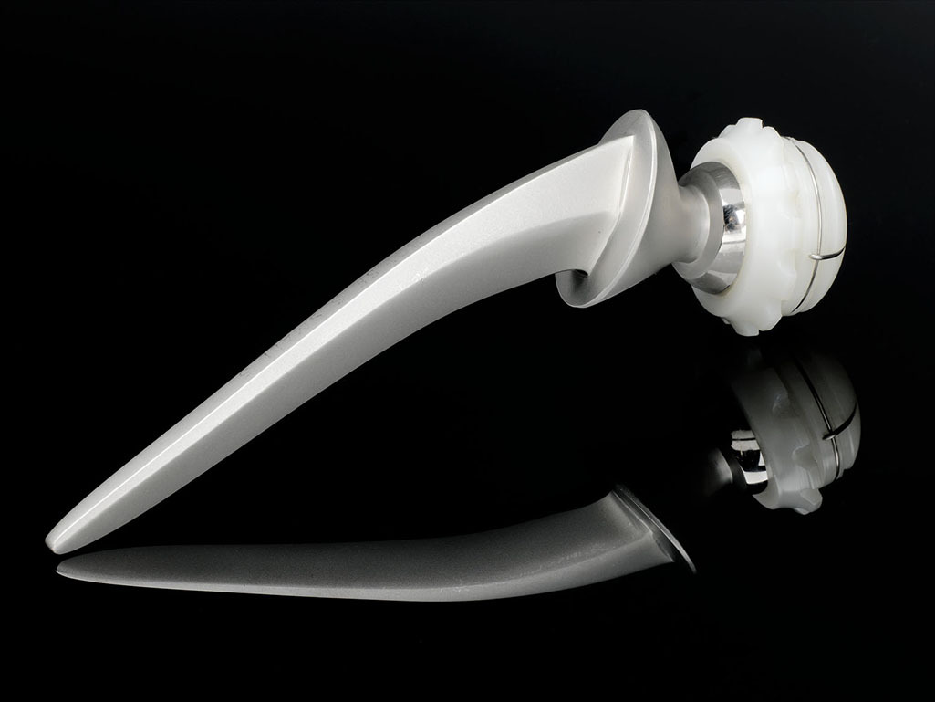
A Charnley-type low-friction prosthesis. A stainless-steel femoral stem with a small-diameter metal head articulating against a white ultra-high-molecular-weight polyethylene cup, the classic metal-on-polyethylene, small-head design. Science Museum London / Science & Society Picture Library, via Wikimedia Commons, CC BY-SA 2.0.
A Charnley-type low-friction prosthesis. A stainless-steel femoral stem with a small-diameter metal head articulating against a white ultra-high-molecular-weight polyethylene cup, the classic metal-on-polyethylene, small-head design. Science Museum London / Science & Society Picture Library, via Wikimedia Commons, CC BY-SA 2.0.
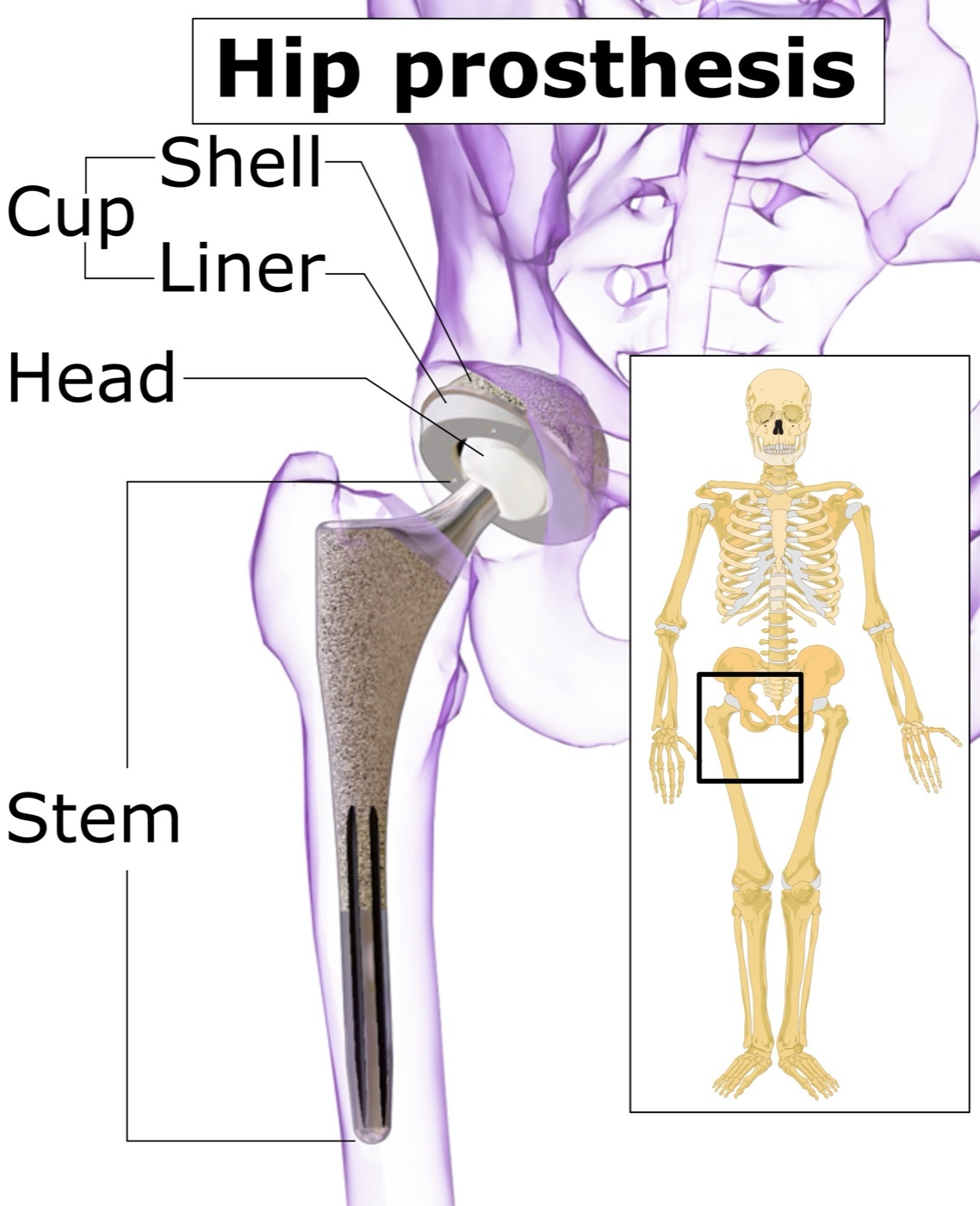
The components of a total hip replacement. The acetabular cup (shell and liner), the modular femoral head, and the stem, shown on a pelvis outline. Mikael Häggström, via Wikimedia Commons, CC BY-SA 4.0.
The components of a total hip replacement. The acetabular cup (shell and liner), the modular femoral head, and the stem, shown on a pelvis outline. Mikael Häggström, via Wikimedia Commons, CC BY-SA 4.0.
Indications and contraindications
The primary indication for THA is disabling pain from end-stage arthritis that has failed conservative care, supported by radiographs of advanced disease, with the diagnosis confirmed on all three axes of history, examination and imaging.[37] Pain is the symptom most reliably relieved, and loss of function in daily life despite non-operative treatment strengthens the indication. Age is weighed against implant life: the operation is “time-limited,” so the older patient is the ideal candidate, while in patients under forty the prosthesis cannot be expected to last a lifetime and a revision should be anticipated, favouring preservation first.[38] Charnley’s own test still applies. If the patient would be no worse off with a resection arthroplasty than they are now, replacement is justified.[39]
There are few absolute contraindications; the principal one is active infection of the hip or systemic sepsis that could seed the implant.[40] Relative contraindications include remote sepsis, a neuropathic (Charcot) joint, an absent or deficient abductor mechanism, severe medical comorbidity or unacceptable anaesthetic risk, morbid obesity (the risk of infection, dislocation and loosening rises sharply above a BMI of 40), poorly-controlled diabetes, current intravenous drug use, prior pelvic irradiation, dementia or heavy alcohol use, and a non-compliant patient with unrealistic expectations.[41]
Part VIII - Surgical approaches
Each approach strikes a different balance between exposure, muscle preservation and dislocation risk.
- Posterior (Moore/Southern; Kocher-Langenbeck): the gluteus maximus is split (no true internervous plane) and the short external rotators (piriformis, gemelli, obturator internus, quadratus femoris) are detached and repaired, while the abductors are preserved. It gives excellent extensile exposure of both femur and acetabulum but historically the highest dislocation rate. The sciatic nerve is the structure at risk, and capsular and rotator repair reduces dislocation to under 1%.[42]
- Direct lateral / transgluteal (Hardinge): splits the abductors, leaving the posterior structures intact and giving a low dislocation rate, at the cost of abductor weakness and a persistent Trendelenburg limp. The superior gluteal nerve is endangered if the split is carried more than ~4 cm above the trochanter.[43]
- Anterolateral (Watson-Jones): between tensor fasciae latae and gluteus medius, an intermediate option.
- Direct anterior (Smith-Petersen / Hueter, with or without a traction table): a true internervous interval (femoral versus superior gluteal territory) that spares muscle and allows early recovery and a low dislocation rate (about 0.6-1.5%). The lateral femoral cutaneous nerve is at risk, femoral exposure is the difficult part, and there is a recognised learning curve.[44]
- Trochanteric osteotomy: now rarely needed for primary surgery, reserved for difficult exposure and abductor tensioning, and complicated by nonunion, migration and hardware problems (chevron and slide variants reduce nonunion).[45]
Component position governs stability: the Lewinnek “safe zone” is 40 ± 10° of cup inclination and 15 ± 10° of anteversion, and most dislocations involve a component placed outside it.[46]
Part IX - Fixation: cemented and cementless
A prosthesis is fixed to bone in one of two ways.
Cemented fixation uses polymethylmethacrylate (PMMA), which acts as a grout, not a glue. It has no adhesive power and works by mechanical interlock at the cement-bone and cement-implant interfaces, spreading load like a viscoelastic shock absorber.[47] Modern “second- and third-generation” technique markedly improved durability: a medullary plug, pulsatile lavage, retrograde filling with a cement gun, pressurisation, and vacuum mixing or centrifugation to reduce porosity.[48] The exothermic polymerisation can reach high temperatures, and instrumentation of the canal can embolise marrow contents (bone-cement implantation syndrome / fat embolism), mitigated by lowering intramedullary pressure with distal suction.[49] Cement is favoured in the older patient and in poor-quality bone, and allows antibiotic loading.[50]
Cementless fixation achieves biological fixation by bone ingrowth (into a three-dimensional porous surface, resisting shear and tension) or ongrowth (onto a roughened surface, resisting shear only).[51] The requirements are an optimal pore size of roughly 100-400 µm, intimate bone contact (gaps over ~2 mm and micromotion over ~40-150 µm prevent bone ingrowth and produce fibrous tissue), and rigid initial mechanical stability from a press-fit.[52] A hydroxyapatite coating speeds early ingrowth but adds no long-term benefit when apposition is poor.[53] The drawbacks are stress shielding (proximal bone resorption around a stiff stem) and thigh pain. Designs range from extensively porous-coated to proximally-coated, tapered and short metaphyseal stems, and press-fit hemispherical cups (often with screw augmentation).[54] Registries show cemented fixation with a slightly lower early revision rate, but both achieve over 90-95% survival at ten years.[55]
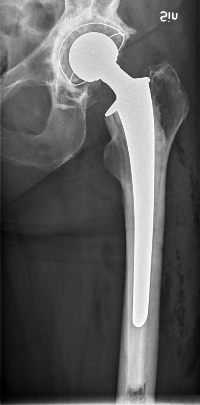
Cementless total hip arthroplasty. Postoperative AP radiograph of a porous-coated, press-fit tapered femoral stem with a modular head and a hemispherical acetabular cup. Mikael Häggström, via Wikimedia Commons (CC0).
Cementless total hip arthroplasty. Postoperative AP radiograph of a porous-coated, press-fit tapered femoral stem with a modular head and a hemispherical acetabular cup. Mikael Häggström, via Wikimedia Commons (CC0).
Part X - Bearing surfaces
The articulating couple determines wear, and wear determines longevity.
Metal-on-polyethylene has been the standard since Charnley. Conventional polyethylene wears at roughly 0.1 mm/year, and its sub-micron wear particles drive the macrophage-mediated osteolysis (particle disease) that causes most late aseptic loosening; the wear threshold for osteolysis is about 0.3 mm/year.[56] Highly cross-linked polyethylene reduces wear by 40-95% and has nearly abolished osteolysis in mid-term studies, at the cost of slightly reduced mechanical strength (so rim fracture is a concern with a malpositioned, steeply inclined cup).[57]
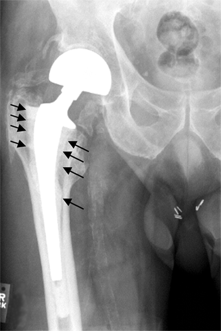
Wear-driven osteolysis (aseptic loosening). Pre-revision radiograph of a cemented femoral stem with peri-implant radiolucent osteolytic lesions (arrows) along the shaft. Shen Z et al., Arthritis Res Ther 2006;8:R70; via Wikimedia Commons, CC BY 2.0.
Wear-driven osteolysis (aseptic loosening). Pre-revision radiograph of a cemented femoral stem with peri-implant radiolucent osteolytic lesions (arrows) along the shaft. Shen Z et al., Arthritis Res Ther 2006;8:R70; via Wikimedia Commons, CC BY 2.0.
Metal-on-metal bearings wear far less by volume (a larger head improving fluid-film lubrication, the opposite of polyethylene) but release cobalt and chromium ions and can provoke an adverse local tissue reaction / pseudotumour (ARMD). Edge loading from a steeply placed cup worsens this, and the high failure rate of large-diameter and resurfacing designs (the DePuy ASR recall) has largely ended their use.[58] Ceramic-on-ceramic has the lowest wear of all and biologically inert debris, but carries a small risk of fracture and of squeaking.[59] Ceramic-on-polyethylene combines an inert, scratch-resistant head with a cross-linked liner. Larger heads improve stability and range of motion (greater jump distance) but, in polyethylene, increase volumetric wear, the trade-off Charnley anticipated.[60]
Part XI - Complications of total hip arthroplasty
The four leading modes of failure are aseptic loosening, infection, dislocation and periprosthetic fracture.[61]
- Dislocation / instability is one of the commonest reasons for revision; most occur within the first year. Risk rises with malpositioned components (outside the Lewinnek zone), the posterior approach (about 3-4% versus under 1.5% anteriorly, and under 1% with capsular repair), small head size, abductor or soft-tissue deficiency, prior surgery, neuromuscular or cognitive impairment, and adverse spinopelvic mechanics. Most settle with closed reduction, but recurrence approaches 60% and may require revision, a dual-mobility or a constrained liner.[62]
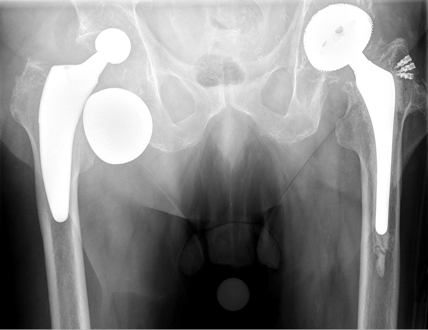
Dislocated total hip replacement. AP pelvis with the right prosthetic femoral head displaced superolaterally, completely out of its acetabular cup. Hellerhoff, via Wikimedia Commons, CC BY-SA 4.0.
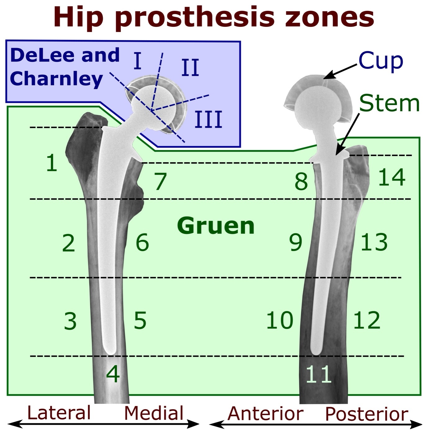
The radiographic zones for describing loosening. Hip-implant radiographs annotated with the Gruen femoral zones and the DeLee-Charnley acetabular zones (I-III), used to localise periprosthetic lucency. Mikael Häggström, via Wikimedia Commons (CC0).
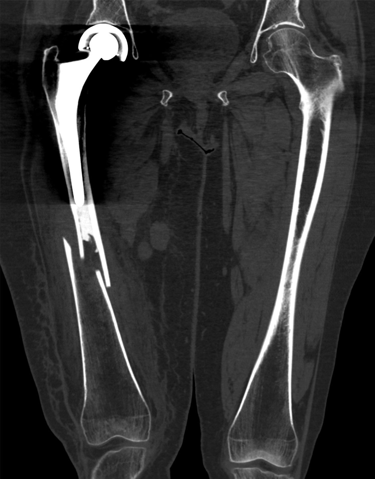
Periprosthetic femoral fracture (Vancouver type C). Coronal CT reconstruction showing an oblique femoral-shaft fracture well distal to the tip of a well-fixed stem. Hellerhoff, via Wikimedia Commons, CC BY-SA 4.0.
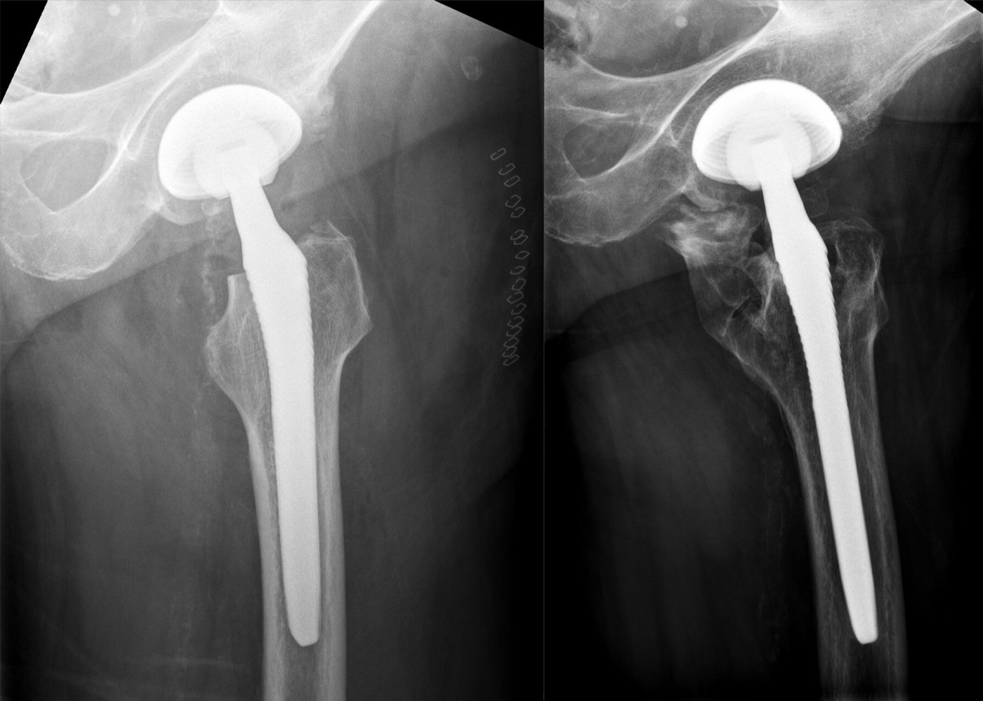
Heterotopic ossification after total hip arthroplasty. Abundant periarticular new bone around the proximal femur and greater trochanter following hip replacement (Brooker-type ossification). Hellerhoff, via Wikimedia Commons, CC BY-SA 4.0.
Heterotopic ossification after total hip arthroplasty. Abundant periarticular new bone around the proximal femur and greater trochanter following hip replacement (Brooker-type ossification). Hellerhoff, via Wikimedia Commons, CC BY-SA 4.0.
Part XII - Outcomes and survivorship
Results are reported with the Harris Hip Score and other instruments (Merle d’Aubigné, Oxford, WOMAC, HOOS). Total hip arthroplasty is durable: Charnley’s cemented prosthesis retained about 90% of components at 25 years, and registry data show cemented fixation surviving about 95% at ten years (versus ~83% for uncemented in the same era), with the best cemented Charnley series reaching 78% survival at a minimum of 35 years.[70] The predictors of longevity are sound component position and fixation, a low-wear bearing, restored offset and leg length, and patient factors (lower demand, controlled weight); high-impact activity shortens implant life.[71]
To tie the topic together: coxarthrosis is the wearing-out of the hip, most often driven by an underlying dysplasia or impingement. Diagnose it from groin pain with loss of internal rotation and the four cardinal radiographic signs, grade it by Tönnis, and treat it up a ladder, from conservative care to osteotomy in the young correctable hip to total hip arthroplasty for end-stage disease. Build the replacement on Charnley’s low-friction principle, position the components within the safe zone, and choose fixation and bearing to the patient. Counsel that, although the result is excellent and lasting, the implant has a finite life and its enemies are infection, dislocation, wear-driven loosening and fracture.
Bulgarian terminology (Боев / Boychev tradition) - glossary
The following Bulgarian equivalents bridge the international literature with the terminology of the Bulgarian school of orthopaedics (in the tradition of Бойчо Бойчев / Boycho Boychev) used in the state examination.
| English term | Bulgarian term (Cyrillic) | Transliteration |
|---|---|---|
| Coxarthrosis (hip osteoarthritis) | Коксартроза | Koksartroza |
| Osteoarthritis / degenerative joint disease | Остеоартроза / дегенеративна ставна болест | Osteoartroza / degenerativna stavna bolest |
| Primary (idiopathic) | Първична (идиопатична) | Parvichna (idiopatichna) |
| Secondary | Вторична | Vtorichna |
| Articular cartilage | Ставен хрущял | Staven hrushtyal |
| Joint-space narrowing | Стесняване на ставната цепка | Stesnyavane na stavnata tsepka |
| Subchondral sclerosis | Субхондрална склероза | Subhondralna skleroza |
| Subchondral cyst (geode) | Субхондрална киста (геода) | Subhondralna kista (geoda) |
| Osteophyte | Остеофит | Osteofit |
| Protrusio acetabuli | Протрузия на ацетабулума | Protruziya na atsetabuluma |
| Antalgic gait | Анталгична (щадяща) походка | Antalgichna (shtadyashta) pohodka |
| Trendelenburg sign | Симптом на Тренделенбург | Simptom na Trendelenburg |
| Fixed flexion deformity | Флексионна контрактура | Fleksionna kontraktura |
| Total hip arthroplasty | Тотална артропластика на тазобедрената става | Totalna artroplastika na tazobedrenata stava |
| Endoprosthesis | Ендопротеза | Endoproteza |
| Bone cement (PMMA) | Костен цимент (полиметилметакрилат) | Kosten tsiment (polimetilmetakrilat) |
| Cementless (biologic) fixation | Безциментна (биологична) фиксация | Beztsimentna (biologichna) fiksatsiya |
| Bone ingrowth | Костно врастване | Kostno vrastvane |
| Bearing surface | Триеща (артикулираща) повърхност | Trieshta (artikulirashta) povarhnost |
| Polyethylene | Полиетилен | Polietilen |
| Osteolysis (particle disease) | Остеолиза (болест на износващите частици) | Osteoliza (bolest na iznosvashtite chastitsi) |
| Aseptic loosening | Асептично разхлабване | Aseptichno razhlabvane |
| Periprosthetic fracture | Перипротезна фрактура | Periproteznata fraktura |
| Dislocation | Луксация (изкълчване) | Luksatsiya (izkalchvane) |
| Periprosthetic infection | Перипротезна инфекция | Periproteznata infektsiya |
| Intertrochanteric osteotomy | Интертрохантерна остеотомия | Intertrohanterna osteotomiya |
| Arthrodesis (fusion) | Артродеза | Artrodeza |
Image attributions
(Figure attributions and licences are listed in the figure MANIFEST and inserted with each image. All images are openly licensed [CC0 / CC BY / CC BY-SA] or used under their stated terms; any non-commercial [NC] item is flagged as such and must not be used in a commercial product.)
References
-
Berry, Surgery of the Hip, p. 440; The Adult Hip (Callaghan), p. 984, p. 1159.
-
Berry, p. 440.
-
The Adult Hip, p. 1161-1166.
-
The Adult Hip, p. 984.
-
Berry, p. 440.
-
Berry, p. 440; The Adult Hip, p. 984.
-
Berry, p. 42, p. 440; The Adult Hip, p. 984.
-
Berry, p. 42; The Adult Hip, p. 984.
-
The Adult Hip, p. 805; Berry, p. 440.
-
The Adult Hip, p. 984.
-
The Adult Hip, p. 984.
-
The Adult Hip, p. 984.
-
The Adult Hip, p. 1163; Berry, p. 455.
-
The Adult Hip, p. 1163, p. 1167-1168.
-
The Adult Hip, p. 1163.
-
The Adult Hip, p. 1168; Berry, p. 456.
-
The Adult Hip, p. 1163.
-
Berry, p. 423, p. 440.
-
Berry, p. 440.
-
Berry, p. 440.
-
Berry, p. 440.
-
Berry, p. 423.
-
The Adult Hip, p. 1164.
-
Berry, p. 440; The Adult Hip, p. 1162, p. 1164.
-
The Adult Hip, p. 1162, p. 1164.
-
Berry, p. 751-752.
-
Berry, p. 751-752.
-
Berry, p. 745.
-
The Adult Hip, p. 1161.
-
Berry, p. 780, p. 785.
-
Charnley, Low Friction Arthroplasty of the Hip, p. 11.
-
Charnley, p. 11, p. 22; Berry, p. 98.
-
Charnley, p. 12-15.
-
Charnley, p. 14-15.
-
Charnley, p. 22, p. 339; Berry, p. 285.
-
Charnley, p. 21.
-
The Adult Hip, p. 1163, p. 1167.
-
The Adult Hip, p. 1161-1166.
-
The Adult Hip, p. 1166.
-
The Adult Hip, p. 1168.
-
The Adult Hip, p. 1168-1170.
-
Berry, p. 277-283.
-
Berry, p. 273-275.
-
Berry, p. 264-271.
-
Berry, p. 285-296.
-
Berry, p. 893.
-
Berry, p. 91, p. 865; The Adult Hip, p. 1103.
-
The Adult Hip, p. 1109-1110; Berry, p. 870-872.
-
Berry, p. 85, p. 873.
-
The Adult Hip, p. 1112.
-
Berry, p. 158; The Adult Hip, p. 1079-1080.
-
Berry, p. 158-160, p. 847; The Adult Hip, p. 1082-1084.
-
Berry, p. 849; The Adult Hip, p. 1087.
-
Berry, p. 876-889; The Adult Hip, p. 1084-1085.
-
The Adult Hip, p. 1091, p. 1110.
-
Berry, p. 200, p. 922.
-
Berry, p. 915-919.
-
Berry, p. 921-929.
-
Berry, p. 935-940.
-
Berry, p. 921-923.
-
Berry, p. 1054.
-
Berry, p. 1061, p. 1098-1099, p. 893.
-
Berry, p. 1224, p. 1228-1229.
-
Berry, p. 1092, p. 1099; The Adult Hip, p. 1116.
-
Berry (periprosthetic-fracture classification); The Adult Hip.
-
Berry, p. 1278.
-
Berry, p. 1056.
-
The Adult Hip, p. 1160.
-
Berry (heterotopic ossification).
-
The Adult Hip, p. 1110, p. 1159; Berry, p. 838, p. 867.
-
The Adult Hip, p. 1165; Berry, p. 893.