Contents
- Introduction and scope
- Part I - Definition
- Part II - Classification
- Part III - Developmental coxa vara: epidemiology and etiology
- Part IV - Pathoanatomy
- Part V - Clinical features
- Part VI - Radiographic assessment
- Part VII - Natural history
- Part VIII - Treatment
- Part IX - Complications and outcomes
- Bulgarian terminology (Боев / Boychev tradition) - glossary
- Image attributions
Introduction and scope
Coxa vara is a reduction of the femoral neck-shaft angle below the normal range for age. It is best thought of not as a single disease but as a sign produced by a heterogeneous group of conditions, whether present at birth, developing in early childhood, or acquired from trauma, dysplasia or metabolic disease.[1] The examiner expects the aetiological classification (congenital, developmental, acquired) and a clear account of the prototype, developmental (infantile) coxa vara with its triangular metaphyseal fragment. Above all the examiner wants command of the two measurements that drive management: the neck-shaft angle and the Hilgenreiner-epiphyseal angle (HEA), which both predicts progression and sets the surgical threshold.
The unifying idea is mechanical. As the neck-shaft angle falls, the proximal femoral physis tilts toward the vertical, so the load across it shifts from compression to shear. This overwhelms the abnormal bone of the medial neck and drives a self-perpetuating cycle of worsening varus, neck shortening, trochanteric overgrowth and abductor insufficiency. Treatment aims to restore a more horizontal physis and a normal neck-shaft angle, which re-converts the load to compression, lets the neck defect ossify, and restores the abductor lever arm.
Part I - Definition
Coxa vara is any decrease in the femoral neck-shaft angle below the age-normal value; an angle of about 110° or less is taken as coxa vara.[2] The normal neck-shaft angle is roughly 150° in infancy, declining to about 120-127° at skeletal maturity, so what counts as “varus” depends on age.[3] Varus produces a vertically oriented physis (loaded in shear rather than compression), a shortened neck (coxa breva), a high-riding, overgrown greater trochanter, and a shortened abductor lever arm with abductor insufficiency.[4]
Part II - Classification
The standard aetiological classification (Elmslie, modified by Fairbank; equivalently Beals) has three groups:[5]
- Congenital coxa vara. Present at birth and part of the proximal femoral focal deficiency (PFFD) / congenital short femur spectrum, with significant femoral shortening; a true congenital limb-bud defect (incidence of true PFFD about 1-2 per 100,000).
- Developmental (infantile, “cervical”) coxa vara. The classic primary entity, arising in early childhood from a defect of endochondral ossification of the inferomedial femoral neck, with the characteristic triangular metaphyseal fragment and no other skeletal abnormality. (The term “congenital coxa vara” was historically used for this entity too, which is a recurring source of confusion.)
- Acquired / secondary coxa vara. This follows trauma (femoral-neck fracture malunion or growth arrest), slipped capital femoral epiphysis, the sequelae of avascular necrosis (Legg-Calvé-Perthes disease, post-traumatic, septic, or after treatment of developmental dysplasia of the hip, graded by the Kalamchi-MacEwen classification), the skeletal dysplasias (spondyloepiphyseal, metaphyseal and multiple epiphyseal dysplasias, cleidocranial dysplasia, Morquio), and metabolic bone disease (rickets and renal osteodystrophy, osteogenesis imperfecta, osteopetrosis, and fibrous dysplasia, the “shepherd’s-crook” deformity).[6]
One clinical rule is worth keeping: bilateral coxa vara should raise the suspicion of a skeletal dysplasia or metabolic disorder rather than the isolated developmental form.[7]
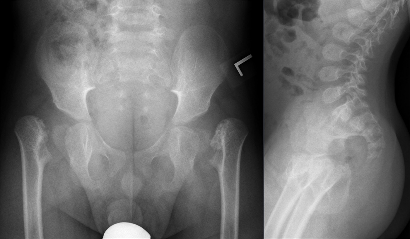
Bilateral coxa vara in a skeletal dysplasia. Antero-posterior pelvis (left) showing bilateral coxa vara with vertical proximal-femoral physes, and a lateral spine (right) with ovoid vertebrae, in spondyloepiphyseal dysplasia. Hellerhoff, Wikimedia Commons (CC BY-SA 4.0).
Bilateral coxa vara in a skeletal dysplasia. Antero-posterior pelvis (left) showing bilateral coxa vara with vertical proximal-femoral physes, and a lateral spine (right) with ovoid vertebrae, in spondyloepiphyseal dysplasia. Hellerhoff, Wikimedia Commons (CC BY-SA 4.0).
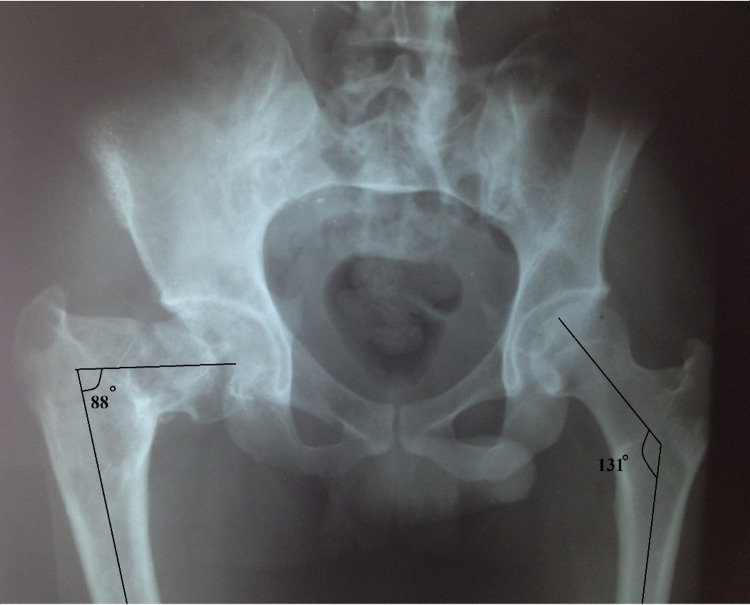
Secondary coxa vara - the “shepherd’s crook” of fibrous dysplasia. Antero-posterior pelvis: the right proximal femur shows severe varus (neck-shaft angle 88°) with ground-glass change, against a normal 131° on the left. Pathak SK et al., Cureus 2021;13(7):e16485, Fig. 2 (CC BY).
Secondary coxa vara - the “shepherd’s crook” of fibrous dysplasia. Antero-posterior pelvis: the right proximal femur shows severe varus (neck-shaft angle 88°) with ground-glass change, against a normal 131° on the left. Pathak SK et al., Cureus 2021;13(7):e16485, Fig. 2 (CC BY).
Part III - Developmental coxa vara: epidemiology and etiology
Developmental coxa vara has an incidence of roughly 1 in 25,000 live births and affects boys and girls equally. It is bilateral in 30-50% of cases, and an autosomal-dominant inheritance with incomplete penetrance has been described.[8] The most widely accepted cause is a primary defect in the endochondral ossification of the medial femoral neck, producing dystrophic bone that fatigues under weight-bearing. An analogy is often drawn with infantile Blount’s disease of the proximal tibia, though the two have not been shown to coexist. Vascular insult and abnormal intrauterine pressure are alternative theories.[9] The term “developmental” was coined by Duncan (1938) after it became clear that some “congenital” cases had normal hips at birth and only developed the varus by 2-3 years of age.[10]
Part IV - Pathoanatomy
The defining lesion is an ossification defect of the inferomedial femoral neck that appears radiographically as a triangular metaphyseal fragment (Fairbank’s triangle) set in an inverted-Y pattern between two radiolucent bands; CT shows it behaves like a Salter-Harris type II separation through the defective neck.[11] Because the physis becomes more vertical, body weight loads it in shear as well as compression. By the Hueter-Volkmann principle the medial physis is restrained while the lesion fails to ossify, so the varus is relentlessly progressive once established.[12] Histology shows disorganised chondrocyte columns, irregular trabeculae and fibrous infiltration of the metaphysis. The net deformity is a short neck with a proximally migrated, overgrown greater trochanter, reduced anteversion and an insufficient abductor lever arm.[13]
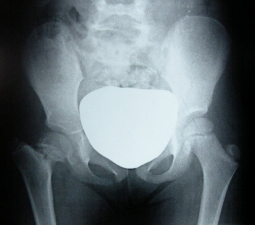
Developmental coxa vara. Antero-posterior pelvis of a child with left-sided coxa vara: the reduced neck-shaft angle, shortened neck and high greater trochanter (the central white shield is gonad protection). Mazzini JP et al., Cases Journal 2009;2:8130, Fig. 1 (CC BY 3.0).
Developmental coxa vara. Antero-posterior pelvis of a child with left-sided coxa vara: the reduced neck-shaft angle, shortened neck and high greater trochanter (the central white shield is gonad protection). Mazzini JP et al., Cases Journal 2009;2:8130, Fig. 1 (CC BY 3.0).
Part V - Clinical features
Developmental coxa vara usually presents after the child begins to walk and before about 6 years of age, typically with a painless limp rather than pain.[14] The hallmark is a positive Trendelenburg sign and a Trendelenburg (abductor) lurch from the shortened abductor lever arm; bilateral disease produces a waddling gait with increased lumbar lordosis (resembling bilateral developmental dysplasia of the hip). The greater trochanter is prominent and high, and hip movement is reduced, most notably abduction (blocked by trochanteric impingement) and internal rotation (from the loss of anteversion).[15] Limb-length discrepancy in unilateral cases is usually mild, rarely more than 3 cm at maturity even untreated. The examination should also look for the disproportion of a skeletal dysplasia and the features of PFFD.
Part VI - Radiographic assessment
The standing antero-posterior pelvic radiograph is the key study. The measurements to know are:
- The Hilgenreiner-epiphyseal angle (HEA, the H-E angle) is the angle between Hilgenreiner’s horizontal line and a line along the proximal femoral (capital) physis; the higher the angle, the more vertical the physis. The normal value is low (about 16° in Weinstein’s series of 100 healthy patients, with ~25° cited in other texts), and in developmental coxa vara it is typically 40-70°.[16] It is the single best predictor of progression and the basis of the surgical threshold (below).
- The neck-shaft angle indicates coxa vara when below about 110°, with surgery generally considered once it falls to 90-100°; it is less reliable than the HEA for predicting progression and recurrence.[17]
- The articulotrochanteric distance (joint surface to trochanteric tip) helps localise the deformity to the physeal/intertrochanteric region (decreased) versus the subtrochanteric region (normal).[18]
- The associated signs: the vertical, widened physis, Fairbank’s triangular fragment, the short neck, reduced anteversion and secondary acetabular dysplasia.[19]
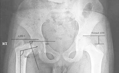
The Hilgenreiner-epiphyseal angle. Antero-posterior pelvis with the HE angle, neck-shaft angle (NSA) and articulo-trochanteric distance (ATD) drawn on the affected hip, the normal ATD shown opposite. Hefny H et al., Strategies Trauma Limb Reconstr 2013;8(3):161-167, Fig. 1 (CC BY).
The Hilgenreiner-epiphyseal angle. Antero-posterior pelvis with the HE angle, neck-shaft angle (NSA) and articulo-trochanteric distance (ATD) drawn on the affected hip, the normal ATD shown opposite. Hefny H et al., Strategies Trauma Limb Reconstr 2013;8(3):161-167, Fig. 1 (CC BY).
The differential (PFFD/congenital short femur, the skeletal dysplasias, and the metabolic forms such as rickets, renal osteodystrophy, osteogenesis imperfecta and fibrous dysplasia) must be excluded before labelling a case “developmental”.
Part VII - Natural history
Weinstein established that the natural history is governed by the HEA.[20] An HEA below 45° is usually stable: the neck defect heals spontaneously and the deformity does not progress. An HEA of 45-59° is an indeterminate “grey zone” requiring close monitoring, and an HEA of 60° or more progresses almost invariably. Untreated severe disease leads to worsening varus and neck shortening, occasionally a pseudarthrosis of the femoral neck with the trochanter ending up above the femoral head, and early degenerative change with a persistent Trendelenburg gait. The chance of restoring a normal hip falls rapidly after about age 8.[21]
Part VIII - Treatment
Observation
A child with an HEA below 45° and no symptoms is observed with serial radiographs until maturity; an HEA of 45-59° is followed closely, since progression is itself an indication to operate. Non-operative measures (bracing, traction) do not alter the natural course.[22]
Valgus osteotomy
The definitive treatment is a valgus (intertrochanteric or subtrochanteric) proximal femoral osteotomy, indicated for an HEA of 60° or more, documented progression, a neck-shaft angle falling to 90-100°, or a symptomatic Trendelenburg gait.[23] Its goals (Borden) are to bring the varus into the normal range, convert physeal loading from shear back to compression (which lets the neck defect ossify), restore the abductor length-tension relationship, and correct any limb-length discrepancy.[24] The key technical targets are:
- Correct the HEA to below about 38°. This is the strongest determinant of success: correction to under 38° gives roughly 95% freedom from recurrence, whereas leaving the HEA above 40° leads to revision in the great majority. The HEA is a far better guide than the neck-shaft angle, correction of which alone does not prevent recurrence.[25]
- Overcorrect the neck-shaft angle into valgus (to about 140-150°), with derotation added for the relative retroversion and, where needed, an adductor release or lateralisation of the distal fragment to improve the abductor lever arm.[26]
The osteotomy is done at the intertrochanteric or subtrochanteric level (neck osteotomies are avoided), and no single technique is proven superior. Named options include the Pauwels Y-shaped intertrochanteric osteotomy (a lateral closing wedge whose angle equals the HEA minus the normal ~16°), the Langenskiöld and Borden valgus osteotomies, and the lateral closing-wedge valgisation osteotomy with trochanteric advancement.[27] Fixation is matched to the child’s size: multiple smooth K-wires (with cerclage or a tension band) in the very young, and a blade plate, paediatric hip screw or a pre-contoured proximal-femoral plate in larger children. Where the bone is generally weak, as in osteogenesis imperfecta, an intramedullary device is used. After adequate valgus correction the triangular neck defect ossifies and the physis horizontalises over the following months.
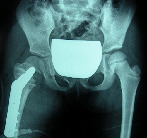
Valgus osteotomy for coxa vara. The same child after a valgus osteotomy of the left proximal femur fixed with an angled (blade) plate, with greater-trochanter epiphysiodesis; the neck-shaft angle is restored. Mazzini JP et al., Cases Journal 2009;2:8130, Fig. 3 (CC BY 3.0).
Valgus osteotomy for coxa vara. The same child after a valgus osteotomy of the left proximal femur fixed with an angled (blade) plate, with greater-trochanter epiphysiodesis; the neck-shaft angle is restored. Mazzini JP et al., Cases Journal 2009;2:8130, Fig. 3 (CC BY 3.0).
Secondary coxa vara
Acquired coxa vara is treated by addressing the underlying disease together with a valgus osteotomy: realignment (with grafting/fixation for a nonunion) after trauma, and valgus osteotomy of the shepherd’s-crook femur in fibrous dysplasia. In osteogenesis imperfecta, where coxa vara is present in around 60% of type III, correction uses a subtrochanteric valgus osteotomy stabilised with a telescoping intramedullary rod (the Fassier technique).[28]
Part IX - Complications and outcomes
The major problem is recurrence, reported in around half of cases overall and determined chiefly by the adequacy of HEA correction (under 38° protects against it) rather than by the patient’s age, the type of osteotomy, the implant, or the aetiology.[29] Premature closure of the proximal femoral physis occurs in the great majority of operated hips (about 89%, usually within 1-2 years), producing trochanteric overgrowth, limb shortening and recurrent varus. This is managed by greater-trochanteric apophysiodesis or transfer and, for recurrent varus, a repeat valgus osteotomy.[30] Other risks are avascular necrosis (the medial femoral circumflex artery must be protected during the osteotomy), persistent abductor weakness and Trendelenburg gait, and residual limb-length discrepancy. The aim that ties the whole topic together is a horizontal physis (HEA under 38°), a normal-to-slightly-valgus neck-shaft angle, and a competent abductor lever arm.[31]
Bulgarian terminology (Боев / Boychev tradition) - glossary
The following Bulgarian equivalents bridge the international literature with the terminology of the Bulgarian school of orthopaedics (in the tradition of Бойчо Бойчев / Boycho Boychev) used in the state examination.
| English term | Bulgarian term (Cyrillic) | Transliteration |
|---|---|---|
| Coxa vara | Кокса вара | Koksa vara |
| Neck-shaft angle | Шийно-диафизарен ъгъл | Shiyno-diafizaren agal |
| Femoral neck | Бедрена шийка | Bedrena shiyka |
| Hip joint | Тазобедрена става | Tazobedrena stava |
| Developmental coxa vara | Предизвикана (развитийна) кокса вара | Predizvikana (razvitiyna) koksa vara |
| Congenital coxa vara | Вродена кокса вара | Vrodena koksa vara |
| Acquired / secondary coxa vara | Придобита (вторична) кокса вара | Pridobita (vtorichna) koksa vara |
| Hilgenreiner-epiphyseal angle | Ъгъл на Хилгенрайнер-епифизата | Agal na Hilgenrayner-epifizata |
| Triangular fragment (Fairbank) | Триъгълен фрагмент (на Феърбанк) | Triagalen fragment (na Fearbank) |
| Growth plate (physis) | Растежна зона (физа) | Rastezhna zona (fiza) |
| Greater trochanter | Голям трохантер | Golyam trohanter |
| Trendelenburg gait | Походка на Тренделенбург | Pohodka na Trendelenburg |
| Abductor insufficiency | Недостатъчност на абдукторите | Nedostatachnost na abduktorite |
| Valgus osteotomy | Валгизираща остеотомия | Valgizirashta osteotomiya |
| Subtrochanteric | Субтрохантерен | Subtrohanteren |
| Limb-length discrepancy | Разлика в дължината на крайниците | Razlika v dalzhinata na kraynitsite |
| Shepherd’s-crook deformity | Деформитет тип „овчарска гега” | Deformitet tip “ovcharska gega” |
| Proximal femoral focal deficiency | Проксимален фокален бедрен дефицит | Proksimalen fokalen bedren defitsit |
| Avascular necrosis | Аваскуларна некроза | Avaskularna nekroza |
Image attributions
(Figure attributions and licences are listed in the figure MANIFEST and inserted with each image. All images are openly licensed [CC0 / CC BY / CC BY-SA] or used under their stated terms; any non-commercial [NC] item is flagged as such and must not be used in a commercial product.)
References
-
The Pediatric and Adolescent Hip, ch. 7 Coxa Vara, p. 193; Lovell & Winter, Pediatric Orthopaedics, “Other Conditions of the Hip”, p. 1309.
-
Lovell, p. 1309; Pediatric and Adolescent Hip, pp. 193, 198.
-
Pediatric and Adolescent Hip, pp. 196, 198; Lovell, p. 1310. (Some texts quote a slightly higher adult range of ~120-135°.)
-
Pediatric and Adolescent Hip, p. 196; Lovell, p. 1313.
-
Lovell, p. 1309, Table 26-1; Pediatric and Adolescent Hip, pp. 193-195.
-
Pediatric and Adolescent Hip, p. 195, Table 7.1; Lovell, pp. 1311-1312.
-
Lovell, p. 1309.
-
Lovell, p. 1309; Pediatric and Adolescent Hip, p. 197.
-
Lovell, pp. 1309-1310.
-
Pediatric and Adolescent Hip, p. 194.
-
Pediatric and Adolescent Hip, pp. 193, 199; Lovell, p. 1310.
-
Pediatric and Adolescent Hip, p. 196; Lovell, p. 1312.
-
Pediatric and Adolescent Hip, pp. 196-197; Lovell, p. 1313.
-
Pediatric and Adolescent Hip, p. 197; Lovell, p. 1309.
-
Lovell, pp. 1309-1310; Pediatric and Adolescent Hip, pp. 197-198.
-
Lovell, p. 1310; Pediatric and Adolescent Hip, p. 198.
-
Pediatric and Adolescent Hip, p. 198; Lovell, p. 1310; Campbell’s, p. 1379.
-
Pediatric and Adolescent Hip, pp. 198-199.
-
Pediatric and Adolescent Hip, p. 199; Lovell, Table 26-2.
-
Pediatric and Adolescent Hip, p. 197; Lovell, p. 1312.
-
Lovell, p. 1312; Campbell’s, p. 1379.
-
Pediatric and Adolescent Hip, p. 199; Lovell, p. 1312.
-
Pediatric and Adolescent Hip, p. 199; Lovell, p. 1313; Campbell’s, p. 1379.
-
Lovell, p. 1312.
-
Pediatric and Adolescent Hip, pp. 200, 205; Lovell, pp. 1323-1324.
-
Campbell’s, p. 1380; Lovell, p. 1322.
-
Lovell, pp. 1315-1322; Tachdjian valgus-osteotomy technique; Pediatric and Adolescent Hip, pp. 200-204.
-
Sabharwal, Pediatric Lower Limb Deformities, pp. 270-272, 575; Pediatric and Adolescent Hip, pp. 712-715; Lovell, p. 1312.
-
Pediatric and Adolescent Hip, p. 200; Lovell, pp. 1322-1324.
-
Lovell, p. 1324.
-
Lovell, pp. 1322-1324; Pediatric and Adolescent Hip, pp. 199-200.