Contents
- Introduction and scope
- Part I - Embryology of the spine: the shared foundation
- Part II - Congenital scoliosis
- Part III - Congenital kyphosis
- Part IV - Klippel-Feil syndrome
- Part V - Sprengel deformity
- Part VI - Cervical ribs and thoracic outlet syndrome
- Part VII - Spina bifida and myelomeningocele
- Bulgarian terminology (Боев / Boychev tradition) - glossary
- Image attributions
Introduction and scope
This summary covers the congenital malformations of the spine and the cervico-thoracic junction that the state-examination syllabus groups together: congenital scoliosis and congenital kyphosis (deformities arising from anomalous vertebral development), Klippel-Feil syndrome (congenital cervical fusion), Sprengel deformity (the congenital high scapula), cervical ribs and the thoracic outlet syndromes they cause, and the dysraphic spectrum culminating in spina bifida / myelomeningocele. These conditions share a common embryological thread: a disturbance of somitogenesis, neurulation, or mesodermal migration during the first eight weeks of intra-uterine life. They also cluster clinically, so a child found to have one is screened for the others.
Three principles run through the whole topic. The first is that a congenital vertebral anomaly is present at birth, but the deformity it produces develops over time as the spine grows asymmetrically; the rate and direction of that growth, not the size of the curve on the first film, determine the prognosis. The second is that a bony vertebral anomaly is a marker for anomalies of the structures that form alongside the vertebrae (the spinal cord, heart, kidneys and limbs), so the radiograph begins a work-up rather than ending it. The third is that the neurological axis must be imaged before any corrective spine surgery, because an unrecognised tethered cord, diastematomyelia or Chiari malformation turns a routine correction into a paralysing one.
Part I - Embryology of the spine: the shared foundation
A working grasp of normal spinal development makes every malformation in this topic intelligible, because each is a named failure of a specific developmental step.
During the third and fourth weeks of gestation the paraxial mesoderm on either side of the notochord and neural tube segments into paired blocks called somites, in a strict head-to-tail sequence governed by a molecular “segmentation clock” oscillating through the Notch signalling pathway.[1] Each somite differentiates into a ventral sclerotome, which gives rise to the vertebra, and a dorsal dermatomyotome, which gives rise to the overlying muscle and dermis. A key re-segmentation step then occurs: the caudal half of one sclerotome fuses with the cephalic half of the next to build a single vertebral body, so that each definitive vertebra is assembled from two adjacent somites and the segmental nerves and vessels come to lie at the level of the disc.
Two distinct things can therefore go wrong, and they map directly onto the two great categories of congenital vertebral malformation:
- A failure of formation, in which one part of a vertebra never develops, producing a wedge vertebra or a hemivertebra.
- A failure of segmentation, in which two or more vertebrae fail to separate, producing a block vertebra (if symmetrical) or an unsegmented bar (if one-sided).
The neural tube closes over the same window (by days 27-28), and the scapula forms opposite the lower cervical somites in the third week and normally descends to the thorax by the eighth. This shared timetable explains the constant clinical companions of this topic: why a child with fused cervical vertebrae (Klippel-Feil) so often has an undescended scapula (Sprengel), a congenital scoliosis, and a renal anomaly, all stamped in during the same few embryonic weeks.[2]
Part II - Congenital scoliosis
Definition and the central concept
Congenital scoliosis is a lateral curvature of the spine caused by congenital vertebral malformations that interrupt the normal, balanced longitudinal growth of the spine. The malformation is always present at birth, but (and this is the examiner’s favourite point) the scoliosis itself need not be: it develops and progresses as the child grows, because one side of the spine grows while the other is tethered or deficient.[3]
This distinguishes it sharply from idiopathic scoliosis, in which there is no vertebral malformation. The congenital curve is typically short, sharply angular and rigid, and a large curve can be present in a very young child. Conversely, a curve appearing in the first months of life is not necessarily congenital; it may be infantile idiopathic scoliosis, which has no underlying anomaly and a quite different (often resolving) natural history.[4]
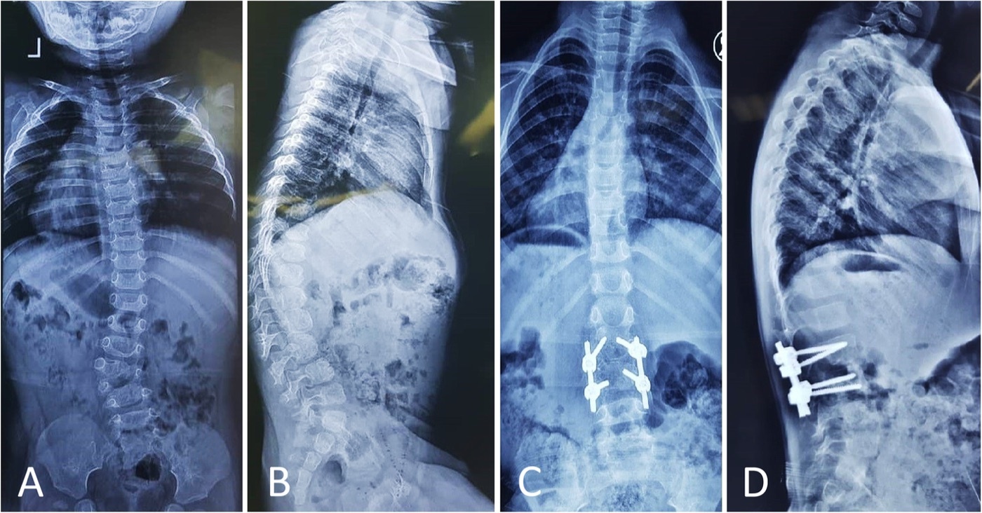
Congenital scoliosis from a lumbar hemivertebra. Standing antero-posterior (A) and lateral (B) radiographs of an 18-month-old boy with a fully segmented lumbar hemivertebra producing a 39° congenital curve; (C, D) after posterior instrumented correction. Zarei M et al., J Orthop Surg Res 2021;16:271, Fig. 3 (CC BY 4.0).
Congenital scoliosis from a lumbar hemivertebra. Standing antero-posterior (A) and lateral (B) radiographs of an 18-month-old boy with a fully segmented lumbar hemivertebra producing a 39° congenital curve; (C, D) after posterior instrumented correction. Zarei M et al., J Orthop Surg Res 2021;16:271, Fig. 3 (CC BY 4.0).
Epidemiology and aetiology
The incidence is roughly 1 in 1,000 live births, with a female predominance of about 2.5 : 1.[5] The cause is multifactorial, combining genetic susceptibility with intra-uterine insult. The best-characterised experimental teratogen is carbon monoxide (which binds haemoglobin with 200-300 times the affinity of oxygen): exposure of pregnant mice on the ninth gestational day produces spinal malformations in around 70% of offspring, showing that a brief hypoxic insult at the moment of somitogenesis can reproduce the human malformation.[6] Other implicated insults are maternal hypoxia, alcohol (fetal alcohol syndrome), the anticonvulsants valproic acid and phenytoin, retinoic acid, hyperthermia, and maternal diabetes. Candidate genes lie in the somitogenesis pathways already named (DLL3, TBX6, MESP2, LFNG, HES7, PAX1, WNT3A, T (Brachyury)), and familial recurrence is around 3%; an isolated hemivertebra is usually sporadic, whereas multiple anomalies carry a 5-10% sibling risk.[7]
Classification - the core examination material
The basic system is the MacEwen classification as modified by Winter and colleagues (1968) and adopted by the Scoliosis Research Society. It recognises three categories: failure of formation, failure of segmentation, and mixed (often unclassifiable). Pure forms are uncommon, though; most patients have a mixture, with one type predominating.[8]
Failure of formation
Part of a vertebra fails to develop. The spectrum runs from partial to complete:
- Wedge vertebra: both pedicles are present but one side of the body is hypoplastic, giving a trapezoidal shape; this is a partial unilateral failure of formation.
- Hemivertebra: the complete form, in which only one pedicle and
one half of the vertebral body are present. A hemivertebra is not
an extra wedge of bone inserted into the spine; it is the normally
formed half of a vertebra whose opposite half failed to
develop.[9] The hemivertebra is sub-classified by the state of the
discs above and below it, because that determines its growth
potential and hence its danger:
- Fully segmented (“free”) hemivertebra: normal discs and growth plates both above and below. It therefore continues to grow longitudinally on the convexity and is progressive.
- Semisegmented hemivertebra: a functional disc on one side only, fused on the other; intermediate, less progressive.
- Non-segmented hemivertebra: fused to both adjacent vertebrae, with no functional disc on either side; little growth potential, so it is benign and essentially non-progressive on its own.
- Incarcerated hemivertebra: “tucked into” a recess formed by compensatory notching of the adjacent vertebrae, so it sits within the line of the spine and causes little or no curve (benign).
- Hemimetameric shift (segmental shift): a hemivertebra on one side balanced by a contralateral hemivertebra one or more segments away; the two may cancel out the coronal balance but can still create two progressive curves.[10]
High-yield contrast: a fully segmented hemivertebra has growth plates above and below and is progressive; a non-segmented hemivertebra is fused and benign.
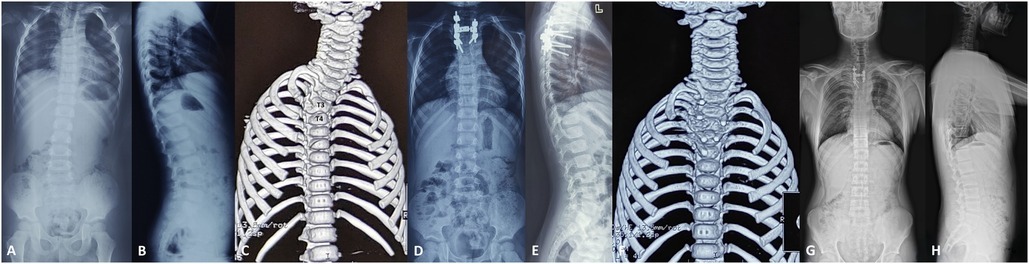
Hemivertebra on three-dimensional CT (failure of formation). Panel C shows a right T3-T4 hemivertebra in a 3-year-old; the surrounding panels are the corresponding pre-operative radiographs. Zhang C et al., Front Surg 2025;12:1473800, Fig. 2 (CC BY 4.0).
Hemivertebra on three-dimensional CT (failure of formation). Panel C shows a right T3-T4 hemivertebra in a 3-year-old; the surrounding panels are the corresponding pre-operative radiographs. Zhang C et al., Front Surg 2025;12:1473800, Fig. 2 (CC BY 4.0).
Failure of segmentation
An abnormal bony bridge (“bar”) tethers growth on the affected side:
- Block vertebra: bilateral, symmetrical failure of segmentation (the disc is absent on both sides). The tether is symmetrical, so it produces little or no deformity; this is the most benign anomaly of all.
- Unilateral unsegmented bar: segmentation fails on one side only, often across several segments and involving both the bodies and the posterior elements. The tethered concave side cannot grow while the convex side continues to elongate, producing a rigid, relentlessly progressive curve, the single worst pure anomaly.[11]
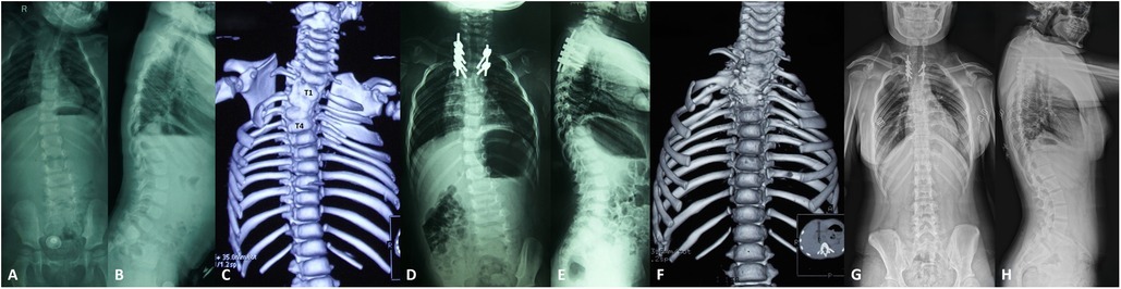
Unilateral unsegmented bar on three-dimensional CT (failure of segmentation). Panel C shows a left T1-T4 unsegmented bar in a 2-year-old - the most progressive of the pure anomalies. Zhang C et al., Front Surg 2025;12:1473800, Fig. 1 (CC BY 4.0).
Unilateral unsegmented bar on three-dimensional CT (failure of segmentation). Panel C shows a left T1-T4 unsegmented bar in a 2-year-old - the most progressive of the pure anomalies. Zhang C et al., Front Surg 2025;12:1473800, Fig. 1 (CC BY 4.0).
Mixed / unclassifiable
Formation and segmentation defects coexist. The archetype, and the most progressive anomaly of all, is the unilateral unsegmented bar with a contralateral hemivertebra: a rigid concave tether on one side, an actively growing hemivertebra pushing on the other.[12] Roughly 10% of malformations cannot be cleanly classified, particularly when they are not yet fully ossified in infancy.
For completeness, Winter’s numbered scheme runs: I unclassifiable; II rib fusion; III unilateral partial failure of formation (wedge); IV unilateral complete failure of formation (hemivertebra); V bilateral failure of segmentation (block); VI unilateral failure of segmentation (bar).[13] The modern Kawakami CT-based three-dimensional classification adds the important concept of discordancy (anterior and posterior anomalies occurring at different levels), which matters greatly when planning the level of surgery.[14]
Natural history and the ranking of progression risk
Two landmark studies define the natural history: Winter, Moe and Eilers (1968) and McMaster and Ohtsuka (1982).[15] Progression depends on (1) the type of anomaly, (2) its location, and (3) the growth remaining, and, crucially, not on the initial size of the curve, since some small curves progress fastest.
The ranking of progression risk, from worst to most benign, is one of the most testable facts in the whole topic:
| Anomaly | Typical progression |
|---|---|
| Unilateral unsegmented bar + contralateral hemivertebra (worst) | ≈ 5-10°/year |
| Unilateral unsegmented bar alone | ≈ 5°/year |
| Double convex (two) hemivertebrae | high |
| Fully segmented (free) hemivertebra | moderate |
| Wedge vertebra | mild-moderate |
| Block vertebra (most benign) | < 1-2°/year |
In McMaster’s series, only 11% of curves were non-progressive, while about 75% progressed significantly; the risk of reaching more than 40° by maturity ranges across untreated series from roughly 37% to 84%.[16]
Two further modifiers matter. By region, for any given anomaly deterioration is least in the upper thoracic spine, greater in the mid-thoracic, and worst at the thoracolumbar junction; a small cervicothoracic curve, although it progresses less, produces disproportionate cosmetic deformity (head tilt, shoulder asymmetry).[17] By age, the spine has two periods of rapid growth, infancy (the first five years) and the adolescent growth spurt (roughly ages 10-14), separated by a relatively quiescent mid-childhood interval; deformity that is clinically evident in the first year of life carries the worst prognosis.[18]
Associated anomalies - the mandatory work-up
Because the vertebrae form alongside the spinal cord and the urogenital, cardiac and limb systems, congenital vertebral malformations are accompanied by other malformations in up to 60% of patients.[19] Every child therefore needs a structured screen.
- Intraspinal (neuraxial) anomalies, about 20-40%. This is the most important association for the surgeon. With plain radiographs and myelography McMaster found 18%, but with MRI the figure rises to 30-38%. The commonest single lesion is diastematomyelia (split cord), present in around 20% of congenital scoliosis, followed by tethered cord, intraspinal lipoma, syringomyelia and Chiari malformation. The yield is highest with lumbosacral hemivertebrae. Because physical examination alone misses many of these, MRI of the whole spine is mandatory before any corrective surgery: an unrecognised tethered cord or diastematomyelic spur turns corrective distraction into a cord injury.[20]
- Genitourinary anomalies, about 20-40%. Most often unilateral renal agenesis, but also duplex collecting systems, horseshoe or ectopic kidney, obstruction and reflux; usually silent. Screen all patients with a renal ultrasound.[21]
- Cardiac anomalies, about 10-26%. Most commonly atrial and ventricular septal defects. Screen with echocardiography.[22]
- VACTERL / VATER association (Vertebral, Anal atresia, Cardiac, Tracheo-Esophageal fistula, Renal, Limb) and named syndromes such as Klippel-Feil, Sprengel, Goldenhar, Jarcho-Levin (spondylothoracic dysostosis), Alagille, trisomy 18, diabetic embryopathy.[23]
- Pulmonary consequences. For a given Cobb angle, vital-capacity loss is about 15% greater than in idiopathic scoliosis, and the concept of thoracic insufficiency syndrome, the inability of the thorax to support normal lung growth and function, dominates management of the young child with fused ribs.[24]
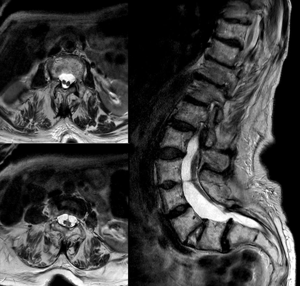
Diastematomyelia (split-cord malformation). Axial (left) and sagittal (right) T2-weighted MRI in which the cord is divided into two hemicords - the commonest intraspinal anomaly accompanying congenital scoliosis, and a reason whole-spine MRI precedes correction. Hellerhoff, Wikimedia Commons (CC BY-SA 3.0).
Diastematomyelia (split-cord malformation). Axial (left) and sagittal (right) T2-weighted MRI in which the cord is divided into two hemicords - the commonest intraspinal anomaly accompanying congenital scoliosis, and a reason whole-spine MRI precedes correction. Hellerhoff, Wikimedia Commons (CC BY-SA 3.0).
Clinical and radiological evaluation
Examination records sitting and standing height, trunk and shoulder balance, head tilt, curve rigidity and a careful neurological assessment of the lower limbs (power, sensation, abdominal and deep-tendon reflexes). The skin of the back is inspected for markers of dysraphism (a hairy patch, naevus, haemangioma, lipoma, dimple or abnormal pigmentation), and the feet for cavus, asymmetric calves or a unilaterally small foot, all of which point to an occult cord anomaly. The absence of neurological signs does not exclude intraspinal pathology.[25]
Imaging proceeds in three tiers:
- Plain radiographs: supine in the infant, standing once the child stands. Cobb measurement is difficult because the endplates and pedicles are distorted; the cardinal rule is always to compare with the earliest film, since the inter-observer error can exceed the annual change.[26]
- CT with three-dimensional reconstruction: the best modality for the bony anatomy and for planning resection; reserved for complex cases rather than routine follow-up because of the radiation dose (low-dose biplanar EOS imaging mitigates this).[27]
- MRI: the standard for the neural axis; image the whole spine at presentation and always before correction.[28]
Treatment
The governing principle is that it is easier to prevent a deformity than to correct one, so documented progression is treated early; the goals at maturity are a balanced spine, maximised pulmonary function, optimised trunk height and no neurological deterioration.[29]
- Observation: serial radiographs every 4-6 months for low-risk, balanced anomalies (block vertebra, hemimetameric shift, non-segmented hemivertebra), always compared with the earliest film.[30]
- Bracing is largely ineffective for the short, rigid congenital curve itself; it has a role only in controlling long, flexible compensatory curves above or below the anomaly, and only the Milwaukee brace has documented benefit. It is inappropriate for a unilateral bar with contralateral hemivertebra or for congenital kyphosis.[31]
- In-situ posterior fusion (growth arrest) is the classic operation for a short, rigid, progressive curve in a balanced spine, the textbook indication being an early-diagnosed unilateral unsegmented bar. Because the fused segment is already devoid of growth, “nature makes the spine short, not the surgeon.”[32]
- Convex hemiepiphysiodesis / hemiarthrodesis (convex growth arrest) arrests convex growth to let the concavity slowly correct the curve; ideal in a child under five with an isolated, progressive lumbar curve under 40-50° and no significant kyphosis.[33]
- Hemivertebra excision is the one operation that gives immediate correction over a short segment; ideal for a young child with an isolated, progressive hemivertebra at the thoracolumbar, lumbar or lumbosacral junction causing coronal imbalance. Modern posterior-only excision with transpedicular instrumentation (Ruf and Harms) achieves around 75% correction with a low neurological complication rate.[34]
- Growth-friendly constructs are used for long curves in the very young child, where early fusion would stunt thoracic and pulmonary growth: growing rods (distraction-based, lengthened roughly every six months) and VEPTR (Vertical Expandable Prosthetic Titanium Rib) for congenital scoliosis with fused ribs / thoracic insufficiency syndrome.[35]
- Vertebral column resection / osteotomy at maturity is reserved for severe, rigid, multiplanar deformity; powerful but carrying a high neurological risk, mandating multimodality cord monitoring.[36]
Safety note. Congenital scoliosis is the most common cause of paraplegia induced by corrective instrumentation; dysraphism must be excluded and released before correction, and cord monitoring is mandatory.[37]
Part III - Congenital kyphosis
Definition and why it matters
Congenital kyphosis is a sagittal-plane deformity arising from anomalous vertebral development. It is rarer than congenital scoliosis but far more dangerous, because its great complication is paraplegia: congenital kyphosis is the most common cause of paraplegia among the non-infectious spinal deformities (worldwide, tuberculous kyphosis remains the leading cause overall).[38]
The Winter classification
Winter, Moe and Wang (1973) divided congenital kyphosis into three types, and the distinction is high-yield because it inverts the rule learned for scoliosis:
- Type I, failure of formation. The anterior part of the vertebral body is absent or hypoplastic while the posterior elements develop normally, so the spine collapses forward over a sharp apex. This is the dangerous type and the most common type, and it is the one that causes congenital paraplegia.
- Type II, failure of segmentation. A progressively ossifying anterior unsegmented bar tethers anterior growth while the posterior column continues to grow, producing a gradual, less angular kyphosis. It is less progressive, less deforming, and does not cause paraplegia; it is commonest at the thoracolumbar junction and often forces a compensatory lumbar hyperlordosis that can be more symptomatic than the kyphosis itself.
- Type III, mixed.[39]
High-yield reversal: in scoliosis the worst anomaly is a failure of segmentation (the unilateral bar); in kyphosis the worst anomaly is a failure of formation (Winter type I).
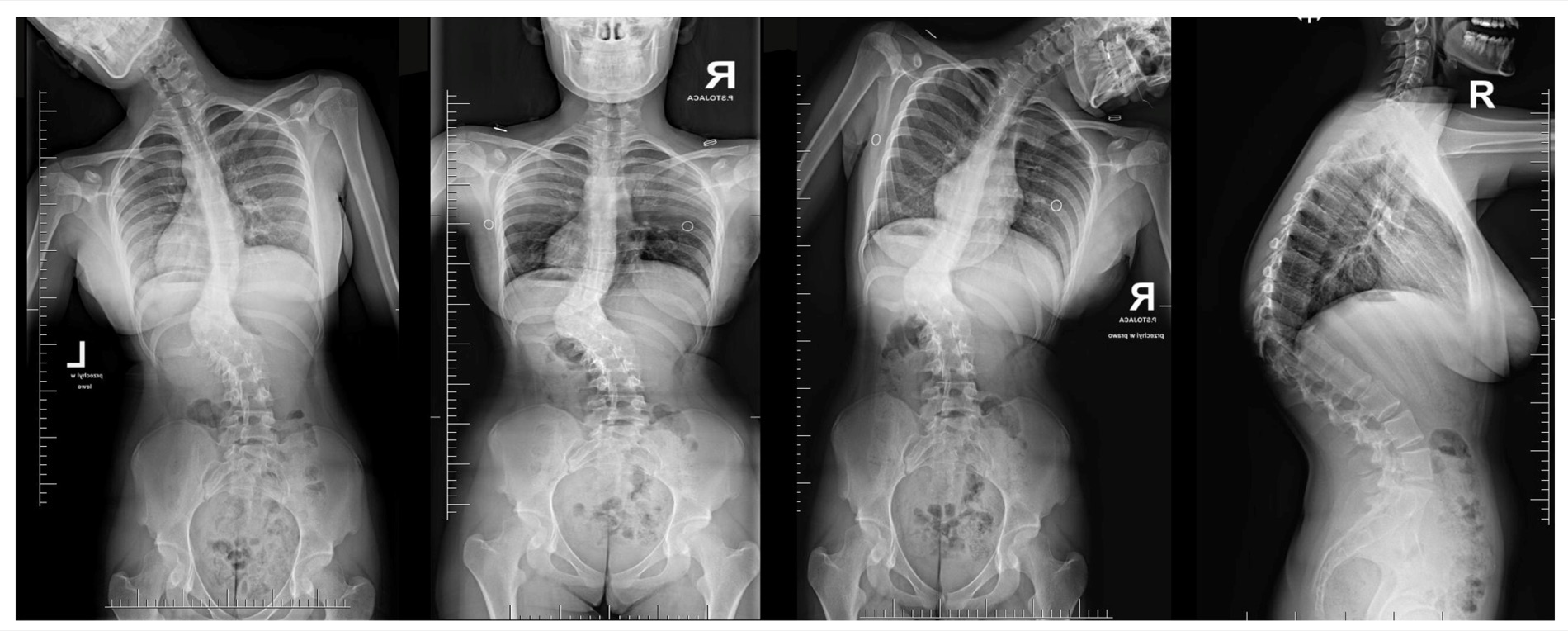
Congenital kyphosis. Standing antero-posterior, side-bending and lateral radiographs of a severe, rigid congenital kyphosis; the sharp angular kyphus is seen on the lateral (right-hand) view. Grabala P et al., Medicina (Kaunas) 2024;60:897, Fig. 2 (CC BY 4.0).
Congenital kyphosis. Standing antero-posterior, side-bending and lateral radiographs of a severe, rigid congenital kyphosis; the sharp angular kyphus is seen on the lateral (right-hand) view. Grabala P et al., Medicina (Kaunas) 2024;60:897, Fig. 2 (CC BY 4.0).
Why type I causes paraplegia
In a failure of formation the anterior column collapses and the spinal cord is draped, under tension, over a sharp angular apex. The risk is greatest when the apex lies between T4 and T9, the “watershed” zone of the cord’s blood supply, and it rises during the adolescent growth spurt; progressive anterior collapse there produces cord compression and ischaemia.[40]
Treatment
There is no effective non-operative treatment: bracing, exercise and manipulation do not alter the natural history. Management is therefore surgical and, for type I, early.
- Small curves in the young child: posterior fusion (in situ), historically combined with casting, can achieve gradual correction while anterior growth potential remains.
- Larger curves: a combined anterior (release ± strut graft) and posterior approach, most effective if done early; modern posterior-only techniques (pedicle subtraction osteotomy, the “eggshell” decancellation procedure, posterior vertebral column resection) increasingly replace the anterior approach.
- Any neurological sign mandates decompression as well as stabilisation.[41]
The mirror-image deformity, congenital lordosis (from a posterior unsegmented bar), is correctable only by a two-stage procedure and produces a restrictive ventilatory defect.
Part IV - Klippel-Feil syndrome
Definition, eponym and embryology
Klippel-Feil syndrome is the congenital fusion (failure of segmentation) of two or more cervical vertebrae. Maurice Klippel and André Feil published the first complete description in 1912, in a patient with a strikingly short neck, a low posterior hairline and severe restriction of neck movement, in whom post-mortem revealed a complete cervical fusion; Feil added the original classification in 1919.[42] It results from a failure of normal segmentation of the cervical somites during the third to eighth weeks of gestation and has an incidence of about 0.7%; familial cases occur, and the implicated pathways are again the somitogenesis genes (PAX1, GDF6, the Notch pathway).[43]
The classic triad
The textbook presentation is a triad of (1) a short neck, (2) a low posterior hairline, and (3) limited neck motion. The examiner’s point is that the full triad is present in fewer than half of patients; the most consistent single finding is limitation of neck movement (rotation and lateral bending are lost before flexion-extension, which is often preserved through hypermobility at the unfused segments). Obvious facial asymmetry, torticollis or neck webbing (in the extreme, pterygium colli) occurs in fewer than 20%.[44]
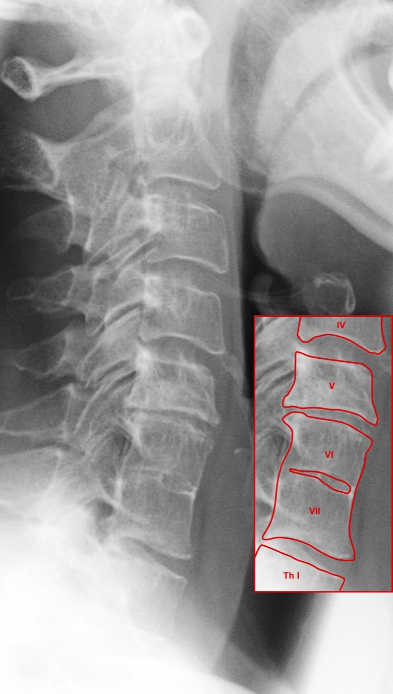
Klippel-Feil syndrome. Lateral cervical radiograph; the annotated inset marks congenital fusion at C6-C7 (vertebrae VI-VII), with the disc space obliterated. Dr. Jochen Lengerke, Wikimedia Commons (File:Klippel osteoch.jpg; licence “Copyrighted free use” - freely reusable, not a named Creative Commons licence).
Klippel-Feil syndrome. Lateral cervical radiograph; the annotated inset marks congenital fusion at C6-C7 (vertebrae VI-VII), with the disc space obliterated. Dr. Jochen Lengerke, Wikimedia Commons (File:Klippel osteoch.jpg; licence “Copyrighted free use” - freely reusable, not a named Creative Commons licence).
Classification
The original Feil classification is anatomical: type I, a single massive block of fused cervical and upper-thoracic vertebrae; type II, fusion of only one or two cervical segments (the commonest); type III, cervical fusion combined with lower thoracic or lumbar fusion.[45] Feil’s scheme has limited clinical utility, so the more recent Samartzis classification (type I single-level fusion; type II multiple non-contiguous fusions; type III multiple contiguous fusions) was devised to correlate the fusion pattern with the risk of adjacent-segment symptoms.
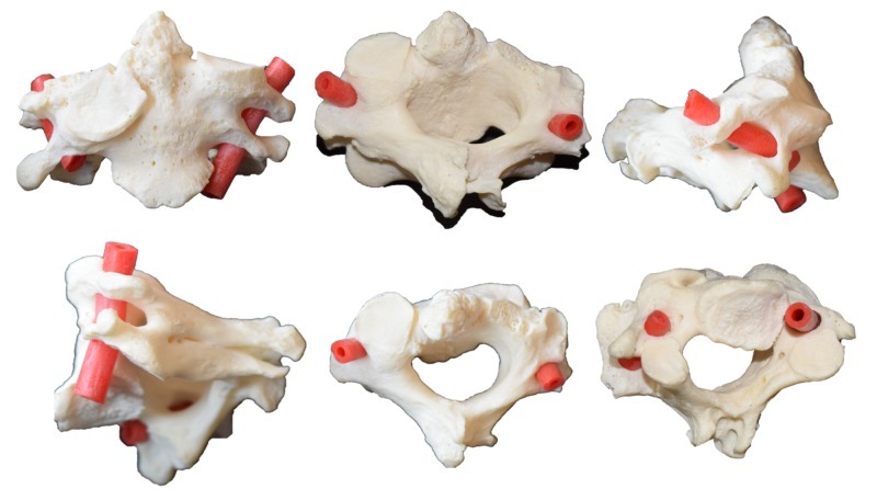
Cervical block vertebra (osteological specimen). Six views of congenitally fused C2-C3 vertebrae - the bony substrate of Klippel-Feil failure of segmentation; the red tubing marks the course of the vertebral artery. Tubbs RS et al., Cureus 2018;10:e3038, Fig. 1 (CC BY 3.0).
Cervical block vertebra (osteological specimen). Six views of congenitally fused C2-C3 vertebrae - the bony substrate of Klippel-Feil failure of segmentation; the red tubing marks the course of the vertebral artery. Tubbs RS et al., Cureus 2018;10:e3038, Fig. 1 (CC BY 3.0).
The central danger - hypermobility at the open segments
The cardinal concept is that, atlantoaxial anomalies aside, the symptoms of Klippel-Feil arise not from the fused segments but from the open (non-fused) segments adjacent to them, which become compensatorily hypermobile. Over time this hypermobility produces instability and premature degenerative change, and can culminate in neurological injury (even sudden quadriplegia after minor trauma) or vertebral-artery compromise. The highest-risk pattern is a long fused block immediately adjacent to a single open segment (for example fusion of C1-C2 and C3-C4 leaving an isolated, heavily loaded C2-C3), whereas short, low-cervical fusions are well compensated and low-risk. Most symptomatic patients present in the second or third decade.[46]
Associated anomalies - the work-up
Klippel-Feil clusters with the other malformations of this topic, and the screen mirrors that for congenital scoliosis:
| Association | Approximate frequency | Screen |
|---|---|---|
| Scoliosis (congenital or idiopathic-like) | ~60% | spinal radiographs |
| Renal anomaly (e.g. unilateral agenesis) | ~35% | renal ultrasound |
| Sprengel deformity | ~30% | clinical / radiograph |
| Sensorineural deafness | ~30% | audiology |
| Synkinesis (mirror movements) | ~20% | clinical |
| Congenital heart disease | ~14% | clinical / echo |
[47] Mirror movements reflect cervicomedullary neuroschisis; an omovertebral bone may accompany the Sprengel deformity.
Imaging and management
Plain antero-posterior and lateral films are supplemented by flexion-extension lateral views to assess instability, always before any general anaesthetic, because an unstable cervical spine may demand awake fibre-optic intubation. CT (especially at C1-C2) and MRI define the bony and neural anatomy, and MRI is performed before any spinal procedure to exclude an associated Chiari malformation, tethered cord or diastematomyelia.[48]
Most patients are managed non-operatively, with counselling to avoid contact and collision sports when there is instability or stenosis. Surgery (decompression and arthrodesis) is reserved for instability with neurological compromise, myelopathy or progressive deformity; operating for cosmetic reasons alone is generally unwarranted and carries real risk.[49]
Part V - Sprengel deformity
Definition and pathoanatomy
Sprengel deformity (congenital high or undescended scapula) is the failure of the scapula to complete its normal fetal descent. The scapula forms opposite the lower cervical somites in the third week and should descend to the thoracic wall by the eighth; arrest of that descent leaves a scapula that is small, high and rotated, with its inferior pole pointing toward the spine and its superomedial angle riding up toward the neck.[50] In a third or so of cases an omovertebral bone, an anomalous bony, cartilaginous or fibrous bridge, connects the superomedial scapula to the spinous process, lamina or transverse process of a lower cervical vertebra (C4-C7), further tethering shoulder elevation.[51] Sprengel deformity is strongly associated with Klippel-Feil syndrome and with congenital scoliosis.
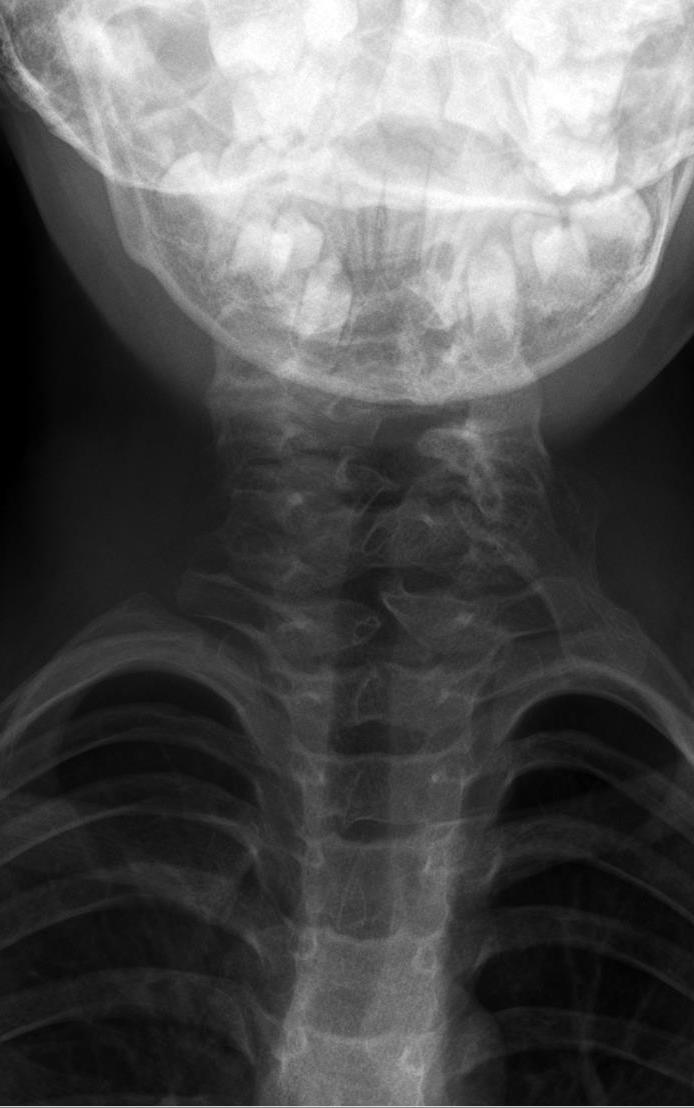
Sprengel deformity (congenital high scapula). Antero-posterior cervico-thoracic radiograph of an 8-month-old in a documented case of Sprengel deformity with an omovertebral bone (os omocervicale) bridging the scapula to the cervical spine. Hellerhoff, Wikimedia Commons (CC BY-SA 4.0).
Sprengel deformity (congenital high scapula). Antero-posterior cervico-thoracic radiograph of an 8-month-old in a documented case of Sprengel deformity with an omovertebral bone (os omocervicale) bridging the scapula to the cervical spine. Hellerhoff, Wikimedia Commons (CC BY-SA 4.0).
Grading and treatment
Severity is graded by the Cavendish classification: grade 1 (very mild, invisible when dressed); grade 2 (mild, a visible lump in the web of the neck when dressed); grade 3 (moderate, the shoulder elevated 2-5 cm); grade 4 (severe, the shoulder grossly elevated with the superomedial angle near the occiput).
Mild deformities (Cavendish 1-2) are observed. Functionally or cosmetically significant deformities are treated by surgical repositioning of the scapula, ideally between about three and eight years of age. The two named operations are:
- the Woodward procedure, a midline approach that detaches the origin of the trapezius and rhomboid sheet from the spinous processes and reattaches it more inferiorly, lowering the scapula en bloc; and
- the Green procedure, an extraperiosteal detachment of the scapular muscles from the bone, with excision of the omovertebral bar and of the prominent supraspinous portion of the scapula, and distal repositioning.
In both, the brachial plexus is the structure at risk during distal transfer of the scapula: a protective clavicular osteotomy (and moving the scapula by direct pressure rather than by traction on the arm) guards against plexus stretch injury.[52]
Part VI - Cervical ribs and thoracic outlet syndrome
The cervical rib
A cervical rib is a supernumerary rib arising from the transverse process of C7, a persistent and ossified costal element. Its prevalence is about 0.5-1%, it is commoner in women and is frequently bilateral, and the great majority, around 90-95%, are entirely asymptomatic.[53] The Sargent classification describes the spectrum, from a mere elongation of the C7 transverse process to a complete extra rib articulating with the first rib by a fibrous, cartilaginous or bony connection. When symptomatic, the rib (or, more often, a fibrous band running from its tip to the first rib) compresses the lower brachial plexus or the subclavian artery in the scalene triangle.
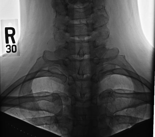
Cervical ribs. Antero-posterior radiograph showing bilateral, symmetric supernumerary ribs arising from the C7 transverse processes - the bony lesion that can narrow the thoracic outlet. Huntsville Hospital Imaging, Wikimedia Commons (public domain).
Cervical ribs. Antero-posterior radiograph showing bilateral, symmetric supernumerary ribs arising from the C7 transverse processes - the bony lesion that can narrow the thoracic outlet. Huntsville Hospital Imaging, Wikimedia Commons (public domain).
Thoracic outlet syndrome - definition and subtypes
Thoracic outlet syndrome (TOS) is the compression of one or more of the three neurovascular structures (the brachial plexus, the subclavian artery and the subclavian vein) as they cross the apex of the thorax. The principal sites are the scalene (interscalene) triangle (bounded by the anterior and middle scalene muscles and the first rib, transmitting the plexus and artery) and the costoclavicular space (between the first rib and the clavicle, transmitting the vein). The term was coined by Peet in 1956.[54]
Three subtypes are distinguished by the structure compressed:
- Neurogenic TOS, by far the commonest, around 95% (Mackinnon quotes 98%) of cases, and itself divided into a rare true form and a common disputed form (below).
- Arterial (vascular) TOS, uncommon but dangerous.
- Venous TOS, the Paget-Schroetter “effort thrombosis.”
TOS predominantly affects young to middle-aged women (about three times as often as men).[55]
Neurogenic TOS - “true” versus “disputed”
- True neurogenic TOS is rare and objectively definable. It is a lower-trunk (C8-T1) lesion, almost always unilateral and seen in young to middle-aged women, in whom a rudimentary cervical rib or an elongated C7 transverse process, with a taut fibrous band (“scalenus sickle”), angulates the lower trunk. The clinical picture is pain along the medial forearm, sensory blunting over the medial forearm and little finger, and wasting of the thenar (and other intrinsic) hand muscles: the classic Gilliatt-Sumner hand. Gilliatt estimated the incidence of cervical-rib syndrome with frank muscle wasting at about one per million per year. The diagnosis is confirmed by nerve-conduction studies.[56]
- Disputed (non-specific) neurogenic TOS is far commoner: pain, paraesthesiae and numbness on the ulnar side of the forearm and hand, supraclavicular tenderness, and a labile radial pulse on shoulder bracing, but without objective motor wasting and with normal or equivocal neurophysiology. Much of this symptom complex reflects postural muscle imbalance rather than true nerve compression, and it is treated conservatively first.[57]
A useful caveat: TOS coexists with cubital- and carpal-tunnel compression often enough that the double-crush phenomenon (Upton and McComas) must be considered. Intrinsic-muscle atrophy occurs in fewer than 5% of all TOS and signals long-standing severe disease.[58]
Vascular TOS
- Arterial TOS results from the subclavian artery being distorted over a cervical or first rib, producing stenosis, post-stenotic dilatation, a true aneurysm with intimal damage and, finally, distal embolisation; the limb is cold, blanching and easily fatigued (upper-limb claudication), and the gravest complication is retrograde embolisation to the brain. Signs include a pulse deficit, a supraclavicular bruit or pulsatile mass and an inter-arm blood-pressure difference of 20 mmHg or more.[59]
- Venous TOS (Paget-Schroetter syndrome, “effort thrombosis”) is exertional thrombosis of the subclavian-axillary vein after strenuous overhead activity, presenting with a swollen, cyanotic, aching arm and distended superficial veins; it is treated by thrombolysis followed by prompt first-rib resection.[60]
Provocative tests, investigation and treatment
The named provocative manoeuvres are worth knowing precisely: Adson’s test (the seated patient takes a deep breath, extends the neck and rotates the head toward the affected side, and a positive test obliterates the radial pulse); the Wright (hyperabduction) test; the Roos test / Elevated Arm Stress Test (EAST) (arms abducted to 90° and externally rotated, opening and closing the hands for three minutes to reproduce symptoms); the Halstead (costoclavicular / military-posture) test; and Spurling’s test, which reproduces radicular pain and points instead to cervical-disc disease. The pulse-based tests are non-specific (many normal people lose the radial pulse), so reproduction of the patient’s symptoms is more reliable than pulse loss.[61]
Investigation begins with a plain radiograph (for the cervical rib, an elongated C7 transverse process or old clavicular callus). Electrodiagnostic studies are typically normal in TOS except in the true neurogenic form (where they are diagnostic) or where there is concurrent distal entrapment; vascular imaging (arteriography or venography, or their CT/MR/duplex equivalents) is used when a vascular form is suspected.[62]
Treatment is conservative first: physiotherapy directed at posture and scalene/pectoral muscle balance succeeds in roughly half to four-fifths of patients. Surgical decompression is reserved for true neurogenic TOS, for vascular TOS, and for conservative failure with a correctable lesion; it comprises scalenectomy with first-rib (and any cervical-rib) resection. Scalenotomy alone has a high recurrence rate (up to about 65%), so a complete first-rib resection is the more durable operation; the phrenic and long-thoracic nerves, the subclavian vessels and (on the left) the thoracic duct are at risk. Recovery of already-wasted hand intrinsics after decompression is rare, so surgery in true neurogenic TOS is undertaken mainly for pain relief.[63]
Part VII - Spina bifida and myelomeningocele
The dysraphic spectrum - definitions
“Spina bifida” and “spinal dysraphism” cover the malformations that arise from failure of neural-tube closure or of the mesoderm that should cover it. The spectrum is divided embryologically into open forms (exposed neural tissue) and closed/occult forms (skin-covered):
- Spina bifida occulta: a defect of the posterior vertebral arch only, with no herniation of the contents; it is common and usually harmless, but an overlying skin marker (a dimple, hairy patch, lipoma or haemangioma) may signal a tethered cord beneath.
- Meningocele: herniation of meninges only, with no neural tissue; neurology is usually normal.
- Myelomeningocele: herniation of the neural placode together with meninges through a deficient posterior arch, with no skin cover; this is the common, severe, neurologically devastating form, producing motor and sensory paralysis, and bladder and bowel dysfunction, below the lesion.[64]
- Rachischisis / myelocele: the most open form, a flat exposed placode.
- Lipomyelomeningocele: a skin-covered subcutaneous lipoma connected to the conus, tethering the cord; patients have normal intelligence and no Chiari malformation, and, importantly, its incidence is not reduced by folate.[65]
- Diastematomyelia and tethered cord lie within this spectrum as tethering lesions.
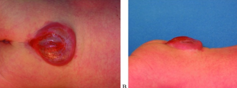
Lumbosacral myelomeningocele. Posterior (left) and lateral (right) clinical photographs of the membrane-covered neural-placode sac before repair. DeJong PM et al., Eplasty 2016;16:ic51, Fig. 1 (CC BY).
Lumbosacral myelomeningocele. Posterior (left) and lateral (right) clinical photographs of the membrane-covered neural-placode sac before repair. DeJong PM et al., Eplasty 2016;16:ic51, Fig. 1 (CC BY).
Embryology, epidemiology and prevention
Myelomeningocele results from failure of the neural folds to fuse during neurulation, at about 26-28 days’ gestation.[66] Its birth prevalence is around 1 per 1,000, and it remains the most severely disabling birth defect compatible with survival; survival itself has been transformed, from about 10% in the 1950s to at least 75% reaching early adulthood today, largely through the introduction of the ventricular shunt.[67]
The single most important public-health fact is prevention by periconceptional folic acid:
- All women capable of pregnancy: 0.4 mg (400 µg) folic acid daily, which prevents an estimated 50-70% of neural-tube defects.
- Women with a previously affected pregnancy (high risk): 4 mg (often quoted as 4-5 mg) daily.
Mandatory folic-acid fortification of grain (USA, 1998) was followed by a fall in birth prevalence of roughly 23%, and European supplementation policies produced comparable declines.[68] Antenatal detection rests on maternal serum alpha-fetoprotein (raised in over 95% of anencephaly and 65-80% of open defects, though not in closed defects) and ultrasound.
A major recent advance is fetal (in-utero) repair. The Management of Myelomeningocele Study (MOMS), a randomised controlled trial, was stopped early in 2010 for efficacy: prenatal closure (at 19-25 weeks) roughly halved the need for shunting and improved motor function and the likelihood of independent walking at 30 months, while lessening the Chiari II malformation. The trade-off was greater prematurity and maternal morbidity.[69]
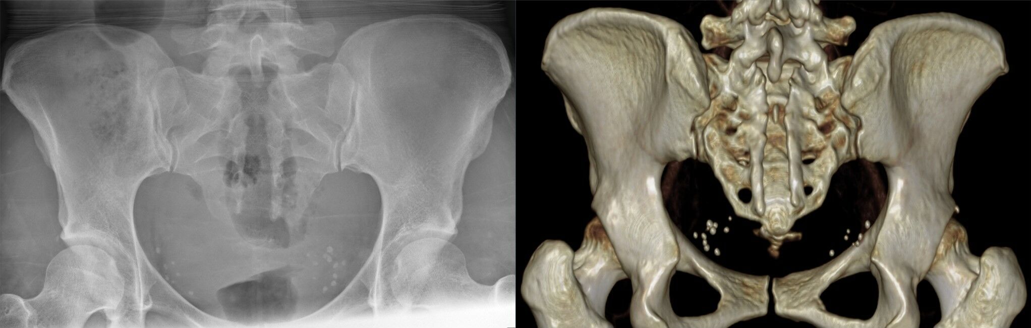
Bony spina bifida (sacral dysraphism). Antero-posterior radiograph (left) and three-dimensional CT reconstruction (right) showing long-segment failure of closure of the posterior sacral arches. Hellerhoff, Wikimedia Commons (CC BY-SA 4.0).
Bony spina bifida (sacral dysraphism). Antero-posterior radiograph (left) and three-dimensional CT reconstruction (right) showing long-segment failure of closure of the posterior sacral arches. Hellerhoff, Wikimedia Commons (CC BY-SA 4.0).
The neurosegmental level - the master prognostic factor
Everything in the orthopaedic management of myelomeningocele is organised around the neurosegmental (motor) level, which dictates the muscles that work, the pattern of deformity, the bracing needed and the prognosis for walking. The motor level should remain constant for life; any loss of strength signals a tethered cord until proven otherwise.[70] The standard functional grouping (Swaroop and Dias) is:
| Functional level | Key working muscles | Bracing | Adult ambulation |
|---|---|---|---|
| Thoracic / high-lumbar (L3 and above) | no functional quadriceps | HKAFO / RGO in childhood | most become wheelchair-dependent as adults |
| Low lumbar (L4-L5) | quadriceps and medial hamstrings (≥ grade 3) | AFOs + crutches | most are community ambulators |
| High sacral (S1-S3) | quadriceps and gluteus medius | AFOs | community ambulators (gluteal lurch) |
| Low sacral (S3-S5) | quadriceps, gluteus medius and gastrocsoleus | none / minimal | near-normal gait |
[71] The critical watershed is L3 versus L4: the medial hamstring (innervated at L4) is needed for community walking, so L4 patients generally walk in the community while L3 patients generally do not. Quadriceps and iliopsoas strength are the strongest individual predictors, and sitting balance is an independent predictor of community ambulation. The classic four-tier Hoffer ambulation classification (community, household, non-functional (exercise/therapeutic), and non-ambulator) captures the same gradient and is the eponymous scheme to quote.[72]
The principle of muscle imbalance and deformity by level
Deformities in myelomeningocele are either congenital (present at birth: kyphosis, teratologic hip dislocation, clubfoot, vertical talus), developmental (from unopposed muscle action, the muscle-imbalance principle), or iatrogenic (a postoperative tethered cord). The developmental deformities are predictable from the level: at the mid-lumbar (L3-L4) level, strong hip flexors and adductors acting without paralysed extensors and abductors drive the hip out of joint; at L4-L5, working ankle dorsiflexors without plantarflexors produce a calcaneus foot.[73]
Hip
Hip instability affects up to half of children in the first decade, and is greatest at the mid-lumbar levels for the reason just given. The cardinal teaching is the relocation controversy: the work of Feiwell and others showed that ambulation depends on the neurological level, not on whether the hip is in joint, and that aggressive relocation gives no gain in walking, range or comfort while carrying a high rate of stiffness, fracture and even neurological loss. The modern goals are therefore a level pelvis and mobile, flexible hips rather than a radiographically reduced one: contracture release for the thoracic/high-lumbar hip, soft-tissue balancing only for the unilateral low-lumbar dislocation, and, as the one genuine exception, careful consideration of concentric reduction in the high-functioning sacral-level child, whose working abductors make a dislocation functionally costly.[74]
Knee
Flexion and extension contractures predominate. Flexion contracture is commoner at higher levels and, beyond about 20°, produces a high-energy crouch gait and blocks bracing; it is treated by radical knee-flexor release. Extension contracture is usually congenital and bilateral; it is treated first by serial casting and, if necessary, by V-Y quadricepsplasty. Late valgus instability in low-lumbar and sacral ambulators, driven by external tibial torsion, is corrected by rotational osteotomy.[75]
Foot and ankle
Some foot deformity is present in almost every patient, and the goal is a plantigrade, supple, braceable foot that will not break down. Two principles dominate: tendon excision is more reliable than tendon transfer or lengthening in an insensate foot, and arthrodesis (especially triple arthrodesis) is avoided because a stiff insensate foot ulcerates. The common deformities are equinovarus (clubfoot), the commonest, rigid and prone to relapse; calcaneus / calcaneovalgus (commonest at L4-L5); congenital vertical talus; and cavovarus (at sacral level and with lipomeningocele).[76]
Spine
Spinal deformity is very common and its frequency rises with the level of the lesion. Scoliosis is present in 60-90% overall, approaching universality at the thoracic level, with the three best predictors being motor level, ambulatory status and the last intact laminar arch; curves over 40° progress at around 12-13° per year. Kyphosis, the rigid congenital gibbus of the thoracic-level child, often exceeding 80° at birth, causes recurrent skin ulceration over its apex and is treated by kyphectomy when it interferes with sitting or skin integrity. Bracing temporises but does not halt progression, and surgery (usually combined anterior-posterior instrumented fusion, often to the pelvis) carries the highest complication rate of all deformity surgery.
The single most important spinal teaching is that a worsening curve must trigger a search for a tethered cord, hydromyelia/syringomyelia, Chiari II malformation or shunt malfunction before it is attributed to simple progression; treating the neuraxial cause may obviate or precede the fusion, and shunt function must be confirmed before any cord manipulation.[77]
The whole child - issues the orthopaedic surgeon must know
Myelomeningocele is a multisystem disease, and several non-orthopaedic problems determine orthopaedic decisions:
- Arnold-Chiari II malformation and hydrocephalus. A Chiari II malformation is present in virtually all patients, and hydrocephalus in around 90%, usually requiring a ventriculoperitoneal (VP) shunt. Deterioration in a child with a shunt is shunt malfunction until proven otherwise, and shunt function must be checked before attributing new weakness to a tethered cord or Chiari, and before any intraoperative cord manipulation.[78]
- Tethered cord and hydromyelia. Because MRI shows a low-lying cord in almost all repaired patients, the diagnosis of a symptomatic tethered cord rests on clinical deterioration (most often a progressive scoliosis, but also new weakness, spasticity, back pain or urological change) after excluding shunt failure and hydromyelia.[79]
- Neurogenic bladder and bowel. Almost universal; most patients need clean intermittent catheterisation to protect the upper urinary tracts, and bowel programmes (including the Malone antegrade continence enema) for continence. Constipation can raise intra-abdominal pressure enough to cause shunt malfunction.[80]
- Insensate skin and pressure sores. A constant threat (reported in up to 80% over time), which underlies the plantigrade-foot and no-arthrodesis rules; feet, casts and orthoses must be inspected relentlessly.[81]
- Latex allergy. Reported in up to about 30% of patients (with anaphylaxis in a significant minority), driven by repeated surgical exposure; every patient with myelomeningocele must be managed in a strictly latex-free environment from birth, since this both prevents reactions and reduces sensitisation.[82]
- Pathological fractures. The osteopenic, insensate lower limb fractures readily (in up to 20% of patients, and up to 29% in the six months after spine surgery). The classic and dangerous trap is that a metaphyseal or diaphyseal fracture presents as a warm, swollen, red limb that mimics infection (with fever, raised white-cell count and ESR) but is usually painless; recognising it avoids a needless infection work-up. Most heal quickly with simple immobilisation, using a removable splint rather than a heavy spica.[83]
The thread through all of this is that myelomeningocele care is multidisciplinary, with orthopaedics, neurosurgery, urology, rehabilitation, physiotherapy, orthotics, wound care and social support working together, and that the orthopaedic surgeon’s job is less to normalise anatomy than to keep the child upright, mobile, seated comfortably and free of skin breakdown.
Bulgarian terminology (Боев / Boychev tradition) - glossary
The following Bulgarian equivalents and transliterations are provided to bridge the international literature with the terminology of the Bulgarian school of orthopaedics (in the tradition of Бойчо Бойчев / Boycho Boychev) used in the state examination.
| English term | Bulgarian term (Cyrillic) | Transliteration |
|---|---|---|
| Congenital scoliosis | Вродена сколиоза | Vrodena skolioza |
| Congenital kyphosis | Вродена кифоза | Vrodena kifoza |
| Hemivertebra | Полупрешлен (хемивертебра) | Polupreshlen (hemivertebra) |
| Vertebra / vertebral body | Прешлен / тяло на прешлена | Preshlen / tyalo na preshlena |
| Failure of formation | Дефект във формирането | Defekt vav formiraneto |
| Failure of segmentation | Дефект в сегментацията | Defekt v segmentatsiyata |
| Block vertebra | Блоков прешлен | Blokov preshlen |
| Unsegmented bar | Несегментирана костна ивица (мост) | Nesegmentirana kostna ivitsa (most) |
| Curvature of the spine | Изкривяване на гръбначния стълб | Izkrivyavane na grabnachniya stalb |
| Spine / vertebral column | Гръбначен стълб | Grabnachen stalb |
| Klippel-Feil syndrome | Синдром на Клипел-Файл | Sindrom na Klipel-Fayl |
| Short neck | Къс врат | Kas vrat |
| Cervical vertebrae | Шийни прешлени | Shiyni preshleni |
| Sprengel deformity (high scapula) | Болест на Шпренгел (вродено високо разположена лопатка) | Bolest na Shprengel (vysoko razpolozhena lopatka) |
| Scapula | Лопатка | Lopatka |
| Cervical rib | Шийно ребро | Shiyno rebro |
| Thoracic outlet syndrome | Синдром на горната гръдна апертура | Sindrom na gornata gradna apertura |
| Brachial plexus | Брахиален сплит (раменен сплит) | Brahialen split (ramenen split) |
| Spina bifida | Спина бифида (разцепен гръбнак) | Spina bifida (raztsepen grabnak) |
| Myelomeningocele | Миеломенингоцеле | Mielomeningotsele |
| Meningocele | Менингоцеле | Meningotsele |
| Spina bifida occulta | Скрита спина бифида | Skrita spina bifida |
| Tethered cord | Фиксиран (привързан) гръбначен мозък | Fiksiran (privarzan) grabnachen mozak |
| Hydrocephalus | Хидроцефалия | Hidrotsefaliya |
| Ventriculoperitoneal shunt | Вентрикулоперитонеален шънт | Ventrikuloperitonealen shant |
| Neurogenic bladder | Неврогенен пикочен мехур | Nevrogenen pikochen mehur |
| Clubfoot (equinovarus) | Вродено криво стъпало (еквиноварус) | Vrodeno krivo stapalo (ekvinovarus) |
| Paralysis | Парализа | Paraliza |
| Folic acid | Фолиева киселина | Folieva kiselina |
Image attributions
(Figure attributions and licences are listed in the figure MANIFEST and inserted with each image. All images are openly licensed [CC0 / CC BY / CC BY-SA] or used under their stated terms; any non-commercial [NC] item is flagged as such and must not be used in a commercial product.)
References
-
Lovell & Winter, Pediatric Orthopaedics, congenital-scoliosis chapter, pp. 718-719: sclerotome induction by Sonic Hedgehog from the notochord/floor plate, segment boundaries set by FGF8, rostro-caudal identity by the Hox gene clusters (HoxA-D, 39 genes), and Pax1/Meox1 mediating sclerotome subdivision.
-
Bedi & Hensinger, in Rothman-Simeone The Spine, congenital-cervical-anomalies chapter, p. 571-572: somite segmentation at 20-30 days, scapular descent by the 8th week, and the resulting co-occurrence of cervical fusion, Sprengel deformity and genitourinary anomalies.
-
Akbarnia, The Growing Spine, congenital-scoliosis chapter (Yazici, Yilmaz, Kawakami), pp. 187, 190; Lovell, p. 717. The deformity is the mechanical consequence of unbalanced growth around a fixed structural anomaly.
-
Moe’s Textbook of Scoliosis (R. B. Winter), congenital chapter, p. 274.
-
Akbarnia, p. 187. Lovell quotes school-screening figures of 0.5-1 per 1,000 and lower figures (0.1-0.3 per 1,000) from antenatal-ultrasound series, p. 717.
-
Akbarnia, p. 187; Lovell, p. 719.
-
Akbarnia, p. 187; Lovell, p. 719; Moe’s, p. 276 (Wynne-Davies).
-
Akbarnia, p. 188; Lovell, p. 721; Moe’s, p. 274.
-
Moe’s, p. 274.
-
Akbarnia, p. 188; Moe’s, p. 274-276; Lovell, p. 722, 728.
-
Akbarnia, p. 190; Moe’s, p. 279.
-
Akbarnia, p. 188; Lovell, p. 721.
-
Akbarnia, pp. 189-190.
-
Akbarnia, pp. 188, 190.
-
Akbarnia, p. 190.
-
Moe’s, p. 283; Lovell, p. 721.
-
Akbarnia, pp. 190-191.
-
Lovell, pp. 721, 725.
-
Lovell, p. 719.
-
Akbarnia, pp. 192-193; Lovell, p. 719; Moe’s, pp. 278-279.
-
Akbarnia, pp. 192, 194; Lovell, p. 719.
-
Akbarnia, p. 194; Moe’s, p. 279.
-
Akbarnia, p. 187; Lovell, p. 719.
-
Lovell, pp. 720-721, 724.
-
Akbarnia, pp. 191-192; Moe’s, p. 278.
-
Akbarnia, pp. 191-192; Lovell, pp. 725-726.
-
Akbarnia, p. 192; Lovell, p. 724.
-
Akbarnia, p. 192; Moe’s, p. 278.
-
Lovell, p. 728.
-
Akbarnia, p. 194; Lovell, p. 725.
-
Akbarnia, p. 194; Moe’s, p. 287; Lovell, p. 726.
-
Moe’s, p. 293.
-
Akbarnia, p. 195; Lovell, p. 731.
-
Akbarnia, p. 198; Lovell, pp. 730-737.
-
Akbarnia, p. 197; Lovell, pp. 750-752.
-
Lovell, pp. 743-746.
-
Moe’s, p. 293.
-
Moe’s, p. 287.
-
Moe’s, pp. 274, 287; Lovell, p. 722; Akbarnia, sagittal-plane chapter, p. 409.
-
Moe’s, p. 287.
-
Akbarnia, sagittal chapter, pp. 412-413.
-
Bedi & Hensinger, in Rothman-Simeone The Spine, p. 570.
-
Rothman, pp. 570-571; Lovell, p. 882.
-
Rothman, pp. 571-572.
-
Lovell, pp. 882-883.
-
Rothman, pp. 572-573; Lovell, p. 883.
-
Rothman, pp. 571-572 (Hensinger series); Lovell, p. 882.
-
Lovell, p. 883.
-
Lovell, p. 883.
-
Rothman, p. 572; Tachdjian / Herring, congenital-high-scapula section, p. 184.
-
Tachdjian, pp. 185, 188.
-
Tachdjian, pp. 184-195.
-
Colbert, in Mackinnon, Nerve Surgery, thoracic-outlet chapter, p. 330; Birch, Surgical Disorders of the Peripheral Nerves, p. 294.
-
Mackinnon, pp. 328-329.
-
Mackinnon, p. 331.
-
Birch, pp. 296-297.
-
Birch, p. 297; Mackinnon, p. 328.
-
Mackinnon, pp. 331, 333.
-
Birch, pp. 294-296; Mackinnon, pp. 331, 344.
-
Mackinnon, pp. 343, 348-349.
-
Mackinnon, pp. 332, 335-336.
-
Mackinnon, pp. 339-344; Birch, pp. 296-297.
-
Mackinnon, pp. 344-351; Birch, pp. 296-298.
-
Akbarnia, myelomeningocele chapter (Karlin), pp. 247, 266-267; Lovell, congenital-spine/myelomeningocele chapter (Swaroop & Dias), p. 574.
-
Lovell, p. 602.
-
Lovell, p. 575; Akbarnia, p. 267.
-
Akbarnia, pp. 247, 268, 270; Lovell, p. 574.
-
Lovell, pp. 574-575. The 0.4 mg general and < 1 mg total-folate figures are explicit in the source; the 4 mg high-risk dose is standard teaching.
-
Akbarnia, p. 270.
-
Lovell, p. 582.
-
Lovell, pp. 578-580.
-
Lovell, p. 578; Hoffer et al. 1973, cited in Akbarnia ref list p. 262.
-
Lovell, pp. 582, 586, 596.
-
Lovell, pp. 586-588.
-
Lovell, pp. 589-591.
-
Lovell, pp. 592-598.
-
Akbarnia, pp. 250-253; Lovell, pp. 583-586.
-
Akbarnia, pp. 248-249, 268; Lovell, p. 576.
-
Lovell, p. 576.
-
Lovell, p. 576.
-
Lovell, pp. 577, 601.
-
Akbarnia, p. 250; Lovell, p. 577.
-
Lovell, pp. 577, 586, 598-599.