Contents
- Scope and orientation
- Part I - Classification, embryology and epidemiology
- Part II - Failure of formation (deficiencies)
- Part III - Failure of differentiation
- Part IV - Duplication and overgrowth
- Part V - Amniotic-band (constriction-ring) disease and associated syndromes
- Bulgarian terminology glossary
- Figure credits and licences
Scope and orientation
Congenital anomalies of the hand are common and varied. This summary works through them by the classic categories (failure of formation, failure of differentiation, duplication, overgrowth and undergrowth), then covers the amniotic-band (constriction-ring) disease named in the syllabus and the major associated syndromes.
A few organising ideas carry the topic:
- Classification frames everything. The Swanson/IFSSH seven-category scheme is the traditional one. The modern OMT (Oberg-Manske-Tonkin) classification reorganises anomalies by the developmental mechanism and the disturbed limb-bud axis.
- The commonest anomalies are syndactyly and polydactyly, and most anomalies arise during the first trimester (weeks 4-8) of limb-bud signalling.
- Some anomalies are red flags for systemic disease. A radial deficiency or a hypoplastic thumb mandates screening for Fanconi anaemia, Holt-Oram, TAR and VACTERL. Recognising Fanconi early can be life-saving.
- Amniotic-band disease is a deformation, not a malformation: an extrinsic band constricts a normally-formed part, so the anatomy proximal to the ring is normal. That is the point distinguishing it from symbrachydactyly.
A Bulgarian glossary, a viva self-test appendix, and image credits follow the clinical sections.
Part I - Classification, embryology and epidemiology
I.1 Classification
The Swanson / IFSSH classification (adopted by the ASSH and IFSSH) sorts anomalies into seven categories: I failure of formation, II failure of differentiation/separation, III duplication, IV overgrowth, V undergrowth, VI constriction-ring syndrome, and VII generalised skeletal abnormalities (Rayan & Upton list the order of IV and V the other way round).[1] By category, failure of differentiation (~35%) and duplication (~33%) are the commonest groups.[2] Its weakness is that many anomalies fit several categories or none (cleft hand has deficiency + syndactyly + polydactyly), and its assumed embryology has not been borne out.[3]
The modern OMT (Oberg-Manske-Tonkin) classification (now the IFSSH-adopted scheme) reorganises anomalies by dysmorphology into malformations (abnormal formation/differentiation, subdivided by the affected axis: proximodistal/AER, radioulnar/ZPA-SHH, dorsoventral/WNT), deformations (extrinsic moulding: constriction-ring syndrome, trigger digit), and dysplasias (abnormal growth: macrodactyly, tumours), with a syndromic tag.[4]
I.2 Embryology and epidemiology
The limb bud appears at about day 26 and is patterned by three signalling centres: the apical ectodermal ridge (AER, FGFs) controls the proximodistal axis (its loss gives transverse deficiency; failed interdigital apoptosis gives syndactyly); the zone of polarising activity (ZPA, Sonic hedgehog) controls the anteroposterior/radioulnar axis (loss → ulnar deficiency; ectopic anterior Shh → mirror hand/polydactyly); and WNT signalling controls the dorsoventral axis. Development is largely complete by the end of week 8.[5] Congenital upper-limb anomalies occur in about 1 in 500-600 live births, are commoner in boys and often bilateral, and the commonest individual anomalies are polydactyly and syndactyly.[6]
Part II - Failure of formation (deficiencies)
II.1 Radial longitudinal deficiency (radial club hand)
This is the commonest congenital longitudinal deficiency (~1/30,000), a preaxial failure that affects the whole radial border: bone, muscle, nerve (an anomalous radially-placed median nerve) and the thumb.[7] The Bayne & Klug classification grades it: I short distal radius, II hypoplastic (miniature) radius, III partial radial absence, IV complete absence (the commonest); milder proximal “N/O” types were later added.[8]
About two-thirds are syndromic or have systemic anomalies, so a radial deficiency mandates a work-up for the four classic associations: VACTERL, Holt-Oram (TBX5, cardiac), Fanconi anaemia (test chromosomal breakage in infancy, fatal without marrow transplant), and TAR (thrombocytopenia-absent radius, in which the thumb is always present).[9] Management begins with stretching and splinting in the first months, then centralisation or radialisation of the carpus on the ulna, then thumb reconstruction. One rule matters: a stiff extended elbow (lacking 90° of flexion) contraindicates wrist surgery, because that posture lets the hand reach the mouth.[10]
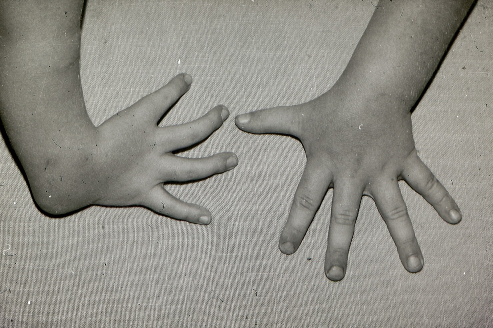
Radial longitudinal deficiency: the affected hand (image left) has four digits with an absent thumb; the other hand is normal. (Mehlauge, Wikimedia Commons; CC BY-SA 3.0.)
Radial longitudinal deficiency: the affected hand (image left) has four digits with an absent thumb; the other hand is normal. (Mehlauge, Wikimedia Commons; CC BY-SA 3.0.)
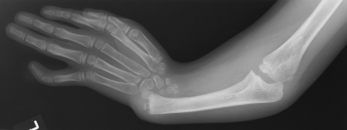
Radiograph of radial club hand (TAR syndrome): the radius is absent, the single ulna is bowed, and the hand is radially deviated off the forearm. (Ahmad R, BMC Nephrol 2007; CC BY 2.0.)
Radiograph of radial club hand (TAR syndrome): the radius is absent, the single ulna is bowed, and the hand is radially deviated off the forearm. (Ahmad R, BMC Nephrol 2007; CC BY 2.0.)
II.2 Thumb hypoplasia
Thumb hypoplasia is graded by the Blauth classification (I-V), and the decision turns on the carpometacarpal (CMC) joint: types I-II and IIIA (stable CMC) are reconstructed (opponensplasty, ulnar-collateral reconstruction, first-web release), whereas IIIB (unstable/absent CMC), IV (floating thumb, pouce flottant) and V (absent thumb) are treated by ablation and pollicisation of the index finger.[11] Type V is the commonest, and the components to address are the small size, IP stiffness, MCP/UCL instability, thenar underdevelopment and first-web contracture.[12]
II.3 Ulnar longitudinal deficiency
Much rarer than radial deficiency (~1/100,000; a roughly 10:1 radial-to-ulnar ratio). Its associations are musculoskeletal, not systemic, so no cardiac/renal/haematologic screen is needed.[13] The Bayne scheme parallels the radial one (I hypoplasia → IV with radiohumeral synostosis); the hand is ulnarly deviated with absent ulnar rays, frequent thumb anomalies and an unstable or fused elbow. The most useful operations are syndactyly release and thumb realignment; ulnar-anlage excision is now rarely done.[14]
II.4 Transverse deficiency, symbrachydactyly and cleft hand
The commonest transverse deficiency is the congenital below-elbow amputation (transverse arrest at the proximal forearm; ~1/20,000, usually unilateral and isolated), often managed without a prosthesis since prostheses do not reliably improve function.[15] Symbrachydactyly (“short-fingered hand,” with rudimentary nubbins that retain nails and pulps) is a unilateral failure of formation that is the hand manifestation of Poland syndrome; it is distinguished from amniotic-band disease by its abnormal proximal anatomy, whereas band disease has normal proximal anatomy.[16] Typical cleft hand (ectrodactyly) is a central longitudinal deficiency, usually bilateral and familial, part of the split-hand/split-foot spectrum (p63, Dlx, EEC syndrome), and is graded by the Manske & Halikis first-web classification; it is distinct from the atypical cleft hand, which is symbrachydactyly.[17] Phocomelia is the intercalary (segmental) deficiency famously linked to thalidomide.[18]
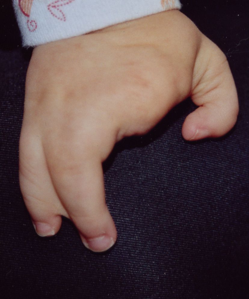
Typical cleft hand (ectrodactyly) in a one-year-old: a central V-shaped cleft divides the hand. (Aurélie & Sylvain Mulard, Wikimedia Commons; CC BY-SA 3.0.)
Typical cleft hand (ectrodactyly) in a one-year-old: a central V-shaped cleft divides the hand. (Aurélie & Sylvain Mulard, Wikimedia Commons; CC BY-SA 3.0.)
Part III - Failure of differentiation
III.1 Syndactyly
Syndactyly (webbed fingers, ~1/3,000) is among the commonest hand anomalies, a failure of the normal distal-to-proximal interdigital apoptosis. It is classified as simple (skin only) vs complex (bony fusion) and complete vs incomplete, with complicated forms (interposed bones, as in Apert); it is commonest in the third web, more frequent in boys, often bilateral, and frequently autosomal dominant.[19] The strong syndromic association is Apert syndrome (FGFR2 acrocephalosyndactyly), and Poland syndrome.[20] The surgical principles: release border or unequal-length digits early (thumb-index, ring-small; ~9 months) to prevent deviation, delay simple central webs to 12-18 months, never release both sides of a digit at once (vascular risk), reconstruct the commissure with a dorsal flap, use interdigitating zigzag flaps, and resurface the residual defects with full-thickness skin grafts. Web “creep” and, rarely, digital necrosis are the main complications.[21]
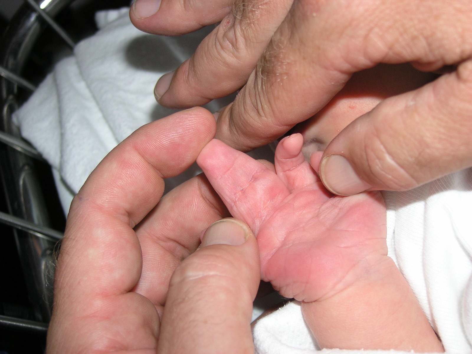
Complete complex syndactyly of a newborn hand: two central fingers are fused into a single soft-tissue mass sharing one nail. (Wikimedia Commons; public domain.)
Complete complex syndactyly of a newborn hand: two central fingers are fused into a single soft-tissue mass sharing one nail. (Wikimedia Commons; public domain.)
III.2 Camptodactyly and clinodactyly
Camptodactyly is a non-traumatic flexion contracture of the PIP joint (usually the little finger), with infantile and adolescent forms, driven by anomalous FDS/lumbrical anatomy and soft-tissue contracture. Most cases are managed by stretching and splinting, with surgery reserved for a fixed contracture over ~60°.[22] Clinodactyly is a coronal-plane (radioulnar) angulation, usually radial deviation of the little finger from an abnormally shaped middle phalanx (the delta phalanx / longitudinally bracketed epiphysis), associated with Down syndrome. Mild cases are observed; an angulation over ~30° impairing function is treated by osteotomy, or, in the young, by physiolysis/bracket resection.[23] The triphalangeal thumb is a related delta-phalanx anomaly, autosomal dominant, with Holt-Oram among its associations.[24]
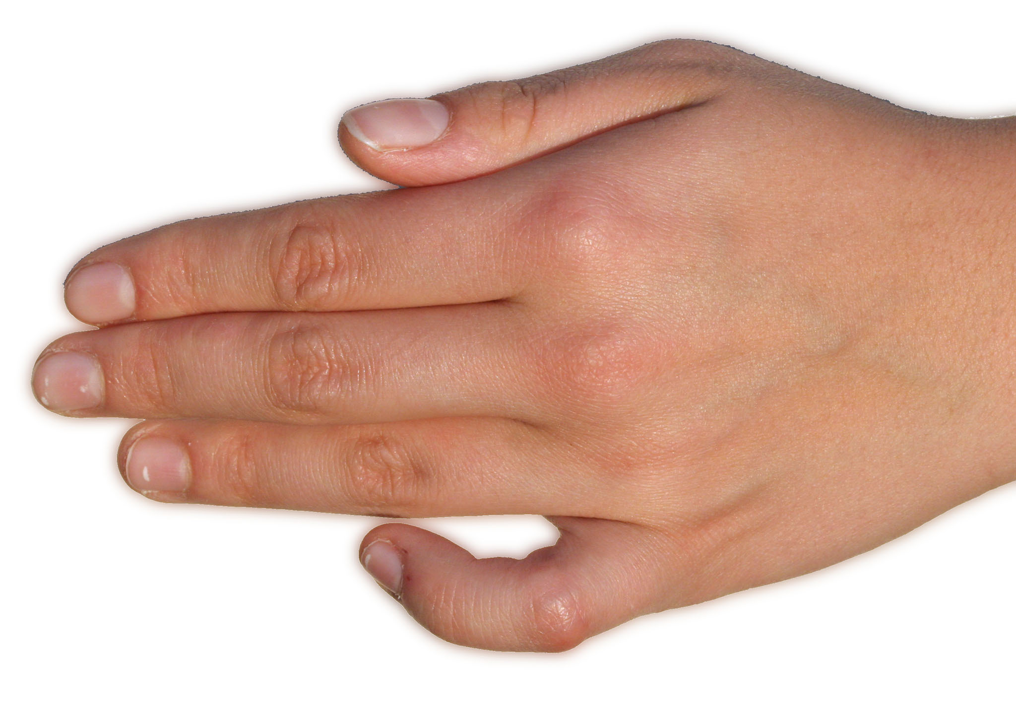
Camptodactyly: a fixed flexion contracture of the little finger at the PIP joint while the other digits extend. (Armin Kübelbeck, Wikimedia Commons; CC BY-SA 3.0.)
Camptodactyly: a fixed flexion contracture of the little finger at the PIP joint while the other digits extend. (Armin Kübelbeck, Wikimedia Commons; CC BY-SA 3.0.)
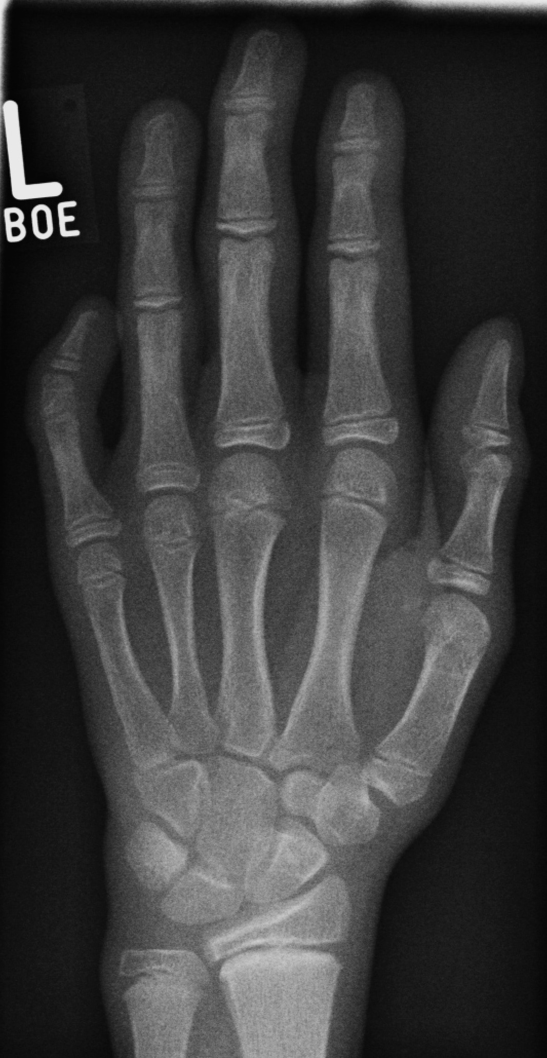
Radiograph of little-finger clinodactyly with a short middle phalanx (brachymesophalangy) and radial angulation. (Tomograph, Wikimedia Commons; CC BY-SA 3.0.)
Radiograph of little-finger clinodactyly with a short middle phalanx (brachymesophalangy) and radial angulation. (Tomograph, Wikimedia Commons; CC BY-SA 3.0.)
III.3 Madelung deformity
Madelung deformity is a dysplasia of the volar-ulnar distal-radial physis producing excessive volar and ulnar tilt of the distal radius, a dorsally prominent ulnar head, and proximal/volar carpal migration (a “dinner-fork” wrist), classically in adolescent girls. It is associated with Léri-Weill dyschondrosteosis and the SHOX gene, and an abnormal volar Vickers ligament tethers the radius. Mild cases are observed. The young symptomatic wrist is treated by Vickers-ligament release and physiolysis, the established deformity by a radial dome osteotomy with ulnar shortening as needed.[25]
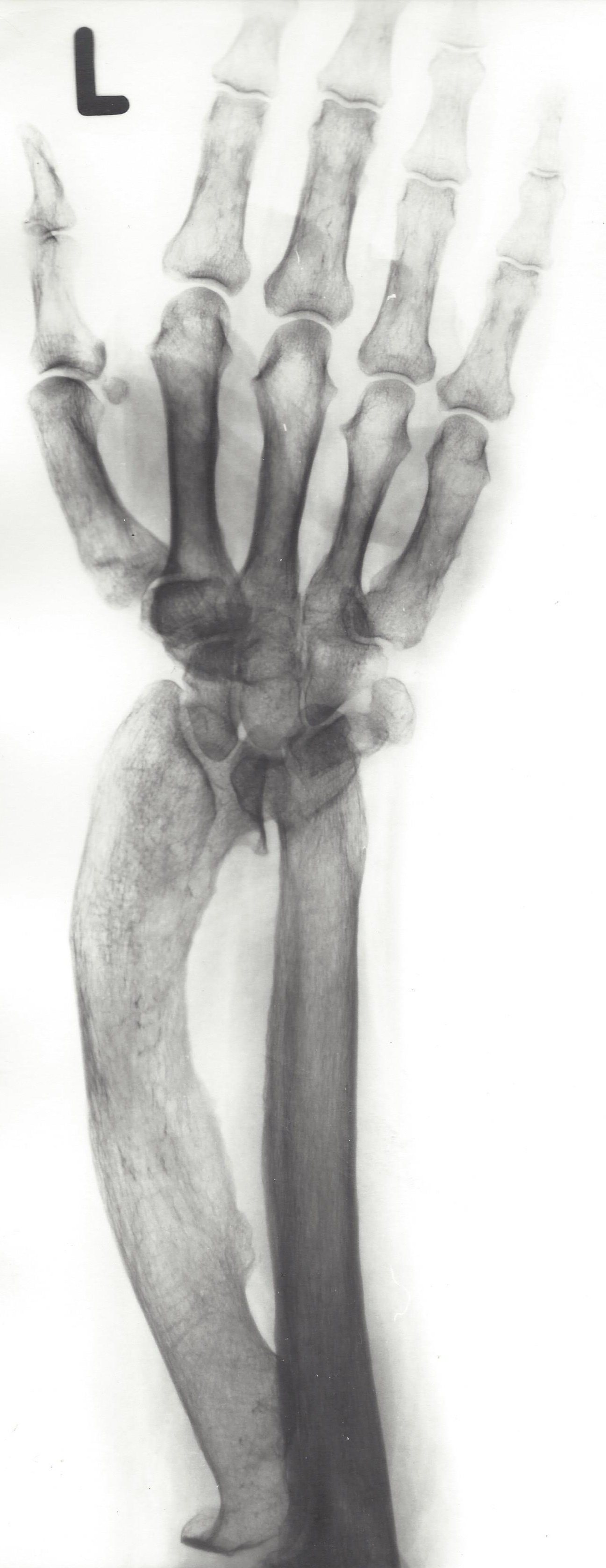
Radiograph of Madelung deformity: increased ulnar and volar tilt of the distal radius with a dorsally prominent ulna and the carpus wedged into the radioulnar gap. (Wikimedia Commons; CC BY-SA 3.0.)
Radiograph of Madelung deformity: increased ulnar and volar tilt of the distal radius with a dorsally prominent ulna and the carpus wedged into the radioulnar gap. (Wikimedia Commons; CC BY-SA 3.0.)
III.4 Synostosis, trigger thumb and the arthrogrypotic hand
Congenital radioulnar synostosis fixes the forearm in pronation (often bilateral, autosomal dominant, associated with Klinefelter); attempts to restore rotation usually fail, so a derotation osteotomy is used for a functionally limiting position.[26] Symphalangism (fused IP joint, sparing the thumb, with a telltale absent flexion crease) and carpal coalition (commonest lunotriquetral) are other failures of segmentation.[27] Congenital trigger thumb is a thickening of the flexor pollicis longus at the A1 pulley (the Notta nodule) presenting as a fixed flexed IP joint. Many resolve spontaneously, with A1-pulley release for persistent cases.[28] The arthrogrypotic hand shows stiff joints, a thumb-in-palm and ulnarly-deviated (“windblown”) fingers, managed by early splinting and selective releases/transfers.[29]
Part IV - Duplication and overgrowth
IV.1 Polydactyly
Polydactyly (an extra digit, one of the two commonest hand anomalies) is grouped as preaxial (radial/thumb), postaxial (ulnar) and central.[30]
Preaxial polydactyly (thumb duplication) is graded by the Wassel classification (I-VII), where the type number equals the number of abnormal bones: I bifid distal phalanx, II duplicated distal phalanx, III bifid proximal phalanx, IV duplicated proximal phalanx (the commonest, ~40-45%), V bifid metacarpal, VI duplicated metacarpal, and VII triphalangeal (linked to Holt-Oram/autosomal-dominant forms).[31] The key principle is that neither thumb is normal: it is a split, not a true duplication, so simple amputation fails. Reconstruction combines components (Bilhaut-Cloquet for the equal-sized I/II) or ablates the smaller (usually radial) duplicate and reconstructs the collateral ligament, the abductor pollicis brevis insertion and the skeletal alignment.[32]
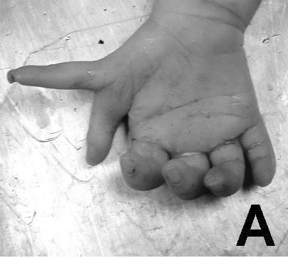
Preaxial polydactyly (thumb duplication) of an infant hand. (Tantbirojn P et al., J Med Case Rep, via Wikimedia Commons; CC BY 2.0.)
Preaxial polydactyly (thumb duplication) of an infant hand. (Tantbirojn P et al., J Med Case Rep, via Wikimedia Commons; CC BY 2.0.)
Postaxial polydactyly (small-finger duplication) is much commoner in Black/African populations (where it is usually a benign isolated autosomal-dominant trait) and more often syndromic in white/Asian populations; it is divided into type A (a well-formed articulated digit) and type B (a rudimentary pedunculated nubbin). Type B is treated by suture ligation or excision; type A needs formal reconstruction (reconstructing the ulnar collateral ligament and abductor digiti minimi).[33] Central polydactyly is rarer, usually hidden within syndactyly (synpolydactyly, the HOXD13 anomaly). Mirror hand (ulnar dimelia) is the rare duplication of the ulna with multiple fingers and no thumb, managed by pollicisation and reduction.[34]
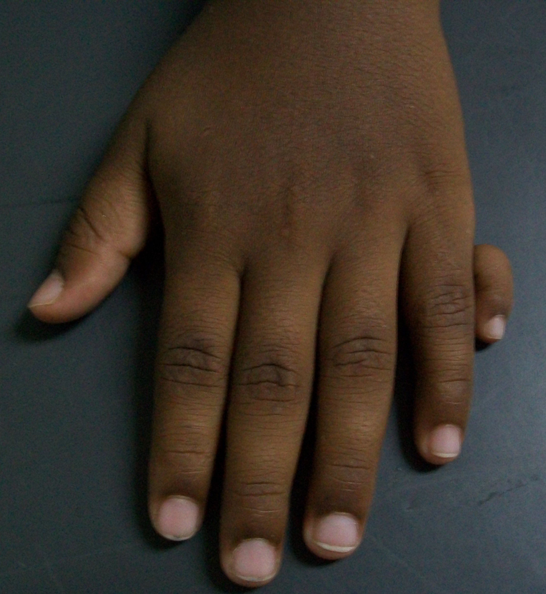
Postaxial polydactyly: a supernumerary digit on the ulnar (small-finger) border of a child’s hand. (Bobjgalindo, Wikimedia Commons; CC BY-SA 4.0.)
Postaxial polydactyly: a supernumerary digit on the ulnar (small-finger) border of a child’s hand. (Bobjgalindo, Wikimedia Commons; CC BY-SA 4.0.)
IV.2 Macrodactyly (overgrowth)
Macrodactyly is congenital enlargement of all the structures of a digit, in a static form (grows proportionately) or a progressive form (macrodystrophia lipomatosa). Its commonest cause is lipofibromatous infiltration of a digital nerve in its territory (“nerve-territory-oriented macrodactyly,” most often the median-nerve territory → index/long finger), and it is also seen with neurofibromatosis, overgrowth syndromes (Proteus, and the PIK3CA-related CLOVES spectrum) and Klippel-Trenaunay.[35] It is difficult to treat and never normalised. Options, chosen for progressive deformity, neuropathy or disabling size, include staged soft-tissue debulking, nerve decompression, epiphysiodesis to arrest length, shortening osteotomy, and ray amputation.[36]
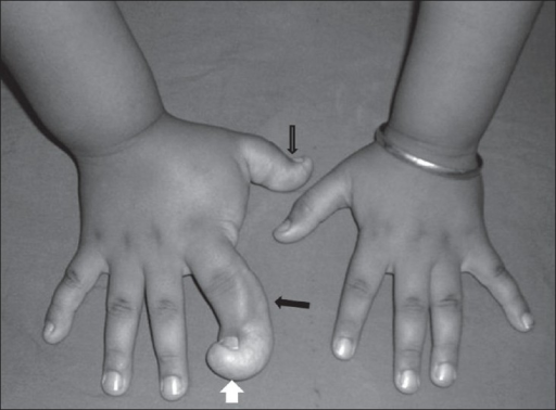
Macrodactyly of the hand (macrodystrophia lipomatosa): grossly enlarged digits (arrows). (Singla V et al., Indian J Radiol Imaging 2008; CC BY, PMC2747455.)
Macrodactyly of the hand (macrodystrophia lipomatosa): grossly enlarged digits (arrows). (Singla V et al., Indian J Radiol Imaging 2008; CC BY, PMC2747455.)
Part V - Amniotic-band (constriction-ring) disease and associated syndromes
V.1 Amniotic-band / constriction-ring syndrome
This is the syllabus “амниотична болест”: a group of circumferential constriction grooves, congenital amputations and acrosyndactyly, with many synonyms (amniotic band syndrome, constriction-ring syndrome, Streeter dysplasia, ADAM complex).[37] Two aetiological theories compete: the intrinsic (Streeter) germ-plasm theory and the extrinsic (Torpin) early-amnion-rupture sequence, in which free fibrous strands encircle and constrict the developing parts. The extrinsic theory is now favoured.[38] It is sporadic with no heredity, occurs in about 1/1,200 to 1/15,000 births, is often multiple/bilateral, and is associated with clubfoot and craniofacial clefts.[39]
The Patterson classification has four grades: type 1 a simple constriction ring; type 2 a ring with distal deformity ± lymphoedema; type 3 a ring with acrosyndactyly (distal fusion with proximal epithelialised sinuses, a “bunch-of-grapes” appearance); type 4 intrauterine (congenital) amputation.[40] The central/longer digits are preferentially affected, and the defining feature is that the anatomy proximal to the ring is normal, which distinguishes it from symbrachydactyly.[41]
Management is by severity: an emergency neonatal release for a deep band threatening circulation, and elective excision of the ring with Z-plasties and defatting (a simple straight-line closure is avoided, and a deep circumferential band is often released in two stages to protect the vascularity). Acrosyndactyly is separated, and absent digits are reconstructed by toe transfer or distraction, feasible here because the proximal anatomy is normal.[42]
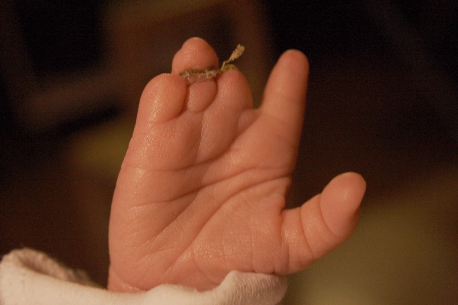
Amniotic-band sequence in a newborn hand: the fingertips are distally truncated (intrauterine congenital amputation). (Moscowmom, Wikimedia Commons; public domain.)
Amniotic-band sequence in a newborn hand: the fingertips are distally truncated (intrauterine congenital amputation). (Moscowmom, Wikimedia Commons; public domain.)
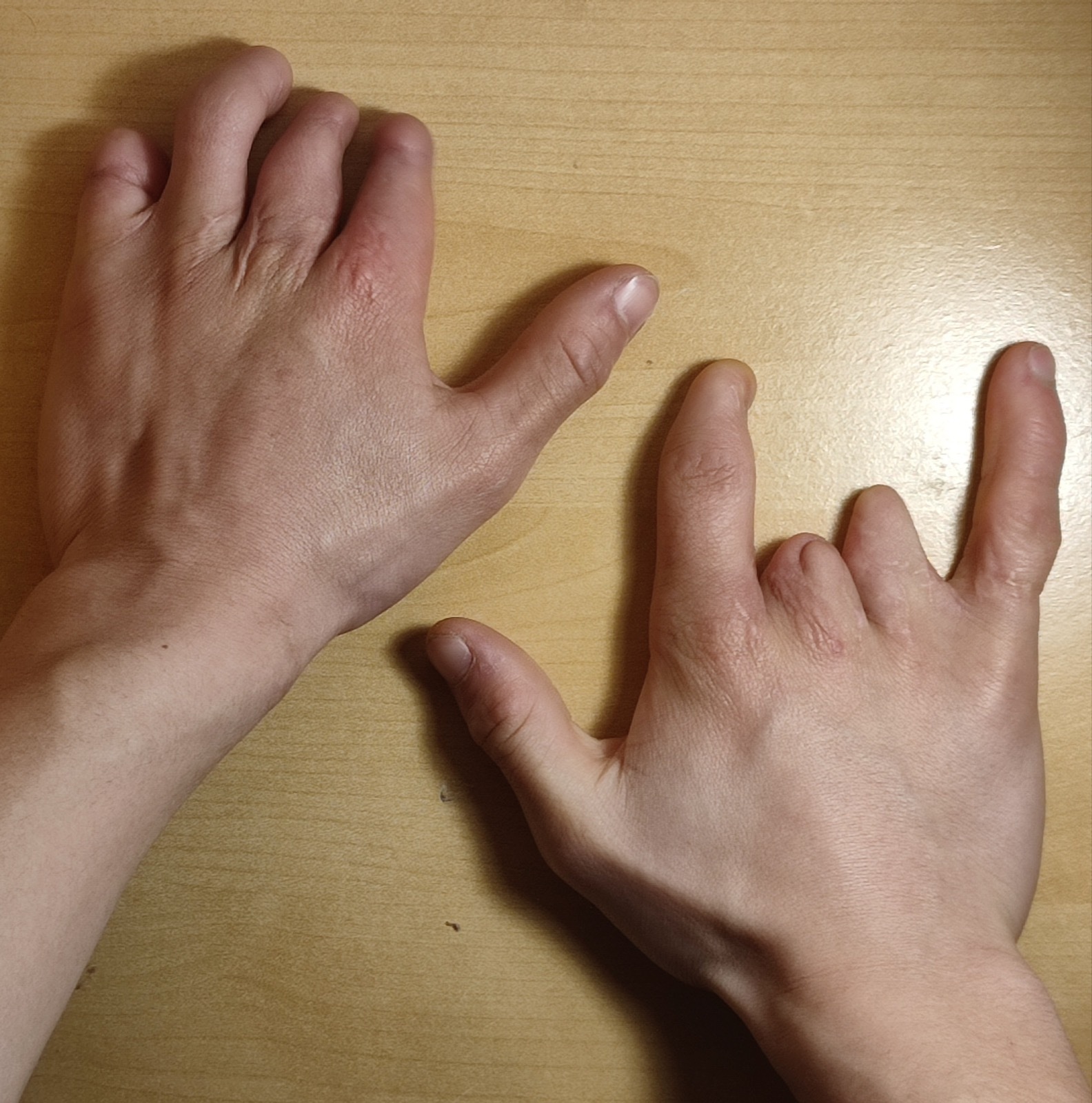
Constriction-ring (amniotic-band) syndrome: foreshortened central digits with stumps and basal acrosyndactyly. (Fyrosth, Wikimedia Commons; CC0.)
Constriction-ring (amniotic-band) syndrome: foreshortened central digits with stumps and basal acrosyndactyly. (Fyrosth, Wikimedia Commons; CC0.)
V.2 Associated syndromes
- Apert syndrome: FGFR2 acrocephalosyndactyly, with craniosynostosis, midface hypoplasia and complex syndactyly of all digits (the “spade/mitten/rosebud” hand by the Upton classification).[43]
- Poland syndrome: absence of the sternal head of pectoralis major with ipsilateral symbrachydactyly; mostly sporadic, attributed to a subclavian-artery disruption sequence.[44]
- Holt-Oram syndrome: autosomal dominant (TBX5), with radial-ray deficiency and a cardiac (ASD/VSD) defect.[45]
- Fanconi anaemia: autosomal recessive, with radial/thumb deficiency and progressive pancytopenia (the thumb is more affected than the radius); diagnosed by chromosomal-breakage testing, fatal without marrow transplant.[46]
- TAR (thrombocytopenia-absent radius): autosomal recessive (1q21.1/RBM8A), with bilateral absent radii but the thumbs always present, and thrombocytopenia at birth.[47]
- VACTERL association: vertebral, anal, cardiac, tracheo-oesophageal, renal and (preaxial) limb anomalies (≥3 features), with radial dysplasia in ~65%.[48]
- Briefly: Down syndrome (clinodactyly, simian crease), synpolydactyly (HOXD13), and Cornelia de Lange (limb reduction, Kirner deformity).[49]
Bulgarian terminology glossary
For consistency with the Bulgarian state-examination vocabulary (and the operative terminology of Boychev, Хирургическа ортопедия):
| English | Bulgarian |
|---|---|
| Congenital hand anomalies | Вродени аномалии на ръката (китката) |
| Failure of formation / differentiation | Нарушено формиране / нарушена диференциация |
| Syndactyly | Синдактилия (срастнали пръсти) |
| Polydactyly (preaxial / postaxial / central) | Полидактилия (преаксиална / постаксиална / централна) |
| Thumb duplication | Удвояване на палеца |
| Macrodactyly | Макродактилия (гигантизъм на пръст) |
| Camptodactyly / clinodactyly | Камптодактилия / клинодактилия |
| Radial longitudinal deficiency (radial club hand) | Радиален лонгитудинален дефицит (лъчева “сопеста” ръка) |
| Thumb hypoplasia | Хипоплазия на палеца |
| Cleft hand (ectrodactyly) | Цепнатина на ръката (ектродактилия, “рачешка” ръка) |
| Pollicisation | Полицизация (превръщане на пръст в палец) |
| Madelung deformity | Деформитет на Madelung |
| Radioulnar synostosis | Радиоулнарна синостоза |
| Amniotic-band / constriction-ring syndrome | Синдром на амниотичните прищипвания / констрикционни пръстени (амниотична болест) |
| Acrosyndactyly | Акросиндактилия |
| Congenital (intrauterine) amputation | Вродена (вътреутробна) ампутация |
| Apert / Poland / Holt-Oram syndrome | Синдром на Apert / Poland / Holt-Oram |
| Fanconi anaemia | Анемия на Fanconi |
Figure credits and licences
All images were independently opened and visually verified to depict the stated entity before use; licences were confirmed against the Wikimedia Commons API or the NCBI PMC Open-Access service. All are public-domain/CC0, CC BY or CC BY-SA (no non-commercial files).
- Radial club hand (clinical) -
radial_clubhand_absent_thumb_clinical.jpg. Mehlauge, Wikimedia Commons. CC BY-SA 3.0. - Radial deficiency / absent radius (radiograph) -
radial_deficiency_absent_radius_xray_TAR.jpg. Ahmad R, BMC Nephrol 2007;8:5. CC BY 2.0. - Cleft hand (ectrodactyly) -
clefthand_ectrodactyly_1yr.jpg. Aurélie & Sylvain Mulard, Wikimedia Commons. CC BY-SA 3.0. - Complete complex syndactyly -
syndactyly_newborn_complex_clinical.jpg. Wikimedia Commons. Public domain. - Camptodactyly -
camptodactyly_clinical.jpg. Armin Kübelbeck, Wikimedia Commons. CC BY-SA 3.0. - Clinodactyly (radiograph) -
clinodactyly_littlefinger_radiograph.jpg. Tomograph, Wikimedia Commons. CC BY-SA 3.0. - Madelung deformity (radiograph) -
madelung_radiograph_left.jpg. Röntgeninstitut, Städtisches Krankenhaus Kiel, Wikimedia Commons. CC BY-SA 3.0. - Preaxial polydactyly (thumb duplication) -
polydactyly_preaxial_thumb_clinical.jpg. Tantbirojn P et al., J Med Case Rep, via Wikimedia Commons. CC BY 2.0. - Postaxial polydactyly -
polydactyly_postaxial_clinical_boy.jpg. Bobjgalindo, Wikimedia Commons. CC BY-SA 4.0. - Macrodactyly of the hand -
macrodactyly_hand_clinical_PMC2747455.png. Singla V et al., Indian J Radiol Imaging 2008 (PMC2747455). CC BY. - Amniotic-band congenital amputation -
amnioticband_baby_hand_clinical.jpg. Moscowmom, Wikimedia Commons. Public domain. - Constriction-ring syndrome -
constriction_ring_hands_clinical.jpg. Fyrosth, Wikimedia Commons. CC0.
CC BY/CC BY-SA images require attribution (and, for SA, that a redistributed derivative keep the same licence); the CC0/public-domain images are free of restriction.
References
-
Rayan & Upton, Congenital Hand Anomalies and Associated Syndromes, p.10; Waters & Bae, Pediatric Hand and Upper Limb Surgery, p.17.
-
Rayan & Upton, pp.10-11.
-
Rayan & Upton, pp.10-11; Waters & Bae, p.17.
-
Waters & Bae, p.17 (founded on Manske & Oberg 2009 and Tonkin 2006); the full OMT tree is the standard published scheme.
-
Waters & Bae, pp.10-16.
-
Rayan & Upton, pp.10-11; Waters & Bae, p.10.
-
Rayan & Upton, p.132; Waters & Bae, p.132.
-
Rayan & Upton, p.132; Waters & Bae, p.132.
-
Rayan & Upton, pp.132, 137-143; Waters & Bae, pp.102, 131.
-
Rayan & Upton, pp.132-134; Waters & Bae, pp.131-137.
-
Waters & Bae, pp.102-103.
-
Waters & Bae, p.102.
-
Rayan & Upton, p.159; Waters & Bae, p.141.
-
Rayan & Upton, p.159; Waters & Bae, pp.142-145.
-
Waters & Bae, pp.97-98.
-
Buck-Gramcko, Congenital Malformations of the Hand and Forearm, pp.31-32; Waters & Bae, p.99.
-
Waters & Bae, pp.77-82; Buck-Gramcko, p.31.
-
Buck-Gramcko, p.24; Waters & Bae, p.133.
-
Waters & Bae, pp.21-22.
-
Waters & Bae, pp.22, 31-32.
-
Waters & Bae, pp.22-32.
-
Waters & Bae, pp.59-66; Buck-Gramcko, pp.324-326.
-
Waters & Bae, pp.58-66.
-
Waters & Bae, pp.58-59; Rayan & Upton, pp.326-328.
-
Rayan & Upton, pp.200-204; Waters & Bae, pp.147-153.
-
Buck-Gramcko, p.44.
-
Rayan & Upton, pp.213-215, 299-304.
-
Standard description; the dedicated trigger-thumb chapter was outside the page-cited extracts.
-
Buck-Gramcko, pp.318, 328-330.
-
Waters & Bae, p.16; Buck-Gramcko, p.46.
-
Waters & Bae, pp.41-43.
-
Waters & Bae, pp.41-50.
-
Waters & Bae, pp.35-39.
-
Waters & Bae, pp.52-57; Rayan & Upton, pp.206-209.
-
Waters & Bae, pp.68-69; Buck-Gramcko, pp.206-210.
-
Waters & Bae, pp.69-75.
-
Rayan & Upton, p.479; Waters & Bae, p.90.
-
Rayan & Upton, p.479; Waters & Bae, p.90.
-
Rayan & Upton, p.479; Waters & Bae, pp.90-91; Buck-Gramcko, p.26.
-
Rayan & Upton, p.479; Waters & Bae, p.91; Buck-Gramcko, p.26.
-
Rayan & Upton, pp.481, 368; Waters & Bae, p.91.
-
Waters & Bae, pp.91-96; Buck-Gramcko, pp.204-205.
-
Rayan & Upton, pp.371-375.
-
Rayan & Upton, pp.440-442.
-
Buck-Gramcko, pp.26-27; TBX5 is the established gene.
-
Buck-Gramcko, pp.26-27; Rayan & Upton, p.138.
-
Rayan & Upton, pp.137-139.
-
Rayan & Upton, pp.141-143.
-
Rayan & Upton, pp.102-104, 356-364, 382-383.