Contents
- Scope and orientation
- Part I - Definition, epidemiology and aetiology
- Part II - Pathoanatomy
- Part III - Clinical assessment and classification
- Part IV - The Ponseti method
- Part V - Alternatives and surgery
- Bulgarian terminology glossary
- Figure credits and licences
Scope and orientation
Congenital clubfoot (talipes equinovarus) is one of the commonest congenital deformities of the limb. This summary covers its pathoanatomy and causes, the clinical assessment and severity scoring, and the Ponseti method, which moved treatment away from extensive surgery toward manipulation, casting, a small tenotomy and bracing.
A few organising ideas run through the topic:
- The deformity is a combination, captured by the mnemonic CAVE: Cavus, Adductus of the forefoot, Varus of the hindfoot, and Equinus of the ankle. It is a three-dimensional malalignment of the tarsus, not an embryonic malformation or a true dislocation.
- Distinguish the rigid (true) foot from the postural (positional) one, which corrects with little or no treatment.
- The Ponseti method corrects in a fixed order (cavus first, then adductus and varus together by abducting the foot around the talar head, then equinus last by a tenotomy), and the cardinal error is to pronate the foot.
- Relapse is the rule unless the foot-abduction brace is worn. Brace non-compliance is the leading cause of recurrence; dynamic supination from a relapse is treated by recasting and, later, a tibialis-anterior transfer.
A Bulgarian glossary, a viva self-test appendix, and image credits follow the clinical sections.
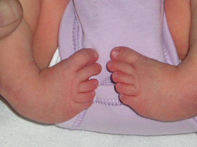
Bilateral congenital talipes equinovarus in a newborn: both feet are inverted and adducted, with the soles turned toward the midline. (Brachet Youri, Wikimedia Commons; CC BY-SA 3.0.)
Bilateral congenital talipes equinovarus in a newborn: both feet are inverted and adducted, with the soles turned toward the midline. (Brachet Youri, Wikimedia Commons; CC BY-SA 3.0.)
Part I - Definition, epidemiology and aetiology
I.1 Definition and terminology
Clubfoot is a congenital, three-dimensional malalignment of the bones and joints of the foot and ankle, combining four components captured by the mnemonic CAVE: cavus of the forefoot, adductus of the forefoot on the midfoot, varus (inversion and adduction) of the hindfoot, and equinus of the ankle.[1] It is not an embryonic malformation and not a true dislocation: a normally developing foot turns into a clubfoot during the second trimester, so (like developmental hip dysplasia and idiopathic scoliosis) it is best regarded as a developmental deformation.[2] The deformities are not passively correctable, and this is what separates a true clubfoot from a postural one.[3] The working categories are idiopathic (typical), postural/positional, neurogenic and syndromic/teratologic.[4]
I.2 Epidemiology
- Incidence is about 1 per 1000 live births in Caucasians, with marked ethnic variation: roughly half as frequent in Japanese, about three times as frequent in South-African Black populations, and highest (about six times) in Polynesians/Maori.[5]
- Sex: boys predominate, about 2:1 to 3:1 (sources differ; Ponseti gives 3:1, Lovell/Wenger about 2:1).[6]
- Bilateral in roughly 40-50%.[7]
- Heritability is clear: the risk to a subsequent sibling is about 1 in 30-35, monozygotic-twin concordance is about 33% versus ~3% for dizygotic twins, and the offspring risk is about 3-4% with one affected parent (about 30% if both are affected).[8]
I.3 Aetiology
The cause is unknown and almost certainly multifactorial/polygenic, with clubfoot probably a common endpoint of several mechanisms.[9] The classical theories are intrauterine moulding (now doubted, since clubfoot is not more frequent in twins or large babies), a subclinical neuromuscular defect (supported by the high incidence in spina bifida and arthrogryposis), and a primary germ-plasm / arrested-development defect (Irani & Sherman found a consistently short, medially-deviated talar neck).[10] Ponseti’s biological model is a retracting fibrosis: excess collagen synthesis in the medial and posterior ligaments, tendons and muscles, with an overpull of tibialis posterior, that may persist to age 3-4 years and drive relapse.[11] Genetic factors include a strong family history and variants in the PITX1-TBX4 pathway; maternal smoking is a dose-dependent environmental risk (and interacts more than additively with a family history).[12]
Part II - Pathoanatomy
Ponseti’s account, from fetal and neonatal specimens, is the standard description.[13] The deformity is a medial rotation of the calcaneus-forefoot unit around the talus, combining bony positional changes with medial and posterior soft-tissue contractures.
Bony/positional changes:
- Talus: in severe equinus (plantar flexion), with the neck deviated medially and plantarward and a wedge-shaped head whose antero-lateral surface is left uncovered (so it is palpable on the dorsolateral foot).[14]
- Navicular: the site of greatest distortion, severely medially displaced (subluxed) onto the medial talar head, its tuberosity nearly touching the medial malleolus.[15]
- Calcaneus: in equinus, varus and adduction beneath the talus, so the talar and calcaneal axes become parallel (the radiographic loss of divergence, a reduced Kite angle); its posterior tuberosity is also rotated laterally.[16]
- Cuboid medially displaced on the calcaneus, the first metatarsal more plantar-flexed than the lateral rays (producing the cavus), and the forefoot adducted and supinated.[17]
Soft-tissue contractures form a medial and posterior “knot”: a hypertrophic tibialis posterior tendon (the chief deforming force on the navicular), the contracted deltoid, spring (calcaneonavicular) and tibionavicular ligaments medially, and a tight, thick tendo Achilles with contracted posterior ankle/subtalar ligaments.[18] The interosseous talocalcaneal ligament, by contrast, is not part of this contracted knot (Ponseti describes it as thin and loosely textured; Staheli calls it normal).[19]
The biological key to treatment is that infant ligament collagen is “crimped” (wavy) and stretches gently with manipulation, the crimp reforming within days. The tendo Achilles is the exception: it is non-stretchable and therefore requires a tenotomy.[20]
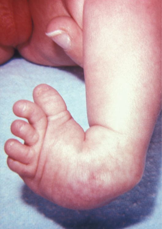
Clinical clubfoot in an infant: the foot is in equinus with hindfoot varus and forefoot adductus, the sole turned medially. (OpenStax College, Wikimedia Commons; CC BY 3.0.)
Clinical clubfoot in an infant: the foot is in equinus with hindfoot varus and forefoot adductus, the sole turned medially. (OpenStax College, Wikimedia Commons; CC BY 3.0.)
Part III - Clinical assessment and classification
III.1 Examination
The diagnosis is clinical and made at birth from the four CAVE components; radiographs are not needed for diagnosis in the newborn.[21] The classic signs are a fixed equinovarus foot that does not correct passively, a single deep posterior ankle crease and a deep medial midfoot crease, an “empty heel” (the calcaneus is pulled up and hard to palpate), a palpable lateral talar head, and calf atrophy with a slightly short foot.[22]
The key distinction is rigid (true) versus postural (positional): a postural foot is very flexible and corrects passively, completely, or with one or two casts, whereas a true clubfoot is not passively correctable.[23] Every newborn also needs a general examination: motor/sensory testing (to exclude a neurogenic foot), the spine (for dysraphism) and the hands (adducted contracted thumbs suggest arthrogryposis). Routine hip imaging for a coexisting DDH is debated (Lovell/Mosca find no association, while Wenger images the hips).[24]
III.2 Severity scoring
Two systems are in current use, each applied at the start of treatment:
- Pirani score (0-6): six clinical signs, each scored 0 / 0.5 / 1, split into three hindfoot (posterior crease, emptiness of the heel, rigidity of equinus) and three midfoot (curvature of the lateral border, medial crease, position of the lateral head of the talus). Serial scores monitor the progress of casting and help decide when to do the tenotomy.[25]
- Diméglio (Bensahel) score (0-20): grades the reducibility of the four components (equinus, varus, forefoot/calcaneal derotation around the talus, forefoot adduction), each 0-4, plus four one-point items (posterior crease, medial crease, cavus, muscle condition); severity runs from grade I (benign) to grade IV (very severe/resistant).[26] The Diméglio system is the more reproducible of the older comparative schemes (Harrold & Walker, Catterall, Ponseti & Smoley are historical).[27]
III.3 Imaging
Radiographs are limited in the newborn because the tarsals are largely cartilaginous (the navicular does not ossify until 3-4 years). When used, weight-bearing/stress views show the talocalcaneal “parallelism” (a reduced Kite angle), loss of the normal talus-first-metatarsal alignment, and adducted metatarsals.[28]
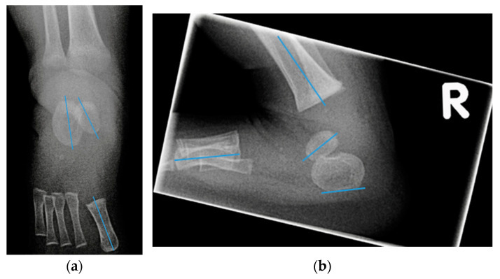
Clubfoot radiographs in an infant - (a) antero-posterior and (b) lateral, with axis lines drawn for the talus, calcaneus and first metatarsal: the talocalcaneal (Kite) angle is reduced (parallelism) and the metatarsals are adducted. (Moerman et al., Children 2022;9:865; CC BY 4.0.)
Clubfoot radiographs in an infant - (a) antero-posterior and (b) lateral, with axis lines drawn for the talus, calcaneus and first metatarsal: the talocalcaneal (Kite) angle is reduced (parallelism) and the metatarsals are adducted. (Moerman et al., Children 2022;9:865; CC BY 4.0.)
III.4 Idiopathic versus syndromic/teratologic clubfoot
The idiopathic foot occurs in an otherwise normal child. Non-idiopathic clubfeet (associated with arthrogryposis, myelomeningocele, diastrophic dysplasia and amniotic-band (constriction) syndrome) are stiffer and more resistant, relapse more often, and have an outcome that depends more on the underlying disorder than on the foot. They still begin with the Ponseti method, but correction takes longer (e.g. 9-15 casts in arthrogryposis), tendons are released rather than lengthened at surgery, and insensate myelodysplastic feet need extra care to avoid casting ulcers.[29]
Part IV - The Ponseti method
IV.1 Principles and timing
Treatment starts in the first one to two weeks of life to exploit the elasticity of the infant tissues.[30] The whole foot (except the equinus) is corrected simultaneously by abducting it in supination around the head of the talus, which is stabilised by counter-pressure over its lateral aspect.[31] The cavus is corrected first, by supinating the forefoot and elevating (dorsiflexing) the first ray to realign it with the hindfoot.[32]
The cardinal error is to pronate the foot (“Thou shall not pronate”): pronation worsens the cavus, jams the adducted calcaneus against the talus and blocks the heel from coming out of varus. The heel varus corrects only by abducting (externally rotating) the calcaneus under the talus, not by everting it (Kite’s misconception).[33]
IV.2 Casting
The order of correction follows CAVE: cavus, then adductus and varus together by abduction, and equinus last.[34] Above-knee (long-leg) plaster casts are applied with the knee flexed 90° and changed weekly; a below-knee cast is useless because it slips on the chubby infant leg.[35] The foot is progressively abducted to about 60-70° with the heel brought to slight valgus, and the counter-pressure is on the lateral head of the talus, never on the calcaneocuboid joint (Kite’s error) or the lateral malleolus.[36] Most feet need about five to six casts (range up to ~8) to correct everything but the equinus; if a foot is not corrected by six or seven casts, the technique is faulty.[37]
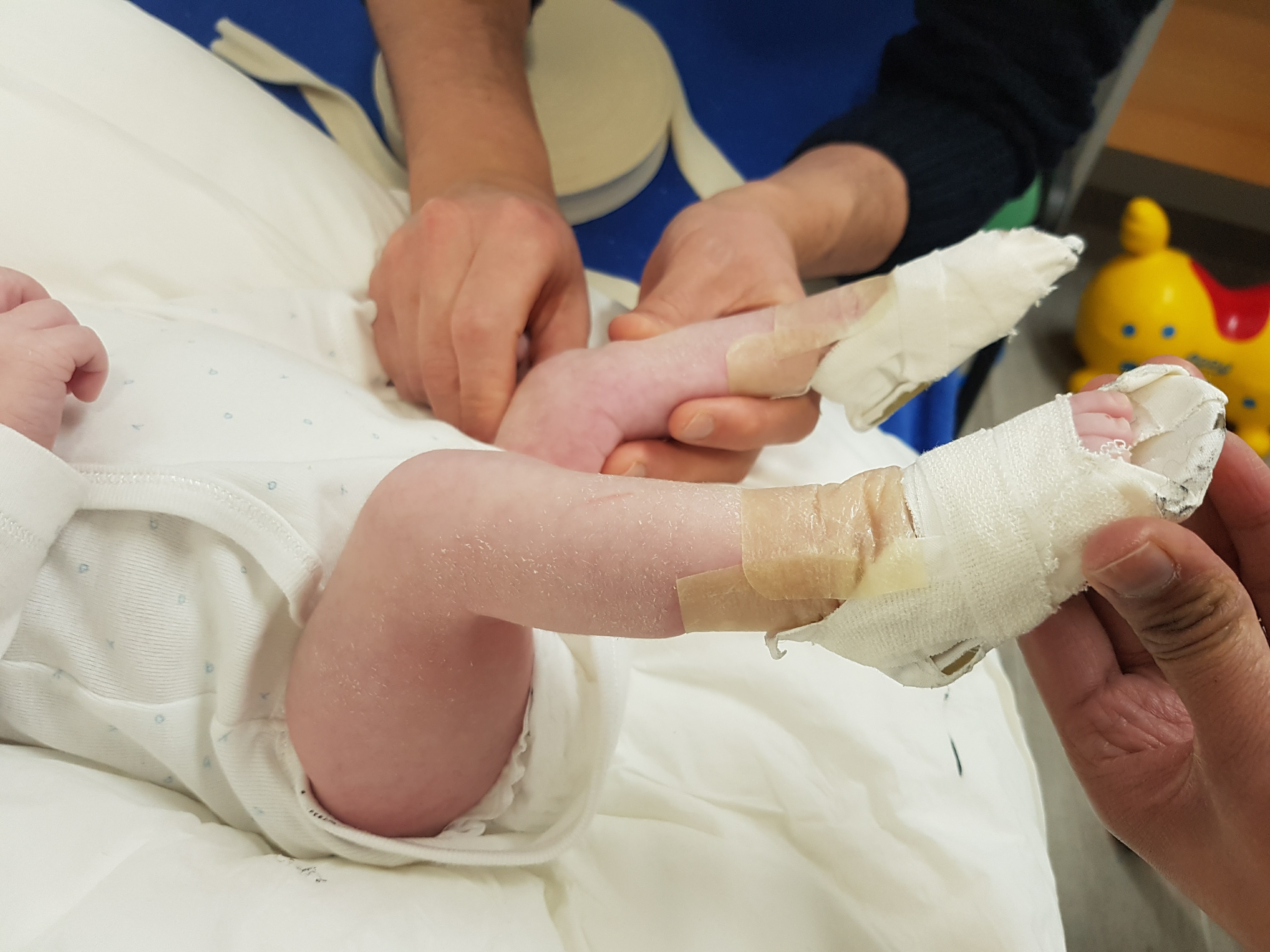
Application of a long-leg (above-knee) plaster cast in the Ponseti method, the foot held abducted around the talar head. (Orthopediatrie, Wikimedia Commons; CC BY-SA 4.0.)
Application of a long-leg (above-knee) plaster cast in the Ponseti method, the foot held abducted around the talar head. (Orthopediatrie, Wikimedia Commons; CC BY-SA 4.0.)
IV.3 Percutaneous Achilles tenotomy
Residual equinus persists in the great majority (~85%, “nearly all”) of feet because the tendo Achilles will not stretch, so a percutaneous tenotomy is performed once the other components are corrected but ankle dorsiflexion remains under about 10-15°.[38] It is an office procedure under local anaesthetic: a blade divides the tendon about 1.5-2 cm above the calcaneus (entered from the medial side, the posterior tibial neurovascular bundle lying just anteromedial), and dorsiflexion increases by 10-20° with a palpable pop.[39] A final cast holds the foot in maximal dorsiflexion and abduction for about three weeks while the tendon regenerates at the correct length.[40]
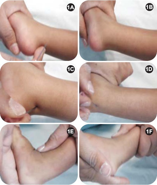
Percutaneous needle tenotomy of the tendo Achilles in clubfoot (six-panel clinical series): the needle/blade at the medial heel, then the foot dorsiflexing as correction is achieved. (Patwardhan et al., J Orthop Case Rep 2012;2(1):35; CC BY-NC-SA 3.0 - non-commercial.)
Percutaneous needle tenotomy of the tendo Achilles in clubfoot (six-panel clinical series): the needle/blade at the medial heel, then the foot dorsiflexing as correction is achieved. (Patwardhan et al., J Orthop Case Rep 2012;2(1):35; CC BY-NC-SA 3.0 - non-commercial.)
IV.4 The foot-abduction brace
The maintenance phase uses a foot-abduction brace (Denis Browne “boots-and-bar”): open-toe shoes on a bar the width of the child’s shoulders, set at about 60-70° of external rotation on the clubfoot side and 30-40° on a normal foot, with the bar bent to hold dorsiflexion.[41] It is worn full-time (≈23 hours/day) for about three months, then at night and naptime until age 3-4 years.[42] Brace non-compliance is the leading cause of relapse: recurrence occurs in over 80% of non-compliant families versus about 6% of compliant ones.[43]
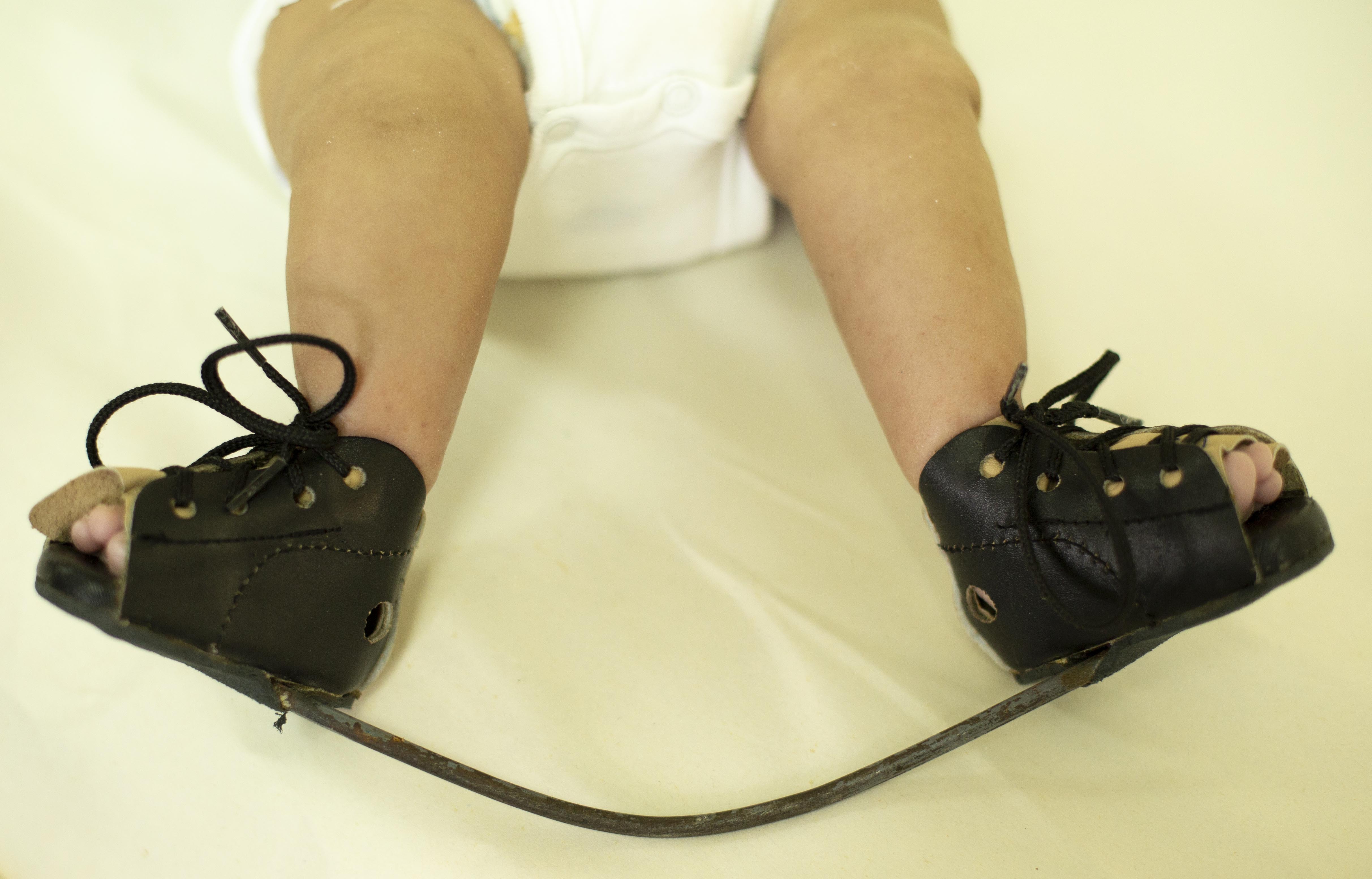
Foot-abduction brace (boots-and-bar) worn by an infant in the Ponseti maintenance phase. (Bassgrab75, Wikimedia Commons; CC BY-SA 4.0.)
Foot-abduction brace (boots-and-bar) worn by an infant in the Ponseti maintenance phase. (Bassgrab75, Wikimedia Commons; CC BY-SA 4.0.)
IV.5 Relapse and the tibialis-anterior transfer
Clubfoot has a stubborn tendency to relapse (from the same retracting fibrosis), usually before age five and rarely after seven.[44] The first sign is dynamic supination of the forefoot (a strong tibialis anterior overpowering the weaker peroneals), with recurrent heel varus. Relapse is treated first by repeat casting (one to three casts) to regain correction, then strict bracing.[45] A persistent dynamic supination after the second or third year is corrected by a tibialis-anterior tendon transfer to the lateral (third) cuneiform, done once the lateral cuneiform ossifies (after about age 2.5-3, ideally 3-5 years), the whole tendon transferred (never split, never to the cuboid). Recurrent equinus is treated by serial casts and, if needed, a repeat tenotomy.[46]
IV.6 Outcomes
Initial correction is achieved in over 90-95% of idiopathic feet.[47] Long-term results are good: a mobile, plantigrade, painless foot, though the calf and foot remain slightly small (a unilateral clubfoot is on average ~1.3 cm shorter with a ~2.3 cm smaller calf). In the Laaveg & Ponseti series (mean 18.8-year follow-up) 74% were excellent or good with no patient having pain on walking, and the Cooper & Dietz 30-year follow-up found function no different from people born with normal feet.[48] These results are superior to extensive surgical release, which leaves stiff, weak, often arthritic and painful feet in adult life.[49]
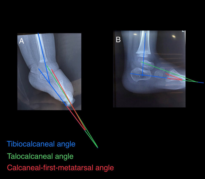
Lateral clubfoot radiographs before (A) and one year after (B) tendo-Achilles tenotomy, with the tibiocalcaneal, talocalcaneal and calcaneal-first-metatarsal angles labelled, showing correction of the hindfoot. (Tahririan et al., Sci Rep 2023;13:11734; CC BY 4.0.)
Lateral clubfoot radiographs before (A) and one year after (B) tendo-Achilles tenotomy, with the tibiocalcaneal, talocalcaneal and calcaneal-first-metatarsal angles labelled, showing correction of the hindfoot. (Tahririan et al., Sci Rep 2023;13:11734; CC BY 4.0.)
Part V - Alternatives and surgery
V.1 The French functional (physiotherapy) method
The main non-operative alternative is the French functional method: daily manipulation with adhesive taping/splinting and continuous passive motion by a physiotherapist over several months. In compliant families its results approach the Ponseti method, but it is far more labour-intensive. The Ponseti method remains the worldwide standard because it is effective, cheap and easy to disseminate.[50]
V.2 The role of surgery
The Ponseti method has greatly reduced the need for extensive surgery. Maximum cast correction always precedes any operation.[51] The only operations usually needed are the tendo-Achilles tenotomy and the tibialis-anterior transfer.[52]
When casting genuinely fails (the unusually rigid, severe or syndromic foot), a limited, “à la carte” release is preferred over the now largely abandoned extensive posteromedial release: only the structures that did not correct are released (a posterior release of the Achilles, posterior ankle/subtalar capsule and calcaneofibular ligament, with a circumferential postero-plantar-medial release through a Cincinnati incision for the most resistant feet), so as to preserve as much joint integrity as possible.[53] Extensive release is avoided because it leaves a stiff, weak, flat, arthritic and painful foot in the long term.[54]
V.3 Salvage and the older/neglected foot
A neglected clubfoot is still started on Ponseti casting, which can correct feet well into late childhood. Residual cavovarus is assessed with the Coleman lateral block test and treated with soft-tissue and osteotomy procedures; triple arthrodesis is a salvage for the rigid deformed foot in the older child (over about 9-10 years), and talectomy or an Ilizarov frame is reserved for the severely rigid, relapsed or arthrogrypotic foot.[55] Surgically corrected feet remain prone to recurrence throughout childhood (about 25-50%).[56]
Bulgarian terminology glossary
For consistency with the Bulgarian state-examination vocabulary (and the operative terminology of Boychev, Хирургическа ортопедия):
| English | Bulgarian |
|---|---|
| Congenital clubfoot (talipes equinovarus) | Вродено еквиноварусно ходило (pes equinovarus congenitus) |
| Cavus / adductus / varus / equinus | Кавус / аддукция / варус / еквинус |
| Forefoot / midfoot / hindfoot | Преден / среден / заден отдел на стъпалото |
| Rigid (true) vs postural (positional) clubfoot | Ригидно (истинско) срещу постурално (позиционно) еквиноварусно ходило |
| Talus / navicular / calcaneus | Талус (скочна кост) / ладиевидна кост / калканеус (петна кост) |
| Pirani score | Скор на Pirani |
| Diméglio classification | Класификация на Diméglio |
| Ponseti method | Метод на Ponseti |
| Manipulation and casting | Манипулация и гипсова имобилизация |
| Percutaneous Achilles tenotomy | Перкутанна ахилотомия (тенотомия на Ахилесовото сухожилие) |
| Foot-abduction brace (Denis Browne) | Абдукционна шина за стъпалата (по Denis Browne) |
| Relapse / recurrence | Рецидив (повторна поява на деформитета) |
| Tibialis anterior tendon transfer | Транспозиция на сухожилието на m. tibialis anterior |
| Posteromedial release | Постеромедиално освобождаване (release) |
| Triple arthrodesis | Тройна артродеза |
| Arthrogryposis | Артрогрипоза |
Figure credits and licences
All images were independently opened and visually verified to depict the stated entity before use; licences were confirmed against the Wikimedia Commons API or the NCBI PMC Open-Access service.
- Bilateral newborn clubfoot -
clubfoot_bilateral_newborn_BrachetYouri.jpg. Brachet Youri, Wikimedia Commons. CC BY-SA 3.0. - Clinical clubfoot (single foot) -
813_Clubfoot.jpg. OpenStax College (Anatomy & Physiology), Wikimedia Commons. CC BY 3.0. - Clubfoot radiographs (AP + lateral) -
clubfoot_radiograph_AP_lateral_PMC9221593.jpg. Moerman S et al., Children (Basel) 2022;9(6):865. CC BY 4.0. - Ponseti casting -
clubfoot_PBVE_plaquette_Orthopediatrie.jpg. Orthopediatrie, Wikimedia Commons. CC BY-SA 4.0. - Percutaneous Achilles tenotomy -
tenotomy_percutaneous_needle_NC_PMC4844500.jpg. Patwardhan S, Shyam A, Sancheti P, J Orthop Case Rep 2012;2(1):35-36. CC BY-NC-SA 3.0 - non-commercial use only. - Foot-abduction brace (boots-and-bar) -
FAB_brace_Bassgrab75.jpg. Bassgrab75, Wikimedia Commons. CC BY-SA 4.0. - Pre/post-tenotomy radiographs -
clubfoot_radiograph_pre_post_tenotomy_PMC10359333.jpg. Tahririan MA et al., Sci Rep 2023;13:11734. CC BY 4.0.
Licensing note for distribution: image 5 (tenotomy) is CC BY-NC-SA, acceptable for a personal, non-commercial study document, but it must be removed or replaced if this material is ever used commercially, and any derivative shares the same licence. The CC BY-SA images likewise require that a redistributed derivative carry the same licence.
References
-
Lovell & Winter, p.4315; Ponseti, Congenital Clubfoot: Fundamentals of Treatment, p.11; Staheli & Ponseti, Clubfoot: Ponseti Management, p.8.
-
Staheli & Ponseti, p.4; Wenger & Rang, p.162.
-
Mosca, Pediatric Foot & Ankle, pp.71-72.
-
Lovell & Winter, p.4311; Staheli & Ponseti, p.8.
-
Ponseti, p.9; Lovell & Winter, p.4313.
-
Ponseti, p.9; Lovell & Winter, p.4313; Wenger & Rang, p.162.
-
Ponseti, p.9; Lovell & Winter, p.4313.
-
Ponseti, p.9; Lovell & Winter, p.4313; Staheli & Ponseti, pp.7, 28.
-
Wenger & Rang, pp.161-162; Lovell & Winter, p.4313.
-
Wenger & Rang, pp.161-162; Lovell & Winter, p.4314.
-
Staheli & Ponseti, p.4.
-
Lovell & Winter, p.4314.
-
Ponseti, pp.16-23.
-
Ponseti, pp.18-20; Staheli & Ponseti, p.4.
-
Ponseti, pp.18-19.
-
Ponseti, p.20; Wenger & Rang, p.163.
-
Ponseti, p.20.
-
Ponseti, pp.16, 21-23.
-
Ponseti, p.21; Staheli & Ponseti, p.4.
-
Staheli & Ponseti, pp.4-5.
-
Lovell & Winter, p.4317; Mosca, p.72.
-
Lovell & Winter, pp.4315-4316; Wenger & Rang, p.163.
-
Lovell & Winter, p.4311; Wenger & Rang, p.163; Staheli & Ponseti, p.8.
-
Lovell & Winter, pp.4316-4317; Mosca, p.72; Wenger & Rang, pp.163-164.
-
Staheli & Ponseti, p.27; Lovell & Winter, p.4318.
-
Lovell & Winter, pp.4318-4320.
-
Lovell & Winter, p.4318.
-
Lovell & Winter, pp.4320-4322; Wenger & Rang, pp.166-168.
-
Lovell & Winter, p.4311; Mosca, pp.72-75; Staheli & Ponseti, pp.7, 23.
-
Staheli & Ponseti, pp.6, 28.
-
Staheli & Ponseti, pp.5, 9; Ponseti, p.76.
-
Staheli & Ponseti, p.10; Ponseti, pp.72-75.
-
Staheli & Ponseti, pp.5, 13; Ponseti, pp.71-72.
-
Ponseti, pp.71-73; Staheli & Ponseti, p.7.
-
Ponseti, pp.76, 79; Staheli & Ponseti, p.10.
-
Ponseti, pp.72, 77; Staheli & Ponseti, pp.11, 13.
-
Ponseti, p.87; Staheli & Ponseti, pp.5-6.
-
Ponseti, pp.82-83; Staheli & Ponseti, pp.5, 7, 14.
-
Ponseti, pp.90-91; Staheli & Ponseti, p.14; Mosca, pp.158-163.
-
Ponseti, pp.83, 91; Staheli & Ponseti, p.15.
-
Staheli & Ponseti, p.16; Ponseti, p.87.
-
Staheli & Ponseti, pp.16, 29.
-
Staheli & Ponseti, pp.16, 20.
-
Ponseti, p.106.
-
Staheli & Ponseti, pp.20-21.
-
Ponseti, pp.91-92; Staheli & Ponseti, pp.21, 24; Mosca, p.176.
-
Staheli & Ponseti, p.7.
-
Ponseti, pp.116-125; Staheli & Ponseti, pp.5, 7.
-
Ponseti, pp.83, 125; Staheli & Ponseti, p.7.
-
Staheli & Ponseti, pp.6, 31 - the French method is referenced (Richards et al., JBJS Am 2008) but described from standard sources rather than the page-cited extracts.
-
Ponseti, pp.81, 93; Staheli & Ponseti, p.23.
-
Ponseti, p.89.
-
Mosca, pp.192, 204-206; Ponseti, p.95.
-
Ponseti, p.89; Staheli & Ponseti, p.7.
-
Ponseti, pp.95-104; Staheli & Ponseti, pp.22-23.
-
Staheli & Ponseti, p.23.