Contents
- Introduction and scope
- Part I - Definition and historical background
- Part II - Epidemiology and etiology
- Part III - Classification
- Part IV - Clinical assessment
- Part V - Natural history of ambulation and gait
- Part VI - Management of tone and the general principles of treatment
- Part VII - The paralytic (spastic) hip
- Part VIII - The paralytic (spastic) foot and ankle
- Part IX - The spastic knee
- Part X - The neuromuscular spine
- Part XI - The upper limb in spastic hemiplegia
- Bulgarian terminology (Боев / Boychev tradition) - glossary
- Image attributions
Introduction and scope
The syllabus item is “Cerebral palsy (CP). Paralytic dislocation of the hip. Paralytic foot.”, covering the orthopaedic management of the commonest cause of physical disability in childhood, with particular weight on its two most important deforming problems: the spastic (paralytic) hip and the spastic (paralytic) foot. This summary therefore gives a complete account of cerebral palsy as a disease, from its definition and causes through classification, assessment and the management of tone, and then treats the paralytic hip and the paralytic foot in depth, with shorter sections on the spastic knee, the neuromuscular spine and the upper limb.
A few ideas run through the whole topic. The first is that the brain lesion is static but the musculoskeletal deformity is progressive: the encephalopathy does not worsen, but spasticity, weakness and absent normal loading make muscles grow more slowly than bones, so contractures and bony deformities develop and evolve throughout growth. Cerebral palsy has aptly been called a “short-muscle disease.”[1] The second is that function, and the risk of every deformity, tracks the GMFCS level: the Gross Motor Function Classification System predicts walking, hip displacement and the whole pattern of orthopaedic problems, and it frames every treatment decision. The third is that the goal of orthopaedic care is not a normal child but a functional one. Where walking is possible that means a level pelvis with located, mobile hips, plantigrade braceable feet and an efficient balanced gait; where it is not, it means comfortable sitting and easy care.
Part I - Definition and historical background
The widely accepted modern definition (Rosenbaum, Bax and colleagues, 2006/2007) is: “Cerebral palsy describes a group of permanent disorders of the development of movement and posture, causing activity limitation, that are attributed to non-progressive disturbances that occurred in the developing fetal or infant brain. The motor disorders of cerebral palsy are often accompanied by disturbances of sensation, perception, cognition, communication and behaviour, by epilepsy, and by secondary musculoskeletal problems.”[2]
The key qualifiers are that CP is a group of disorders, not one disease; the causal disturbance is non-progressive (a “static encephalopathy”) and occurred in the developing (fetal or infant) brain; and the motor disorder is permanent but not unchanging, in Mercer Rang’s phrase.[3] Historically, William Little gave the first description (1843; the spastic diplegia of prematurity was long called “Little’s disease”), William Osler coined the term cerebral palsy (1889), and Sigmund Freud argued that many cases originate before birth, a view borne out by modern epidemiology.[4]
Part II - Epidemiology and etiology
The prevalence of cerebral palsy is about 2-2.5 per 1,000 live births and has remained stable despite advances in obstetric and neonatal care.[5] The diagnosis often cannot be confirmed reliably until about 4 years of age, because some early neurological signs resolve.
The single most important fact about causation is that most cerebral palsy originates before birth, not during delivery: at least 70% has prenatal antecedents and only some 10-20% relates to perinatal events.[6] The principal risk factors are:
- Prematurity and low birth weight, the strongest predictor. The CP rate in survivors born before 33 weeks is roughly 30 times that of term infants; very preterm births are under 2% of survivors yet contribute 20-25% of all CP. Periventricular leukomalacia (PVL) is the characteristic white-matter lesion of prematurity and typically produces spastic diplegia.[7]
- Multiple gestation (roughly four times the risk in twins, far higher in triplets), partly through assisted reproduction.[8]
- Intrauterine infection / chorioamnionitis and the TORCH organisms; maternal infection sharply raises the risk even at normal birth weight.[9]
- Kernicterus (Rh/ABO incompatibility, hyperbilirubinaemia) classically causing dyskinetic (athetoid) CP with deafness, now rare since anti-D prophylaxis and phototherapy.[10]
- Perinatal asphyxia, a genuine but minority cause, and postnatal insults (meningitis, head injury, near-drowning) up to about age 2-3 years.[11]
On MRI, about 43% of cases show white-matter injury of prematurity. Smaller proportions show basal-ganglia lesions, cortical/subcortical lesions, malformations or focal infarcts, and about 12% are normal.[12] A useful aphorism is that “the genotype may load the gun and the environment pulls the trigger.”
Part III - Classification
Cerebral palsy is classified in several complementary ways, all of which the examiner expects.
By physiologic (motor) type
- Spastic (pyramidal), much the commonest at 60-85% of cases. There is a velocity-dependent (“clasp-knife”) increase in tone, with hyperreflexia, clonus and an extensor plantar response, from a lesion affecting the corticospinal system; it is associated with prematurity and PVL.[13]
- Dyskinetic (extrapyramidal), in athetoid, dystonic and choreoathetoid forms. This is the second commonest movement disorder, usually term-born with basal-ganglia or diffuse lesions; dystonia produces variable, action-induced twisting postures with full passive range when relaxed.[14]
- Ataxic, with incoordination, dysmetria and a wide-based gait. Resting tone is normal, so hip dysplasia, scoliosis and contractures do not occur.[15]
- Hypotonic and mixed (commonly spastic plus dystonic, especially in term quadriplegics).[16]
A vital refinement is the upper-motor-neuron syndrome’s positive versus negative features. The positive features (spasticity, hyperreflexia, clonus, co-contraction) are the release phenomena we can treat; the negative features (weakness, loss of selective motor control, impaired balance, sensory deficits) are what actually determine whether and how a child walks. Clinicians instinctively focus on the treatable positives, but the untreatable negatives govern the prognosis. Spasticity does not arise from corticospinal injury alone, which causes only loss of fine distal control, but from damage to the brainstem reticular and vestibular pathways that normally inhibit the stretch reflex.[17]
By topographic distribution
Hemiplegia, diplegia and quadriplegia (total body involvement) are the principal patterns, with monoplegia and triplegia as rarer variants. The functional rule of thumb is that almost all hemiplegics walk independently, about 80% of diplegics walk (with or without aids), and only about 20% of quadriplegics walk, and then only with assistance.[18] A pure paraplegia with no upper-limb involvement should prompt a search for a spinal-cord lesion or hereditary spastic paraplegia rather than CP.[19] Because the diplegia-quadriplegia distinction is imprecise, a functional classification was needed.
GMFCS - the Gross Motor Function Classification System
The GMFCS (Palisano and Rosenbaum, 1997) grades gross motor function into five levels, emphasising sitting and walking, with age-band descriptors. After the sixth birthday:[20]
- Level I: walks without limitations; runs and jumps, with reduced speed, balance and coordination.
- Level II: walks in most settings; may need a handheld device in challenging environments or wheeled mobility for long distances.
- Level III: walks indoors with a handheld mobility device; uses a wheelchair for community distances.
- Level IV: self-mobility is limited; uses powered or attendant-pushed wheeled mobility in most settings.
- Level V: transported in a manual wheelchair, with limited ability to maintain head and trunk posture.
The GMFCS is valid, reliable and stable (it can be assigned reliably by age 4-6 and rarely changes), and it predicts much of the clinical picture: motor “growth curves” plateau earlier at higher levels, and hip-displacement risk rises almost linearly from essentially 0% at level I to about 90% at level V.[21] Companion tools include the GMFM (the criterion measure of gross motor function), the MACS (manual ability) and CFCS (communication) classifications, and the Functional Mobility Scale (FMS), scored over distances of 5, 50 and 500 metres.[22]
Part IV - Clinical assessment
The history records the birth and developmental milestones (independent sitting by about 6 months, walking by about 12); an established hand preference before the age of one is abnormal and suggests a hemiplegia.[23] Examination assesses strength and, crucially, selective motor control, the type and degree of tone, the range of motion and deformity at each joint, torsional and bony alignment, and balance and equilibrium reactions. Tone is graded with the Ashworth (or modified Ashworth) scale and the velocity-dependent component with the Tardieu test.[24]
The central distinction is between spasticity, dystonia and contracture, and between dynamic and fixed deformity. Spasticity is velocity-dependent and elicited by passive stretch at varying speed; dystonia is action- and posture-induced and is confirmed by observation; a dynamic contracture can be abolished by relaxation or anaesthesia, whereas a fixed (myostatic) contracture cannot. This drives the treatment ladder summarised by Rang and Graham: stage 1, spasticity with dynamic contracture (physiotherapy, orthoses, botulinum toxin); stage 2, fixed musculotendinous contracture (tendon lengthening); stage 3, contracture with bony torsion and joint instability (lengthening plus rotational osteotomy and, where necessary, arthrodesis).[25] The Silfverskiöld test, comparing ankle dorsiflexion with the knee extended versus flexed, separates an isolated gastrocnemius contracture from a combined gastrocnemius-soleus contracture and is performed both clinically and intra-operatively.[26] In ambulant children, instrumented three-dimensional gait analysis is essential to plan surgery, because the static examination cannot predict dynamic gait.[27]
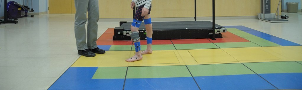
Instrumented gait analysis. A child with reflective/marker straps walks across the instrumented walkway of a motion-analysis laboratory, generating the kinematic, kinetic and dynamic-EMG data used to plan multilevel surgery. NIH Clinical Center (public domain), via Wikimedia Commons.
Instrumented gait analysis. A child with reflective/marker straps walks across the instrumented walkway of a motion-analysis laboratory, generating the kinematic, kinetic and dynamic-EMG data used to plan multilevel surgery. NIH Clinical Center (public domain), via Wikimedia Commons.
Part V - Natural history of ambulation and gait
Natural history of walking
The strongest practical predictors of walking are the ability to sit independently by age two and the GMFCS level; head control by 9 months and crawling by 30 months are also favourable, while bilateral involvement, dystonia, an IQ below 50, epilepsy and severe visual impairment are unfavourable.[28] Across all CP, roughly half walk without aids, a sixth walk with aids, and a third do not walk. Function is not static across life: gait and walking ability tend to decline through adolescence and adulthood as body mass rises and flexibility is lost, and up to a fifth of previously ambulant individuals become non-ambulatory by middle age, with those at GMFCS IV at greatest early risk.[29]
Gait
Normal gait requires stance stability, swing clearance, prepositioning of the foot, adequate step length and energy efficiency. The pivotal mechanism in the leg is the plantarflexion-knee-extension couple: in mid-stance the soleus restrains forward advance of the tibia, keeping the ground-reaction force anterior to the knee so the knee extends without quadriceps effort; losing this couple produces crouch. Because the plantarflexors are five to six times stronger than the dorsiflexors, equinus dominates when spasticity is global, and “muscle balance” must be understood as balance against the ground-reaction force across the whole sagittal-plane chain, not as agonist-against-antagonist.[30] Torsional and foot malalignments (femoral anteversion, external tibial torsion, an unstable valgus foot) shorten and misalign the foot lever, the so-called lever-arm dysfunction, and degrade the couple.
The recognisable gait patterns are classified for both topographies. In spastic hemiplegia, the Winters-Gage-Hicks types I-IV describe progressively proximal involvement: type I, a swing-phase drop foot (managed with an AFO); type II, fixed equinus in stance; type III, added knee involvement; type IV, added hip involvement.[31] In spastic diplegia, the Rodda-Graham sagittal patterns run from true equinus, through jump gait and apparent equinus, to crouch gait. These classifications feed directly into single-event multilevel surgery (SEMLS), which corrects all the contractures, torsions and instabilities in one operation and so replaces the old “birthday syndrome” of repeated single-level operations.[32]
Part VI - Management of tone and the general principles of treatment
Interventions can be mapped on a “spasticity compass” of focal versus generalised and temporary versus permanent.[33]
- Physiotherapy and occupational therapy are the most widely used measures (biomechanical, neurodevelopmental/Bobath, and motor-learning approaches, plus constraint-induced movement therapy for the hemiplegic arm); the evidence for changing function is modest, and therapy alone is inadequate rehabilitation after major surgery.[34]
- Orthoses, chiefly the ankle-foot orthosis (AFO) in its several forms: the posterior-leaf-spring AFO for a swing-phase drop foot, the articulated AFO to allow controlled dorsiflexion, the solid AFO for severe spasticity, and the ground-reaction (floor-reaction) AFO to restore the plantarflexion-knee-extension couple in crouch or after an over-lengthened heel cord.[35]
- Oral agents: baclofen (GABA-B agonist), diazepam, tizanidine and dantrolene reduce generalised tone but cause sedation and weakness, and are used mainly for comfort and care at GMFCS IV-V; levodopa and trihexyphenidyl are used for dystonia.[36]
- Botulinum toxin A chemically denervates a spastic muscle by cleaving SNAP-25, giving a temporary, reversible relaxation lasting about 12-16 weeks. It is an adjunct, not an alternative to surgery, best for a dynamic contracture in the absence of a fixed one, started young (commonly 1-3 years for spastic equinus and repeated until gross motor function plateaus at 4-6 years), most often into the gastrocnemius. The most serious adverse events are dysphagia and aspiration in severely affected children.[37]
- Intrathecal baclofen (ITB) via a programmable pump treats generalised spasticity and dystonia at a fraction of the oral dose; it is reversible but carries catheter, infection and withdrawal risks.[38]
- Selective dorsal rhizotomy (SDR) divides a proportion (about a third) of the dorsal sensory rootlets from L1-S2 to permanently reduce spasticity. The ideal candidate is an ambulant child with spastic diplegia from prematurity/PVL, good underlying strength and selective control, around 4-8 years old; it reliably reduces lower-limb spasticity without unmasking weakness, but most children still need later orthopaedic surgery.[39]
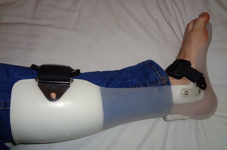
Ankle-foot orthosis (AFO). A hinged AFO with a calf shell, ankle joint and footplate, of the kind used to control dynamic equinus and a swing-phase drop foot. Photo: Pagemaker787, via Wikimedia Commons (CC BY-SA 4.0).
Ankle-foot orthosis (AFO). A hinged AFO with a calf shell, ankle joint and footplate, of the kind used to control dynamic equinus and a swing-phase drop foot. Photo: Pagemaker787, via Wikimedia Commons (CC BY-SA 4.0).
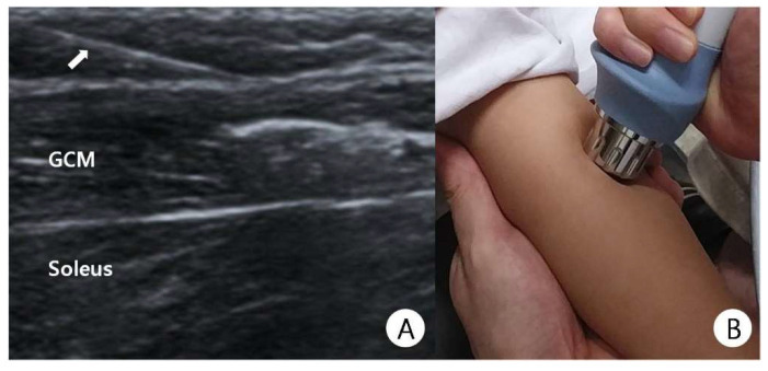
Botulinum toxin A injection. (A) Ultrasound of the calf during a sonography-guided injection of the medial gastrocnemius (GCM; soleus below, needle at the arrow); (B) the same child receiving adjunctive shock-wave therapy. From Kwon & Kwon, Children (Basel) 2021;8:1059 (CC BY 4.0).
Botulinum toxin A injection. (A) Ultrasound of the calf during a sonography-guided injection of the medial gastrocnemius (GCM; soleus below, needle at the arrow); (B) the same child receiving adjunctive shock-wave therapy. From Kwon & Kwon, Children (Basel) 2021;8:1059 (CC BY 4.0).
The orthopaedic principle is to manage tone and dynamic deformity early, then correct fixed deformity at the right time: usually preventive hip surgery under age 6, and contracture/torsion surgery (often as SEMLS) between about 6 and 12 years, planned by GMFCS level and, in ambulators, by gait analysis.[40]
Part VII - The paralytic (spastic) hip
Pathomechanics
The hip in cerebral palsy is normal at birth and displaces over time, which makes it fundamentally different from developmental dysplasia (DDH), where the hip is abnormal at birth.[41] The newborn hip has a neck-shaft angle of about 150° and high femoral anteversion, which normally remodel toward about 130° once the child stands and walks with active abductors. In cerebral palsy two things go wrong: the spastic, overpowering hip flexors and adductors act unopposed by weak extensors and abductors, while delayed or absent weight-bearing prevents normal remodelling. The result is persistent coxa valga and excessive anteversion, with the hip held in flexion-adduction-internal rotation driving the head posterosuperiorly, so that it migrates laterally and progresses from subluxation to dislocation. The force across the spastic hip is greatly increased, the acetabular deficiency is posterosuperior, and the chronically dislocated hip becomes arthritic and painful in the adolescent or adult.[42] Coxa valga is therefore a response to the abnormal forces, not a primary cause.
Spastic hip disease. Antero-posterior pelvic series of a non-ambulant child (GMFCS V): (A) dislocation of the right hip with subluxation of the left; (B, C) after femoral and pelvic osteotomy of the right hip (blade plate) with soft-tissue release. From Terjesen, Acta Orthop 2019;90:614-621 (CC BY 4.0).
Spastic hip disease. Antero-posterior pelvic series of a non-ambulant child (GMFCS V): (A) dislocation of the right hip with subluxation of the left; (B, C) after femoral and pelvic osteotomy of the right hip (blade plate) with soft-tissue release. From Terjesen, Acta Orthop 2019;90:614-621 (CC BY 4.0).
Risk by GMFCS, surveillance and measurement
Hip-displacement risk rises almost linearly with GMFCS: roughly 0% at level I, 15% at II, 41% at III, 69% at IV and 90% at level V, with non-ambulant total-body-involvement children at greatest risk.[43] Because the hip dislocates silently and gradually, structured hip surveillance is the cornerstone of management: clinical screening of abduction (a radiograph if abduction with hips and knees extended falls below about 45°) and serial pelvic radiographs at intervals set by GMFCS level. Formal surveillance programmes (the Swedish CPUP and the Australian Victoria programmes) have demonstrably reduced the rate of painful dislocation and the need for salvage surgery.[44]
The single most useful radiographic measure is the Reimers migration percentage (MP), the percentage of the femoral head lying lateral to Perkins’ line (dropped from the lateral acetabular margin). A normal hip measures under about 25-30%; 30-40% is at-risk/early subluxation; 40-60% is established subluxation; and 100% is dislocation. A steadily rising migration percentage on serial films (subluxation tends to increase at roughly 2% per month while the MP is under about 50-60%) is the signal to intervene.[45] Other measures (acetabular index, Wiberg’s centre-edge angle, Shenton’s line) add little to spastic-hip monitoring, and CT best defines the direction of displacement and the anteversion. The progressive radiographic picture can be graded with the Melbourne Cerebral Palsy Hip Classification System.[46]
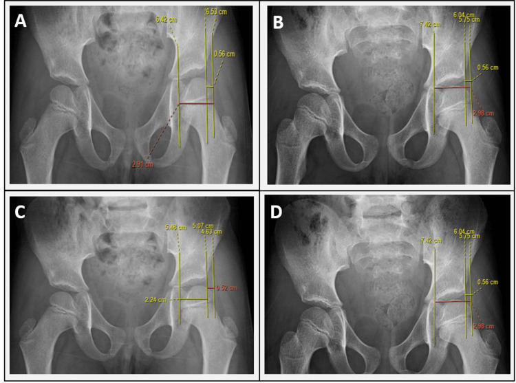
Reimers migration percentage (MP). Antero-posterior pelvis with the construction drawn: Hilgenreiner’s horizontal line, the vertical Perkins’ lines from the lateral acetabular margins, and the distances (A = head lateral to Perkins’ line, B = head width) giving MP = A/B × 100%; the right hip is migrating laterally. From Sajeev et al., Cureus 2025;17(12):e98870 (CC BY 4.0).
Reimers migration percentage (MP). Antero-posterior pelvis with the construction drawn: Hilgenreiner’s horizontal line, the vertical Perkins’ lines from the lateral acetabular margins, and the distances (A = head lateral to Perkins’ line, B = head width) giving MP = A/B × 100%; the right hip is migrating laterally. From Sajeev et al., Cureus 2025;17(12):e98870 (CC BY 4.0).
Treatment
Treatment follows the triad prevention → reconstruction → salvage, with the indication set by the migration percentage and the child’s age.[47]
- Prevention (hip at risk / early subluxation, MP ~25-60% in a young child): soft-tissue release. Open lengthening of adductor longus and gracilis (with adductor brevis as needed) and of the iliopsoas (myofascial recession over the brim in possible ambulators, tenotomy at the lesser trochanter in non-ambulators) normalises roughly 80% of hips when done before about age 8 and while the MP is under 40-50%; bilateral (symmetric) surgery is essential to avoid driving the other hip into a windswept posture. Botulinum toxin is only a short-term temporiser and does not prevent displacement, and the historic obturator neurectomy is now avoided (or limited to the anterior branch in non-ambulators) because it causes fixed abduction/windblown deformity.[48]
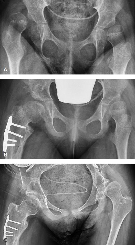
- Reconstruction (established subluxation, MP >40% with age >8, or MP >60%/dislocated). Three components must each be addressed: soft-tissue release; a femoral varus derotation (shortening) osteotomy (VDRO) reducing the neck-shaft angle to about 100-110° and anteversion to about 10-15°, with shortening to lower the joint reaction force; and, where the acetabulum is dysplastic, an acetabular osteotomy directed at the posterosuperior deficiency, namely the Dega / San Diego (incomplete transiliac) or Pemberton osteotomy. Open reduction with capsulorrhaphy is added when the hip is dislocated. The Salter innominate osteotomy is contraindicated in CP because it provides coverage in the wrong (anterior) direction. Full three-component reconstruction achieves a stable, located, painless hip in about 95% of cases.[49] The windswept (windblown) deformity, with one hip adducted and one abducted and pelvic obliquity, requires asymmetric bilateral surgery and is managed together with any coexisting scoliosis.[50]
- Salvage of the neglected, painful, dislocated hip in the adolescent or adult. The goals are pain relief and comfortable sitting, not weight-bearing. The options, none wholly reliable, include proximal femoral resection (the Castle procedure), the McHale procedure (valgus osteotomy with femoral head/neck resection), a Schanz valgus osteotomy, interposition arthroplasty, hip arthrodesis (only for unilateral disease), and, in the small subset of well-controlled ambulators, total hip arthroplasty or resurfacing. The clear message is that surveillance and timely reconstruction prevent the painful dislocation that salvage can only palliate.[51]
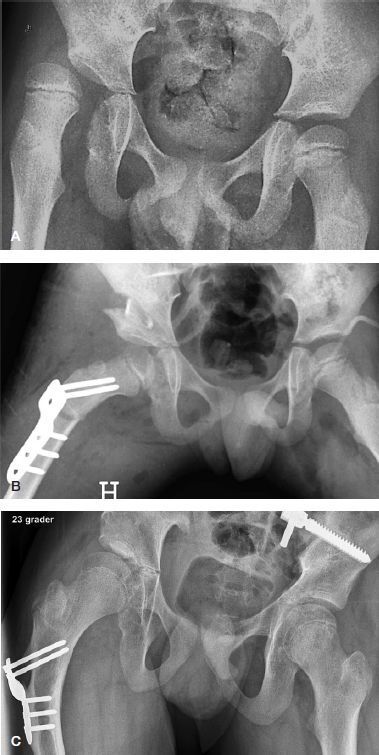
Reconstruction of the spastic hip. Antero-posterior pelvis of a child (GMFCS V) with right-hip subluxation (MP 67%): (A) pre-operative; (B) after a femoral varus-derotation-shortening osteotomy (blade plate) with pelvic osteotomy and soft-tissue release; (C) follow-up with maintained coverage. From Terjesen, Acta Orthop 2019;90:614-621 (CC BY 4.0).
Reconstruction of the spastic hip. Antero-posterior pelvis of a child (GMFCS V) with right-hip subluxation (MP 67%): (A) pre-operative; (B) after a femoral varus-derotation-shortening osteotomy (blade plate) with pelvic osteotomy and soft-tissue release; (C) follow-up with maintained coverage. From Terjesen, Acta Orthop 2019;90:614-621 (CC BY 4.0).
The principal complications across all stages are avascular necrosis, recurrence/redislocation (especially after isolated soft-tissue surgery or isolated VDRO, and at GMFCS IV-V), heterotopic ossification (notably in quadriplegia), and fractures of the osteopenic limb.[52]
Part VIII - The paralytic (spastic) foot and ankle
General principles
The aim is a plantigrade, braceable, stable and painless foot that provides a good rigid lever for the plantarflexion-knee-extension couple. Management depends on distinguishing dynamic from fixed and flexible from rigid deformity, on the Silfverskiöld test to apportion gastrocnemius versus soleus, and on the modern principle of preferring muscle balancing and joint-sparing osteotomies over arthrodesis in the growing spastic foot. Arthrodesis is reserved for rigid or recurrent deformity, because a stiff foot loses shock absorption and overloads adjacent joints.[53] The two commonest patterns reflect the topography: equinus and equinovalgus (planovalgus) in diplegia and quadriplegia, and equinovarus in hemiplegia.
Equinus
Equinus, the commonest deformity, begins as dynamic overactivity and becomes a fixed contracture (usually by age 5-7). The treatment ladder runs from stretching and AFOs through botulinum toxin with casting to surgical lengthening. The choice of lengthening is dictated by the Silfverskiöld test: a gastrocnemius recession (Strayer, Vulpius or Baumann), safest in diplegia, for an isolated gastrocnemius contracture, versus a tendo-Achilles lengthening (open Z-lengthening, or the Hoke/White sliding techniques) only when both gastrocnemius and soleus are contracted.[54] The cardinal danger is over-lengthening, which produces a calcaneus deformity and crouch gait. The soleus is exquisitely sensitive - a tendo-Achilles lengthening of about 1 cm reduces its strength by roughly half - so an isolated tendo-Achilles lengthening in a diplegic child can convert equinus into a far worse crouch (reported in up to 40% on long-term follow-up). The maxim is that “a little equinus is better than a calcaneus.”[55]
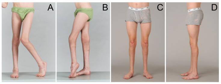
Spastic equinus. Standing clinical photographs (waist-down) of a boy with bilateral spastic CP: (A, B) pre-operative toe-walking equinus with knee recurvatum; (C, D) plantigrade feet after gastrocsoleus lengthening as part of single-event multilevel surgery. From Ma et al., Medicina (Kaunas) 2021;57:98 (CC BY 4.0).
Spastic equinus. Standing clinical photographs (waist-down) of a boy with bilateral spastic CP: (A, B) pre-operative toe-walking equinus with knee recurvatum; (C, D) plantigrade feet after gastrocsoleus lengthening as part of single-event multilevel surgery. From Ma et al., Medicina (Kaunas) 2021;57:98 (CC BY 4.0).
Equinovarus
Equinovarus is commoner in hemiplegia and results from overactivity of the tibialis posterior and/or tibialis anterior against weaker peroneals. Dynamic EMG identifies the culprit (the “confusion test” implicates a phasically overactive tibialis anterior). For a flexible, purely dynamic deformity the options are a split posterior tibial tendon transfer (SPOTT) or a split anterior tibial tendon transfer (SPLATT), combined with a gastrocnemius recession; an intramuscular tibialis posterior lengthening is used for a mild contracture. Complete (non-split) transfer of either tibialis tendon is unpredictable and should be avoided. A fixed hindfoot varus needs a calcaneal (Dwyer) osteotomy, and a rigid, severe or recurrent deformity is salvaged by triple arthrodesis.[56]
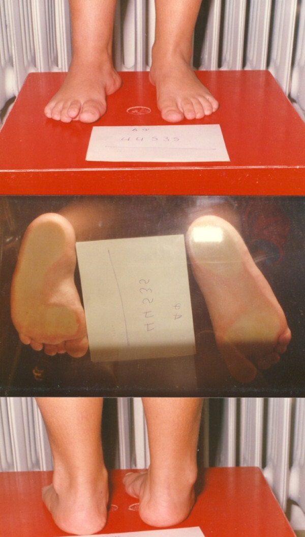
Clinical assessment of the spastic varus foot. Standing dorsal view (top), a podoscope view of the soles through glass (middle) and a posterior heel view (bottom) in a child with spastic equinovarus. From Vlachou & Dimitriadis, J Foot Ankle Res 2010;3:28 (CC BY 2.0).
Clinical assessment of the spastic varus foot. Standing dorsal view (top), a podoscope view of the soles through glass (middle) and a posterior heel view (bottom) in a child with spastic equinovarus. From Vlachou & Dimitriadis, J Foot Ankle Res 2010;3:28 (CC BY 2.0).
Planovalgus (equinoplanovalgus)
Planovalgus is the commonest foot deformity in diplegia and quadriplegia. The hindfoot falls into valgus, the midfoot breaks and the forefoot abducts and supinates, usually with an associated equinus (which masks a tight gastrocnemius) and external tibial torsion; secondary problems include ankle valgus, hallux valgus and a dorsal bunion. Weight-bearing radiographs guide treatment. After correcting the equinus and any tibial torsion, a flexible deformity in a community ambulator is best treated by a lateral column (calcaneal) lengthening osteotomy (Evans, as popularised by Mosca), which corrects all three segments by ligamentotaxis while preserving subtalar motion; lower-function or markedly hypermobile feet are better served by a subtalar arthrodesis (the Grice extra-articular or Dennyson-Fulford technique), and a rigid severe deformity by triple arthrodesis.[57] The medial column and forefoot supination must be corrected at the same time, or medial-border overload persists.
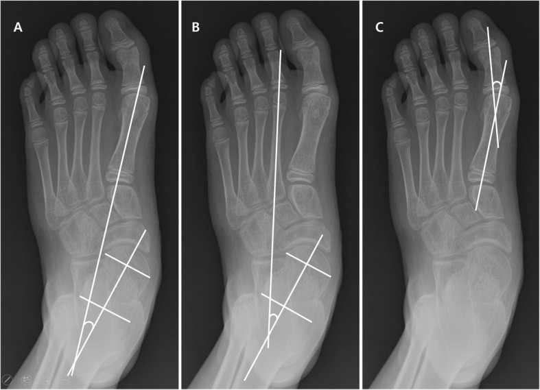
Planovalgus deformity - radiographic assessment. Antero-posterior foot radiographs with the talo-first-metatarsal, talo-second-metatarsal and hallux-valgus angles drawn to grade the planovalgus deformity of cerebral palsy. From Min et al., BMC Musculoskelet Disord 2020;21:141 (CC BY 4.0).
Planovalgus deformity - radiographic assessment. Antero-posterior foot radiographs with the talo-first-metatarsal, talo-second-metatarsal and hallux-valgus angles drawn to grade the planovalgus deformity of cerebral palsy. From Min et al., BMC Musculoskelet Disord 2020;21:141 (CC BY 4.0).
Other foot problems
Hallux valgus in the adolescent (associated with planovalgus) is most reliably corrected by first metatarsophalangeal arthrodesis; the dorsal bunion (an elevated first ray with a plantarflexed hallux, from a strong tibialis anterior and weak peroneus longus) is corrected by soft-tissue rebalancing with a first MTP fusion in severe cases. Ankle valgus, almost always secondary to planovalgus and external tibial torsion, is treated in the growing child by medial malleolar guided growth (hemiepiphysiodesis) and at maturity by supramalleolar osteotomy; significant tibial torsion is corrected by a supramalleolar derotation osteotomy, but tibial-tendon surgery should not be combined with a rotational osteotomy, as overcorrection results.[58]
Part IX - The spastic knee
The two characteristic problems are crouch gait and stiff-knee gait. Crouch gait, meaning persistent excessive knee flexion in stance, is driven less by hamstring tightness (its role is overestimated, and the popliteal angle predicts it poorly) than by plantarflexor (soleus) weakness, often iatrogenic after a heel-cord lengthening, and by lever-arm dysfunction such as external tibial torsion, frequently with patella alta and an extensor lag. Management corrects the lever arm and lengthens only the muscles shown to be tight on gait analysis (commonly the iliopsoas over the brim, and the medial hamstrings if genuinely short), while taking care that lengthening the hamstrings against a tight psoas does not increase the anterior pelvic tilt. Fixed crouch in the older child is treated by a distal femoral extension osteotomy combined with patellar tendon advancement (shortening).[59] Stiff-knee gait, from an out-of-phase rectus femoris, is improved by a rectus femoris transfer to a knee flexor (semitendinosus or gracilis) with early mobilisation.[60] Patella alta with recurrent dislocation, which prevents walking, is realigned surgically.[61]
Part X - The neuromuscular spine
Spinal deformity is common in total-body-involvement children at GMFCS IV-V, and the GMFCS level is its strongest predictor. The typical curve is a long, C-shaped, collapsing scoliosis with pelvic obliquity, presenting earlier than idiopathic scoliosis, progressing faster (and continuing after maturity), and closely linked to hip displacement and windswept deformity. Bracing and seating improve comfort but do not halt progression. A progressive curve is treated by long posterior instrumented fusion to the pelvis (a unit rod, Galveston technique, or iliac/S2-alar-iliac screws); the complication rate is high, so preoperative optimisation of nutrition, respiration, bleeding risk and seizure medication is essential, and the surgery improves sitting, care and quality of life.[62]
Part XI - The upper limb in spastic hemiplegia
The characteristic posture is shoulder adduction and internal rotation, elbow flexion, forearm pronation, wrist and finger flexion, and a thumb-in-palm deformity; it is predictable in hemiplegia (and therefore amenable to surgery) but variable in dystonia. Function is graded with the House classification, and the level of sensation limits what surgery can achieve. The goals range from improving fine bimanual control in high-functioning children to easing hygiene and dressing in the most involved. Procedures (often combined as an upper-limb SEMLS) include elbow-flexor and pronator lengthening, the transfer of flexor carpi ulnaris to the wrist/finger extensors (the most useful single procedure), wrist arthrodesis for severe fixed deformity, and a staged thumb-in-palm release; botulinum toxin is a useful adjunct, avoiding the long finger flexors where grip must be preserved.[63]
Bulgarian terminology (Боев / Boychev tradition) - glossary
The following Bulgarian equivalents bridge the international literature with the terminology of the Bulgarian school of orthopaedics (in the tradition of Бойчо Бойчев / Boycho Boychev) used in the state examination.
| English term | Bulgarian term (Cyrillic) | Transliteration |
|---|---|---|
| Cerebral palsy | Детска церебрална парализа (ДЦП) | Detska tserebralna paraliza (DTsP) |
| Spasticity | Спастичност | Spastichnost |
| Spastic | Спастичен | Spastichen |
| Muscle tone | Мускулен тонус | Muskulen tonus |
| Dystonia | Дистония | Distoniya |
| Athetosis | Атетоза | Atetoza |
| Ataxia | Атаксия | Ataksiya |
| Hemiplegia | Хемиплегия | Hemiplegiya |
| Diplegia | Диплегия | Diplegiya |
| Quadriplegia (total body) | Тетраплегия (квадриплегия) | Tetraplegiya (kvadriplegiya) |
| Contracture | Контрактура | Kontraktura |
| Paralytic hip dislocation | Паралитична луксация на тазобедрената става | Paralitichna luksatsiya na tazobedrenata stava |
| Hip joint | Тазобедрена става (ТБС) | Tazobedrena stava (TBS) |
| Hip subluxation | Сублуксация на тазобедрената става | Subluksatsiya na tazobedrenata stava |
| Migration percentage (Reimers) | Миграционен индекс (по Раймерс) | Migratsionen indeks (po Raymers) |
| Coxa valga | Кокса валга | Koksa valga |
| Femoral anteversion | Антеверзия на бедрената шийка | Anteverziya na bedrenata shiyka |
| Varus derotation osteotomy | Варизираща деротационна остеотомия | Varizirashta derotatsionna osteotomiya |
| Paralytic foot | Паралитично ходило (стъпало) | Paralitichno hodilo (stapalo) |
| Equinus | Еквинус (конско стъпало) | Ekvinus (konsko stapalo) |
| Equinovarus | Еквиноварус | Ekvinovarus |
| Planovalgus (pes valgus) | Плановалгус (плоско-валгусно стъпало) | Planovalgus (plosko-valgusno stapalo) |
| Tendo-Achilles lengthening | Удължаване на ахилесовото сухожилие | Udalzhavane na ahilesovoto suhozhilie |
| Triple arthrodesis | Тройна артродеза | Troyna artrodeza |
| Gait | Походка | Pohodka |
| Crouch gait | Приклекнала походка | Prikleknala pohodka |
| Orthosis / brace | Ортеза | Orteza |
| Botulinum toxin | Ботулинов токсин | Botulinov toksin |
| Selective dorsal rhizotomy | Селективна дорзална ризотомия | Selektivna dorzalna rizotomiya |
| Scoliosis | Сколиоза | Skolioza |
Image attributions
(Figure attributions and licences are listed in the figure MANIFEST and inserted with each image. All images are openly licensed [CC0 / CC BY / CC BY-SA] or used under their stated terms; any non-commercial [NC] item is flagged as such and must not be used in a commercial product.)
References
-
Lovell & Winter, Pediatric Orthopaedics, ch. 14 Cerebral Palsy (Graham, Thomason, Novacheck), pp. 506-507; the static-encephalopathy / progressive-musculoskeletal-pathology concept is central.
-
Bleck & Horstmann, Orthopaedic Management in Cerebral Palsy, 2nd ed., p. 14; Lovell, p. 503.
-
Bleck, p. 14; Lovell, p. 506. There is no laboratory or genetic test that proves or excludes the diagnosis. It is a clinical diagnosis of a non-progressive motor disorder of early cerebral origin; transient motor delay, progressive brain disease and spinal-cord lesions are excluded.
-
Bleck, pp. 12-13; Lovell, p. 503.
-
Bleck, p. 36; the European SCPE register gives 2.08 per 1,000 (Gage, The Identification and Treatment of Gait Problems in Cerebral Palsy, p. 183); Lovell, p. 505, notes a wider regional range.
-
Lovell, pp. 503, 506. Neither electronic fetal monitoring nor rising Caesarean rates have reduced the prevalence of CP, which suggests that intrapartum asphyxia accounts for only a minority of cases.
-
Bleck, pp. 43-44; Lovell, pp. 505, 507; Gage, pp. 121-122.
-
Lovell, p. 505.
-
Bleck, p. 44.
-
Bleck, p. 41; Gage, p. 118.
-
Bleck, p. 37; Lovell, pp. 503, 505.
-
Lovell, p. 506 (Bax et al. European CP study).
-
Lovell, pp. 507-508; Bleck, pp. 25-27.
-
Lovell, p. 508; Bleck, pp. 18, 26.
-
Bleck, p. 27; Lovell, p. 508.
-
Lovell, p. 508.
-
Lovell, pp. 505-506; Gage, pp. 103-111.
-
Lovell, p. 509.
-
Bleck, p. 25.
-
Gage, p. 185; Lovell, pp. 509-512.
-
Lovell, pp. 509-512.
-
Gage, pp. 165-167; Lovell, pp. 514-515.
-
Lovell, pp. 504, 513.
-
Bleck, pp. 32-33; Gage, pp. 198-200.
-
Lovell, pp. 507-508; Gage, p. 387.
-
Lovell, pp. 522-524; Bleck p. 32.
-
Gage, pp. 193-197; Lovell, pp. 515-516.
-
Gage, pp. 183-185.
-
Gage, pp. 187-190.
-
Gage, pp. 341, 569-570; Lovell, pp. 520-521.
-
Gage, pp. 170-171; Lovell, pp. 518-520.
-
Lovell, pp. 520, 530-531.
-
Lovell, p. 516.
-
Lovell, p. 516.
-
Gage, pp. 346-351.
-
Gage, pp. 367-373; Lovell, pp. 516-517.
-
Gage, pp. 378-388; Lovell, pp. 517-518.
-
Gage, pp. 397-404; Lovell, pp. 516-517.
-
Gage, pp. 411-423; Lovell, pp. 516-517.
-
Lovell, pp. 518, 530-531.
-
Miller, Cerebral Palsy, pp. 538, 543; Lovell, p. 540.
-
Miller, pp. 538-544; Gage, pp. 506-510; Lovell, p. 540.
-
Lovell, pp. 518, 539, 555; Gage, pp. 507-508.
-
Miller, pp. 545-548; Lovell, p. 540.
-
Miller, pp. 545-547, 554-555; Lovell, p. 544.
-
Miller, pp. 545-555; Gage, p. 511.
-
Miller, p. 548; Lovell, p. 545.
-
Miller, pp. 549-561; Lovell, pp. 541-546; Gage, pp. 514-515.
-
Miller, pp. 562-575; Lovell, pp. 552-555; Gage, pp. 521-524.
-
Lovell, p. 556; Miller, pp. 526-527, 566.
-
Lovell, p. 556; Miller, pp. 581-582; Gage, p. 525.
-
Miller, pp. 560-581; Gage, p. 524; Lovell, p. 554.
-
Gage, pp. 528-530; Lovell, p. 507; Miller, pp. 723-724.
-
Miller, pp. 728-732; Lovell, pp. 522-524; Mosca, Principles and Management of Pediatric Foot and Ankle, pp. 79-80, 88.
-
Gage, p. 562 (Delp et al.: a 1 cm tendo-Achilles lengthening diminishes soleus strength by ~50%); Lovell, pp. 521, 533-534; Miller, pp. 723, 732-736.
-
Miller, pp. 738-756; Lovell, pp. 528-530; Gage, pp. 532-545.
-
Miller, pp. 755-775; Gage, pp. 534-540; Lovell, pp. 533-535; Mosca, pp. 110-111.
-
Miller, pp. 713-722, 768; Mosca, pp. 83-84, 102-103; Lovell, pp. 531, 558-559.
-
Gage, pp. 569-583; Lovell, pp. 520, 535, 544-548.
-
Lovell, pp. 519, 537-538; Gage, p. 516.
-
Miller, pp. 708-709.
-
Miller, Cerebral Palsy (spine chapter); Bleck & Horstmann (treatment).
-
Lovell, pp. 521, 525-528; Bleck, pp. 531-533.