Contents
- Introduction and scope
- Part I - Blount’s disease (tibia vara)
- Part II - Madelung deformity
- Bulgarian terminology (Боев / Boychev tradition) - glossary
- Image attributions
Introduction and scope
This topic pairs two named developmental growth-plate disorders that the syllabus groups together: Blount’s disease (tibia vara), a growth disturbance of the posteromedial proximal tibial physis producing progressive bow-leg, and Madelung deformity, a growth disturbance of the volar-ulnar distal radial physis producing a characteristic wrist deformity. They sit at different sites but share a theme: asymmetric, localised physeal arrest under mechanical load and genetic influence. The treatment logic is similar too. Recognise the disorder early, measure the deformity, and act on the physis or with an osteotomy before things become fixed. Walter Blount himself described tibia vara as “an osteochondrosis similar to coxa plana and Madelung deformity,” which is presumably why the two sit together in the konspekt.[1]
Examiners reward two skills. For Blount’s, you need to distinguish it from physiologic genu varum and to use the metaphyseal-diaphyseal angle and Langenskiöld staging. For Madelung, you need to recognise the Vickers ligament / volar-ulnar physeal bar, the dyschondrosteosis (Léri-Weill / SHOX) association, and the place of physiolysis in the growing child.
Part I - Blount’s disease (tibia vara)
Definition, terminology and classification by age
Blount’s disease (tibia vara) is a growth disorder of the posteromedial proximal tibial physis and epiphysis that produces a progressive three-dimensional deformity of varus, internal tibial torsion and procurvatum (flexion).[2] Walter Blount (1937) called it osteochondrosis deformans tibiae and described the abrupt angulation just below the physis, the irregular physeal line and the wedge-shaped epiphysis with a medial metaphyseal “beak”; Langenskiöld (1952) gave the classic six-stage radiographic classification. Current thinking regards it as an acquired disorder of the proximal tibia driven by abnormal compressive load rather than a true osteochondrosis or epiphyseal dysplasia.[3]
It is classified by age of onset:[4]
- Infantile (1-3 years): the commonest form and the main cause of pathologic genu varum. It is often bilateral (about 60%), shows the most severe epiphyseal and physeal distortion, and can progress through the Langenskiöld stages to a medial physeal bar and intra-articular deformity.
- Juvenile (≈4-10 years): an intermediate, disputed entity, less common than the other two.
- Adolescent / late-onset (≥10 years): usually an obese child with unilateral complaints, frequently with a contributing distal-femoral varus, but without a discrete physeal bar or medial plateau depression.
The infantile-versus-adolescent age cut-off differs slightly between texts (Campbell uses 8 years), but the practical division is between a young, plateau-distorting form and an older, obesity-driven one.
Epidemiology and etiology
The consistent associations are early walking (independent ambulation before about 10-12 months), obesity, African or African-American ancestry and a family history of bowing; the adolescent form is overwhelmingly one of obese adolescents.[5] The disease does not occur in non-ambulatory children, since weight-bearing is obligatory. The mechanism is biomechanical: by the Hueter-Volkmann principle, excessive compressive load on the posteromedial proximal tibial physis suppresses medial growth while the lateral physis continues, producing progressive varus. Finite-element modelling shows that a 2-year-old in the 90th weight percentile with 20° of varus generates enough medial physeal compression during single-limb stance to retard growth. In the obese adolescent the “fat-thigh” effect (large thighs blocking hip adduction, so the foot cannot be placed under the body’s centre of mass) creates a varus knee moment even without pre-existing bowing.[6] Vitamin-D deficiency and the modern obesity epidemic are contributing factors, and adolescent Blount’s may coexist with slipped capital femoral epiphysis.
Pathoanatomy
In the infantile form the medial proximal tibial metaphysis shows depression, beaking and fragmentation (the last being pathognomonic of progressive disease), with a short, wedge-shaped, medially sloping epiphysis. The medial physis progressively slows, reorients vertically and may form a bony bar, and end-stage disease shows a depressed medial articular plateau with lateral tibial subluxation and early arthritis.[7] The distal femur is usually normal. In the adolescent form the histological changes affect the whole physis (worse medially) and resemble those of slipped epiphysis, growth inhibition is posteromedial, there is no discrete bar and no plateau depression, and a contributing distal-femoral varus is typical (the femur contributes 34-76% of the genu varum in some series).[8] One mimic worth knowing is proximal focal fibrocartilaginous dysplasia, which causes an abrupt varus at the medial metaphyseal-diaphyseal junction (at the pes anserinus) with a normal physis and often resolves spontaneously.[9]
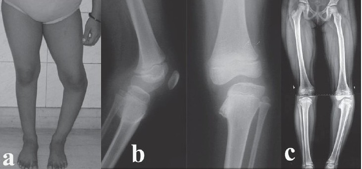
Infantile Blount’s disease (Langenskiöld IV). Clinical varus of the leg (a), with lateral and antero-posterior knee radiographs (b) showing medial metaphyseal beaking and depression of the medial tibial epiphysis, and a long-leg alignment film (c) with medial mechanical-axis deviation. Umrani SP, Aroojis AJ, Indian J Orthop 2008;42(3):351-354, Fig. 1 (CC BY).
Infantile Blount’s disease (Langenskiöld IV). Clinical varus of the leg (a), with lateral and antero-posterior knee radiographs (b) showing medial metaphyseal beaking and depression of the medial tibial epiphysis, and a long-leg alignment film (c) with medial mechanical-axis deviation. Umrani SP, Aroojis AJ, Indian J Orthop 2008;42(3):351-354, Fig. 1 (CC BY).
Clinical features and the differential from physiologic genu varum
The high-yield clinical task is to separate early infantile Blount’s from physiologic genu varum, which resolves. Normal alignment follows a predictable course (Salenius and Vankka): the newborn knee is in about 10-15° of varus, which corrects to neutral by roughly 14 months, swings to a maximum physiologic valgus of 8-10° at 3-4 years, and settles to the adult 5-7° of valgus by 6-7 years. So varus that persists or worsens after age 2 is abnormal and must be investigated.[10] Several features point to Blount’s rather than physiologic bowing: a focal varus at the proximal tibia (rather than the diffuse whole-limb bowing of physiologic varus), a lateral thrust of the knee during stance, obesity, medial-lateral knee laxity, and a positive cover-up test in the toddler (the upper tibia is neutral or varus when the foot and lower leg are covered).[11] The adolescent presents as an obese teenager with bowing, knee pain and sometimes a lateral thrust; check the hips for slipped epiphysis and the airway for obstructive sleep apnoea before surgery.
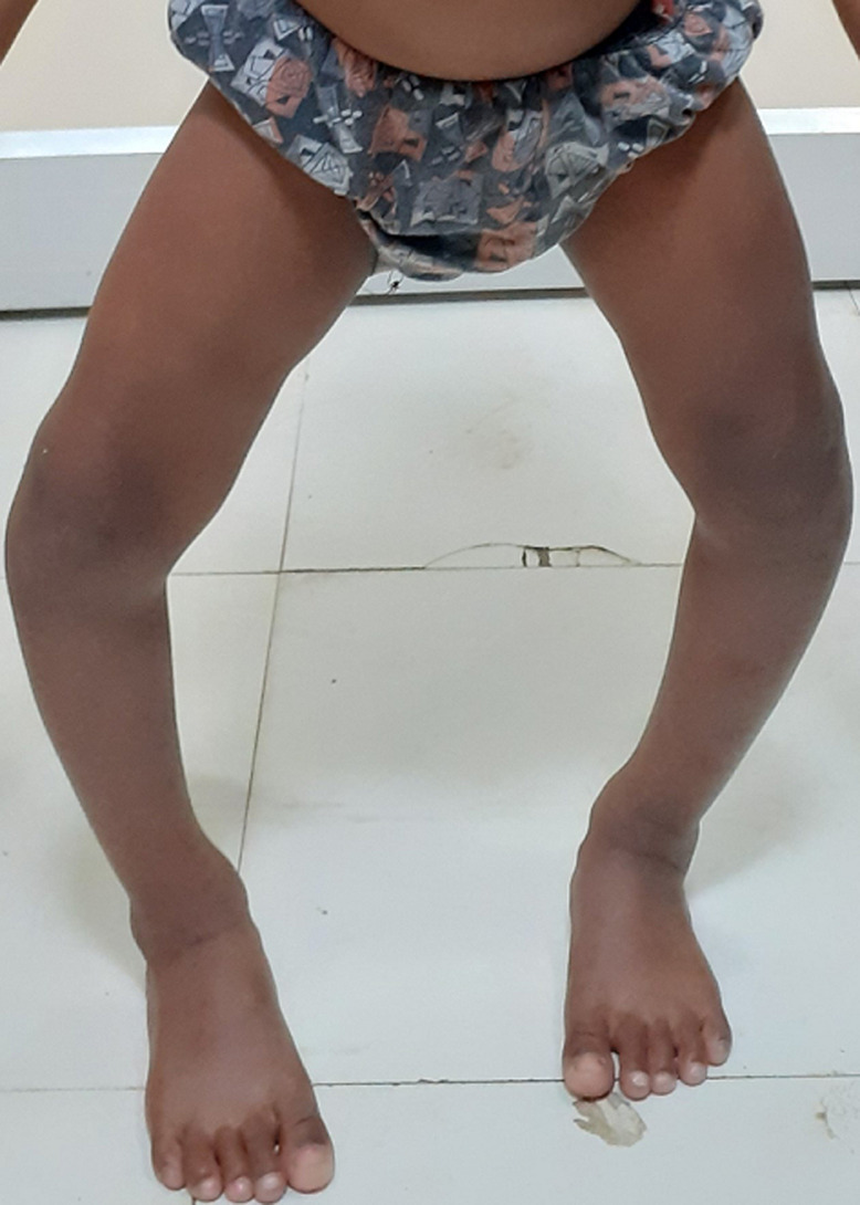
Clinical bow-leg (genu varum). Standing toddler with marked bilateral genu varum - the presentation that must be distinguished from physiologic bowing. Vagha K et al., Pan Afr Med J 2022;42:161, Fig. 1 (CC BY 4.0).
Clinical bow-leg (genu varum). Standing toddler with marked bilateral genu varum - the presentation that must be distinguished from physiologic bowing. Vagha K et al., Pan Afr Med J 2022;42:161, Fig. 1 (CC BY 4.0).
Radiographic assessment and measurement
The two measurements to know are:
- The metaphyseal-diaphyseal angle (the Drennan / Levine-Drennan angle) is the angle between a line across the two beaks of the proximal tibial metaphysis and a line perpendicular to the long axis of the tibia. Below about 10-11° is normal/physiologic, above 16° is essentially diagnostic of Blount’s, and 11-16° is indeterminate and followed; Bowen found that all children with an angle over 16° progressed.[12] The femoral-to-tibial angle ratio helps localise the bowing (a ratio above 1 favours physiologic, below 1 favours tibial Blount’s).
- The Langenskiöld classification (stages I-VI) charts the progressive infantile changes, running from mild medial metaphyseal beaking (I) through depression (II), fragmentation (III), early bar formation as the physis becomes vertical (IV), a cleft/double-contour medial epiphysis with plateau depression (V), to a complete medial physeal bar (VI).[13] Both the stage and the age at which it occurs are prognostic; North American children (more often obese and of African-American descent) progress faster than Langenskiöld’s original Finnish series.
Standing full-length hip-to-ankle radiographs (patellae forward) assess the mechanical axis, and MRI or CT is used to define a physeal bar and the articular cartilage in advanced or recurrent disease (Greene’s criteria for obtaining a CT: age over 5, a medial physeal slope of 50-70°, Langenskiöld grade IV, and weight over the 95th percentile).[14]
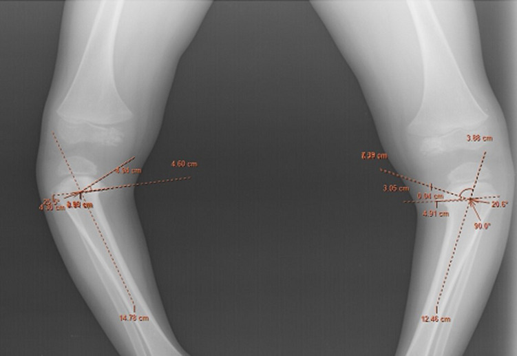
The metaphyseal-diaphyseal (Levine-Drennan) angle. Weight-bearing radiograph of both proximal tibiae with the angle constructed and measured (here ~21° and ~26°), confirming bilateral tibia vara. Vagha K et al., Pan Afr Med J 2022;42:161, Fig. 2 (CC BY 4.0).
The metaphyseal-diaphyseal (Levine-Drennan) angle. Weight-bearing radiograph of both proximal tibiae with the angle constructed and measured (here ~21° and ~26°), confirming bilateral tibia vara. Vagha K et al., Pan Afr Med J 2022;42:161, Fig. 2 (CC BY 4.0).
Treatment
Infantile Blount’s
- Observation for indeterminate cases, re-examined every 3-4 months.
- Bracing with a varus-correcting knee-ankle-foot orthosis (KAFO) is offered to children under 3 with Langenskiöld stage I-II disease; improvement should appear within 12 months. Its evidence is weak (best for unilateral disease, poor in the obese and bilateral), and it is not started after age 3 or continued once stage III develops.[15]
- Proximal tibial (and fibular) valgus osteotomy is the mainstay for persistent or progressive disease and works best when done before about age 4-5, deliberately overcorrecting into slight valgus (and lateralising the axis) to unload the diseased medial physis. Recurrence is under 20% in young children but around 80% when surgery is delayed past age 5.[16]
- Guided growth (lateral tension-band plating / hemiepiphysiodesis) tethers the lateral physis to correct varus gradually in a child with growth remaining; solid stainless-steel screws or sturdier plates are preferred because the metaphyseal screw is prone to fracture in these (often heavy) patients.[17]
- Medial physeal bar resection (epiphysiolysis), with interposition and combined with a corrective osteotomy, is used for an established bar with growth remaining (best in the young, non-obese child with a small bar), and can restore medial growth in selected cases.[18]
- Medial plateau elevation (an arcuate proximal tibial osteotomy) combined with a metaphyseal realignment osteotomy and lateral epiphysiodesis is required for the depressed plateau of Langenskiöld V-VI, and gradual correction with an Ilizarov or hexapod frame handles severe, multiplanar or relapsed deformity.[19]
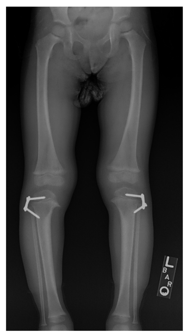
Guided growth. Lateral proximal-tibial tension-band (“8-plate”) plates tether the lateral physis to correct the varus gradually in a child with growth remaining. Robbins CA, Children (Basel) 2021;8(7):566, Fig. 12 (CC BY 4.0).
Guided growth. Lateral proximal-tibial tension-band (“8-plate”) plates tether the lateral physis to correct the varus gradually in a child with growth remaining. Robbins CA, Children (Basel) 2021;8(7):566, Fig. 12 (CC BY 4.0).
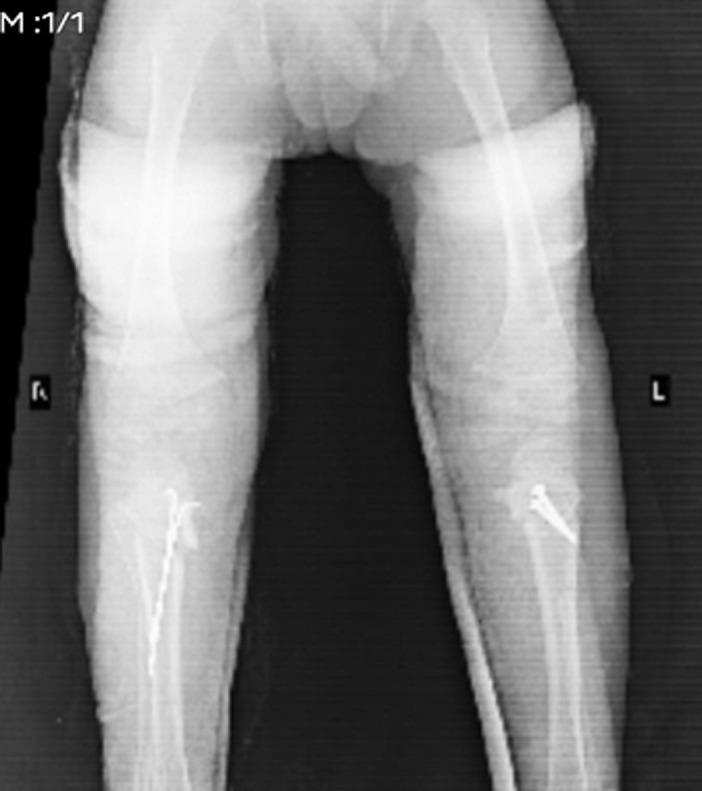
Corrective osteotomy. Post-operative radiograph after proximal tibial osteotomy with internal fixation, the bowing corrected. Vagha K et al., Pan Afr Med J 2022;42:161, Fig. 4 (CC BY 4.0).
Corrective osteotomy. Post-operative radiograph after proximal tibial osteotomy with internal fixation, the bowing corrected. Vagha K et al., Pan Afr Med J 2022;42:161, Fig. 4 (CC BY 4.0).
Adolescent Blount’s
Treatment is almost always surgical and must address the distal-femoral varus, the procurvatum, the internal torsion and the obesity as well as the tibial varus. The options are guided growth (lateral hemiepiphysiodesis) if enough growth remains, an acute proximal tibial osteotomy with stable internal fixation (translated laterally, since the deformity is at physeal level, though the Millis oblique osteotomy avoids translation), or gradual correction with external fixation (a circular/hexapod frame is preferred for the severe and very obese).[20]
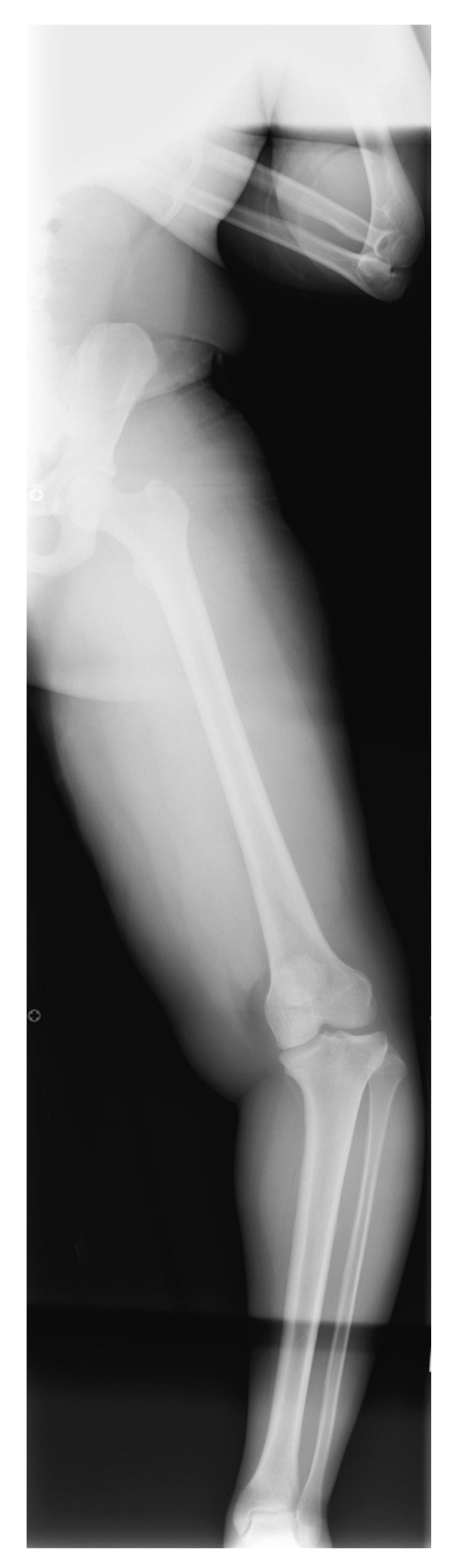
Adolescent (late-onset) Blount’s disease. Full-limb lateral radiograph of a heavy adolescent showing multilevel deformity (distal-femoral and proximal-tibial varus). Robbins CA, Children (Basel) 2021;8(7):566, Fig. 22 (CC BY 4.0).
Adolescent (late-onset) Blount’s disease. Full-limb lateral radiograph of a heavy adolescent showing multilevel deformity (distal-femoral and proximal-tibial varus). Robbins CA, Children (Basel) 2021;8(7):566, Fig. 22 (CC BY 4.0).
Complications and prognosis
The dominant problem is recurrence (favoured by young age with a high Langenskiöld stage, obesity, ligamentous laxity, undercorrection and late surgery), along with physeal bar formation, limb-length inequality and residual deformity. The classic operative complications of high tibial osteotomy are peroneal nerve injury, compartment syndrome and deep infection, and a prophylactic anterior fasciotomy is often added. The long-term concern, particularly in the infantile form, is premature medial-compartment osteoarthritis. The goal of every intervention is to restore a normal or slightly valgus, laterally placed mechanical axis that unloads the diseased medial tibia.[21]
Part II - Madelung deformity
Definition
Madelung deformity is a developmental deformity of the distal radius caused by a growth disturbance of the volar-ulnar (palmar-ulnar) part of the distal radial physis. The unaffected dorsal-radial physis keeps growing while the volar-ulnar portion arrests (often forming a physeal bar), producing increased volar and ulnar tilt of the distal radial articular surface, a short bowed radius, relative overgrowth with dorsal subluxation of the distal ulna, widening of the distal radioulnar joint, and proximal/volar migration (“subsidence”) of the carpus with the lunate wedged into the radioulnar gap, the so-called pyramidalisation of the carpus.[22] The result is the “dinner-fork” or bayonet wrist posture that Otto Madelung described in 1878 (Dupuytren had noted it in 1834). A rarer “reverse” (dorsal) variant exists.
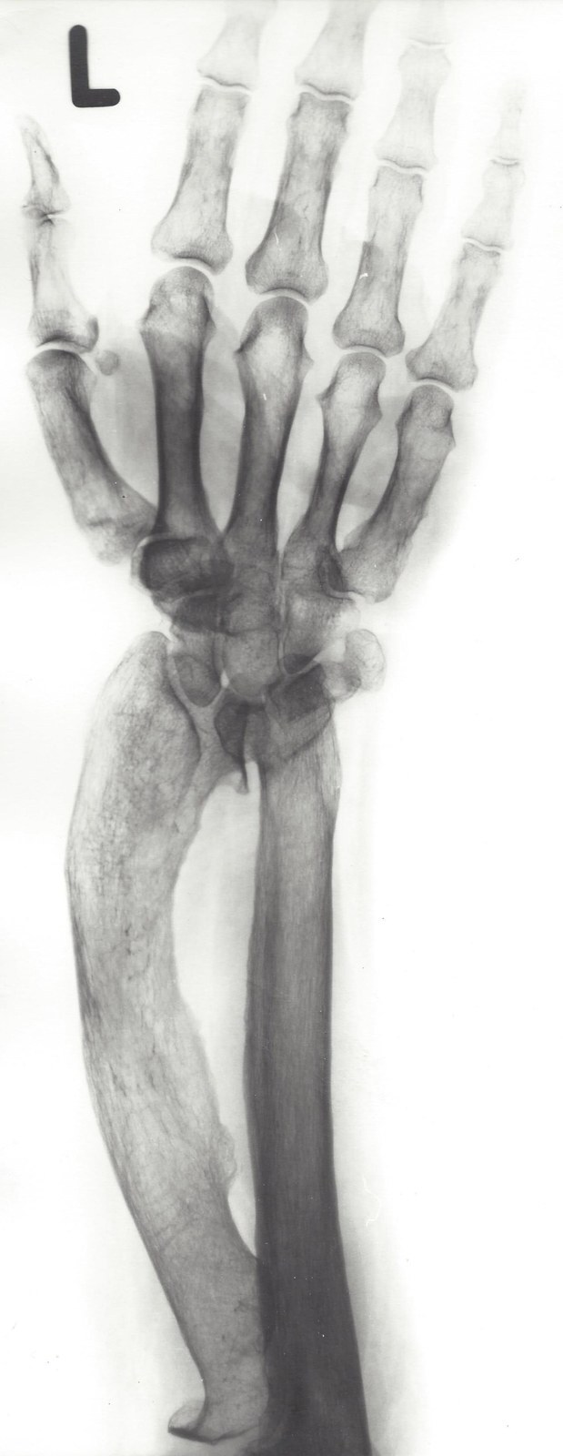
Madelung deformity (PA forearm radiograph). Marked bowing and shortening of the radius, a prominent distal ulna and proximal carpal subsidence (the lunate driven into the radioulnar gap). Röntgeninstitut des Städtischen Krankenhauses Kiel, Wikimedia Commons (CC BY-SA 3.0).
Madelung deformity (PA forearm radiograph). Marked bowing and shortening of the radius, a prominent distal ulna and proximal carpal subsidence (the lunate driven into the radioulnar gap). Röntgeninstitut des Städtischen Krankenhauses Kiel, Wikimedia Commons (CC BY-SA 3.0).
Epidemiology and genetics
Madelung deformity is uncommon, making up about 1.7% of congenital upper-limb differences in Flatt’s series. It presents in adolescence (8-14 years) around the growth spurt, is female-predominant (about 4:1) and frequently bilateral, and is inherited as an autosomal-dominant trait with variable expression and incomplete penetrance.[23] Its strongest association is with dyschondrosteosis (Léri-Weill syndrome), and the underlying gene in the classic female form is SHOX (short-stature homeobox), a pseudoautosomal gene at Xp22.3; SHOX defects also underlie the short stature of Turner syndrome.[24] Madelung-like deformities can also be secondary to distal-radial physeal trauma (the repetitive-loading wrist of the gymnast), infection, multiple hereditary exostoses, Ollier disease or a mucopolysaccharidosis.
Pathoanatomy - the Vickers ligament
The characteristic lesion is the Vickers ligament, an abnormally thick (5-7 mm), short volar radiolunate ligament that tethers the lunate to the volar-ulnar distal radius and is associated with the physeal bar at the same site.[25] Whether the ligament is the primary cause or a secondary tether is debated. Waters argues the underlying driver is the volar-ulnar physeal dysplasia, since a congenital tether would produce far greater deformity in infancy, whereas the original Vickers concept framed the band as a traction tether pulling the lunate proximally. Either way, arrest of volar-ulnar radial growth drives the multiplanar deformity. The operation that removes it, release of the ligament with excision of the physeal bar, is named physiolysis after Vickers and Nielsen (1992).
Clinical features
The typical patient is an adolescent (often female) with bilateral, progressive wrist deformity and activity-related wrist pain, usually dorsal-ulnar and worsening through adolescence. There is limited extension and supination (and radial deviation), a prominent dorsal distal ulna, and frequently a cosmetic complaint.[26] Distal hand and elbow function are preserved; the distal radioulnar joint may be unstable; and spontaneous extensor-tendon rupture over the prominent ulna is a rare presentation.
Radiographic features and measurement
The radiograph shows a triangular distal radial epiphysis with increased volar and ulnar inclination, a short bowed radius, dorsal subluxation of the distal ulna with positive ulnar variance and a widened distal radioulnar joint, scalloping of the lunate fossa, and proximal/volar migration of the lunate with a pyramidal carpus.[27] Because the radial landmarks are abnormal, McCarroll based the measurements on the ulna. The diagnostic thresholds are an ulnar tilt ≥33°, lunate subsidence ≥4 mm, a lunate fossa angle ≥40° and palmar carpal displacement ≥20 mm, of which ulnar tilt and lunate subsidence are the most reliable.[28] A physeal bar at the lunate facet may be seen on CT.
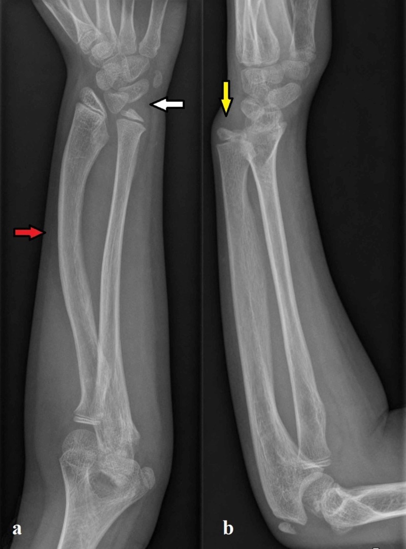
Madelung deformity - radiographic signs. (a) PA wrist: increased radial inclination, triangularisation of the carpus (white arrow) and a bowed, shortened radius (red arrow); (b) lateral wrist: volar tilt of the distal radius with dorsal subluxation of the ulnar head (yellow arrow). De Leucio A et al., Cureus 2020;12(2):e7100, Fig. 1 (CC BY).
Madelung deformity - radiographic signs. (a) PA wrist: increased radial inclination, triangularisation of the carpus (white arrow) and a bowed, shortened radius (red arrow); (b) lateral wrist: volar tilt of the distal radius with dorsal subluxation of the ulnar head (yellow arrow). De Leucio A et al., Cureus 2020;12(2):e7100, Fig. 1 (CC BY).
Treatment
- Non-operative care: observation, splinting and activity modification for the mild or asymptomatic wrist.
- In the skeletally immature wrist with growth remaining, the procedure of choice is Vickers ligament release with physiolysis (excision of the volar-ulnar physeal bar with fat or muscle interposition), which untethers the radius, arrests progression and allows remodelling; a dome osteotomy may be added.[29]
- For established deformity, a corrective radial osteotomy (Waters favours a concave-distal “dome” osteotomy) restores the radial inclination and corrects the volar tilt. It is combined with Vickers ligament release and a procedure to rebalance the ulna, either distal ulnar epiphysiodesis if growth remains or an ulnar shortening osteotomy for positive variance.[30]
- In the mature or severe wrist, distal-ulnar salvage procedures such as the Darrach resection or the Sauvé-Kapandji procedure address persistent ulnar-sided and distal radioulnar problems.[31]
Outcomes are good for pain relief but only modest for range of motion, and recurrence is common in the younger child (mitigated by always releasing the Vickers ligament and by epiphysiodesis or ulnar shortening). The superficial radial nerve is at risk during pin placement.[32]
Léri-Weill dyschondrosteosis (the companion syndrome)
Léri-Weill dyschondrosteosis (first described in 1929) is the autosomal-dominant SHOX disorder (about 50% penetrance, more severe in females) whose hallmark is mesomelia plus Madelung deformity with disproportionate short stature (adult height 135-170 cm). The mesomelic (middle-segment) shortening affects the forearm and the lower leg (tibia and fibula), sometimes with mild genu varum or ankle valgus. Intelligence is normal, and some patients have growth-hormone deficiency that responds to supplementation. The homozygous form is the much more severe Langer mesomelic dysplasia.[33]
Bulgarian terminology (Боев / Boychev tradition) - glossary
The following Bulgarian equivalents bridge the international literature with the terminology of the Bulgarian school of orthopaedics (in the tradition of Бойчо Бойчев / Boycho Boychev) used in the state examination.
| English term | Bulgarian term (Cyrillic) | Transliteration |
|---|---|---|
| Blount’s disease | Болест на Блаунт | Bolest na Blaunt |
| Tibia vara | Тибия вара (варусна подбедрица) | Tibiya vara (varusna podbedritsa) |
| Genu varum (bow-leg) | Варусно коляно (О-образни крака) | Varusno kolyano (O-obrazni kraka) |
| Genu valgum (knock-knee) | Валгусно коляно (Х-образни крака) | Valgusno kolyano (H-obrazni kraka) |
| Proximal tibial physis | Проксимална тибиална растежна зона | Proksimalna tibialna rastezhna zona |
| Growth plate (physis) | Растежна зона (физа) | Rastezhna zona (fiza) |
| Metaphyseal-diaphyseal angle | Метафизо-диафизарен ъгъл | Metafizo-diafizaren agal |
| Tibial torsion | Торзия на тибията | Torziya na tibiyata |
| Osteotomy | Остеотомия | Osteotomiya |
| Guided growth / hemiepiphysiodesis | Направлявана растеж / хемиепифизиодеза | Napravlyavan rastezh / hemiepifiziodeza |
| Bracing / orthosis | Ортеза (шина) | Orteza (shina) |
| Obesity | Затлъстяване | Zatlastyavane |
| Madelung deformity | Деформитет на Маделунг | Deformitet na Madelung |
| Distal radius | Дистален радиус | Distalen radius |
| Distal radioulnar joint | Дистална радиоулнарна става | Distalna radioulnarna stava |
| Lunate (bone) | Полулунна кост | Polulunna kost |
| Ulnar variance | Улнарна вариация | Ulnarna variatsiya |
| Dyschondrosteosis (Léri-Weill) | Дисхондростеоза (синдром на Лери-Вайл) | Dishondrosteoza (sindrom na Leri-Vayl) |
| Mesomelia | Мезомелия | Mezomeliya |
| Wrist | Китка | Kitka |
Image attributions
(Figure attributions and licences are listed in the figure MANIFEST and inserted with each image. All images are openly licensed [CC0 / CC BY / CC BY-SA] or used under their stated terms; any non-commercial [NC] item is flagged as such and must not be used in a commercial product.)
References
-
Campbell’s Operative Orthopaedics, Tibia Vara (Blount Disease), p. 1439.
-
Sabharwal, Pediatric Lower Limb Deformities, ch. 1, p. 29; Campbell’s, p. 1440; Lovell & Winter, Pediatric Orthopaedics, lower-extremity chapter, p. 1385.
-
Sabharwal, ch. 27, p. 506; Campbell’s, p. 1439; Lovell, p. 1363.
-
Sabharwal, pp. 29, 506; Lovell, pp. 1363, 1383; Campbell’s, p. 1439.
-
Sabharwal, p. 29; Lovell, pp. 1362-1364, 1383; Campbell’s, p. 1440.
-
Sabharwal, p. 29; Lovell, pp. 1364, 1383.
-
Campbell’s, p. 1440; Lovell, pp. 1367, 1374.
-
Lovell, p. 1385; Campbell’s, p. 1441.
-
Sabharwal, p. 30; Lovell, p. 1362.
-
Lovell, pp. 1359-1364; Sabharwal, p. 28.
-
Lovell, pp. 1359-1362; Sabharwal, pp. 36-37.
-
Sabharwal, p. 45; Campbell’s, p. 1440; Lovell, pp. 1362-1363.
-
Lovell, pp. 1365, 1374; Campbell’s, p. 1440.
-
Sabharwal, pp. 42-46; Campbell’s, p. 1441; Lovell, p. 1367.
-
Lovell, p. 1365; Sabharwal, p. 509.
-
Lovell, pp. 1365-1372; Campbell’s, p. 1442.
-
Sabharwal, pp. 509-510; Lovell, p. 1365.
-
Lovell, pp. 1367-1378; Sabharwal, pp. 510-511.
-
Lovell, pp. 1374-1382; Campbell’s, pp. 1442-1451.
-
Lovell, pp. 1387-1392; Sabharwal, pp. 513-517; Campbell’s, p. 1443.
-
Sabharwal, pp. 508-512; Lovell, pp. 1367, 1385; Campbell’s, pp. 1441-1451.
-
Waters & Bae, Pediatric Hand and Upper Limb Surgery, ch. 15, p. 148; Rayan & Upton, Congenital Hand Anomalies, ch. 14, p. 200; Lovell, ch. 7, pp. 224-225.
-
Waters & Bae, pp. 147-148; Rayan & Upton, p. 200.
-
Waters & Bae, pp. 147, 155; Lovell, p. 225; Rayan & Upton, p. 204.
-
Waters & Bae, pp. 147, 150; Rayan & Upton, p. 200.
-
Waters & Bae, p. 148; Rayan & Upton, p. 200; Lovell, p. 225.
-
Rayan & Upton, pp. 200-201; Lovell, p. 225.
-
Waters & Bae, p. 148; Rayan & Upton, pp. 200-202.
-
Waters & Bae, pp. 149-150, 153.
-
Waters & Bae, pp. 148-153; Lovell, p. 225.
-
Waters & Bae, pp. 153, 155 (Darrach distal-ulnar resection); the Sauvé-Kapandji procedure is the standard distal-radioulnar-joint-sparing salvage alternative.
-
Waters & Bae, p. 153; Lovell, p. 225.
-
Rayan & Upton, p. 204; Lovell, pp. 224-226.